Clinicians › Wrist
Wrist Arthroscopy
Diagnostic and therapeutic wrist arthroscopy — what it is, when it's used, and recovery.
For patients: a plain-language version of this topic is available. See the patient guide.
Overview
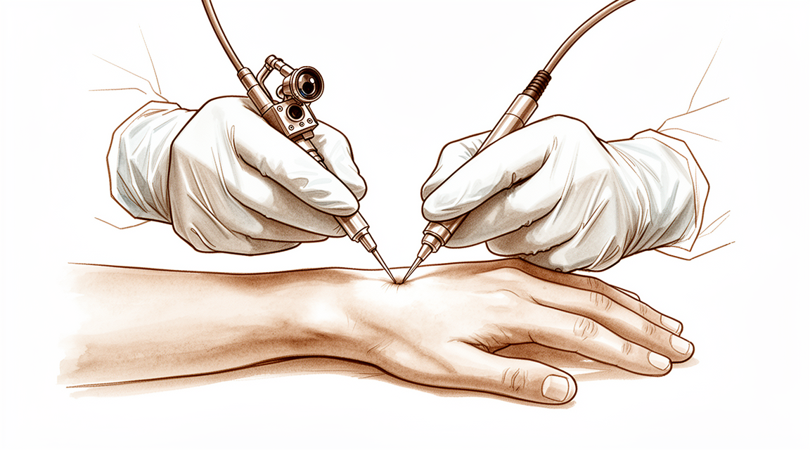
Wrist arthroscopy is an essential diagnostic and therapeutic tool for the orthopaedic surgeon, with an ever-expanding list of indications and procedures [1]. It has taken up a place in the armamentarium of the hand surgeon [3] and evolved into a critical instrument for managing TFCC pathology, carpal instability, fracture reduction assistance, and salvage procedures [38]. The procedure is advantageous for accurate assessment of articular surfaces and detection of concomitant soft-tissue injuries [69]. Indications continue to grow with the description of volar portals for the radiocarpal and scaphotrapezial trapezoid joint [5], while innovations such as new portals and smaller arthroscopes are expanding its applications [38]. Although wrist arthroscopy is here to stay with proven superiority in diagnosis, its value in treatment is evolving and requires more clinical studies to refine indications and outcomes [13].
The procedure is safe, with a low rate of major and minor complications [8] that are believed to be uncommon [4]. These complications are minor and transient, serving as a crucial tool for diagnosing intra-articular pathology and treating multiple wrist conditions [6]. Complications may largely be avoided by paying meticulous attention to detail and by anticipating potential problems before they occur [15]. Wrist arthroscopy technique emphasizes the use of traction for joint distention and the availability of various portals and instruments for diagnostic and therapeutic procedures [28].
Clinical outcomes vary by indication. Participants who underwent arthroscopic investigation for persistent wrist pain improved on average by approximately 50% at one year, though most continued to have some pain and disability [9]. Nonrandomized studies show satisfactory results for arthroscopically assisted reduction of distal radius fractures [69]. Arthroscopic arthrolysis is a reliable and reproducible technique that becomes a therapeutic indication of choice for the treatment of wrist stiffness after trauma or surgery [64]. Wrist arthroscopy is a feasible treatment option for translunate perilunate injury with advantages including lower risk of arthrofibrosis and avascular necrosis [169]. The most common indications for repeat wrist arthroscopy were ligamentous instability and osteoarthritis from dynamic impaction [20]. The authors do not feel that the routine use of wrist arthroscopy as a replacement or supplement to arthrography can be justified [57]. There was a decrease in the utilization of wrist arthroscopy among surgeons without hand and upper extremity fellowship training, with nearly all procedures performed by surgeons who had completed such a fellowship [48].
Anatomy & Pathophysiology
Bony Anatomy
The wrist constitutes the anatomic region between the forearm and hand, encompassing the distal radioulnar, radiocarpal, and ulnocarpal joints along with the eight carpal bones [84]. The carpus comprises eight ossicles divided into a proximal row (scaphoid, lunate, triquetrum, pisiform) and a distal row (trapezium, trapezoid, capitate, hamate) [87]. The distal radius features two concave articular facets for the scaphoid and lunate, separated by the scapholunate ridge, while the sigmoid notch along the ulnar border provides a shallow concavity for the articulating ulnar head [87]. The distal ulna is covered with hyaline cartilage on its dorsal, lateral, palmar, and distal surfaces, and the ulnar styloid projects distally with a base containing the fovea for triangular fibrocartilage complex insertion [87].
Vascular supply to the carpus is critical for healing. The scaphoid receives its primary supply from a branch of the radial artery at the dorsal ridge, with smaller vessels entering the palmar tubercle to supply the distal 30% [87]. The lunate possesses both dorsal and palmar vascular supply in 80% of wrists, whereas only a palmar supply is present in 20% [87]. The capitate head often relies on a retrograde vascular supply [87]. The pisiform functions as a sesamoid bone within the flexor carpi ulnaris tendon and serves as the origin for the abductor digiti minimi [87].
Geometric parameters influence wrist stability. The distal radial articular surface exhibits a double obliquity of 12–15 degrees in the lateral view and 15–20 degrees in the anteroposterior view [95]. The carpal articular surface has a smaller diameter of curvature than the radius, with medial and palmar stability dependent on ligamentous and capsular resistance [95]. Consequently, the carpus is more stable in flexion than extension due to its anterior concavity [95]. The triquetrum does not contact the ulnar head directly; a fibro-cartilage disc separates the two bones [95]. The scaphoid presents a long axis inclined by 45 degrees to the long axis of the radius [95]. The lunate sits on the capitate with anterior and posterior horns, forming a line perpendicular to the long axis of the wrist in neutral position [95]. The ulnar head sits proximal to the distal radius and exerts only an indirect effect on wrist stability [95].
Ligaments
The triangular fibrocartilage complex (TFCC) is formed by the central meniscus homolog, dorsal and volar radioulnar ligaments, the floor of the extensor carpi ulnaris tendon sheath, and volar ulnocarpal ligaments [87]. It arises from the radial border of the distal radius and inserts into the base of the ulnar styloid and distal ulna through the ligamentum subcruentum [87]. The dorsal and volar radioulnar ligaments serve as the primary stabilizers of the distal radioulnar joint [87]. Vascular supply to the TFCC is limited to the peripheral 10% to 40% of the volar, ulnar, and dorsal regions [87]. The TFCC attaches to the base of the ulnar styloid, separating the hyaline cartilage-covered ulnar head from the styloid [84]. Chondroligamentous supports attaching the distal radius and ulnar side of the carpus to the distal ulna include the ulnar collateral ligament, dorsal and volar radioulnar ligaments, articular disc, meniscal homologue, extensor carpi ulnaris sheath, and ulnolunate and ulnotriquetral ligaments [84].
Intrinsic ligaments bind the proximal carpal row. The scapholunate interosseous ligament is C-shaped in the sagittal plane, with the dorsal third being the thickest and strongest portion [87]. The volar portion of the lunotriquetral ligament is the thickest [87]. The interosseous ligaments include the scapholunate and lunotriquetral interosseous ligaments connecting the proximal row, and ligaments connecting the trapezium to the trapezoid, trapezoid to the capitate, and capitate to the hamate in the distal row [84]. Structural differences between intrinsic and extrinsic wrist ligaments may account for intrinsic ligaments being stronger and elongating further prior to failure than extrinsic ligaments [114].
Extrinsic ligaments cross the radiocarpal joint. The radial collateral ligament originates from the radius 0 mm from the radial styloid and inserts on the scaphoid waist and distal palmar trapezium [87]. The radioscaphocapitate ligament originates from the radius 4 mm from the radial styloid and inserts on the scaphoid waist and midpalmar capitate [87]. The radiolunatotriquetral ligament originates from the radius 10 mm from the radial styloid and inserts on the lunate and triquetrum [87]. The short radiolunate ligament originates from the volar-ulnar margin of the radius and inserts on the lunate [87]. On the ulnar side, the ulnotriquetral ligament originates from the volar radioulnar ligament and inserts on the triquetrum, while the ulnolunate ligament originates from the volar radioulnar ligament and inserts on the lunate [87]. The ulnocapitate ligament originates from the volar margin of the ulnar head and inserts on the capitate [87].
Dorsal extrinsic ligaments include the dorsal radiocarpal ligament, which originates from the dorsal radius at the Lister tubercle and inserts on the lunate and triquetrum [87]. Specifically, it attaches along the dorsal radial articular margin of the lunate fossa from the Lister tubercle to the lesser sigmoid notch, spans the lunotriquetral joint, and inserts on the dorsal surface of the triquetrum [84]. The dorsal intercarpal ligament originates from the triquetrum and inserts on the scaphoid, trapezoid, and capitate [87]. It is attached to the distal, dorsal surface of the triquetrum and passes across the midcarpal joint to attach to the dorsal surfaces of the scaphoid waist and the trapezoid [84]. The extrinsic or crossing ligaments also include the radial collateral ligament from the radial styloid to the scaphoid waist, the ulnar collateral ligament from the base of the ulnar styloid to the pisiform, and the transverse carpal ligament [84]. Volar extrinsic ligaments on the radial side include the radioscapocapitate ligament, radiolunotriquetral ligament, and radioscapolunate ligament, while the ulnar side comprises the ulnolunate and ulnotriquetral components of the TFCC [84]. The space of Poirier is a relatively thin area on the palmar side of the carpus between the radiolunotriquetral ligament and the radioscapocapitate ligament, overlying the palmar surface of the lunate [84].
Kinematics and Biomechanics
The wrist functions as a two-joint system linking the hand to the forearm around the highly mobile bones of the proximal carpal row [88]. The two principal articulations are the radiocarpal and midcarpal joints, situated proximal and distal to the mobile proximal carpal row [88]. The proximal carpal row has no muscular or tendinous attachments and is an intercalary segment [87]. With ulnar deviation, the proximal row extends relative to the forearm/distal row, and with radial deviation, the proximal row flexes relative to the forearm/distal row [87].
Load distribution and motion partitioning are specific to wrist position. With axial loading through the neutral wrist, approximately 80% of forces are transmitted through the distal radius (60% scaphoid facet, 40% lunate facet) and 20% through the distal ulna [87]. With wrist flexion, 60% of the motion is midcarpal and 40% is radiocarpal, while with wrist extension, 33% of the motion is midcarpal and 66% is radiocarpal [87]. In normal wrists, motion occurred equally at the midcarpal and radiocarpal joints [112]. The joints within the proximal carpal row govern 35% of the wrist extension-flexion arc and 33% of the wrist radial-ulnar deviation arc [119].
Biomechanical behavior is predominantly determined by articular geometry rather than ligament constraints, as the wrist behaves kinematically consistent but kinetically variable [82]. Despite complex carpal bone anatomy and kinematics, computed fiber elongations were found to vary linearly with wrist position [86]. Most carpal ligaments lengthen only during one half of a full movement cycle, constraining either dorsal- or palmar-directed, or ulnar- or radial-directed motion [105]. Wrist ligaments coordinate the positioning of the bones in the mid-range of carpal motions and restrict further motion in extreme positions of the wrist joint [125]. A stable wrist is defined as one which, while being loaded within a physiological range of stress, does not deviate from a stable state of equilibrium at any point within the whole physiological range of motion [102]. Conversely, a wrist joint should be considered biomechanically unstable when it is not able to bear loads and does not exhibit normal kinematics throughout its arc of motion [81].
Traction (distraction) changes normal carpal kinematics, and motion of the wrist combined with traction induces abnormal patterns of motion [90]. Wrist position and sectioning stage significantly influenced the displacement of the radiocarpal unit relative to the ulna [118]. The longitudinal 'columnar' concept of wrist kinematics does not fit in with many of the recent findings related to carpal instabilities [83]. The kinematic consequences of resecting the distal scaphoid include diminished flexion-extension motion of the proximal row during deviations at the expense of increased radioulnar translational motion [51]. The experimental kinetic behavior of the chronic scapholunate advanced collapse wrist is similar to that with a recent scapholunate dissociation without carpal collapse associated [127]. Among fusion types, radioscapholunate fusion shows the most biomechanically similar behavior compared with the healthy wrist [115]. Four-dimensional CT complements conventional imaging and arthroscopy by providing functional information on wrist biomechanics and should be used selectively when dynamic instability is suspected and conventional imaging is inconclusive [49].
Pathophysiology
Scapholunate interosseous ligament injury and extrinsic ligament complex attenuation lead to palmar flexion of the scaphoid and extension of the lunate, resulting in dorsal intercalated segment instability [62]. In scapholunate advanced collapse, the radioscaphoid joint becomes incongruous, leading to alteration in normal radioscaphoid contact forces and development of arthrosis [62]. As the scaphoid flexes and the scapholunate diastasis increases, the capitate migrates proximally [62]. Altered intercarpal contact forces result in arthrosis at the capitolunate joint [62]. The styloscaphoid, radioscaphoid, and capitolunate joints are affected by scapholunate advanced collapse arthritic changes, while the radiolunate joint is typically spared because of its spheroid shape [62].
Ulnocarpal impingement is a degenerative condition resulting from a discrepancy in the relative length of the distal articular surfaces of the radius and ulna (positive ulnar variance) [62]. Posttraumatic causes include distal radius fracture with shortening, Galeazzi or Essex-Lopresti fracture (distal radioulnar joint injury), and childhood epiphyseal plate injuries [62]. Congenital causes include dyschondroplasia (Madelung deformity) and naturally occurring positive ulnar variance [62].
The classic pattern of rheumatoid wrist deformity involves the radiocarpal and radioulnar joints with destabilization of the carpus caused by attenuation of the extrinsic wrist ligaments, resulting in ulnar-palmar translocation and wrist supination [146]. Three main pathophysiological factors play the greatest role in rheumatoid wrist deformation: cartilage destruction, synovial expansion, and ligamentous laxity [146]. Cartilage thinning is caused by cytochemical effects with continuous degradation [146]. Bony erosion arises due to synovial expansion, particularly at the site of vascular penetration into the bone such as the radial origin of the Testut ligament [146]. The scapholunate interval starts to dissociate and continues to disintegrate the internal carpal architecture [146]. The force vector across the rheumatoid wrist predominately acts in a palmar-ulnar direction [146]. Flexion of the scaphoid through the weakening of the scapholunate ligament with subsequent collapse of the radial column is described in rheumatoid arthritis [146]. Stretching of the wrist ulnar collateral ligament attenuates ulnar column support, leading to a typical carpal supination pattern [146]. Carpal supination contributes to radial deviation of the metacarpals and accentuates ulnar deforming forces on the fingers at the metacarpophalangeal joints [146]. Volar flexion of the lunate relative to the scaphoid in early-to-midstage rheumatoid wrists is caused by intrinsic ligament laxity, mainly of the scapholunate ligament [146]. In later stages, the capitate tends to flex dorsally due to midcarpal instability as a result of extrinsic ligament weakening, leading to a significant decrease in carpal height [146].
In juvenile arthritis, uncontrolled joint synovitis stretches essential ligaments and eventually erodes cartilage and bone [149]. Uncontrolled synovitis results in a progressive shift of the carpus ulnarward and volarward, leaving the ulna dorsally dislocated and creating a dorsal wrist step-off [149]. Destruction of joint mechanics is accompanied by loss of active and passive wrist extension and weakness of grip [149]. If disease spreads to the distal radioulnar joint, forearm rotation becomes limited [149].
Displaced radius fractures in nonosteoporotic patients have a high incidence of associated soft tissue injuries [170]. Associated injuries in distal radius fractures affect the long-term outcome, and arthroscopic evaluation is paramount to establish a correct and complete diagnosis and facilitate early treatment [170]. After falls, arthroscopy can diagnose triangular fibrocartilage complex injuries, intercarpal scapholunate and lunotriquetral ligament tears, and chondral lesions [170]. Undetected associated injuries may explain the absence of improved outcome in studies comparing volar locking plate fixation and early mobilization versus external fixation [170].
Normal wrist kinematics requires integrity of highly complex anatomy and synchronous interaction between the proximal and distal carpal rows [184]. Unimpeded multiplanar motion is contingent on uncompromised stability provided by critical intrinsic and extrinsic ligamentous structures [184]. The scapholunate and lunotriquetral interosseous ligaments tightly bind the carpal bones of the proximal row, while volarly situated extrinsic radiocarpal ligaments support and guide the proximal carpus [184]. The proximal carpal row functions as an intercalated segment, solely dependent on intact bony and ligamentous anatomy for maintenance of stability, which renders it highly vulnerable to injury [184]. Excessive radiocarpal hyperextension and ulnar deviation coupled with intercarpal supination is the principal pathologic force that disrupts the key osseous and ligament components of the wrist in perilunate injuries [184]. Perilunate injuries comprise a predictable spectrum of pathology incurred from variation of the basic deforming force [184].
Tears of the lunotriquetral ligament have been implicated in the pathogenesis of volar intercalated segmental instabilities [178]. The lunotriquetral ligament consists of dorsal, proximal, and palmar subregions, with the palmar subregion being the most important for maintaining stability [178].
Classification
TFCC Injuries
Palmer: The Palmer classification categorizes triangular fibrocartilage complex (TFCC) tears into traumatic (Class 1) or degenerative (Class 2) categories [30]. Subtypes are defined by the specific location of the injury within the TFCC [30]. Class 1A injuries involve central perforation or tear [30]. Class 1B injuries consist of ulnar avulsion with or without ulnar styloid fracture [30]. Class 1C injuries involve distal avulsion affecting the origins of the ulnolunate and ulnotriquetral ligaments [30]. Class 1D injuries involve radial avulsion of the dorsal and/or volar radioulnar ligaments [30]. Class 2A tears are characterized by TFCC wear or thinning [30]. Class 2B tears involve TFCC wear plus lunate and/or ulnar chondromalacia [30]. Class 2C tears consist of TFCC perforation plus lunate and/or ulnar chondromalacia [30]. Class 2D tears include TFCC perforation, lunate and/or ulnar chondromalacia, and lunotriquetral ligament disruption [30]. Class 2E tears involve TFCC perforation, lunate and/or ulnar chondromalacia, lunotriquetral ligament disruption, and ulnocarpal and distal radioulnar joint arthritis [30]. The class and location of a TFCC tear have important implications for treatment [30]. The original Palmer classification is based on clinical examination, routine x-ray films, wrist arthrograms, wrist arthroscopy, and wrist arthrotomy [25].
Atzei: Atzei's classification is used for the detailed classification of proximal carpal TFCC tears [160].
Scapholunate Ligament Injuries
Geissler: The Geissler classification grades scapholunate ligament lesions based on arthroscopic findings [182]. Grade 1 injuries consist of attenuation or hemorrhage of the membranous portion of the ligament with a smooth and intact scapholunate interval [182]. Grade II lesions consist of an attenuated fibrocartilaginous membrane with a step-off between the scaphoid and lunate where a 1-mm probe may be placed [182]. Grade III lesions consist of a step-off visualized from both the radiocarpal and midcarpal joints where a 1-mm probe may be inserted and freely rotated in the scapholunate interval [182]. Grade IV lesions consist of complete disruption of the ligament allowing a 2.7-mm arthroscope to be passed between the radiocarpal and midcarpal joints [182].
Scapholunate Instability
Four-Stage System: A four-stage classification system for scapholunate instability includes predynamic instability, dynamic instability, static instability, and scapholunate advanced collapse (SLAC) [182].
Kienbock's Disease
Staging: Kienbock's disease can be classified into Stage IIIA (without carpal collapse) or Stage IIIB (with carpal collapse) by comparison to the contralateral wrist [70]. Arthroscopic classification of Kienbock's disease allows surgeons to tailor surgical reconstruction to anatomical findings [155].
Cartilage Lesions
Outerbridge: Cartilage lesions in the wrist are classified according to the Outerbridge system [167].
Diagnostic Reliability and Limitations
Other Considerations: Current arthroscopic classification systems have limitations regarding diagnostic reproducibility [143]. The diagnostic accuracy of MRI for detailed TFCC injury classification, such as Atzei's classification, is lower compared to wrist arthroscopy [160]. Grading of cartilage lesions has higher reliability than grading of scapholunate ligament or TFCC lesions based on digital photograph documentation [167].
Clinical Presentation
Diagnostic Indications
Wrist arthroscopy is indicated for the evaluation of chronic wrist pain of uncertain etiology that has persisted for more than 3 months and is unresponsive to conservative treatment [21]. It is also indicated for the assessment of acute ligamentous injuries, including scapholunate, lunotriquetral, and triangular fibrocartilage complex (TFCC) injuries [21]. Further indications include the evaluation of carpal instability [21], the assessment of chondral lesions [21], and the evaluation of associated soft tissue injury in fracture conditions, including distal radius, scaphoid, ulnar styloid, and other carpal bone fractures [21]. The procedure is indicated for the assessment of scaphoid healing in delayed union and nonunion [21] and for the staging of posttraumatic arthritis, including scapholunate advanced collapse (SLAC), scaphoid nonunion advanced collapse (SNAC), and distal radius fractures [21]. Additional indications encompass the evaluation of monoarticular arthritis and synovial biopsy [21] and the evaluation of Kienböck disease [21]. The most common indications for repeat wrist arthroscopy are ligamentous instability and osteoarthritis from dynamic impaction [20].
Clinical Examination Principles
A careful history and physical examination are prerequisites before using wrist arthroscopy for either diagnosis or treatment [32]. The natural inclination to study radiographs or special imaging studies prior to a thorough history and physical examination should be avoided to prevent cognitive bias that can affect thinking and decision making [56]. Physical examination must be preceded by a thorough investigation of the patient’s medical history, with special emphasis on the mechanism of injury and acuity [56]. Patients should be asked for details about the location, duration, and characteristics of pain, including aggravating and relieving factors and previous treatments [56]. For chronic problems, it is important to inquire about the patient’s jobs and hobbies, and whether there has been exposure to repetitive stress, vibrating tools, or potentially dangerous instruments [56]. Elucidating a history of ligamentous laxity or multiple joint instabilities is important, especially in younger patients presenting with chronic wrist pain [56].
Palpation for areas of maximal tenderness is one of the most useful tools in the diagnosis of wrist pathology, especially in patients with chronic dysfunctions [56]. In acute dislocations, tenderness is seldom elicited at specific points but rather in a diffuse manner due to extensive soft tissue damage [56]. A careful assessment of neural and vascular status is imperative, with particular attention paid to the median and ulnar nerves [56]. A thorough set of provocative maneuvers should be performed to rule out alternative or concurrent diagnoses, not just to concentrate on the suspected diagnosis [56]. The examination should begin in a nontender area and proceed rotationally around the carpus, ending at the most symptomatic area [56].
Bilateral grip and pinch strength are useful to uncover underlying pathology in chronic cases [56]. Strength may be diminished due to muscle atrophy, pain inhibition, or learned behaviors [56]. Rapid alternating grip assessment may be helpful in determining voluntary effort [56]. A local injection of anesthetic to a painful joint or selected tendon sheath may help normalize dynamometer readings and narrow the diagnostic spectrum [56]. Sensory testing should always accompany an examination of suspected nerve compression, using threshold or density testing [56]. Patients without a positive provocative sign on examination seldom yield positive findings at wrist arthroscopy [63]. Arthroscopic findings need to correlate with clinical examination [63].
Specific Clinical Signs and Maneuvers
The ulnar fovea sign is a provocative maneuver aimed at eliciting abnormal tenderness in the fovea region of the wrist, bounded by the ulnar styloid [103]. It is used in the context of a symptomatic patient with no identifiable pathology on other clinical and radiologic investigations who continues to suffer troubling pain despite appropriate nonsurgical interventions [103]. Watson’s test, or the scaphoid shift test, is designed to show scaphoid instability by causing a dorsal subluxation of the scaphoid accompanied by a painful click [174]. The ballotment tests, or shear tests, consist of demonstrating abnormal movements between adjacent bones by exerting pressure in opposite directions [174]. The scapholunate ballotment test involves stressing the scapholunate joint by volar pressure on the tubercle of the scaphoid and dorsal pressure on the lunate [174]. Reagan’s test for lunotriquetral instability involves applying pressure of the examiner’s thumb on the lunate and pressure of the index on the triquetrum [174].
The midcarpal joint "pivot shift" test consists of supinating and volar subluxing the distal row of the carpus [174]. The radio-carpal and midcarpal joint anteroposterior drawer test involves applying axial traction while stabilizing the forearm and applying anteroposterior force to elicit a drawer at the radio-carpal and then midcarpal joint [174]. A marked drawer test at the midcarpal joint is a sign of congenital laxity [174]. Triquetral hamate instability is demonstrated with the wrist straight with ulnar deviation, producing a firm block after a range of about 20 degrees [174]. Forcing a sharp click accompanied by discrete posterior movement of the wrist in triquetral hamate instability indicates the proximal row has moved from the VISI position to the DISI position [174]. An exaggeration of the normal ulna head prominence is seen in dorsal subluxation or articular effusion of the distal radioulnar joint [174]. Pain precipitated by pronosupination while the ulna head is pressed volarward and the pisiform pressed dorsally is usually indicative of some form of ulnar impingement or abutment syndrome [174]. Significant pain precipitated by holding the hand in full ulnar deviation and holding the ulna head forward by the examiner’s thumb suggests distal radioulnar joint pathology [174].
Imaging and Diagnostic Utility
Wrist arthroscopy is more sensitive than arthrography for evaluating TFCC tears and interosseous ligament tears [32]. The presence of certain apparent joint abnormalities, such as a ligamentous disruption diagnosed by arthrography or MR imaging, may not represent a clinically significant process [32]. Wrist arthroscopy remains the gold standard for the diagnosis and treatment of longitudinal split tears of the ulnotriquetral ligament [16]. Wrist arthroscopy remains the gold standard for diagnosing TFCC pathologies despite technical progress in imaging modalities [36]. MRI findings are often discordant when compared with diagnostic arthroscopy for determining the cause of ulnar wrist pathologies [55]. Arthroscopy is a simple method that enables new diagnostic possibilities in rare but difficult cases of posttraumatic pain of the wrist, particularly when clinical and radiological examinations fail to provide a diagnosis [45]. The increased use of arthroscopic examination of the wrist is advisable in wrist trauma to establish a correct diagnosis, prognosis, and treatment of all the patient’s lesions, possibly preventing degenerative arthritis [27]. Wrist arthroscopy is an effective adjunctive tool in the diagnosis of wrist derangements [29].
Wrist arthroscopy provides views of and access to the intraarticular spaces of the wrist that are otherwise difficult to achieve without widely open approaches [19]. Diagnostic arthroscopy is a useful adjunct in the diagnosis and treatment of intra-articular wrist pathology after careful history and physical examination [10]. A thorough understanding of the 3-D anatomy of the wrist is essential to optimize efficiency and to minimize complications associated with improper instrument placement [10]. The diagnosis of dorsal capsule tears of the wrist is arthroscopic [121]. Ulnar-sided wrist pain is a common cause of upper extremity disability with a complex differential diagnosis [106, 137]. Determining the etiology of ulnar-sided wrist pain is often challenging due to overlapping history and physical examination findings [129]. A detailed history, systematic physical examination with provocative maneuvers, and appropriate diagnostic imaging are essential for the diagnosis of ulnar-sided wrist pain [129]. Disorders of the distal radioulnar joint are a common source of ulnar-sided wrist pain [128].
Outcomes and Prognosis
Most patients who underwent arthroscopic investigation for persistent wrist pain continued to have some pain and disability at one year [9]. Patients with persistent wrist pain undergoing arthroscopy may expect approximately 50% improvement in pain and disability within one year [35]. Moderate levels of pain and disability often persist in patients with persistent wrist pain undergoing arthroscopy [35].
Investigations
Diagnostic Role and Indications: Wrist arthroscopy serves as a valuable diagnostic and therapeutic adjunct for myriad wrist disorders, providing access to intraarticular spaces that are difficult to achieve without widely open approaches [7, 11, 19]. Its use is advisable in wrist trauma to establish correct diagnosis, prognosis, and treatment of all lesions, potentially preventing degenerative arthritis [27]. Specific indications include evaluation of ligamentous injuries, examination of joint articular surfaces, removal of loose bodies, biopsy of synovium, irrigation and debridement, and confirmation or supplementation of wrist arthrography [52]. The procedure is particularly well suited to directly visualize and treat multiple causes of ulnar-sided wrist pain simultaneously [17].
Gold Standard Status: Arthroscopy is the gold standard for detecting triangular fibrocartilage complex (TFCC) tears [30] and remains the gold standard for diagnosing TFCC pathologies despite technical progress in imaging, although MR arthrography may become a real alternative in the future [36]. It is also considered the gold standard for evaluating intercarpal ligament injuries and instability, as well as for examining patients with wrist pain of unknown origin [52]. Furthermore, wrist arthroscopy is the gold standard for diagnosing wrist ligament pathology [188] and should be considered particularly when scapholunate or lunotriquetral interosseous ligament involvement is suspected based on history and physical examination, even in the face of negative MRI findings [165]. Arthroscopy can test the scapholunate joint dynamically and detect articular lesions long before they appear on radiographs [187]. It has been found to be more accurate than arthrography in identifying the location and size of triangular fibrocartilage and interosseous ligament injuries [52], and more accurate than triple-injection cinearthrography in detecting tears of the dorsal sensory branch of the ulnar nerve during arthroscopic repair of the triangular fibrocartilage [52].
Comparison with Imaging Modalities: MRI is a useful adjunct for determining the cause of ulnar wrist pathologies, but findings are often discordant when compared with diagnostic arthroscopy [55]. Given only a fair correlation between arthroscopy and MRI, the two methods cannot be concluded to be equivalent for assessing wrist cartilage, preserving an important role for arthroscopy in the assessment of a painful degenerative wrist [144]. While 3T MRI proved of good value in diagnosing cartilage lesions, especially in the distal carpal row, wrist arthroscopy provided therapeutic options [142]. Negative results of MRI or clinical provocative tests are unable to safely rule out clinically relevant tears to the TFCC and other wrist ligaments, necessitating further diagnostic evaluation with wrist arthroscopy [164]. Consequently, wrist ligament pathology cannot be ruled out by a negative magnetic resonance imaging [188]. A stable distal radioulnar joint upon clinical examination and normal MRI findings do not rule out foveal TFCC injury, requiring a high index of clinical suspicion when managing patients with ulnar-sided wrist pain [175]. MRI-detectable carpal lesions at the time of radial fracture are common, but only a few decompensate later to become symptomatic and of therapeutic relevance [172]. Single compartment direct wrist magnetic resonance arthrography provides high diagnostic accuracy for full-thickness triangular fibrocartilage lesions, intrinsic carpal ligament tears, and chondral lesions in the radiocarpal joint, but is much less accurate in diagnosing synovitis of the radiocarpal joint [183]. MRI demonstrated an identifiable dorsal abnormality in 84% of patients with dorsal wrist pain associated with weight bearing on the extended wrist [171]. Arthrography and MRI have limitations and are not always reliable for routine work-up of wrist lesions [140]. Conventional radiography should be the first imaging modality to exclude or diagnose wrist pathology; when inconclusive, high resolution 3 Tesla MRI is advised [141]. Although arthrography is a well-accepted diagnostic modality, normal arthrographic findings do not necessarily rule out the possibility of internal derangement of the wrist [189].
Diagnostic Limitations and Outcomes: Diagnostic wrist arthroscopy for a wrist with normal radiological alignment has poor interobserver agreement [150]. Participants who underwent arthroscopic investigation for persistent wrist pain improved on average by approximately 50% at one year; however, most continued to have some pain and disability [9]. The section on evaluation includes discussions on imaging and wrist arthroscopy, with a caution noted regarding the anecdotal presentation of arthroscopy [2]. A thorough understanding of the 3-D anatomy of the wrist is essential to optimize efficiency and to minimize complications associated with improper instrument placement during diagnostic arthroscopy [10]. Detailed knowledge of anatomy is essential to minimize complications associated with wrist arthroscopy [11].
Treatment
Non-Operative
Wrist arthroscopy is recommended in cases of chronic wrist pain where clinical suspicion is strong and the operating team is well-versed with the technique [176]. For patients with neutral to positive ulnar variance and persistent ulnar-sided wrist pain despite conservative treatment, wrist arthroscopy combined with an ulnar shortening procedure is indicated to unload the ulnocarpal joint [145].
Operative
Indications: Wrist arthroscopy serves both diagnostic and therapeutic roles. Diagnostic indications include the evaluation of associated soft tissue injury in fracture conditions such as distal radius, scaphoid, and ulnar styloid fractures [21], as well as the staging of posttraumatic arthritis including scapholunate advanced collapse (SLAC) and scaphoid nonunion advanced collapse (SNAC) [21]. Therapeutic indications span a wide range of pathologies. Ablative procedures include arthroscopic TFCC debridement [21], debridement of ligament tears (scapholunate, lunotriquetral, extrinsic, or intrinsic) [21], synovectomy for inflammatory, septic, gouty arthritis, or posttraumatic synovitis [21], ganglionectomy for dorsal and volar ganglions [21], removal of loose bodies [21], capsulotomy or capsulectomy [21], lavage [21], arthrolysis [21], scaphoidectomy [21], radial styloidectomy [21], the wafer procedure [21], proximal row carpectomy [21], proximal hamate excision [21], and debridement of chondral and osteochondral lesions [21]. Reparative procedures include arthroscopic repair of peripheral TFCC tears [21], TFCC foveal avulsion [21], scapholunate ligament injury [21], lunotriquetral ligament injury [21], and drill or abrasion chondroplasty [21]. Arthroscopic-assisted reduction and internal fixation (ARIF) is indicated for distal radius fractures [21] and scaphoid fractures [21]. Reconstructive procedures include TFCC reconstruction with tendon graft [21], scapholunate ligament reconstruction with tendon graft [21], bone grafting for scaphoid nonunion [21], limited carpal fusion [21], and intraosseous bone cysts or ganglions [21], as well as osteochondral grafting [21].
Surgical Approach / Technique: The patient is positioned supine on the operating table [21]. A traction device is applied to distract the wrist joint, using either an overhead traction boom or a dedicated sterilizable wrist traction device [21]. Traction force of 10 to 12 lb is applied through plastic finger traps over the index and middle fingers, or preferably the middle three fingers [21]. Overdistraction or use of wire finger traps may cause postoperative finger joint pain or localized contusion to soft tissue or digital nerves [21]. Nylon finger traps are more comfortable and atraumatic to the patient, especially in awake cases [21]. For arthroscopy over the scaphotrapeziotrapezoid joint, an additional trap and traction can be put on the thumb [21]. When an overhead traction boom is employed, countertraction is provided by securing the arm to the hand table, and the operated limb is draped free up to the elbow level [21]. When a dedicated wrist traction device is used, the limb is draped up to the axilla level and the lower arm is wrapped to the basal plate of the device close to the elbow level [21]. A traction device should be sterilizable and allow flexible positioning of the wrist intraoperatively in varying degrees of extension, flexion, and radial and ulnar deviation [21].
Joint visibility is maintained by saline inflow, as the small volume of the wrist makes fluid distention impractical compared to knee or shoulder joints [21]. The main maneuver in creating the working space is controlled traction, while saline serves to maintain a clear view by removing intraarticular debris and provides a hemostatic role when performed without a tourniquet [21]. Continuous irrigation is achieved with a 3 L bag of normal saline suspended 1.5 m above the patient and instilled under gravity [21]. Caution should be used to avoid extravasation of fluid that may lead to compartment syndrome [21]. A pressure control device is not essential for wrist arthroscopy [21].
Standard wet wrist arthroscopy is performed through the 3/4, 4/5, and midcarpal portals [217]. Comprehensive assessment of both the radiocarpal and midcarpal joints is made during wrist arthroscopy, and where possible, the pisotriquetral and head of the ulna are also assessed [217]. Tears of the intercarpal ligaments are classified according to Geissler et al, and chondral pathology is graded according to the modified Outerbridge classification [217]. For diagnostic examination of scapholunate dissociation, the hand is placed in the upright position with traction assistance [191]. Once the diagnosis of scapholunate dissociation and the absence of degenerative changes is confirmed, the extremity is taken out of traction and placed in pronation on the hand table for surgical repair [191]. The surgical approach utilizes a dorsal longitudinal incision made over the dorsum of the wrist just ulnar to Lister's tubercle [191]. The skin incision is carried down to the extensor retinaculum, and the dorsal venous complex of the wrist should be saved whenever possible [191]. A step-cut incision is made in the extensor retinaculum, leaving a radial and an ulnar-based flap [191]. The terminal branch of the posterior interosseous nerve is identified at the floor of the fourth dorsal compartment lying immediately to the ulnar side of the septum separating the third and fourth compartments [191]. A segment of the terminal branch of the posterior interosseous nerve is harvested, making sure to resect the nerve proximal to the distal end of the radius [191]. An oblique incision is made through the wrist capsule for scapholunate dissociation repair [191].
For established scaphoid nonunion management, wrist arthroscopy is performed under general anesthetic augmented with a peripheral nerve block and using a tourniquet [217]. The wrist is suspended using finger traps, and a 5-lb counterweight is used to provide distraction of the carpus [217]. Standard wet wrist arthroscopy is performed through the 3/4, 4/5, and midcarpal portals [217]. Comprehensive assessment of both the radiocarpal and midcarpal joints is made during wrist arthroscopy [217]. Tears of the intercarpal ligaments are classified according to Geissler et al, and chondral pathology is graded according to the modified Outerbridge classification [217].
Adjuncts: Tourniquet use is optional and is often unnecessary for diagnostic and uncomplicated therapeutic procedures performed under local anesthesia without sedation [21]. The PSLA (Portal Site Local Anesthesia) technique without a tourniquet is a feasible and preferred mode of anesthesia for wrist arthroscopic surgery in the hands of experienced wrist arthroscopists [211]. The PSLA technique offers a success rate of 95% with high patient satisfaction and no complications in selected patients [186]. Wrist arthroscopy can also be performed under general anesthesia augmented with a peripheral nerve block and using a tourniquet [217].
Dry wrist arthroscopy allows for larger portals and thus larger instruments, circumventing problems associated with fluid instillation [215]. The dry technique facilitates the combination of open procedures with arthroscopic visualization, allowing for more precise fracture reduction [215]. The absence of fluid distention in dry wrist arthroscopy facilitates soft tissue dissection and easier hardware placement [215].
Other Considerations: Wrist arthroscopy has allowed for the development of less invasive procedures to treat specific triangular fibrocartilage complex injury patterns [12]. Arthroscopy is particularly well suited to directly visualize and treat multiple causes of ulnar-sided wrist pain simultaneously [17]. The arthroscopic wafer procedure offers advantages over open procedures in unloading the ulnar side of the wrist, including no incision on the ulnar border of the forearm and no possibility of nonunion [41]. Arthroscopic arthrolysis is a reliable and reproducible technique that is a therapeutic indication of choice for the treatment of wrist stiffness after trauma or surgery [64]. Arthroscopic wrist arthrolysis after wrist fracture is a safe procedure that significantly improved pain and wrist flexion-extension [136].
Early detection of carpal instability nondissociative following acute wrist fractures may preserve wrist function by capsular repair, whereas cases with fixed deformity and residual joint incongruity may be best managed with a limited radiocarpal arthrodesis [203]. A salvage operation, such as a partial wrist fusion, should be considered when there is clinical and radiographic evidence of wrist arthritis in chronic scapholunate ligament injury [40]. Total wrist fusion should only be used for exceptional circumstances in the treatment of advanced carpal collapse [200].
Complications
General Safety and Incidence: Wrist arthroscopy is considered a safe procedure with minor and transient complications [6]. However, a systematic review suggests that the previously documented rate of wrist arthroscopy complications may be underestimating the true incidence [162]. In a multicenter study of 10,107 arthroscopies, the global incidence of complications was in keeping with the literature, but the incidence of serious complications was much higher than previously reported [237].
Infection (PJI): Postoperative infection after wrist arthroscopy is uncommon but clinically relevant [138]. This complication is particularly relevant in elderly, male patients with systemic comorbidities or those undergoing synovectomy [138]. Arthroscopy is a sophisticated surgical procedure which may end up with a wrist fusion if infection occurs, a very rare complication [195]. Administering preoperative antibiotics for routine wrist arthroscopy does not appear to lower the surgical site infection rate [218].
Nerve Palsy: In a review of wrist arthroscopy in juvenile idiopathic arthritis, neurapraxia of the superficial radial nerve in one patient and injury to the posterior interosseous nerve were reported [202]. Conversely, in a series of arthroscopic treatment for scapholunate ligament lesions associated with intra-articular distal radius fractures, there were no infections or nerve injuries [159].
Wound Complications: Overdistraction or use of wire finger traps during wrist arthroscopy may cause postoperative finger joint pain or localized contusion to soft tissue or digital nerves [21]. Caution should be used during wrist arthroscopy to avoid extravasation of fluid that may lead to compartment syndrome [21].
Other Considerations: Detailed knowledge of anatomy is essential to minimize complications in wrist arthroscopy [11].
Recovery
Light activity (weeks): The mean time for return to work was 5.5 weeks after arthroscopy and therapy for isolated triangular fibrocartilage complex perforations [151].
Full activity (months): Return to play may be delayed in athletes with concomitant ulnar-sided wrist injuries following arthroscopic treatment of triangular fibrocartilage wrist injuries [193].
Other Considerations: Once ulnar impaction becomes symptomatic, it remains so and recurs, especially on return to the athletic sport [219]. Patients experience good functional outcomes with the majority experiencing a reduction in pain with the ability to return to work following arthroscopic wrist debridement and radial styloidectomy for advanced scapholunate advanced collapse wrist [205]. Hand coordination following tendon transfer surgery demonstrated a graded recovery, with coordinated movement between wrist and finger joints improving at weeks 8 and 12 [154].
Key Evidence
- [L5] Wrist arthroscopy is an essential diagnostic and therapeutic tool for the orthopaedic surgeon with an ever-expanding list of indications and procedures. [1] (10.1016/j.arthro.2007.11.002)
- [Paper] The section on evaluation includes discussions on imaging and wrist arthroscopy, with a caution noted regarding the anecdotal presentation of arthroscopy. [2] (10.1016/0363-5023(89)90207-4)
- [L4] Wrist arthroscopy has taken up a place in the armamentarium of the hand surgeon. [3] (10.1055/s-0033-1351355)
- [L5] Complications associated with wrist arthroscopy are fortunately believed to be uncommon. [4] (10.1016/s0749-0712(21)00187-6)
- [Paper] The indications for wrist arthroscopy are growing with the description of volar portals for the radiocarpal and scaphotrapezial trapezoid joint. [5] (10.1097/00130911-200603000-00007)
- [L4] From these data it appears that wrist arthroscopy is a valuable means of diagnosis and treatment of wrist disorders. [7] (10.1016/s0266-7681(96)80100-8)
- [L4] Wrist arthroscopy is a safe procedure with a low rate of major and minor complications. [8] (10.1016/j.jhsa.2003.12.020)
- [L2] Participants who underwent arthroscopic investigation for persistent wrist pain improved on average by approximately 50% at one year; however, most continued to have some pain and disability. [9] (10.1016/j.jht.2012.03.001)
- [L5] [10] (10.1016/j.hcl.2017.06.004)
- [L5] Wrist arthroscopy has grown from a diagnostic tool to a valuable adjunctive procedure for myriad wrist disorders, but detailed knowledge of anatomy is essential to minimize complications. [11] (10.1016/j.jhsa.2008.07.015)
- [L4] [12] (10.1097/00130911-199712000-00002)
- [L5] Wrist arthroscopy is here to stay with proven superiority in diagnosis, but its value in treatment is evolving and requires more clinical studies to refine indications and outcomes. [13] (10.1054/jhsb.2001.0617)
- [L5] As with any surgical procedure, complications associated with wrist arthroscopy may largely be avoided by paying meticulous attention to detail and by anticipating potential problems before they occur. [15] (10.1016/s0749-0712(21)00029-9)
- [L4] Wrist arthroscopy remains the gold standard for the diagnosis and treatment of this condition. [16] (10.1016/j.hcl.2010.07.004)
- [L5] Arthroscopy is particularly well suited to both directly visualize and treat multiple causes of ulnar-sided wrist pain simultaneously. [17] (10.1016/j.hcl.2013.09.001)
- [L5] Wrist arthroscopy can be a useful tool in one’s armamentarium in the diagnosis and treatment of wrist pathology, providing views of and access to the intraarticular spaces of the wrist that are otherwise difficult to achieve without widely open approaches. [19] (10.1016/j.eats.2024.103223)
- [L4] The most common indications for repeat wrist arthroscopy were ligamentous instability and osteoarthritis from dynamic impaction. [20] (10.1055/s-0033-1364090)
- [L4] This classification is based on the clinical examination, routine x-ray films, wrist arthrograms, wrist arthroscopy, and wrist arthrotomy. [25] (10.1016/0363-5023(89)90174-3)
- [L4] The increased use of arthroscopic examination of the wrist is advisable in wrist trauma in order to establish a correct diagnosis, prognosis and treatment of all the patient’s lesions, possibly preventing degenerative arthritis. [27] (10.1016/j.hansur.2016.10.029)
- [L5] Wrist arthroscopy is an effective adjunctive tool in the diagnosis of wrist derangements. [29] (10.1016/j.hcl.2016.08.005)
- [L5] [32] (10.5435/00124635-200105000-00006)
- [L5] The commentary concludes that patients with persistent wrist pain undergoing arthroscopy may expect approximately 50% improvement in pain and disability within one year, though moderate levels of pain and disability often persist. [35] (10.1016/j.jht.2012.04.002)
- [L5] Wrist arthroscopy remains the 'gold standard' for diagnosing TFCC pathologies despite technical progress in imaging modalities, although MR arthrography may have the potential to become a real alternative in the future. [36] (10.1007/s00402-015-2153-6)
- [L5] Wrist arthroscopy has evolved into an essential diagnostic and therapeutic tool with a wide list of indications, including management of TFCC pathology, carpal instability, fracture reduction assistance, and salvage procedures, with innovations like new portals and smaller arthroscopes expanding its applications. [38] (10.5435/jaaos-20-11-725)
- [L5] A salvage operation, such as a partial wrist fusion, should be considered when there is clinical and radiographic evidence of wrist arthritis. [40] (10.1016/j.hcl.2015.04.013)
- [L4] The arthroscopic “wafer” procedure offers several advantages over open procedures in unloading the ulnar side of the wrist, including no incision on the ulnar border of the forearm and no possibility of nonunion. [41] (10.1097/00130911-200009000-00007)
- [L4] Arthroscopy is a simple method that enables new diagnostic possibilities in rare but difficult cases of posttraumatic pain of the wrist, particularly when clinical and radiological examinations fail to provide a diagnosis. [45] (10.1007/bf00420331)
- [L4] Although nearly all procedures were performed by surgeons who had completed a hand and upper extremity fellowship, there was a decrease in the utilization of wrist arthroscopy among surgeons without hand and upper extremity fellowship training. [48] (10.5435/jaaosglobal-d-25-00158)
- [L5] Four-dimensional CT complements conventional imaging and arthroscopy by providing functional information on wrist biomechanics and should be used selectively when dynamic instability is suspected and conventional imaging is inconclusive. [49] (10.1530/eor-2026-0051)
- [L4] The kinematic consequences of resecting the distal scaphoid are different: the flexion-extension motion of the proximal row during deviations is diminished at the expense of increased radioulnar translational motion. [51] (10.1097/00130911-199909000-00004)
- [L2] While MRI is a useful adjunct for determining the cause of ulnar wrist pathologies, findings are often discordant when compared with diagnostic arthroscopy. [55] (10.1016/j.jhsa.2024.04.015)
- [L4] The authors do not feel that the routine use of wrist arthroscopy as a replacement or supplement to arthrography can be justified. [57] (10.1016/0266-7681(94)90256-9)
- [L4] Arthroscopic arthrolysis is a reliable and reproducible technique that becomes a therapeutic indication of choice for the treatment of wrist stiffness after trauma or surgery. [64] (10.1016/j.main.2006.07.002)
- [L5] Wrist arthroscopy is advantageous for accurate assessment of articular surfaces and detection of concomitant soft-tissue injuries, with nonrandomized studies showing satisfactory results for arthroscopically assisted reduction of distal radius fractures. [69] (10.1016/j.arthro.2007.10.006)
- [L5] The author states that their classification of Kienbock’s disease in stages IIIA (without carpal collapse) or IIIB (with carpal collapse) is done comparing to the contralateral wrist. [70] (10.1177/17531934231205707)
- [L5] A wrist joint should be considered biomechanically unstable when it is not able to bear loads and does not exhibit normal kinematics throughout its arc of motion. [81] (10.1053/jhsu.1999.0866)
- [L5] However, the wrist behaves kinematically consistent but kinetically variable, implying that mechanical behavior is predominantly determined by articular geometry rather than ligament constraints. [82] (10.1002/jor.1100100620)
- [L5] The longitudinal 'columnar' concept of wrist kinematics does not fit in with many of the recent findings related to carpal instabilities. [83] (10.1016/s0363-5023(83)80025-2)
- [L5] Despite complex carpal bone anatomy and kinematics, computed fiber elongations were found to vary linearly with wrist position. [86] (10.1016/j.jhsa.2012.04.025)
- [L5] Traction (distraction) changes normal carpal kinematics and motion of the wrist combined with traction induces abnormal patterns of motion. [90] (10.1053/jhsu.1999.jhsu24a0113)
- [L5] A stable wrist can be defined as one which, while being loaded within a physiological range of stress, does not deviate from a stable state of equilibrium at any point within the whole physiological range of motion. [102] (10.1016/s0266-7681(05)80043-9)
- [L2] [103] (10.1016/j.jhsa.2007.01.022)
- [L5] Most carpal ligaments lengthen only during one half of a full movement cycle, constraining either dorsal- or palmar-directed, or ulnar- or radial-directed motion. [105] (10.1002/jor.1100090509)
- [L5] Ulnar-sided wrist pain is a common cause of upper extremity disability with a complex differential diagnosis. [106] (10.1016/j.jhsa.2012.04.036)
- [L4] In normal wrists, motion occurred equally at the midcarpal and radiocarpal joints. [112] (10.1016/s0266-7681(97)80050-2)
- [L5] These findings suggest that structural differences between the intrinsic and extrinsic ligaments of the wrist may in part account for the biomechanical observations that the intrinsic ligaments are stronger and elongate further prior to failure than the extrinsic ligaments. [114] (10.1016/s0266-7681(95)80041-7)
- [L5] Wrist position and sectioning stage significantly influenced the displacement of the radiocarpal unit relative to the ulna. [118] (10.1002/jor.22669)
- [L5] The joints within the proximal carpal row govern 35% of the wrist extension-flexion arc and 33% of the wrist radial-ulnar deviation arc. [119] (10.1016/0363-5023(90)90101-v)
- [L4] The diagnosis is arthroscopic. [121] (10.1016/j.main.2015.10.091)
- [L5] Wrist ligaments co-ordinate the positioning of the bones in the mid-range of carpal motions, and restrict further motion in extreme positions of the wrist joint. [125] (10.1016/0266-7681(93)90015-8)
- [L5] The experimental kinetic behavior of the chronic SLAC wrist is similar to the one with a recent scapholunate dissociation without carpal collapse associated. [127] (10.1177/1558944716660555w)
- [L5] Disorders of the distal radioulnar joint are a common source of ulnar-sided wrist pain, but increased understanding of anatomy and pathology has facilitated accurate diagnosis and successful treatment in most cases. [128] (10.5435/00124635-199503000-00005)
- [L5] Determining the etiology of ulnar-sided wrist pain is often challenging due to overlapping history and physical examination findings; a detailed history, systematic physical examination with provocative maneuvers, and appropriate diagnostic imaging are essential for diagnosis. [129] (10.5435/jaaos-d-16-00407)
- [L4] The procedure is safe, required minimal invasive surgery, and significantly improved pain and wrist flexion-extension. [136] (10.1016/j.arthro.2006.11.001)
- [L5] Ulnar-sided wrist pain is a common cause of upper-extremity disability with a complex differential diagnosis. [137] (10.1016/j.jhsa.2008.08.026)
- [L3] Postoperative infection after wrist arthroscopy is uncommon but clinically relevant, particularly in elderly, male patients with systemic comorbidities or undergoing synovectomy. [138] (10.1016/j.otsr.2026.104771)
- [L5] Arthrography and MRI have limitations and are not always reliable for routine work-up of wrist lesions. [140] (10.1016/s0749-0712(21)00076-7)
- [L2] Conventional radiography should be the first imaging modality to exclude or diagnose wrist pathology; when inconclusive, high resolution 3 Tesla MRI is advised. [141] (10.1177/1753193416683876)
- [L3] 3T MRI proved to be of good value in diagnosing cartilage lesions, especially in the distal carpal row, whereas wrist arthroscopy provided therapeutic options. [142] (10.1007/s00402-017-2747-2)
- [L4] These findings highlight limitations of current arthroscopic classification systems and underscore the need for improved education and classification optimization to enhance diagnostic reproducibility. [143] (10.1016/j.jhsa.2026.06.011)
- [L3] With only a fair correlation between arthroscopy and MRI, it cannot be concluded that the two methods are equivalent for assessing wrist cartilage and, as such, wrist arthroscopy still has an important role to play in the assessment of a painful degenerative wrist. [144] (10.1177/1753193408090395)
- [L5] Wrist arthroscopy and an ulnar shortening procedure are indicated for patients with neutral to positive ulnar variance and persistent ulnar-sided wrist pain despite conservative treatment to unload the ulnocarpal joint. [145] (10.1016/j.hcl.2017.07.002)
- [L4] Diagnostic wrist arthroscopy for a wrist with normal radiological alignment has poor interobserver agreement. [150] (10.1016/j.jhsa.2022.07.001)
- [L4] The mean time for return to work was 5.5 weeks after arthroscopy and therapy. [151] (10.1016/s0894-1130(12)80089-5)
- [L4] Hand coordination following tendon transfer surgery demonstrated a graded recovery, with coordinated movement between wrist and finger joints improving at weeks 8 and 12. [154] (10.1177/1753193420987523)
- [L4] The classification allows surgeons to tailor surgical reconstruction to anatomical findings, such as performing a proximal row carpectomy or radio-scapholunate fusion. [155] (10.1097/00130911-200603000-00003)
- [L4] [159] (10.1007/s001670050172)
- [L4] In more detailed classification of TFCC injuries, such as pc-TFCC tears classified by Atzei's classification, the diagnostic accuracy of MRI remains lower compared to wrist arthroscopy. [160] (10.1186/s12891-023-07140-z)
- [L4] This systematic review suggests that the previously documented rate of wrist arthroscopy complications may be underestimating the true incidence. [162] (10.1016/j.arthro.2012.01.008)
- [Letter] Negative results of MRI or clinical provocative tests are still unable to safely rule out the possibility of clinically relevant tears to the TFCC and other wrist ligaments, which makes further diagnostic evaluation with wrist arthroscopy necessary. [164] (10.1016/j.arthro.2015.08.001)
- [L5] Until further advances and refinements are made with noninvasive MRI techniques, the gold standard for diagnosis of wrist ligamentous pathology remains diagnostic wrist arthroscopy and should be considered particularly if involvement of the SLIL or LTIL is suspected on the basis of history and physical examination, even in the face of negative MRI findings. [165] (10.1016/j.arthro.2024.05.014)
- [Paper] [167] (10.1016/s0363-5023(09)60080-0)
- [L4] Wrist arthroscopy is a feasible treatment option with advantages including lower risk of arthrofibrosis and avascular necrosis. [169] (10.1055/s-0040-1712518)
- [L5] [170] (10.1016/j.hcl.2017.07.015)
- [L3] MRI demonstrated an identifiable dorsal abnormality in 84% of patients with dorsal wrist pain associated with weight bearing on the extended wrist. [171] (10.1055/s-0037-1599829)
- [L3] MRI-detectable carpal lesions at the time of the radial fracture are common, but only a few of them seem to decompensate later, give symptoms and became of therapeutic relevance. [172] (10.1007/s00402-015-2357-9)
- [L4] Having a stable distal radioulnar joint upon clinical examination and normal MRI findings does not rule out foveal TFCC injury, and a high index of clinical suspicion is needed when managing patients with ulnar sided wrist pain. [175] (10.1177/17531934231206426)
- [L4] It may be recommended in cases of chronic wrist pain where the clinical suspicion is strong and the operating team is well-versed with the technique. [176] (10.1177/1558944716660555ig)
- [L4] [178] (10.1097/bth.0b013e318031d656)
- [Paper] [182] (10.1016/j.hcl.2009.08.006)
- [L3] Single compartment direct wrist magnetic resonance arthrography can provide high diagnostic accuracy for full-thickness triangular fibrocartilage lesions, intrinsic carpal ligament tears and chondral lesions in the radiocarpal joint, but that it is much less accurate in diagnosing synovitis of the radiocarpal joint. [183] (10.1177/1753193417695180)
- [L4] [184] (10.1016/s0749-0712(21)00585-0)
- [L4] PSLA technique is a feasible mode of anesthesia in selected patients for wrist arthroscopy, offering a success rate of 95% with high patient satisfaction and no complications. [186] (10.1055/s-0032-1326726)
- [L4] Wrist arthroscopy is not only able to test the scapholunate joint in a dynamic way, but can also detect articular lesions a long time before the radiographs. [187] (10.1016/s0363-5023(03)80340-4)
- [L5] Wrist ligament pathology cannot be ruled out by a negative magnetic resonance imaging, and arthroscopic surgery is considered the gold standard for diagnosing wrist ligament pathology. [188] (10.1016/j.arthro.2015.07.006)
- [L3] Although arthrography of the wrist is a well accepted diagnostic modality in the evaluation of pain in the wrist, this study suggests that normal arthrographic findings do not necessarily rule out the possibility of internal derangement of the wrist. [189] (10.2106/00004623-199603000-00005)
- [L5] [191] (10.1097/00130911-200009000-00002)
- [L4] Return to play may be delayed in athletes with concomitant ulnar-sided wrist injuries. [193] (10.1177/0363546508325921)
- [L5] [195] (10.1054/jhsb.2001.0578)
- [L4] Total wrist fusion should only be used for exceptional circumstances. [200] (10.1054/jhsb.2000.0434)
- [L4] [202] (10.1055/s-0038-1639508)
- [L4] Early detection may preserve wrist function by capsular repair, whereas cases with fixed deformity and residual joint incongruity may be best managed with a limited radiocarpal arthrodesis. [203] (10.1016/j.jhsa.2019.11.018)
- [L4] Patients experience good functional outcomes with the majority experiencing a reduction in pain with the ability to return to work. [205] (10.1177/1558944717725383)
- [L4] The PSLA technique without a tourniquet is a feasible and preferred mode of anesthesia for wrist arthroscopic surgery in the hands of experienced wrist arthroscopists. [211] (10.1016/j.hcl.2017.06.001)
- [L5] [215] (10.1016/j.jhsa.2014.08.042)
- [Paper] [217] (10.1055/s-0039-1692929)
- [L4] Administering preoperative antibiotics for routine wrist arthroscopy does not appear to lower the surgical site infection rate. [218] (10.1016/j.jhsa.2018.03.040)
- [L5] Once ulnar impaction becomes symptomatic, it remains so and recurs, especially on return to the athletic sport. [219] (10.1016/j.hcl.2012.05.021)
- [L4] Although the global incidence of complications was in keeping with the literature, the incidence of serious complications was much higher than previously reported. [237] (10.1055/s-0036-1584163)
See Also
- Distal Radius Fracture
- TFCC Injury
- Proximal Row Carpectomy
- Scapholunate Ligament Injury
- Partial Wrist Fusion (Four-Corner and Capitolunate Arthrodesis)
- Total Wrist Fusion
References
[1] New Advances in Wrist Arthroscopy. Arthroscopy. 2008. DOI: 10.1016/j.arthro.2007.11.002
[2] The wrist and its disorders. The Journal of Hand Surgery. 1989. DOI: 10.1016/0363-5023(89)90207-4
[3] Trends in Wrist Arthroscopy. Journal of Wrist Surgery. 2013. DOI: 10.1055/s-0033-1351355
[4] COMPLICATIONS OF WRIST ARTHROSCOPY. Hand Clinics. 1999. DOI: 10.1016/s0749-0712(21)00187-6
[5] Uses and Abuses of Wrist Arthroscopy. Techniques in Hand & Upper Extremity Surgery. 2006. DOI: 10.1097/00130911-200603000-00007
[6] Chapter 39 Wrist Arthroscopy. 2019.
[7] The Value of Wrist Arthroscopy. Journal of Hand Surgery. 1996. DOI: 10.1016/s0266-7681(96)80100-8
[8] Complications of wrist arthroscopy. The Journal of Hand Surgery. 2004. DOI: 10.1016/j.jhsa.2003.12.020
[9] Prognosis and Prognostic Factors for Patients with Persistent Wrist Pain Who Proceed to Wrist Arthroscopy. Journal of Hand Therapy. 2012. DOI: 10.1016/j.jht.2012.03.001
[10] Diagnostic Wrist Arthroscopy. Hand Clinics. 2017. DOI: 10.1016/j.hcl.2017.06.004
[11] Wrist Arthroscopy: Current Concepts. The Journal of Hand Surgery. 2008. DOI: 10.1016/j.jhsa.2008.07.015
[12] Arthroscopic Treatment of Triangular Fibrocartilage Complex Tears. Techniques in Hand & Upper Extremity Surgery. 1997. DOI: 10.1097/00130911-199712000-00002
[13] Arthroscopy: The Giant Leap Forward in Wrist Surgery?. Journal of Hand Surgery. 2001. DOI: 10.1054/jhsb.2001.0617
[15] COMPLICATIONS OF WRIST ARTHROSCOPY AND HOW TO PREVENT THEM. Hand Clinics. 1995. DOI: 10.1016/s0749-0712(21)00029-9
[16] Longitudinal Split Tears of the Ulnotriquetral Ligament. Hand Clinics. 2010. DOI: 10.1016/j.hcl.2010.07.004
[17] Minimally Invasive Approaches to Ulnar-Sided Wrist Disorders. Hand Clinics. 2014. DOI: 10.1016/j.hcl.2013.09.001
[19] Wrist Arthroscopy: Positioning, Portal Placement, and Diagnostic Evaluation. Arthroscopy Techniques. 2024. DOI: 10.1016/j.eats.2024.103223
[20] Revision Wrist Arthroscopy after Failed Primary Arthroscopic Treatment. Journal of Wrist Surgery. 2014. DOI: 10.1055/s-0033-1364090
[21] Green S Operative Hand Surgery. AUTHOR'S PREFERRED METHOD OF TREATMENT: ARTHROSCOPIC PARTIAL WRIST FUSION > SURGICAL TECHNIQUE FOR DIAGNOSTIC ARTHROSCOPY > Setup.
[25] Triangular fibrocartilage complex lesions: A classification. The Journal of Hand Surgery. 1989. DOI: 10.1016/0363-5023(89)90174-3
[27] Identification et traitement les lesions intra-carpiennes. Hand Surgery and Rehabilitation. 2016. DOI: 10.1016/j.hansur.2016.10.029
[28] Wrist Arthroscopy. 2021.
[29] Wrist Arthroscopy for Athletic Injuries. Hand Clinics. 2017. DOI: 10.1016/j.hcl.2016.08.005
[30] Miller S Review Of Orthopaedics. DISTAL RADIOULNAR JOINT, TRIANGULAR FIBROCARTILAGE COMPLEX, AND WRIST ARTHROSCOPY > 2. TFCC tears.
[32] Wrist Arthroscopy: Principles and Clinical Applications. Journal of the American Academy of Orthopaedic Surgeons. 2001. DOI: 10.5435/00124635-200105000-00006
[35] Clinical Commentary in Response to: Prognosis and Prognostic Factors for Patients with Persistent Wrist Pain Who Proceed to Wrist Arthroscopy. Journal of Hand Therapy. 2012. DOI: 10.1016/j.jht.2012.04.002
[36] Update TFCC: histology and pathology, classification, examination and diagnostics. Archives of Orthopaedic and Trauma Surgery. 2015. DOI: 10.1007/s00402-015-2153-6
[38] Advances in Wrist Arthroscopy. Journal of the American Academy of Orthopaedic Surgeons. 2012. DOI: 10.5435/jaaos-20-11-725
[40] Chronic Scapholunate Ligament Injury. Hand Clinics. 2015. DOI: 10.1016/j.hcl.2015.04.013
[41] Arthroscopic Wafer for Ulnar Impaction Syndrome. Techniques in Hand & Upper Extremity Surgery. 2000. DOI: 10.1097/00130911-200009000-00007
[45] Arthroscopic diagnosis of posttraumatic disorders of the wrist. Archives of Orthopaedic and Trauma Surgery. 2033. DOI: 10.1007/bf00420331
[48] Trends in Volume of Wrist Arthroscopy Among American Board of Orthopaedic Surgery Part II Candidates. JAAOS: Global Research and Reviews. 2025. DOI: 10.5435/jaaosglobal-d-25-00158
[49] Dynamic wrist imaging using four-dimensional CT: current concepts, clinical applications, and future perspectives. EFORT Open Reviews. 2026. DOI: 10.1530/eor-2026-0051
[51] Distal Scaphoid Excision in Scaphoid-Trapezium-Trapezoid Arthritis. Techniques in Hand & Upper Extremity Surgery. 1999. DOI: 10.1097/00130911-199909000-00004
[52] Campbell S Operative Orthopaedics 4 Volume Set. NERVE INJURIES AT THE LEVEL OF THE HAND AND WRIST > ARTHROSCOPY OF THE WRIST.
[55] Etiology and Diagnostic Challenges of Ulnar Wrist Pain in Pediatric and Adolescent Patients. The Journal of Hand Surgery. 2024. DOI: 10.1016/j.jhsa.2024.04.015
[56] Green S Operative Hand Surgery. Diagnosis and Treatment > Assessment of the Symptomatic Wrist.
[57] Wrist Arthroscopy. Journal of Hand Surgery. 1994. DOI: 10.1016/0266-7681(94)90256-9
[62] Aaos Comprehensive Orthopaedic Review 3. Arthritides of the Hand and Wrist* > IV. Posttraumatic Arthritis.
[63] Green S Operative Hand Surgery. AUTHOR'S PREFERRED METHOD OF TREATMENT: ARTHROSCOPIC PARTIAL WRIST FUSION > Diagnostic Wrist Arthroscopy.
[64] Arthrolyse arthroscopique du poignet. Chirurgie de la Main. 2006. DOI: 10.1016/j.main.2006.07.002
[69] Current Concepts in Wrist Arthroscopy. Arthroscopy. 2008. DOI: 10.1016/j.arthro.2007.10.006
[70] Re: Luitjens J, Goller SS, Schmitt R, Erber B, Van Scoonhoven J, Hess N. Diagnostic performance of traditional radiographic indices in detection of carpal collapse in Kienbock’s disease. J Hand Surg Eur. 2023, 48: 619–24.. Journal of Hand Surgery (European Volume). 2023. DOI: 10.1177/17531934231205707
[81] Position Statement: Definition of Carpal Instability. The Journal of Hand Surgery. 1999. DOI: 10.1053/jhsu.1999.0866
[82] Strains and forces in selected carpal ligaments during in vitro flexion and deviation movements of the hand. Journal of Orthopaedic Research. 1992. DOI: 10.1002/jor.1100100620
[83] Management of chronic rotary subluxation of the scaphoid by scapho-trapezio-trapezoid arthrodesis. The Journal of Hand Surgery. 1983. DOI: 10.1016/s0363-5023(83)80025-2
[84] Campbell S Operative Orthopaedics 4 Volume Set. NERVE INJURIES AT THE LEVEL OF THE HAND AND WRIST > ANATOMY.
[86] Elongation of the Dorsal Carpal Ligaments: A Computational Study of In Vivo Carpal Kinematics. The Journal of Hand Surgery. 2012. DOI: 10.1016/j.jhsa.2012.04.025
[87] Aaos Comprehensive Orthopaedic Review 3. Anatomy of the Hand and Wrist > VII. The Wrist.
[88] Green S Operative Hand Surgery. WRIST BIOMECHANICS > Carpal Kinematics.
[90] The effects of wrist distraction on carpal kinematics. The Journal of Hand Surgery. 1999. DOI: 10.1053/jhsu.1999.jhsu24a0113
[95] Exam Of The Hand Wrist 2Ed. 1.2 SKELETON OF THE WRIST.
[102] Physical Equilibrium of the Normal Wrist and Its Relation to Clinically Defined “Instability”. Journal of Hand Surgery. 1995. DOI: 10.1016/s0266-7681(05)80043-9
[103] The “Ulnar Fovea Sign” for Defining Ulnar Wrist Pain: An Analysis of Sensitivity and Specificity. The Journal of Hand Surgery. 2007. DOI: 10.1016/j.jhsa.2007.01.022
[105] Human carpal ligament recruitment and three‐dimensional carpal motion. Journal of Orthopaedic Research. 1991. DOI: 10.1002/jor.1100090509
[106] Ulnar-sided Wrist Pain: Evaluation and Treatment of Triangular Fibrocartilage Complex Tears, Ulnocarpal Impaction Syndrome, and Lunotriquetral Ligament Tears. The Journal of Hand Surgery. 2012. DOI: 10.1016/j.jhsa.2012.04.036
[112] A Non-Invasive Method for Studying in Vivo Carpal Kinematics. Journal of Hand Surgery. 1997. DOI: 10.1016/s0266-7681(97)80050-2
[114] The Intrinsic and Extrinsic Ligaments of the Wrist. A Correlation of Collagen Typing and Histologic Appearance. Journal of Hand Surgery. 1995. DOI: 10.1016/s0266-7681(95)80041-7
[115] Load_transfer_through_the_radiocarpal_joint_and_the_effects_of_partial_wrist_art_1753193412441761. 1934.
[118] Effect of wrist position on distal radioulnar joint stability: A biomechanical study. Journal of Orthopaedic Research. 2014. DOI: 10.1002/jor.22669
[119] Segmental motion of the proximal carpal row: Their global effect on the wrist motion. The Journal of Hand Surgery. 1990. DOI: 10.1016/0363-5023(90)90101-v
[121] Dorsal capsule tears of the wrist. Chirurgie de la Main. 2015. DOI: 10.1016/j.main.2015.10.091
[125] Kinematic Analysis of Relative Motion within the Proximal Carpal Row. Journal of Hand Surgery. 1993. DOI: 10.1016/0266-7681(93)90015-8
[127] Kinetics of the Wrist in Scapholunate Advanced Collapse. HAND. 2016. DOI: 10.1177/1558944716660555w
[128] The Distal Radioulnar Joint:Problems and Solutions. Journal of the American Academy of Orthopaedic Surgeons. 1995. DOI: 10.5435/00124635-199503000-00005
[129] Evaluation of Ulnar-sided Wrist Pain. Journal of the American Academy of Orthopaedic Surgeons. 2017. DOI: 10.5435/jaaos-d-16-00407
[136] Arthroscopic Wrist Arthrolysis After Wrist Fracture. Arthroscopy. 2007. DOI: 10.1016/j.arthro.2006.11.001
[137] Ulnar-Sided Wrist Pain: Evaluation and Treatment of Triangular Fibrocartilage Complex Tears, Ulnocarpal Impaction Syndrome, and Lunotriquetral Ligament Tears. The Journal of Hand Surgery. 2008. DOI: 10.1016/j.jhsa.2008.08.026
[138] Incidence and risk factors of postoperative infection after wrist arthroscopy: an 11-year nationwide population-based cohort study in South Korea. Orthopaedics & Traumatology: Surgery & Research. 2026. DOI: 10.1016/j.otsr.2026.104771
[140] THE IMPORTANCE OF THE PHYSICAL EXAMINATION. Hand Clinics. 1997. DOI: 10.1016/s0749-0712(21)00076-7
[141] Diagnostic modalities for distal radioulnar joint. Journal of Hand Surgery (European Volume). 2016. DOI: 10.1177/1753193416683876
[142] High-resolution MRI (3T-MRI) in diagnosis of wrist pain: is diagnostic arthroscopy still necessary?. Archives of Orthopaedic and Trauma Surgery. 2017. DOI: 10.1007/s00402-017-2747-2
[143] Inter- and Intraobserver Reliability of Wrist Arthroscopy for TFCC Tears: An International Study. The Journal of Hand Surgery. 2026. DOI: 10.1016/j.jhsa.2026.06.011
[144] Comparison of MRI and Wrist Arthroscopy for Assessment of Wrist Cartilage. Journal of Hand Surgery (European Volume). 2008. DOI: 10.1177/1753193408090395
[145] Arthroscopic Management of Ulnocarpal Impaction Syndrome and Ulnar Styloid Impaction Syndrome. Hand Clinics. 2017. DOI: 10.1016/j.hcl.2017.07.002
[146] Green S Operative Hand Surgery. WRIST INVOLVEMENT IN RA.
[149] Tachdjian S Pediatric Orthopaedics From The Texas Scottish Rite Hospital For Children E Book. Wrist Involvement.
[150] Is Midcarpal Arthroscopy for Suspected Scapholunate Pathology Associated With Greater Interobserver Agreement and More Frequent Offer of Surgery?. The Journal of Hand Surgery. 2022. DOI: 10.1016/j.jhsa.2022.07.001
[151] Management of isolated triangular fibrocartilage complex perforations of the wrist. Journal of Hand Therapy. 1991. DOI: 10.1016/s0894-1130(12)80089-5
[154] Length changes of scapholunate interosseous ligament during wrist ulnar deviation: a four-dimensional CT imaging study under load in vivo. Journal of Hand Surgery (European Volume). 2021. DOI: 10.1177/1753193420987523
[155] Arthroscopic Assessment and Classification of Kienbock's Disease. Techniques in Hand & Upper Extremity Surgery. 2006. DOI: 10.1097/00130911-200603000-00003
[159] Midterm results of arthroscopic treatment of scapholunate ligament lesions associated with intra‐articular distal radius fractures. Knee Surgery, Sports Traumatology, Arthroscopy. 1999. DOI: 10.1007/s001670050172
[160] Diagnostic value of MRI in traumatic triangular fibrocartilage complex injuries: a retrospective study. BMC Musculoskeletal Disorders. 2024. DOI: 10.1186/s12891-023-07140-z
[162] Complications of Wrist Arthroscopy. Arthroscopy. 2012. DOI: 10.1016/j.arthro.2012.01.008
[164] Regarding “Efficacy of Magnetic Resonance Imaging and Clinical Tests in Diagnostics of Wrist Ligament Injuries: A Systematic Review”. Arthroscopy. 2015. DOI: 10.1016/j.arthro.2015.08.001
[165] Editorial Commentary : Magnetic Resonance Imaging Is Not Inferior to the Gold Standard of Diagnostic Arthroscopy for Identification of Wrist Ligamentous Pathology. Arthroscopy. 2024. DOI: 10.1016/j.arthro.2024.05.014
[167] Interobserver Reliability and Intraobserver Reproducibility of Digital Photograph Documentation of Wrist Arthroscopy. The Journal of Hand Surgery. 2009. DOI: 10.1016/s0363-5023(09)60080-0
[169] Three Cases of Translunate Perilunate Injury Treated with Wrist Arthroscopy. Journal of Wrist Surgery. 2020. DOI: 10.1055/s-0040-1712518
[170] Arthroscopic Evaluation of Associated Soft Tissue Injuries in Distal Radius Fractures. Hand Clinics. 2017. DOI: 10.1016/j.hcl.2017.07.015
[171] Dorsal Wrist Pain in the Extended Wrist-Loading Position: An MRI Study. Journal of Wrist Surgery. 2017. DOI: 10.1055/s-0037-1599829
[172] 1-Year outcome of concomitant intracarpal lesions in patients with dislocated distal radial fractures: a systematic assessment of 78 distal radial fractures. Archives of Orthopaedic and Trauma Surgery. 2015. DOI: 10.1007/s00402-015-2357-9
[174] Exam Of The Hand Wrist 2Ed. Examination.
[175] Foveal triangular fibrocartilage complex pathology: a potentially under-recognized injury. Journal of Hand Surgery (European Volume). 2023. DOI: 10.1177/17531934231206426
[176] Wrist Arthroscopy as a Diagnostic Tool in Chronic Wrist Pain. HAND. 2016. DOI: 10.1177/1558944716660555ig
[178] Distal Radioulnar Joint Arthroscopy and the Volar Ulnar Portal. Techniques in Hand & Upper Extremity Surgery. 2007. DOI: 10.1097/bth.0b013e318031d656
[182] The Diagnosis and Treatment of Scapholunate Instability. Hand Clinics. 2010. DOI: 10.1016/j.hcl.2009.08.006
[183] Diagnostic accuracy of single-compartment magnetic resonance arthrography in detecting common causes of chronic wrist pain. Journal of Hand Surgery (European Volume). 2017. DOI: 10.1177/1753193417695180
[184] PERILUNATE INJURIES. Hand Clinics. 2000. DOI: 10.1016/s0749-0712(21)00585-0
[186] Wrist Arthroscopy under Portal Site Local Anesthesia (PSLA) without Tourniquet. Journal of Wrist Surgery. 2012. DOI: 10.1055/s-0032-1326726
[187] Correlation radiography-arthroscopy in the scapholunate instability. The Journal of Hand Surgery. 2003. DOI: 10.1016/s0363-5023(03)80340-4
[188] Editorial Commentary: Wrist Ligament Injury Diagnosis. Arthroscopy. 2015. DOI: 10.1016/j.arthro.2015.07.006
[189] Comparison of the Findings of Triple-Injection Cinearthrography of the Wrist with Those of Arthroscopy. The Journal of Bone & Joint Surgery*. 1996. DOI: 10.2106/00004623-199603000-00005
[191] Surgical Repair for Scapholunate Dissociation. Techniques in Hand & Upper Extremity Surgery. 2000. DOI: 10.1097/00130911-200009000-00002
[193] Arthroscopic Treatment of Triangular Fibrocartilage Wrist Injuries in the Athlete. The American Journal of Sports Medicine. 2008. DOI: 10.1177/0363546508325921
[195] Diagnostic Arthroscopy: Indications and Interpretation of Findings. Journal of Hand Surgery. 2001. DOI: 10.1054/jhsb.2001.0578
[200] Midcarpal Arthrodesis with Complete Scaphoid Excision and Interposition Bone Graft in the Treatment of Advanced Carpal Collapse (SNAC/SLAC Wrist): Operative Technique and Outcome Assessment. Journal of Hand Surgery. 2000. DOI: 10.1054/jhsb.2000.0434
[202] Wrist Arthroscopy in Juvenile Idiopathic Arthritis: A Review of Current Literature and Future Implications. Journal of Wrist Surgery. 2018. DOI: 10.1055/s-0038-1639508
[203] Carpal Instability Nondissociative Following Acute Wrist Fractures. The Journal of Hand Surgery. 2020. DOI: 10.1016/j.jhsa.2019.11.018
[205] Arthroscopic Wrist Debridement and Radial Styloidectomy for Advanced Scapholunate Advanced Collapse Wrist: Long-term Follow-up. HAND. 2017. DOI: 10.1177/1558944717725383
[211] Wrist Arthroscopy Under Portal Site Local Anesthesia Without Tourniquet and Sedation. Hand Clinics. 2017. DOI: 10.1016/j.hcl.2017.06.001
[215] Dry Wrist Arthroscopy. The Journal of Hand Surgery. 2015. DOI: 10.1016/j.jhsa.2014.08.042
[217] Role of Wrist Arthroscopy in the Management of Established Scaphoid Nonunion. Journal of Hand and Microsurgery. 2020. DOI: 10.1055/s-0039-1692929
[218] Preoperative Antibiotics in Wrist Arthroscopy. The Journal of Hand Surgery. 2018. DOI: 10.1016/j.jhsa.2018.03.040
[219] Central TFC Tear/Ulnar Impaction Injuries in Professional Football Players. Hand Clinics. 2012. DOI: 10.1016/j.hcl.2012.05.021
[237] Complications of Wrist Arthroscopy: A Multicenter Study Based on 10,107 Arthroscopies. Journal of Wrist Surgery. 2016. DOI: 10.1055/s-0036-1584163