Clinicians › Hand
Jersey Finger (Flexor Tendon Avulsion)
FDP avulsion: Leddy-Packer classification and time-critical repair (corpus-synthesised).
For patients: a plain-language version of this topic is available. See the patient guide.
Overview
Jersey finger injuries involve the avulsion of the flexor digitorum profundus tendon and require prompt diagnosis to facilitate timely surgical repair and prevent complications [2]. While isolated ruptures are the standard presentation, simultaneous rupture of both flexor tendons in a finger has been reported, with full functional recovery achieved through a two-stage tendon graft [1]. In complex cases involving simultaneous dislocation of both interphalangeal joints and a flexor tendon tear, repair has been followed by good function [3]. The improvement of primary flexor tendon repair has limited the indications for secondary repairs to neglected initial wound care or deficiencies in surgical technique or rehabilitation [74].
Surgical outcomes for flexor tendon repair have become much more reliable due to improved surgical techniques and rehabilitation protocols [75]. Repair ruptures were documented in most reports with rates ranging from 4%-10% in finger flexors and 3%-17% in FPL of thumbs [5]. The basis for the best results from flexor tendon repair is a strong gap resistant suture technique and early controlled motion in therapy [36]. Active motion regimens are desirable after repair of the flexor tendons in all zones [174], and the partial-range active flexion protocol is recommended as a safe, efficient, and generalizable framework for rehabilitation, particularly where therapist assistance is unavailable [77].
Specific surgical strategies yield satisfactory outcomes depending on the injury zone and mechanism. Flexor tendon repair in zone 1 is classically considered providing good results with an overall satisfactory finger function [30]. Hook plates placed in the distal phalanx emerge as a surgical treatment for FDP avulsion types II and III in flexor zone 1, with excellent clinical outcomes and no associated morbidity [28]. For closed rupture of the flexor tendons in the little finger, both bridge tendon grafts and end-to-side tendon transfers have comparable and satisfactory outcomes [70]. A flexor tendon retrieval technique has been used successfully over a period of 3 years on over 20 flexor tendon injuries [6]. When considering delayed repair, the force required to make a fist should be taken into account when considering the limit of 'safe' tendon shortening [4].
Anatomy & Pathophysiology
Mechanism of Injury
Closed avulsion of flexor tendons from their insertions typically results from sudden hyperextension of a strongly flexing finger [8]. For rupture to occur, the tendon’s tensile strength must be reduced by at least 50% prior to the application of the distracting force [8]. In normal tendons, ruptures predominantly occur at bony insertions or musculocutaneous junctions, where the tissue is structurally weaker [8]. Conversely, tendons weakened by disease or repetitive trauma may rupture at any point along their length [8]. Simultaneous ruptures of both deep and superficial flexor tendons are exceedingly rare; in a study of 80 closed flexor tendon ruptures, 17 patients presented with simultaneous deep and superficial injuries, but only two involved normal tendons [8]. The thumb tendon exhibits a greater tendency to rupture than finger tendons because it provides approximately 50% of hand function and cannot be spared from use [12].
Biomechanics and Tensile Strength
The breaking loads of 50%- and 75%-lacerated tendons far exceed the in vivo forces measured during unresisted active finger movement, which reach up to 34 N [58]. Threshold load levels required to rupture human flexor tendons with major divisions of their cross-sectional area are higher than physiologic load levels measured during active motion [40]. Consequently, rupture of flexor tendons with major divisions (greater than 50% of the tendon width) is unlikely provided protected mobilization is used during the first 4 weeks after injury [40]. However, suturing a partially cut tendon reduces its in vivo tensile strength [40]. The in vivo mean tensile strengths of nonsutured tendons are significantly higher than those of sutured tendons at both 2 and 4 weeks, making sutured tendons more likely to rupture than nonsutured, partially cut tendons [40]. Regarding repair techniques, the average tensile strength of tendon junctures was 4400 grams with double loop locking sutures and 2252 grams with Kessler's technique [165]. The weakest point of the tendon juncture using double loop locking sutures was the sutures themselves [165].
Finger Joint Anatomy and Kinematics
The finger articulations form a triarticular chain that flexes toward the thumb and palm to facilitate grasp [86]. The finger is designed to function in flexion, with the volar tendinous apparatus being considerably stronger than the dorsal extensor apparatus [86]. Flexion ranges are approximately 85 degrees at the metacarpophalangeal joint, 115 degrees at the proximal interphalangeal joint, and 80 degrees at the distal interphalangeal joint [86]. The index finger is capable of less flexion than other fingers because it opposes the thumb [86]. Interphalangeal articulations function uniquely in flexion–extension, with trochlear-shaped articulations that remain closely congruent throughout excursion [86]. Understanding human dexterity requires appreciating the kinetic chains of the hand and the intricate interplay of muscles and ligaments controlling movement [87]. Normal ulnar inclination of the fingers occurs at the metacarpophalangeal joints and is most marked in the index finger [92]. This inclination is due to anatomical factors including asymmetry of the metacarpal heads and collateral ligaments, tendon crossing on the ulnar side, and muscle factors [92]. It is normally limited by capsuloligamentous resistance at the MP joints and by the action of the interosseous muscles [92].
Soft Tissue and Skin Anatomy
Cutaneous striations forming fingerprints reflect the arrangement of papillary ridges in the underlying dermis [88]. These striae play an important role in retaining objects during gripping by preventing sliding over the skin [88]. The concentric arrangement of striae at the pulp ensures a group of striae perpendicular to the exerted force, regardless of direction [88]. Palmar skin is anchored to underlying fascial planes by a system of fibrous tracts [88]. Fingertip soft tissue is tightly anchored, whereas soft tissue between the fingertips and the aponeurosis area is mobile and flexible [89]. This mobile soft tissue overlies the fibrous tendon sheaths and extends from the distal phalanges to the metacarpophalangeal joints [89]. During flexion, the distal phalanx is drawn upon the proximal phalanx, effectively shortening the palmar length of the skeleton [89]. Intrinsic hand muscles are essential for normal finger balance, kinetics, power, and position [102].
Pathophysiology of Complications
Potential complications of untreated partial lacerations of flexor tendons include rupture, triggering, and tendon flap formation with entrapment [40]. The "bulbous scar theory" proposes that allowing a partially severed tendon to go unsutured invites the formation of a bulbous scar [40]. Shortening and closing an injury that leads to proximal migration of the FDP from its insertion at the base of the distal phalanx may result in a lumbrical-plus finger [48]. In a lumbrical-plus finger, the retracted FDP tendon creates tension on the extensor mechanism through the lumbrical, causing paradoxical IP joint extension with active digit flexion [48]. This condition is treated with release of the radial lateral band [48]. The thumb is the most important single digit in the hand and must have maximal mobility at both the metacarpophalangeal and interphalangeal joints for normal grasp and pinch [106].
Classification
Leddy and Packer: This classification categorizes jersey finger injuries based on the presence or absence of a bony fragment, the localization of the distal tendon stump, and associated flexor digitorum superficialis rupture [37]. Type I is defined as tendon retraction into the palm with vinculum ruptures [37]. Type II involves FDP tendon retraction to the level of the proximal interphalangeal joint with preserved vincula [37]. Type III is characterized by retraction to the level of the A4 pulley of the middle phalanx with an associated large bony fragment incarcerated within this pulley; in this type, both vincula are usually intact [37].
Smith et al.: Smith et al. extended the Leddy and Packer classification [37]. Type 1 is characterized by the proximal stump retracting into the palm [149]. Type 2 is characterized by the proximal stump retracting to the level of the proximal interphalangeal joint, with or without an associated avulsion fracture [149]. Type 3 is characterized by a large avulsion fracture fragment holding the proximal end of the tendon at the distal edge of the A4 pulley [149]. Type 4 is characterized by an avulsion fracture with FDP tendon avulsion from the fracture fragment [149]. Type 5 is characterized by an avulsion of the FDP tendon from the distal phalanx and a significant fracture of the distal phalanx [149].
Other Considerations: A proposed modification to the flexor tendon avulsion classification incorporates the injury pattern of simultaneous FDP and FDS avulsion with a large fracture to allow independent classification of each tendon injury [146]. A proposed subtype 'Type 5c' describes a closed avulsion of the flexor digitorum profundus insertion where the fracture is intraarticular but does not extend to the dorsal cortex and the tendon retracts under the A2 pulley [163]. The Jersey finger variety combining an articular fracture of the base of P3 and FDP avulsion is considered exceptional [84].
Clinical Presentation
Mechanism and Epidemiology
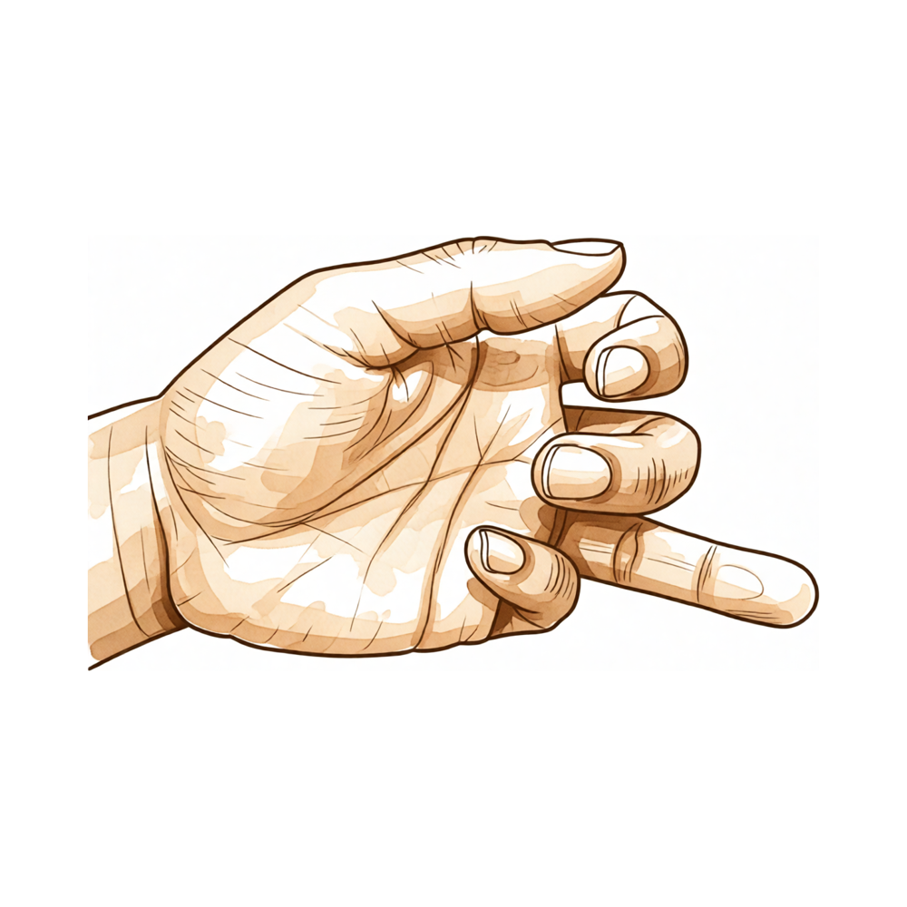
Jersey finger results from forced hyperextension of the distal interphalangeal (DIP) joint while the finger is in active flexion [37]. This mechanism causes rupture of the flexor digitorum profundus (FDP) tendon at its insertion into the distal phalanx, which is its physiologically weakest point [37]. For flexor tendons to rupture, the tensile strength must be reduced by at least 50% before the application of the distracting force [8]. The injury occurs more commonly in the ring finger, accounting for 75% of cases, but can occur in any digit [37]. It is common amongst rugby and American football players [37]. Closed rupture of both flexor tendons in the little finger is rare and may be missed on initial presentation [20].
Physical Examination
The pathognomonic finding in the physical examination of jersey finger is the inability to actively flex the DIP joint [37]. A patient with a zone I rupture of the FDP tendon caused by blunt trauma may present with an inability to flex the distal phalanx and a digit that is passively hyperextended [142]. Clinicians must distinguish acute injury from congenital anomalies; a patient with unilateral absence of the ring finger FDP musculotendinous structure can present with a history and examination suggesting an acute avulsion of the ring finger FDP tendon [33].
Imaging
On radiographs, distal tendon stumps can often be identified if they are attached to a bony fragment [37]. Misleading fractures can occur after profundus tendon avulsions [26]. Real-time ultrasound can be used to determine whether a tendon is ruptured or entrapped in scar tissue [142]. Ultrasound evaluation of flexor tendon lacerations is a described diagnostic modality [19]. Magnetic resonance imaging scanning has been used in the diagnosis of zone II flexor tendon rupture [11].
Investigations
Ultrasound: Ultrasound differentiates between pulley strain, partial rupture, complete rupture, or multiple ruptures [179]. In a case of stage 2 flexor digitorum profundus tendon avulsion, ultrasonography was used to map out the surgical strategy beforehand [176].
Magnetic Resonance Imaging: Magnetic resonance imaging is used to differentiate between pulley strain, partial rupture, complete rupture, or multiple ruptures [179].
Other Considerations: A patient with unilateral absence of the ring finger flexor digitorum profundus musculotendinous structure can present with a history and examination suggesting an acute avulsion, posing a diagnostic challenge [33]. Flexor digitorum profundus avulsion with a concomitant and separate intraarticular fracture of the distal phalanx is a rare and frequently misdiagnosed problem [66]. The Jersey finger variety combining an articular fracture of the base of the proximal phalanx and flexor digitorum profundus avulsion remains exceptional and can often go unnoticed [84].
Central slip ruptures often are missed at the initial presentation, with patients frequently returning with a boutonniere deformity 2 to 3 weeks later [59]. Clinical tests for early diagnosis of boutonniere injury are not always diagnostic because results are clouded by motion of a painful and swollen joint [59]. PIP extensor lag and difficulty of active and passive flexion of the DIP joint may only appear weeks after injury [59].
Treatment
Operative
Indications: The central tenet of modern flexor tendon surgery is to repair and move divided flexor tendons within a few days of injury [35]. The results of correctly rehabilitated primary repairs are likely to be the best attainable [35]. Currently, the basis for the best results from flexor tendon repair are strong gap resistant suture technique and early controlled motion in therapy [36]. The surgical techniques of hand surgeons and the rehabilitation protocols of hand therapists have improved enormously from scientific efforts and the results of flexor tendon repair have become much more reliable [75].
Surgical Approach / Technique: The double armed suture method of repairing flexor tendons allows almost immediate mobilization of the involved digits [119]. Good or excellent results have been obtained in 90% of cases using the double armed suture technique over a 16-year period [119]. In a series of 12 fingers, 92% achieved excellent or good results using the International Federation of Societies for Surgery of the Hand evaluation system with the modified Kleinert splint [10, 17]. Eleven flexors in zone II showed excellent results in 10 and good results in 1 by the Strickland formula using the double loop locking suture technique [45]. The six-strand repair technique is an effective procedure to assure early active motion after flexor pollicis longus tendon injuries and good results can also be achieved by omitting the circumferential suture [153].
Zone-Specific and Avulsion Management: Hook plates placed in the distal phalanx emerge as surgical treatment for FDP avulsion types II and III in flexor zone 1, with excellent clinical outcomes and no associated morbidity [28]. Flexor tendons repair in zone 1 is classically considered providing good results with an overall satisfactory finger function [30]. The Simple Wire Interosseous Fixation Technique (SWIFT) can be safely used to manage other phalangeal avulsion fractures in addition to FDP avulsions with a large bony fragment [85]. The simple interosseus fixation technique is recommended to attach the avulsed FDP tendon to the healthy distal portion of the distal phalanx in patients who do not wish to have a bone graft used or in which bone grafts are contraindicated [148]. This is the first clinical report of a combined rupture of the flexor pulleys and avulsion of the flexor digitorum superficialis tendon, which should be regarded as a single pathology caused by sudden forceful contraction [18]. The authors report a case of simultaneous rupture of both flexor tendons in the ring finger, noting that full recovery of function was achieved by a two-stage tendon graft [1]. Flexor tendon repair was followed by good function in a case of simultaneous dislocation of both interphalangeal joints and flexor tendon tear [3]. The authors present a simple technique using retrograde catheters to successfully reduce delayed flexor tendon avulsions, noting it has been used successfully in eight cases [46]. Secondary therapeutic modalities are varied and depend on finger function and patient requirements [81].
Rehabilitation and Mobilization: Early passive motion appeared to be an effective technique to improve the results of flexor tendon repairs in zone II [44]. In a comparison of 50 digits, the immobilization group had four ruptures with no excellent results, while the early mobilization group had one rupture with 36% excellent and 20% good results [44]. The author recommends the partial-range active flexion protocol as a safe, efficient, and generalizable framework for rehabilitation after flexor tendon repair and other hand disorders, particularly where therapist assistance is unavailable [77]. Early active motion of simple primary flexor tendon repair is now accepted practice, provided that the repair is robust and the patient complies with the rehabilitation programme [115]. When employing an active motion regimen, safety is paramount and the repair must be protected from gapping or rupture by minimising the work of flexion and loading the tendon only within the ‘safe zone’ [115]. Available biomechanical studies concerning finger extension strength, tendon repair strength, tendon shortening, resultant loss of motion, and gliding capability have validated controlled motion as biomechanically sound utilizing contemporary repair techniques [55]. The outcome of a flexor tendon repair is influenced by many factors that cannot be controlled intraoperatively [29]. Healing the flexor tendon takes about 3 months, a period which is sometimes longer than that for which the hand can be kept free of activities or accidents liable to snap the repair [35]. The main obstacle to the restoration of gliding function is the tendency for restrictive adhesions to form between the site of tendon repair and the surrounding tissues [57]. It is necessary that new collagen fibres be laid down in sufficient quantity between the tendon stumps and that they mature to provide adequate tensile strength for the repair [57]. Fibres forming adhesions with surrounding tissues must be kept to a minimum and should be gradually absorbed or become elongated as healing progresses if movement of the tendon is to take place [57]. The Manchester short splint represents a change to splinting practice in the rehabilitation of zone II flexor tendon repairs [115]. A Cochrane review concluded that there was no firm evidence to define the optimum mobilisation strategy [115]. Rehabilitation programmes should be customised according to multiple factors including severity of injury, quality of the repair and the patient characteristics [115].
Secondary Reconstruction and Grafting: The overall results in scarred and in unscarred fingers for staged flexor tendon grafting for isolated profundus loss were no better than the results reported for single-staged flexor tendon grafting through an intact superficialis [76]. The technique of flexor tendon retrieval has been used successfully over a period of 3 years on over 20 flexor tendon injuries [6]. Three patients with zone 2 lacerations of the flexor digitorum profundus of the little finger were found to have proximal coiling of the tendon stump in the palm due to inadequate immobilisation [27]. Factors affecting the results after flexor tendon repair in zone 2 include the mechanism of injury (clean-cut versus crush), associated fractures, the number of digits/tendons injured, age, the experience of the surgeon, and the type of postoperative mobilization programme [23]. A large part of the variance in the outcome of flexor tendon repair in zone 2 is thought to be related to the psychological and biologic characteristics of the patient [23]. Zone 2 lacerations of both flexor tendons of all fingers in the same patient is rare [23]. The responses indicate some general tendencies in the treatment of partial digital flexor tendon lacerations, although wide variation does exist [42]. Rupture of flexor tendons with major divisions (>50% of the width of the tendon) is unlikely as long as protected mobilization is used during the first 4 weeks after injury [40]. Sutures of a partially cut tendon actually result in a reduction of the tensile strength of the tendon in vivo [40]. The in vivo mean tensile strengths of nonsutured tendons are significantly higher than that of the sutured tendons at both 2 and 4 weeks [40]. The bulbous scar theory was proposed by Kleinert, who stated that allowing a partially severed tendon to go unsutured invites the formation of a bulbous scar [40].
Complications
Repair Rupture and Reoperation
Ruptures and adhesions around the repaired tendon requiring tenolysis occur in approximately 5% of cases each [159]. Further surgery for rupture or adhesions is necessary in around 6% of cases [159]. In a series of 50 consecutive digits in Zone II, four ruptures occurred in the immobilization group compared to one rupture in the early mobilization group [44]. Age, smoking, zone of injury, multiple finger injuries, associated flexor digitorum superficialis (FDS) tendon injury, nerve injuries, and delayed treatment have previously been reported to adversely affect outcomes [159]. A separate study identified age, sex, and type of tendon injury as risk factors for reoperation after finger flexor surgery [162].
Adhesions and Functional Limitations
The complications of rupture and adherence of repairs during healing continue to trouble surgeons to an extent that the result of every primary flexor tendon repair remains uncertain [35]. The body does not limit the healing process to injured structures, resulting in all nearby tissues becoming 'spot-welded' together by scar adhesions [35]. This 'spot-welding' can occur anywhere along the length of a flexor tendon but is a particular problem in the digits where flexors move within a fine system [35].
Diagnostic and Presentation Complications
Unilateral absence of the ring finger FDP musculotendinous structure can pose a diagnostic challenge when the history and examination suggest an acute avulsion of the ring finger FDP tendon [33]. All flexor digitorum profundus tendon avulsions should be surgically repaired as soon as possible [26].
Associated Injuries and Variants
In a study of 80 closed flexor tendon ruptures, 17 patients had simultaneous deep and superficial flexor tendon ruptures, only two of which were ruptures of normal tendons [8]. A combined rupture of the flexor pulleys and avulsion of the flexor digitorum superficialis tendon should be regarded as a single pathology caused by sudden forceful contraction [18]. Triggering is a complication that can occur after an unrepaired partially-cut flexor tendon [68].
Recovery
Functional milestones: Outcome data for flexor tendon injuries in the finger and thumb demonstrate variable recovery trajectories depending on the injury pattern and surgical technique. In a series of 99 patients treated soon after injury, 88% achieved excellent or good results at six months following early mobilisation of acute middle slip injuries [180]. For zone II flexors repaired with double loop locking sutures, 10 of 11 fingers showed excellent results and one showed good results by the Strickland formula [45]. The modified Kleinert splint technique yielded excellent or good results in 92% of a series of 12 fingers, assessed using the International Federation of Societies for Surgery of the Hand evaluation system [10]. In cases of simultaneous rupture of both flexor tendons in the ring finger, full recovery of function was achieved via a two-stage tendon graft [1]. Similarly, good function followed flexor tendon repair in a case involving simultaneous dislocation of both interphalangeal joints and a flexor tendon tear [3].
Other Considerations: Specific functional benchmarks have been documented at defined post-operative intervals. Six months after operation, grip strength returned to normal in a patient with traumatic rupture of the profundus tendon proximal to the lumbrical origin [73]. At 6 months, complete and total recovery of active flexion amplitudes of the proximal and distal interphalangeal joints was observed in 4 long fingers following secondary surgery using silicone infusion tubing [154]. Regarding complication rates that may impact recovery, repair ruptures were documented in most reports with rates ranging from 4%-10% in finger flexors [5] and 3%-17% in the FPL of thumbs [5]. Comparative data indicate that overall results in scarred and unscarred fingers for staged flexor tendon grafting were no better than the results reported for single-staged flexor tendon grafting through an intact superficialis [76].
Key Evidence
- [L5] The authors report a case of simultaneous rupture of both flexor tendons in the ring finger, noting that full recovery of function was achieved by a two-stage tendon graft. [1] (10.1016/s0363-5023(96)80330-3)
- [L5] Jersey finger injuries should be promptly diagnosed to allow timely surgical repair and prevention of complications. [2] (10.1016/j.csm.2019.12.004)
- [L5] Flexor tendon repair was followed by good function. [3] (10.1016/s0020-1383(97)00202-7)
- [L5] The force required to make a fist should be taken into account when considering the limit of 'safe' tendon shortening in delayed repair of jersey finger injuries. [4] (10.1177/1753193415585311)
- [L4] Repair ruptures were documented in most reports with rates ranging from 4%-10% in finger flexors and 3%-17% in FPL of thumbs. [5] (10.1016/j.hcl.2004.11.005)
- [L4] The technique has been used successfully over a period of 3 years on over 20 flexor tendon injuries. [6] (10.1016/j.jhse.2007.03.011)
- [L5] [8] (10.1016/j.jhsb.2006.07.014)
- [L4] In a series of 12 fingers, 92% achieved excellent or good results using the International Federation of Societies for Surgery of the Hand evaluation system. [10] (10.1016/0266-7681(88)90084-8)
- [L5] The author posits that the thumb tendon has a greater tendency to rupture than finger tendons because it provides about 50% of hand function and cannot be spared from use, unlike fingers which can share load. [12] (10.1054/jhsb.2000.0421)
- [L4] In this series of 12 fingers, 92% achieved excellent or good results using the International Federation of Societies for Surgery of the Hand evaluation system. [17] (10.1016/0266-7681_88_90084-8)
- [L4] This is the first clinical report of a combined rupture of the flexor pulleys and avulsion of the flexor digitorum superficialis tendon, which should be regarded as a single pathology caused by sudden forceful contraction. [18] (10.1054/jhsb.2001.0559)
- [L4] Closed rupture of both flexor tendons in the little finger is rare and may be missed on initial presentation. [20] (10.1177/1753193409356033)
- [L4] [23] (10.1177/1753193410387333)
- [L4] Therefore all flexor digitorum profundus tendon avulsions should be surgically repaired as soon as possible. [26] (10.1016/0363-5023(92)90465-2)
- [L4] Three patients with zone 2 lacerations of the flexor digitorum profundus of the little finger were found to have proximal coiling of the tendon stump in the palm due to inadequate immobilisation. [27] (10.1016/0266-7681(89)90158-7)
- [L4] Hook plates placed in distal phalanx emerge as surgical treatment for FDP avulsion types II and III in flexor zone 1, with excellent clinical outcomes and no associated morbidity. [28] (10.1177/1558944720957730)
- [L4] The outcome of a flexor tendon repair is influenced by many factors that cannot be controlled intraoperatively. [29] (10.1016/j.jhsa.2022.01.015)
- [L5] Flexor tendons repair in zone 1 is classically considered providing good results with an overall satisfactory finger function. [30] (10.1016/j.main.2014.03.002)
- [L4] This case illustrates a patient with unilateral absence of the ring finger FDP musculotendinous structure, which can pose a diagnostic challenge when the history and examination suggest an acute avulsion of the ring finger FDP tendon. [33] (10.1016/j.jhsa.2016.02.003)
- [L5] [35] (10.1054/jhsb.2002.0800)
- [Paper] Currently, the basis for the best results from flexor tendon repair are strong gap resistant suture technique and early controlled motion in therapy. [36] (10.1197/j.jht.2005.02.015)
- [L4] [37] (10.1016/j.hansur.2017.06.002)
- [L3] [40] (10.1053/jhsu.2000.18486)
- [L4] The responses indicate some general tendencies in the treatment of partial digital flexor tendon lacerations, although wide variation does exist. [42] (10.1016/s0363-5023(05)80023-1)
- [L3] [44] (10.1016/s0363-5023(80)80101-8)
- [L4] Eleven flexors in zone II showed excellent results in 10 and good results in 1 by the Strickland formula. [45] (10.1016/0363-5023(90)90022-j)
- [L4] The authors present a simple technique using retrograde catheters to successfully reduce delayed flexor tendon avulsions, noting it has been used successfully in eight cases. [46] (10.1177/1753193407087867)
- [L4] In addition, available biomechanical studies concerning finger extension strength, tendon repair strength, tendon shortening, resultant loss of motion, and gliding capability have validated controlled motion as biomechanically sound utilizing contemporary repair techniques. [55] (10.1197/j.jht.2005.01.006)
- [Paper] [57] (10.1016/0266-7681(89)90150-2)
- [L5] The breaking loads of both 50%- and 75%-lacerated tendons far exceeded the in vivo forces measured in human flexor tendons during unresisted active finger movement (up to 34 N). [58] (10.1016/s0363-5023(97)80040-8)
- [L5] [59] (10.1016/s0363-5023(03)00427-1)
- [L4] Avulsion of the flexor digitorum profundus with a concomitant and separate intraarticular fracture of the distal phalanx is a rare and frequently misdiagnosed problem. [66] (10.1177/036354658701500413)
- [L5] It provides another mechanical cause for triggering of a partial tendon laceration and yet another example of the complication of triggering after an unrepaired partially-cut flexor tendon. [68] (10.1016/0266-7681(89)90159-9)
- [L3] Both bridge tendon grafts and end-to-side tendon transfers have comparable and satisfactory outcomes after surgical treatment of closed rupture of the flexor tendons in the little finger. [70] (10.1177/17531934211073751)
- [L5] Six months after operation, the patient was pleased with the function of the long finger and his grip strength has returned to normal. [73] (10.1016/0363-5023(90)90067-2)
- [L5] The improvement of primary flexor tendon repair has limited the indications of secondary repairs to neglected initial wound care and deficiencies in the surgical technique or rehabilitation. [74] (10.1016/j.main.2014.05.007)
- [L5] The surgical techniques of hand surgeons and the rehabilitation protocols of hand therapists have improved enormously from these scientific efforts and the results of flexor tendon repair have become much more reliable. [75] (10.1197/j.jht.2005.01.013)
- [L4] The overall results in scarred and in unscarred fingers were no better than the results reported for single-staged flexor tendon grafting through an intact superficialis. [76] (10.1016/0266-7681(86)90267-6)
- [L5] The author recommends the partial-range active flexion protocol as a safe, efficient, and generalizable framework for rehabilitation after flexor tendon repair and other hand disorders, particularly where therapist assistance is unavailable. [77] (10.1177/17531934211037112)
- [L4] Secondary therapeutic modalities are varied and depend on finger function and patient requirements. [81] (10.1016/j.main.2009.07.001)
- [L5] The Jersey finger variety combining an articular fracture of the base of P3 and FDP avulsion remains exceptional and can often go unnoticed. [84] (10.1016/j.main.2007.08.007)
- [L4] In addition, our method can also be safely used to manage other phalangeal avulsion fractures. [85] (10.1097/bth.0b013e31826ce920)
- [Paper] Understanding the mechanics of human dexterity requires an appreciation of the kinetic chains that comprise the hand and the intricate interplay of muscles and ligaments that control its movements. [87] (10.1053/jhsu.2002.28764)
- [L5] The intrinsic muscles of the hand are essential for the normal balance, kinetics, power, and position of the fingers. [102] (10.1197/j.jht.2005.02.008)
- [L4] The thumb is the most important single digit in the hand and must have maximal mobility at both the metacarpophalangeal and interphalangeal joints for normal grasp and pinch. [106] (10.1016/0266-7681(89)90157-5)
- [L4] [115] (10.1177/1758998314533306)
- [L4] [119] (10.1016/0363-5023(92)90129-d)
- [Case_report] [142] (10.1016/j.jhsa.2007.09.018)
- [L5] The authors propose a modification to the flexor tendon avulsion classification to incorporate this injury pattern of simultaneous FDP and FDS avulsion with a large fracture, allowing for independent classification of each tendon injury to guide management. [146] (10.1055/s-0039-1688679)
- [L5] We recommend the use of the simple interosseus fixation technique to attach the avulsed FDP tendon to the healthy distal portion of the distal phalanx particularly in those patients that do not wish to have a bone graft used or in those in which bone grafts are contraindicated. [148] (10.1142/s242483552072008x)
- [L5] [149] (10.1097/bth.0000000000000049)
- [L3] Six-strand repair technique is an effective procedure to assure early active motion after flexor pollicis longus tendon injuries and good results can also be achieved by omitting the circumferential suture. [153] (10.1177/15589447211057295)
- [L5] At 6 months, very significant progress was observed with complete and total recovery of active flexion amplitudes of the proximal and distal interphalangeal joints of 4 long fingers. [154] (10.1016/j.hansur.2018.10.074)
- [L3] [159] (10.1177/1753193416657758)
- [L3] This study identified several risk factors for reoperation after finger flexor surgery, including age, sex, and type of tendon injury. [162] (10.1177/17531934221101563)
- [L4] The authors propose the inclusion of a further subtype, 'Type 5c', for a closed avulsion of the flexor digitorum profundus insertion where the fracture is intraarticular but does not extend to the dorsal cortex and the tendon retracts under the A2 pulley. [163] (10.1054/jhsb.2001.0749)
- [L5] [165] (10.1016/0363-5023(90)90021-i)
- [L5] Active motion regimens are desirable after repair of the flexor tendons in all zones but are not always suitable for all extensor tendon repairs. [174] (10.1016/j.injury.2013.01.022)
- [L5] In this case of stage 2 FDP tendon avulsion, the authors mapped out the surgical strategy beforehand using ultrasonography at the surgeon’s practice. [176] (10.1016/j.hansur.2021.03.019)
- [L5] Ultrasound and magnetic resonance imaging are used to differentiate between pulley strain, partial rupture, complete rupture, or multiple ruptures. [179] (10.1016/j.jhsa.2006.02.011)
- [L4] In a series of 99 patients seen soon after injury, 88% had excellent or good results after six months. [180] (10.1016/0266-7681(90)90079-j)
See Also
References
[1] Simultaneous rupture of both flexor tendons in a finger. The Journal of Hand Surgery. 1996. DOI: 10.1016/s0363-5023(96)80330-3
[2] Evaluation and Treatment of Flexor Tendon and Pulley Injuries in Athletes. Clinics in Sports Medicine. 2020. DOI: 10.1016/j.csm.2019.12.004
[3] Simultaneous dislocation of both interphalangeal joints and flexor tendon tear in a finger. Injury. 1998. DOI: 10.1016/s0020-1383(97)00202-7
[4] The effect of flexor digitorum profundus tendon shortening on jersey finger surgical repair: a cadaveric biomechanical study. Journal of Hand Surgery (European Volume). 2015. DOI: 10.1177/1753193415585311
[5] Clinical Outcomes Associated with Flexor Tendon Repair. Hand Clinics. 2005. DOI: 10.1016/j.hcl.2004.11.005
[6] Flexor Tendon Retrieval—Another Way. Journal of Hand Surgery (European Volume). 2007. DOI: 10.1016/j.jhse.2007.03.011
[8] Multiple Closed Flexor Tendon Avulsions from Their Insertions by a High-Energy Explosion. Journal of Hand Surgery. 2006. DOI: 10.1016/j.jhsb.2006.07.014
[10] The modified kleinert splint in zone II flexor tendon injuries. The Journal of Hand Surgery: Journal of the British Society for Surgery of the Hand. 1988. DOI: 10.1016/0266-7681(88)90084-8
[11] Campbell S Operative Orthopaedics 4 Volume Set. RECONSTRUCTION OF FINGER FLEXORS: SINGLE-STAGE TENDON GRAFT > FLEXOR TENDON INJURIES IN CHILDREN.
[12] Letters to the Editor. Journal of Hand Surgery. 2000. DOI: 10.1054/jhsb.2000.0421
[17] The Modified Kleinert Splint in Zone II Flexor Tendon Injuries. Journal of Hand Surgery. 1988. DOI: 10.1016/0266-7681_88_90084-8
[18] Closed Traumatic Rupture of the Flexor Pulleys of a Long Finger Associated with Avulsion of the Flexor Digitorum Superficialis. Journal of Hand Surgery. 2001. DOI: 10.1054/jhsb.2001.0559
[19] Campbell S Operative Orthopaedics 4 Volume Set. RECONSTRUCTION OF FINGER FLEXORS: SINGLE-STAGE TENDON GRAFT > FLEXOR TENDONS.
[20] Closed ruptures of both flexor tendons of the little finger. Journal of Hand Surgery (European Volume). 2010. DOI: 10.1177/1753193409356033
[23] Zone 2 lacerations of both flexor tendons of all fingers in the same patient. Journal of Hand Surgery (European Volume). 2010. DOI: 10.1177/1753193410387333
[26] Misleading fractures after profundus tendon avulsions: A report of six cases. The Journal of Hand Surgery. 1992. DOI: 10.1016/0363-5023(92)90465-2
[27] Proximal coiling of the profundus tendon after laceration of the finger. The Journal of Hand Surgery: Journal of the British Society for Surgery of the Hand. 1989. DOI: 10.1016/0266-7681(89)90158-7
[28] Hook Plate as a Treatment for Flexor Digitorum Profundus Avulsion Types II and III. HAND. 2020. DOI: 10.1177/1558944720957730
[29] Outcomes of Wide-Awake Flexor Tendon Repairs in 58 Fingers and 9 Thumbs. The Journal of Hand Surgery. 2023. DOI: 10.1016/j.jhsa.2022.01.015
[30] Prise en charge des lésions du tendon fléchisseur profond des doigts en zone 1. Chirurgie de la Main. 2014. DOI: 10.1016/j.main.2014.03.002
[33] Absent Ring Finger Flexor Digitorum Profundus Presenting as a Jersey Finger. The Journal of Hand Surgery. 2016. DOI: 10.1016/j.jhsa.2016.02.003
[35] Primary Flexor Tendon Repair – Operative Repair, Pulley Management and Rehabilitation. Journal of Hand Surgery. 2002. DOI: 10.1054/jhsb.2002.0800
[36] JHT Read for Credit. Journal of Hand Therapy. 2005. DOI: 10.1197/j.jht.2005.02.015
[37] Traumatic avulsion of the flexor digitorum profundus tendon. Jersey finger, a 29 cases report. Hand Surgery and Rehabilitation. 2017. DOI: 10.1016/j.hansur.2017.06.002
[40] Conservative management of zone II partial flexor tendon lacerations greater than half the width of the tendon. The Journal of Hand Surgery. 2000. DOI: 10.1053/jhsu.2000.18486
[42] Clinical management of partially lacerated digital flexor tendons: A surgery of hand surgeons. The Journal of Hand Surgery. 1995. DOI: 10.1016/s0363-5023(05)80023-1
[44] Digital function following flexor tendon repair in Zone II: A comparison of immobilization and controlled passive motion techniques. The Journal of Hand Surgery. 1980. DOI: 10.1016/s0363-5023(80)80101-8
[45] Double loop locking suture: A technique of tendon repair for early active mobilization Part II: Clinical experience. The Journal of Hand Surgery. 1990. DOI: 10.1016/0363-5023(90)90022-j
[46] A New Technique for Reduction of Flexor Tendon Avulsions after Delayed Presentation. Journal of Hand Surgery (European Volume). 2008. DOI: 10.1177/1753193407087867
[48] Miller S Review Of Orthopaedics. 4. Fingertip injuries with tissue loss.
[55] New Perspectives on Extensor Tendon Repair and Implications for Rehabilitation. Journal of Hand Therapy. 2005. DOI: 10.1197/j.jht.2005.01.006
[57] Early mobilisation after flexor tendon repair. The Journal of Hand Surgery: Journal of the British Society for Surgery of the Hand. 1989. DOI: 10.1016/0266-7681(89)90150-2
[58] Partial lacerations of human digital flexor tendons: A biomechanical analysis. The Journal of Hand Surgery. 1997. DOI: 10.1016/s0363-5023(97)80040-8
[59] Ultrasound visualization of central slip injuries of the finger extensor mechanism. The Journal of Hand Surgery. 2003. DOI: 10.1016/s0363-5023(03)00427-1
[66] Flexor digitorum profundus avulsions with associated distal phalanx fractures. The American Journal of Sports Medicine. 1987. DOI: 10.1177/036354658701500413
[68] Triggering secondary to an untreated partially-cut flexor tendon. The Journal of Hand Surgery: Journal of the British Society for Surgery of the Hand. 1989. DOI: 10.1016/0266-7681(89)90159-9
[70] Comparison of bridge graft and end-to-side transfer for treatment of closed rupture of the flexor tendons in the little finger. Journal of Hand Surgery (European Volume). 2022. DOI: 10.1177/17531934211073751
[73] Traumatic rupture of the profundus tendon proximal to the lumbrical origin. The Journal of Hand Surgery. 1990. DOI: 10.1016/0363-5023(90)90067-2
[74] Reconstruction par greffe des tendons fléchisseurs. Chirurgie de la Main. 2014. DOI: 10.1016/j.main.2014.05.007
[75] The Scientific Basis for Advances in Flexor Tendon Surgery. Journal of Hand Therapy. 2005. DOI: 10.1197/j.jht.2005.01.013
[76] Disappointing outcomes in staged flexor tendon grafting for isolated profundus loss. The Journal of Hand Surgery: Journal of the British Society for Surgery of the Hand. 1986. DOI: 10.1016/0266-7681(86)90267-6
[77] Rehabilitation after flexor tendon repair and others: a safe and efficient protocol. Journal of Hand Surgery (European Volume). 2021. DOI: 10.1177/17531934211037112
[81] Avulsion traumatique du fléchisseur profond des doigts. À propos de 20 cas. Chirurgie de la Main. 2009. DOI: 10.1016/j.main.2009.07.001
[84] Avulsion du flexor digitorum profundus combinée à une fracture articulaire de la phalange distale: à propos d'un cas. Chirurgie de la Main. 2007. DOI: 10.1016/j.main.2007.08.007
[85] The Simple Wire Interosseous Fixation Technique (SWIFT) for Reattachment of FDP Avulsions With a Large Bony Fragment. Techniques in Hand & Upper Extremity Surgery. 2012. DOI: 10.1097/bth.0b013e31826ce920
[86] Exam Of The Hand Wrist 2Ed. Movements of the fingers > Articulations of the fingers.
[87] Historical perspective of finger joint motion: The hand-me-downs of our predecessors—the Richard J. Smith Memorial Lecture. The Journal of Hand Surgery. 2002. DOI: 10.1053/jhsu.2002.28764
[88] Exam Of The Hand Wrist 2Ed. Macroscopic appearance of palmar skin > Fingerprints.
[89] Dupuytren S Disease And Related Hyperproliferative Disorders. 2. Palmar Fibromatosis or the Loss of Flexibility of the Palmar Finger Tissue: A New Insight into the Disease Process of Dupuytren Contracture > 2.2 The Skin Anchoring Fibers in the Normal and Diseased Hand > 2.2.1 The External View of the Soft Tissue of the Fingers.
[92] Exam Of The Hand Wrist 2Ed. Movements of the fingers > Normal ulnar inclination of the fingers.
[102] Ulnar Intrinsic Anatomy and Dysfunction. Journal of Hand Therapy. 2005. DOI: 10.1197/j.jht.2005.02.008
[106] Flexor pollicis longus tendon repair: A comparison between dynamic and static splintage. The Journal of Hand Surgery: Journal of the British Society for Surgery of the Hand. 1989. DOI: 10.1016/0266-7681(89)90157-5
[115] The Manchester short splint: A change to splinting practice in the rehabilitation of zone II flexor tendon repairs. Hand Therapy. 2014. DOI: 10.1177/1758998314533306
[119] The double armed suture: Tendon repair with immediate mobilization of the fingers. The Journal of Hand Surgery. 1992. DOI: 10.1016/0363-5023(92)90129-d
[142] Zone I Rupture of the Flexor Digitorum Profundus Tendon Caused by Blunt Trauma: A Case Report. The Journal of Hand Surgery. 2008. DOI: 10.1016/j.jhsa.2007.09.018
[146] An Unusual Pattern of Closed Flexor Tendon Avulsion. Journal of Hand and Microsurgery. 2020. DOI: 10.1055/s-0039-1688679
[148] FDP Avulsion Injury Due to Enchondroma: A Unique Surgical Approach. The Journal of Hand Surgery (Asian-Pacific Volume). 2020. DOI: 10.1142/s242483552072008x
[149] A Novel External Fixator Used to Treat Open Comminuted Fractures of the Middle and Distal Phalanges With Concomitant FDP Avulsion Injury. Techniques in Hand & Upper Extremity Surgery. 2014. DOI: 10.1097/bth.0000000000000049
[153] Six-Strand Flexor Pollicis Longus Tendon Repairs With and Without Circumferential Sutures: A Multicenter Study. HAND. 2022. DOI: 10.1177/15589447211057295
[154] Tubulure de perfusion siliconée en lieu et place de tiges de Hunter dans la chirurgie secondaire des tendons fléchisseurs en zone 2 dans un environnement chirurgical à ressources limitées. Hand Surgery and Rehabilitation. 2018. DOI: 10.1016/j.hansur.2018.10.074
[159] Predictors of outcome after primary flexor tendon repair in zone 1, 2 and 3. Journal of Hand Surgery (European Volume). 2016. DOI: 10.1177/1753193416657758
[162] Risk factors for reoperation after flexor tendon repair: a registry study. Journal of Hand Surgery (European Volume). 2022. DOI: 10.1177/17531934221101563
[163] Reply. Journal of Hand Surgery. 2002. DOI: 10.1054/jhsb.2001.0749
[165] Double loop locking suture: A technique of tendon repair for early active mobilization Part I: Evolution of technique and experimental study. The Journal of Hand Surgery. 1990. DOI: 10.1016/0363-5023(90)90021-i
[174] Rehabilitation of flexor and extensor tendon injuries in the hand: Current updates. Injury. 2013. DOI: 10.1016/j.injury.2013.01.022
[176] Mini-open surgical management of a type-2 jersey finger under WALANT with ultrasound assistance: A case report about a new technique. Hand Surgery and Rehabilitation. 2021. DOI: 10.1016/j.hansur.2021.03.019
[179] Injuries to the Finger Flexor Pulley System in Rock Climbers: Current Concepts. The Journal of Hand Surgery. 2006. DOI: 10.1016/j.jhsa.2006.02.011
[180] Early Mobilisation of Acute Middle Slip Injuries. Journal of Hand Surgery. 1990. DOI: 10.1016/0266-7681(90)90079-j