Clinicians › Shoulder
EnFix Biological Scaffold
For patients: a plain-language version of this topic is available. See the patient guide.
Overview
EnFix implants are procedure-specific allogenic tissue products designed to enhance enthesis healing in rotator cuff repair [1]. As an allograft product, EnFix TAC requires donor screening, aseptic technique, and traceability [17]. The device utilizes patented Demineralized Bone Fiber technology to provide both osteoinductive and osteoconductive properties [1]. Specifically, the demineralized bone matrix implant provides osteoconductive properties to enhance tendon-to-bone healing [16] and provides osteoinductive properties to enhance tendon-to-bone healing [16]. The scaffold is intended to resorb over a period of 3 to 4 months [2].
In preclinical studies, inlay scaffold positioning has been shown to enhance the healing biology of the enthesis [4]. This positioning provides histologic similarities to the native 4-zone configuration of the enthesis [4]. EnFix implants enhance healing at the tendon-bone interface by promoting enthesis reformation [15] and promote Sharpey's fiber formation [15]. Furthermore, the demineralized bone matrix implant promotes enthesis regeneration [16]. These mechanisms represent a significant advance over traditional overlay augmentation products [15].
Anatomy & Pathophysiology
Enthesis & Scaffold Biology
The EnFix scaffold utilizes patented Demineralized Bone Fiber (DBF) technology to provide both osteoinductive and osteoconductive properties [1]. DBF is a form of demineralized bone matrix (DBM) where bone collagen serves as the primary constituent [20]. This matrix contains bone morphogenetic proteins (BMPs) and other growth factors that facilitate healing [20]. Specifically, BMPs stimulate the differentiation of mesenchymal stem cells (MSCs) into osteoblasts and chondrocytes, thereby promoting osteoinduction [20]. The osteoconductive property of DBF enables cells necessary for tissue repair to migrate to the area and provides a structural framework for new tissue formation at the enthesis [20]. Through the porous structure of the DBF scaffold, collagen deposition, neovascularisation, and infiltration of host cells occur [20]. This process achieves the recreation of the four-zone fibrocartilaginous structure of the native enthesis [20].
In preclinical studies, inlay scaffold positioning enhances the healing biology of the enthesis and provides histologic similarities to the native 4-zone configuration [4]. The demineralized bone matrix implant provides osteoconductive and osteoinductive properties to enhance tendon-to-bone healing [16]. Osteoinductive agents induce greater formation of soft tissue and bone ingrowth at the tendon-bone interface, resulting in stronger tendon-to-bone attachment compared with untreated repairs [14]. Experimental evidence supports these mechanisms: DBM use for tendon-to-bone interface healing in rabbit experiments showed good results in gross and histologic analysis [19]. In a canine model, DBM-PRP interposition significantly improved structural healing, biomechanical strength, and histologic integration compared to standard direct repair [54]. Additionally, the subacromial bursa is a major source of endogenous enthesis-related progenitors that contribute to early histological and mechanical maturation after rotator cuff tendon-to-bone repair [23].
Shoulder Anatomy
The shoulder joint is composed of four articulations: sternoclavicular, acromioclavicular, glenohumeral, and scapulothoracic [40]. Normal shoulder motion is approximately two-thirds glenohumeral and one-third scapulothoracic [30]. The scapula is attached to the axial skeleton by the clavicle via the acromioclavicular and sternoclavicular joints [29]. It is separated from the chest wall by thin gliding fibro-fatty tissue and spans second through seventh ribs, serving as an attachment for 17 muscles [29, 41]. The scapula is anteverted on the chest wall approximately 30 degrees relative to the body [41]. The glenoid is connected with the flat body of the scapula by the scapular neck [29]. The coracoid process curves forwards from the superior surface of the scapular neck, while the acromion is a flattened bony process that curves forwards from the scapular spine [29]. The highest concentration of bony mass in the scapula is in the glenoid, scapular neck, and lateral border of the scapular body [29]. Two bony pillars extend between the glenoid and the scapular body to transmit compressive forces from the glenoid fossa: the lateral pillar connects the inferior border of the glenoid with the inferior angle, and the spinal pillar arises from the central part of the glenoid and continues medially to become part of the base of the scapular spine [29]. The weakest bone in the scapula is located primarily in the central part of the biomechanical body, specifically the infraspinous fossa, while the weakest area of the circumference of the biomechanical body is the spinomedial angle [29].
The clavicle is the first bone to ossify at the fifth week of gestation and is the only long bone to ossify by intramembranous ossification [30]. The medial epiphysis of the clavicle is the last ossification center to fuse at age 20 to 25 years [30]. The glenoid is a convex structure of shallow depth shaped like an inverted pear [27]. It is a shallow socket approximately one third the size of the humeral head [28]. The glenoid averages 5 degrees of retroversion in relation to the axis of the scapular body [30, 41]. The glenoid is encircled by the labrum, composed of dense fibrocartilaginous tissue [40]. The labrum provides concavity and up to 50% of marginal glenoid socket depth [30]. It increases the depth of the socket by 50% around the humeral head [40]. The glenoid articular surface and the labrum combine to create a socket approximately 9 mm deep in the superoinferior direction and approximately 5 mm deep in the anteroposterior direction [40]. Adding the glenoid labrum increases the glenoid surface to 75% of the humeral head vertically and 57% of the humeral head horizontally [40]. The glenoid articulates with the humeral head and serves as the attachment for the labrum and joint capsule [27].
The proximal humerus comprises the humeral head, greater tuberosity, lesser tuberosity, and humeral shaft [27]. The articular head of the humerus is spherical with a diameter of 37 to 57 mm [27]. The articular surface of the humeral head is essentially spherical with an arc of approximately 160 degrees covered by articular cartilage [39]. The radius of curvature of the humeral head is approximately 25 mm [39]. The glenoid articular surface radius of curvature is 2 to 3 mm larger than that of the humeral head [39]. The most superior portion of the articular surface of the humeral head averages 8 mm above the greater tuberosity [27]. The superior margin of the humeral head articular surface is normally superior to the top of the greater tuberosity by 8 to 10 mm [39]. Head height is approximately 5.6 cm above the superior border of the pectoralis major tendon [41]. The anatomic neck lies at the junction of the head and the tuberosities and is directly below the humeral head, serving as an attachment for the shoulder capsule [28, 41]. The surgical neck lies below the greater and lesser tuberosities [28]. The bicipital groove lies between the greater and lesser tuberosities and serves as a pathway for the long head of the biceps [27].
Humeral version averages 29.8 degrees with a range of 10 to 55 degrees [27]. Mean humeral retroversion is around 26 degrees in healthy adults [34]. The humeral head is retroverted an average of 30 degrees relative to the transepicondylar axis of the humerus [28, 41]. Humeral retroversion averages 65 degrees in infants and young children [34]. The humeral head is inclined approximately 130 degrees with respect to the humeral shaft [27]. The neck-shaft angle measures an average of 135 degrees [28]. The average neck-shaft angle is 45 degrees with a range of 30 to 50 degrees [39]. The distance from the lateral base of the coracoid process to the lateral margin of the greater tuberosity is called the lateral humeral offset [39]. A significant decrease in lateral humeral offset reduces the lever arms for the deltoid and supraspinatus muscles, while a significant increase in lateral humeral offset causes excessive tension on the soft tissues [39].
The proximal humerus has three ossification centers: for the greater and lesser tuberosities and for the humeral head [34]. The ossification center for the humeral head is usually present at birth [34]. The greater tuberosity ossification center appears by 1 to 3 years of age, and the lesser tuberosity ossification center appears by 5 years of age [34]. The proximal humeral physis closes by 14 to 17 years of age in girls and by 16 to 18 years in boys [34]. The greater tuberosity serves as the attachment site for the supraspinatus, infraspinatus, and teres minor tendons [27]. The lesser tuberosity serves as the attachment site for the subscapularis tendon [27]. The rotator cuff is a sheet of conjoined tendons closely applied over the shoulder capsule [35]. It consists of the subscapularis, supraspinatus, infraspinatus, and teres minor muscles [28]. The teres major is not a rotator cuff muscle [28]. The rotator cuff is made up of subscapularis in front, supraspinatus above, and infraspinatus and teres minor behind [35]. The rotator cuff inserts mainly into the greater tuberosity of the humerus [35]. The subscapularis tendon inserts into the lesser tuberosity [35]. The subscapularis originates from the anterior scapula and inserts anteriorly onto the lesser tuberosity [34]. The supraspinatus, infraspinatus, and teres minor originate from the posterior scapula and attach to the greater tuberosity [34].
The rotator cuff stabilizes the head of the humerus by pulling it firmly into the glenoid whenever the deltoid lifts the arm forwards or sideways [35]. The rotator cuff muscles serve as depressors of the humeral head to allow the deltoid to efficiently abduct the humerus [28]. The rotator cuff stabilizes the glenohumeral joint while allowing greater freedom of motion [39]. It fixes the fulcrum of the upper extremity against which the deltoid can contract and elevate the humerus [39]. The infraspinatus and teres minor are external rotators, while the subscapularis is an internal rotator of the humerus [28]. The tendons of the infraspinatus and supraspinatus muscles join approximately 15 mm proximal to their insertion [40]. The infraspinatus and teres minor fuse near their musculotendinous junctions [40]. The supraspinatus and subscapularis tendons join as a sheath that surrounds the biceps tendon at the entrance of the bicipital groove [40]. The deltoid courses from the clavicle and acromion superiorly into a common tendinous insertion onto the lateral upper third of the humeral shaft [34]. The pectoralis major inserts anteriorly onto the lateral wall of the bicipital groove [34].
The coracoacromial arch is a fibro-osseous canopy formed by the acromion process, coracoid process, and coracoacromial ligament [35, 27]. The rotator cuff, subacromial bursa, and subdeltoid bursa pass underneath the coracoacromial arch [27]. The subacromial bursa separates the rotator cuff tendons from the coracoacromial arch and allows them to glide [35]. The subacromial bursa is one of two bursae in the shoulder region with particular clinical importance [31]. The subscapular bursa lies between the subscapularis tendon and the neck of the scapula and communicates with the joint cavity between the superior and middle glenohumeral ligaments [31]. The capsule of the glenohumeral joint extends from the glenoid rim, progressing laterally toward the surgical neck of the humerus and blending with the tendons of the rotator cuff musculature [34]. Static stability of the glenohumeral joint is maintained by capsulolabral attachments and intracapsular ligamentous thickenings [34]. The rotator cuff and periscapular musculature afford dynamic stability to the glenohumeral joint [34]. The glenohumeral joint depends on static and dynamic stabilizers for movement and stability [39].
The superior glenohumeral ligament is the primary restraint to inferior humeral subluxation in 0 degrees of abduction [40]. The middle glenohumeral ligament limits external rotation when the arm is in the lower and middle ranges of abduction [40]. The inferior glenohumeral ligament is composed of an anterior band, a posterior band, and a thinner intervening axillary pouch [40]. The anteroinferior glenohumeral ligament complex is the main stabilizer to anterior and posterior stresses when the shoulder is abducted 45 degrees or more [40]. The coracohumeral ligament is a thick band of fibrous tissue extending from the coracoid process along the surface of the capsule to the tuberosities [40]. The rotator interval is defined medially by the base of the coracoid, superiorly by the supraspinatus tendon, and inferiorly by the subscapularis tendon [30]. The rotator interval contains the coracohumeral ligament, superior glenohumeral ligament, and intra-articular portion of the long head of the biceps tendon [30].
The humeroscapular motion interface lies between the inner structures of the proximal humerus, rotator cuff, coracohumeral ligament, and biceps tendon sheath and the superficial layer of the acromion, deltoid, coracoacromial ligament, coracoid process, and conjoined tendon [33]. The axillary nerve is a terminal branch coming off the posterior cord of the brachial plexus just proximal to the coracoid process [33]. It passes beneath the conjoined tendon anterior to the subscapularis 3 to 5 mm medial to the musculotendinous junction and enters the quadrilateral space posteriorly [33]. The anterior and middle deltoid muscle receives sole innervation from the anterior branch of the axillary nerve [33]. The posterior deltoid muscle innervation varies, with supply from both branches in 89.1% of cases [33].
The proximal humerus receives its blood supply from the anterior and posterior humeral circumflex branches from the third division of the axillary artery [27]. The major blood supply to the humeral head is through the ascending branch of the anterior humeral circumflex artery [28]. The anterior humeral circumflex artery provides vascular inflow to the humeral head by way of its terminal anterolateral branch known as the artery of Laing [27]. The artery of Laing is also known as the arcuate artery [27]. The ascending branch of the anterior humeral circumflex artery penetrates the head at the bicipital groove and becomes the arcuate artery [28]. The anterolateral ascending branch of the anterior humeral circumflex artery provides the primary blood supply to the humeral head [30]. The terminal intraosseous portion of the anterior humeral circumflex artery enters at the proximal aspect of the intertubercular groove as the arcuate artery [30]. 64% of the humeral head blood supply arises from the posterior humeral circumflex artery [34].
Classification
Regulatory and Material Classification: EnFix implants are classified as procedure-specific allogenic tissue products [1]. These devices utilize patented Demineralized Bone Fiber (DBF) technology [1]. As a form of demineralized bone matrix (DBM), DBF contains bone collagen as the primary constituent, along with bone morphogenetic proteins (BMPs) and other growth factors known to facilitate healing [20]. The BMPs present in DBF stimulate the differentiation of mesenchymal stem cells (MSCs) into osteoblasts and chondrocytes [20].
Biological and Histologic Classification: The demineralized bone matrix implant provides both osteoconductive and osteoinductive properties [16]. The porous structure of DBF functions as a scaffold for collagen deposition, neovascularisation, and infiltration of host cells [20]. In preclinical studies, inlay scaffold positioning provides histologic similarities to the native 4-zone configuration [4]. Furthermore, EnFix implants promote enthesis reformation and Sharpey's fiber formation [15].
Other Considerations: The EnFix TAC allograft is subject to donor screening, aseptic technique, and traceability requirements [17].
Clinical Presentation
EnFix implants facilitate biological integration by promoting enthesis reformation and Sharpey's fiber formation at the tendon-bone interface [15]. The porous structure of the dehydrated bone fragment (DBF) scaffold allows for collagen deposition, neovascularisation, and infiltration of host cells [20]. Bone morphogenetic proteins (BMPs) within the DBF are primarily responsible for stimulating the differentiation of mesenchymal stem cells into osteoblasts and chondrocytes [20].
Radiologic assessment at 6 months postoperatively demonstrated complete healing in the augmented group [20]. This group also exhibited a trend towards more robust tendon quality compared to the non-augmented group [20]. Furthermore, radiologic evaluation showed greater surface area of tendon encroachment onto bone in the augmented group relative to the non-augmented group [20]. Neither the augmented nor non-augmented group experienced any re-tears during the 6-month postoperative period [20].
Clinical outcomes regarding pain, function, active range of motion, and strength were comparable between the augmented and non-augmented cohorts [20]. However, the augmented group showed promising early results with significantly better ASES scores [20], significantly better Constant scores [20], and significantly better UCLA scores [20] compared to the non-augmented group. Additionally, the augmented group demonstrated a trend of achieving greater rotator cuff relative strengths compared to the non-augmented group [20].
In cases of failure, patients experienced significantly less improvement in pain compared to the non-failure group [24]. The failure group also had significantly less improvement in functional scores [24] and significantly less improvement in abduction compared to the non-failure group [24].
Investigations
Preoperative Imaging
Plain radiography: Standardized plain films are almost always sufficient to garner the information needed for shoulder care, and there is information that can be gathered from properly taken plain films that cannot be obtained from CT scans [26]. At least two X-ray views should be obtained for shoulder imaging: an anteroposterior in the plane of the glenoid and an axillary projection with the arm in abduction to show the relationship of the humeral head to the glenoid [44]. The first key radiographic view is the anteroposterior (AP) in the plane of the scapula, which shows the superoinferior position of the humeral head relative to the glenoid, the presence of osteophytes, narrowing of the joint space, and the degree of medial displacement of the humerus [26]. The second key radiographic view is the axillary view taken with the arm in the functional position of elevation in the plane of the scapula, referred to as the "truth view" because it demonstrates glenohumeral relationships in the functional position [26]. The standardized axillary truth view enables the measurement of posterior subluxation or "functional decentering" that is not evident in images taken with the arm at the side [26].
CT: CT imaging is frequently used to evaluate fractures of the shoulder, to assess for bony lesions in recurrent instability cases, or for preoperative templating for shoulder arthritis [50]. CT with three-dimensional reconstructions is the advanced imaging study of choice for determining the extent of glenoid bone loss in the setting of shoulder instability [51].
MRI: MRI is the modality of choice for evaluating the rotator cuff, biceps, and subacromial/subdeltoid bursa [50]. T2-weighted MRI provides better visualization of full thickness rotator cuff tears [50]. MR arthrography is considered the benchmark for evaluation for labral tears and rarely is indicated for evaluation of rotator cuff pathology [50]. The sensitivity of MRI for the detection of full-thickness rotator cuff tears is 100% [51]. The specificity of MRI for the detection of full-thickness rotator cuff tears is 68% [51]. The negative predictive value of MRI for the detection of full-thickness rotator cuff tears is 100% [51].
Ultrasonography: Ultrasonography is a low-cost alternative to MRI and arthrography for evaluating both skeletal and soft-tissue structures of the shoulder [50]. Ultrasonography can provide immediate, real-time visualization of the rotator cuff, biceps tendon, and calcific deposits [50]. The sensitivity of ultrasonography for the detection of full-thickness rotator cuff tears is 98% [51]. The specificity of ultrasonography for the detection of full-thickness rotator cuff tears is 80% [51]. Ultrasonography is highly operator dependent and is not as useful for evaluating labral tears or rotator cuff tears that are very small or larger than 3 cm [50].
Postoperative Imaging
Radiologic assessment at 6 months postoperatively demonstrated complete healing with a trend towards more robust tendon quality and greater surface area of tendon encroachment onto bone in the augmented group compared to the non-augmented group [20]. Neither group experienced any re-tears during the 6-month postoperative period [20]. The 6-month postoperative control scan showed the debridement of the borders of the osteochondrosis dissecans lesion that does not present any signs of progression, with a stable hyaluronate-based scaffold [6].
Treatment
Operative
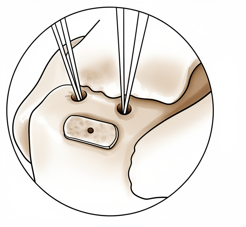
Surgical Approach / Technique: The EnFix biological scaffold is utilized as an interpositional demineralized bone fiber scaffold for the biological enhancement of rotator cuff repair [3]. A single-row technique employing these scaffolds augments rotator cuff biologic healing at the enthesis [11]. This single-row approach provides additional osteoinductive capacity and promotes tendon-bone healing [11]. The interpositional nanofiber scaffold technique allows for optimal placement of the scaffold to maximize its healing properties and achieve full footprint coverage [7].
Implant Selection: EnFix implants represent a significant advance over traditional overlay augmentation products by promoting enthesis reformation [15]. The core material, Demineralized Bone Fiber (DBF), is a form of demineralized bone matrix (DBM) that promotes healing and integration at the enthesis through osteoconductive and osteoinductive properties [20]. The demineralized bone matrix implant provides these osteoconductive and osteoinductive properties to enhance tendon-to-bone healing by promoting enthesis regeneration [16]. Specifically, BMPs within the DBF stimulate the differentiation of mesenchymal stem cells (MSCs) into osteoblasts and chondrocytes, thereby promoting osteoinduction [20]. Concurrently, the osteoconductive property of DBF enables cells necessary for tissue repair to migrate to the area and provides a framework for new tissue formation at the enthesis [20]. EnFix implants enhance healing at the tendon-bone interface by promoting enthesis reformation and Sharpey's fiber formation [15].
Other Considerations: The EnFix scaffold is intended to resorb over a period of 3 to 4 months [2]. A 6-month postoperative control scan showed a stable hyaluronate-based scaffold with no signs of progression of the osteochondrosis dissecans lesion [6]. In preclinical studies, inlay scaffold positioning has been shown to enhance the healing biology of the enthesis while providing histologic similarities to its native 4-zone configuration [4]. The use of demineralized bone matrix for tendon-to-bone interface healing in rabbit experiments showed good results in gross and histologic analysis [19]. Radiologic assessment at 6 months postoperatively demonstrated complete healing in the augmented group using EnFix RC [20]. The augmented group showed a trend towards more robust tendon quality compared to the non-augmented group [20]. Additionally, the augmented group showed a trend towards greater surface area of tendon encroachment onto bone compared to the non-augmented group [20]. The augmented group showed promising early results of significantly better ASES, Constant, and UCLA scores [20]. Finally, the augmented group showed a trend of achieving greater rotator cuff relative strengths [20].
Recovery
Light activity (weeks): The provided evidence does not specify a typical week range for light activity, desk work, driving, or light activities of daily living.
Full activity (months): The provided evidence does not specify a month range for the return to manual work, sport, or full range of motion and strength.
Complete recovery / outcome plateau (months): The provided evidence does not specify a month range for when pain, strength, and final functional outcomes stabilise.
Rehabilitation protocol: The provided evidence does not detail specific physical therapy phasing, immobilisation duration, weight-bearing or range-of-motion progression schedules, or sling/brace removal timing.
Functional milestones: In biologically enhanced demineralized bone matrix augmentation of complex rotator cuff repair, the failure group demonstrated significantly less improvement in pain, functional scores, and abduction compared to the non-failure group [24].
Other Considerations: Inlay scaffold positioning in preclinical studies enhances the healing biology of the enthesis while providing histologic similarities to its native 4-zone configuration [4]. The one-year follow-up evaluation for decellularized human dermis scaffold augmentation assesses the long-term maintenance of good functional results and the possible benefit of adding orthobiological stimuli to the repair [5]. A 6-month postoperative control scan for a hyaluronate-based scaffold in osteochondritis dissecans showed debridement of lesion borders with no signs of progression and a stable scaffold [6]. The OA/Gel macromolecular scaffold exhibited optimal pore architecture of approximately 180 μm, superior cell adhesion and proliferation, and significantly upregulated osteogenic markers at days 7 and 21 [8]. A labral scaffold procedure utilizing dermal allograft is proposed as an option to fill the treatment gap in the management of subcritical glenoid bone loss [9]. The BioBrace biocomposite scaffold in bridging reconstruction provides both structural strength and induction of new host tissue [10]. Demineralized bone fiber scaffolds augment rotator cuff biologic healing at the enthesis by providing additional osteoinductive capacity and promoting tendon-bone healing [11]. Preserving subchondral bone integrity while accessing reparative elements with a hyaluronic acid–based scaffold and bone marrow aspirate concentrate offers a biologically sound alternative that may improve both short-term recovery and long-term joint preservation [12]. Incorporating a fibrin scaffold enriched with concentrated growth factor and stromal vascular fraction during chronic rotator cuff tear repair significantly improves tendon healing by reducing fatty degeneration and enhancing collagen organization [13]. Osteoinductive agents are hypothesized to induce greater formation of soft tissue and bone ingrowth at the tendon-bone interface, resulting in stronger tendon-to-bone attachment compared with untreated repairs [14]. The subacromial bursa is a major source of endogenous enthesis-related progenitors that contribute to the early histological and mechanical maturation after rotator cuff tendon-to-bone repair [23].
Key Evidence
- [L5] The scaffold is intended to resorb over a period of 3 to 4 months. [2] (10.1016/j.eats.2022.08.061)
- [L5] This Technical Note describes the surgical technique for biological enhancement of a rotator cuff repair using an interpositional demineralized bone fiber scaffold. [3] (10.1016/j.eats.2024.103133)
- [L1] Inlay scaffold positioning in preclinical studies has been shown to enhance the healing biology of the enthesis while providing histologic similarities to its native 4-zone configuration. [4] (10.1016/j.arthro.2024.06.048)
- [L1] The one-year follow-up evaluation will assess the long-term maintenance of good functional results and the possible benefit of adding orthobiological stimuli to the repair. [5] (10.1016/j.jseint.2025.101543)
- [L5] The 6-month postoperative control scan showed the debridement of the borders of the osteochondrosis dissecans lesion that does not present any signs of progression, with a stable hyaluronate-based scaffold. [6] (10.1016/j.eats.2024.103225)
- [L5] This technique allows for optimal placement of the scaffold to maximize its healing properties and full footprint coverage. [7] (10.1016/j.eats.2025.103476)
- [Paper] The OA/Gel macromolecular scaffold exhibited optimal pore architecture (~180 μm), superior cell adhesion and proliferation, and significantly upregulated osteogenic markers at days 7 and 21. [8] (10.1186/s13018-026-06945-7)
- [L5] We believe the labral scaffold procedure may be an option to “fill” the treatment gap currently present in the management of subcritical glenoid bone loss. [9] (10.1016/j.eats.2025.103433)
- [L5] The authors propose a technique for utilizing the BioBrace biocomposite scaffold in bridging reconstruction (BRR) to provide both structural strength and induction of new host tissue. [10] (10.1016/j.eats.2025.103699)
- [L5] This technical note describes a single-row technique using demineralized bone fiber scaffolds to augment rotator cuff biologic healing at the enthesis, providing additional osteoinductive capacity and promoting tendon-bone healing. [11] (10.1016/j.eats.2025.103880)
- [L5] By preserving subchondral bone integrity while still accessing reparative elements, this approach offers a biologically sound alternative that may improve both short-term recovery and long-term joint preservation. [12] (10.1016/j.eats.2025.103733)
- [Paper] Incorporating a fibrin scaffold enriched with CGF and SVF during chronic RCT repair significantly improves tendon healing by reducing fatty degeneration and enhancing collagen organization. [13] (10.1016/j.jseint.2025.101475)
- [L5] The study hypothesized that osteoinductive agents would induce greater formation of soft tissue and bone ingrowth at the tendon-bone interface, resulting in stronger tendon-to-bone attachment compared with untreated repairs. [14] (10.2106/JBJS.C.01627)
- [L5] The demineralized bone matrix implant provides osteoconductive and osteoinductive properties to enhance tendon-to-bone healing by promoting enthesis regeneration. [16] (10.1002/atn2.70167)
- [L5] The use of DBM for tendon-to-bone interface healing in rabbit experiments showed good results in gross and histologic analysis. [19] (10.4055/cios20099)
- [Paper] Our findings suggest that SAB is a major source of endogenous enthesis-related progenitors that contribute to the early histological and mechanical maturation after RC tendon-to-bone repair and may be applicable to promoting repair. [23] (10.1016/j.jse.2026.04.007)
- [L4] The failure group had significantly less improvement in pain, functional scores, and abduction compared to the non-failure group. [24] (10.3390/jcm11112956)
- [L2] DBM-PRP interposition significantly improved structural healing, biomechanical strength, and histologic integration compared to standard direct repair in a canine model of large, retracted rotator cuff tears. [54] (10.1002/jor.23680)
See Also
- Rotator Cuff Repair
- Rotator Cuff
- Fractures
- Shoulder Arthritis
- Shoulder Instability
References
[1] EnFix White Paper — Demineralized Bone Fiber Scaffold for Rotator Cuff Repair. 2024.
[2] Rotator Cuff Repair Augmented With Interpositional Nanofiber Scaffold. Arthroscopy Techniques. 2022. DOI: 10.1016/j.eats.2022.08.061
[3] Arthroscopic Transosseous‐Equivalent Double‐Row Rotator Cuff Repair Augmentation With Interpositional Demineralized Bone Fiber Implant. Arthroscopy Techniques. 2024. DOI: 10.1016/j.eats.2024.103133
[4] Inlay Scaffold Augmentation of Rotator Cuff Repairs Enhances Histologic Resemblance to Native Enthesis in Animal Studies: A Systematic Review. Arthroscopy. 2024. DOI: 10.1016/j.arthro.2024.06.048
[5] Efficacy of the augmentation of the rotator cuff primary repair with decellularized human dermis scaffold: preliminary results. JSES International. 2026. DOI: 10.1016/j.jseint.2025.101543
[6] Arthroscopic Single‐Layer Hyaluronate‐Based Scaffold for Osteochondritis Dissecans of the Glenoid. Arthroscopy Techniques. 2024. DOI: 10.1016/j.eats.2024.103225
[7] Bioaugmentation of Rotator Cuff Repair With an Interpositional Nanofiber Scaffold. Arthroscopy Techniques. 2025. DOI: 10.1016/j.eats.2025.103476
[8] Exosome-functionalized alginate/gelatin composite scaffolds: synergistic enhancement of osteogenic differentiation for bone tissue engineering. Journal of Orthopaedic Surgery and Research. 2026. DOI: 10.1186/s13018-026-06945-7
[9] Labral Scaffolding Technique Utilizing Dermal Allograft to Address Subcritical Glenoid Bone Loss in Glenoid Labrum Repair. Arthroscopy Techniques. 2025. DOI: 10.1016/j.eats.2025.103433
[10] Bridging Reconstruction Using a Biocomposite Scaffold for Large/Massive Rotator Cuff Tears: A Technique Guide. Arthroscopy Techniques. 2025. DOI: 10.1016/j.eats.2025.103699
[11] Arthroscopic Single‐Row Rotator Cuff Repair Augmentation With Interpositional Demineralized Bone Fiber Implant. Arthroscopy Techniques. 2025. DOI: 10.1016/j.eats.2025.103880
[12] Calcified Cartilage Zone Debridement With Hyaluronic Acid–Based Scaffold and Bone Marrow Aspirate Concentrate. Arthroscopy Techniques. 2025. DOI: 10.1016/j.eats.2025.103733
[13] Fibrin Scaffold With Concentrated Growth Factor and Stromal Vascular Fraction: A Novel Approach For Repairing Chronic Rotator Cuff Tears in Rabbit Model. JSES International. 2026. DOI: 10.1016/j.jseint.2025.101475
[14] Biologic Augmentation of Rotator Cuff Tendon-Healing with Use of a Mixture of Osteoinductive Growth Factors. J Bone Joint Surg Am. 2007. DOI: 10.2106/JBJS.C.01627
[15] EnFix Shoulder Brochure — Enthesis Failure Syndrome. n.d..
[16] Mini‐Open Biceps Tenodesis Using an Onlay Technique With Enthesis Growth Augment. Arthroscopy Techniques. 2026. DOI: 10.1002/atn2.70167
[17] EnFix TAC Instructions for Use. n.d..
[19] Does Demineralized Bone Matrix Enhance Tendon-to-Bone Healing after Rotator Cuff Repair in a Rabbit Model?. Clinics in Orthopedic Surgery. 2021. DOI: 10.4055/cios20099
[20] EnFix White Paper — Clinical Evidence for DBF Scaffold (Gupta). 2025.
[23] Enthesis-related progenitors recruited from the subacromial bursa contribute to rotator cuff healing in rats. Journal of Shoulder and Elbow Surgery. 2026. DOI: 10.1016/j.jse.2026.04.007
[24] Clinical Outcomes following Biologically Enhanced Demineralized Bone Matrix Augmentation of Complex Rotator Cuff Repair. Journal of Clinical Medicine. 2022. DOI: 10.3390/jcm11112956
[26] Rockwood And Matsen S The Shoulder. Arthroscopic Management of Prearthritic and Arthritic Conditions of the Shoulder and the Postarthroplasty Shoulder > Radiographic Evaluation.
[27] Rockwood And Matsen S The Shoulder. Shoulder and Elbow Specialty Clinic Workers’ Survey > ANATOMY.
[28] A Lange Medical Book Current Diagnosis Treatment In Orthopedics Fifth Edition. 2Musculoskeletal Trauma Surgery > SHOULDER AND ARM INJURIES.
[29] Rockwood And Green S Fractures In Adults. 29: Principles of Nonunion and Bone Defect Treatment > Applied Anatomy Related to Scapular Fractures.
[30] Aaos Comprehensive Orthopaedic Review 3. Anatomy of the Shoulder, Arm, and Elbow > I. Shoulder.
[31] Rockwood And Matsen S The Shoulder. Developmental Anatomy of the Shoulder and Anatomy of the Glenohumeral Joint > Bursae.
[33] Rockwood And Matsen S The Shoulder. Developmental Anatomy of the Shoulder and Anatomy of the Glenohumeral Joint > EDITOR COMMENTARY.
[34] Rockwood And Matsen S The Shoulder. Fractures, Dislocations, and Acquired Problems of the Shoulder in Children > FRACTURES OF THE PROXIMAL HUMERUS.
[35] Apley And Solomon S Concise System Of Orthopaedics And Trauma. DISORDERS OF THE ROTATOR CUFF.
[39] Campbell S Operative Orthopaedics 4 Volume Set. RECONSTRUCTIVE PROCEDURES OF THE SHOULDER AND ELBOW IN ADULTS > ANATOMY AND BIOMECHANICS.
[40] Campbell S Operative Orthopaedics 4 Volume Set. ANTERIOR CRUCIATE LIGAMENT RECONSTRUCTION WITH BONE-PATELLAR TENDON-BONE GRAFT > SHOULDER INJURIES > ANATOMY AND BIOMECHANICS.
[41] Miller S Review Of Orthopaedics. Genetics of musculoskeletal conditions and abnormalities are summarized in Table 1.27 > UPPER EXTREMITY > SHOULDER.
[44] Apley And Solomon S Concise System Of Orthopaedics And Trauma. INVESTIGATION.
[50] Orthopaedic Knowledge Update 13 Ebook Without Multimedia. Shoulder Anatomy and Biomechanics, Clinical Evaluation, Imaging > Clinical Evaluation > Imaging.
[51] Aaos Comprehensive Orthopaedic Review 3. Imaging of the Shoulder and Elbow > I. Shoulder.
[54] Rotator cuff healing using demineralized cancellous bone matrix sponge interposition compared to standard repair in a preclinical canine model. Journal of Orthopaedic Research. 2017. DOI: 10.1002/jor.23680