Distal Clavicle Excision (Mumford Procedure) PDF Evidence
Why this operation has been suggested
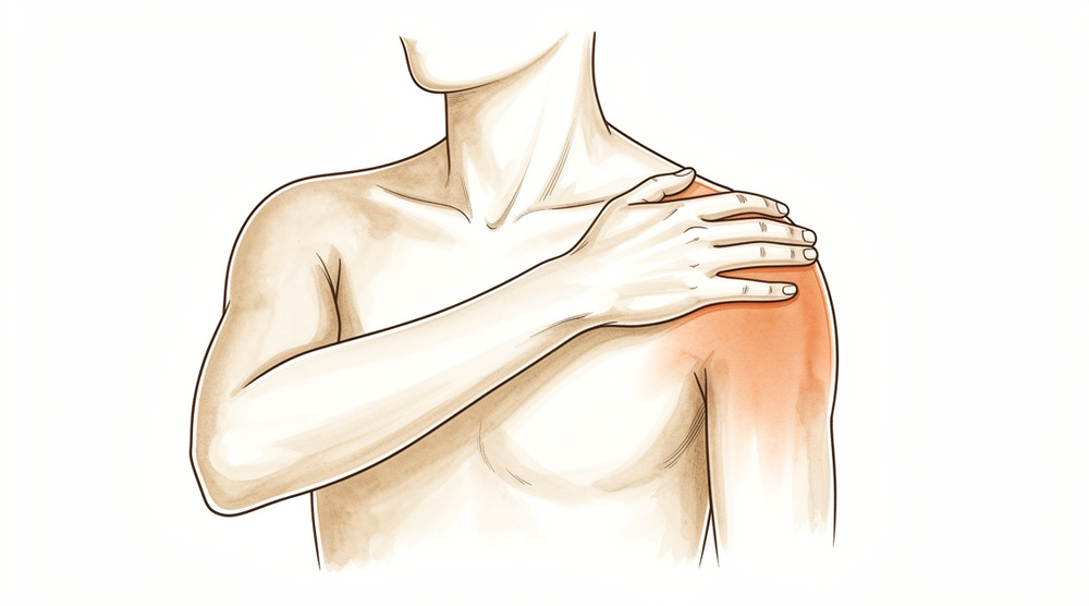
Your surgeon has suggested a distal clavicle excision, also known as a Mumford procedure. This operation removes the outer end of the collarbone to stop it from rubbing against the shoulder blade. You likely need this because you have persistent pain or wear-and-tear arthritis that has not improved with non-surgical treatments.
This surgery is typically offered to patients with old dislocations or chronic pain who do heavy work or frequently raise their arms. The main goal is to relieve your pain and improve your shoulder function. While both open and keyhole methods work well, your surgeon uses the arthroscopic approach. This involves small incisions and a camera to help you return to activities faster with similar long-term results.
Before the operation
You will need to fast before your surgery and stop taking certain medications as your surgeon advises. Please arrange for someone to drive you home and wear comfortable clothing. You may need X-rays, an MRI, blood tests, or an anaesthetic review to check your health and plan the procedure. Your surgeon will perform this operation using an arthroscopic (keyhole) approach with two or three small incisions and a small camera inside the joint. This method helps you return to activities faster while avoiding large scars. Bring a list of all your current medications to your appointment.
On the day
You will arrive at the hospital and meet your surgeon and the anaesthetist. This operation is done under general anaesthetic combined with a regional nerve block. You will be fully asleep for the operation, and the block — an injection that numbs the nerves supplying the arm before you wake up — provides pain relief for the first 12 to 24 hours after surgery. The anaesthetist will meet you before the operation and talk you through both parts.
You will then go to the operating theatre where your surgeon performs the procedure using a keyhole approach. This involves two or three small cuts and a tiny camera inside the joint to guide the work. After the surgery, you will wake up in recovery where the team monitors your comfort as the numbness wears off.
What the operation involves
Your surgeon will perform this surgery using keyhole techniques. They will make two or three small cuts, each about 1 cm long, near your shoulder. Through these openings, a tiny camera and special tools are inserted to see inside the joint. This approach allows your surgeon to access the outer end of your collarbone without making a large cut.
The main goal is to remove a small piece of bone from the outer end of your collarbone. Your surgeon will carefully excise this bone to stop it from rubbing against the shoulder blade. Evidence shows that removing about 5 mm of bone guarantees the bones will not touch again, while removing 2.5 mm was successful in many cases. Your surgeon will place the camera and tools precisely to avoid hurting nearby structures.
After the bone is removed, the small cuts are closed. The surgeon may use dissolving stitches or glue to seal the skin. You can expect to return to activities faster with this keyhole method compared to a larger open cut, while achieving similar long-term results. The procedure focuses on relieving your pain by removing the source of friction in the joint.
After the operation
You will wake up in a recovery area where your team will manage your pain. Your surgeon uses a keyhole technique with two or three small cuts and a tiny camera inside the joint. You will wear a sling and have dressings over the small cuts. You can start moving your fingers and wrist gently right away. Most patients go home the same day, but you must have someone stay with you for the first 24 hours. You can expect to return to driving within 4 weeks and to work within 6 weeks.
Recovery
You will likely feel some pain and swelling in your shoulder for the first few days. This is normal as your body heals from the small keyhole incisions. Your surgeon may suggest ice packs and pain relief to help you stay comfortable. Most people find that the discomfort eases steadily as the swelling goes down.
You will wear a sling to support your arm while you rest. Your physiotherapist will guide you through gentle exercises to keep your shoulder moving without straining it. You can do light daily tasks at home once you feel ready, but avoid lifting anything heavy or reaching overhead. Sleep may be tricky at first; propping yourself up with pillows often helps you find a comfortable position.
As your movement returns and the swelling settles, you will feel more confident in your shoulder. Your surgeon and physiotherapist will tell you exactly when you can resume driving, return to work, or play sports. Your personal timeline may differ from others, so follow their specific advice for your recovery.
What can go wrong
Most patients do well, but problems can occasionally happen. Your surgeon and the team monitor you closely to spot any issue early.
If your shoulder still hurts or feels like it is grinding after surgery, the bone may not have been removed enough. Sometimes the bone can grow back in the same spot. This can cause a deep ache that does not go away with simple painkillers. Tell your surgeon if you feel this way so they can check your healing.
If you notice sudden, sharp pain or a feeling of instability where your collarbone meets your shoulder, too much bone might have been removed. This can make the joint feel loose or wobbly. Report any new clicking or grinding sensation immediately.
In rare cases, a fracture can happen in the collarbone or the bone below it. You might feel a sudden snap or severe pain that prevents you from moving your arm. This is a serious issue that needs urgent attention. Go to the emergency department if you suspect a break.
Your surgeon uses a keyhole approach with two or three small cuts and a tiny camera inside the joint. Even with this careful method, complications can occur. The complications table on this page lists typical rates if you want the specifics.
When to call us
Call your surgeon if you have a fever, increasing redness, or discharge from your small keyhole incisions. Go to emergency care if you feel sudden severe pain, notice your leg is swollen and painful, or have trouble breathing. Contact us immediately if you lose feeling in your arm or cannot move your shoulder. These signs need urgent checks to keep you safe.
Evidence & references
title: "Distal Clavicle Excision (Mumford Procedure)" slug: distal-clavicle-excision region: shoulder audience: patient mesh_terms: ["Acromioclavicular Joint"] article_count: 750 model_used: qwen3.5-35b-a3b-q8 generated_at: '2026-05-18T14:16:59+00:00' key_articles: - title: "Editorial Commentary: The “Mumford” & Sons: For Distal Clavicle Excisions, What Are Our Young Surgeons Doing, and How Well Are They Doing It?" ref_num: 1 evidence_tier: paper evidence_level: 5 doi: 10.1016/j.arthro.2018.03.004 year: 2018 - title: "Open Versus Arthroscopic Distal Clavicle Resection" ref_num: 2 evidence_tier: paper evidence_level: 3 doi: 10.1016/j.arthro.2009.12.007 year: 2010 - title: "Surgical Treatment of Symptomatic Acromioclavicular Joint Problems" ref_num: 3 evidence_tier: paper evidence_level: 3 doi: 10.1097/blo.0b013e31802f5450 year: 2007 - title: "Predicting reduction loss risk after acromioclavicular joint dislocation treated with the endobutton device" ref_num: 5 evidence_tier: paper evidence_level: 3 doi: 10.1186/s12891-025-09190-x year: 2025 - title: "Open Versus Arthroscopic Acromioclavicular Joint Resection: A Retrospective Comparison Study" ref_num: 6 evidence_tier: paper evidence_level: 4 doi: 10.1016/j.arthro.2009.06.010 year: 2009 - title: "Acromioclavicular dislocation after arthroscopic distal clavicle resection: a case report" ref_num: 7 evidence_tier: case_report evidence_level: 4 doi: 10.1016/j.jse.2010.08.032 year: 2011 - title: "Painful Conditions of the Acromioclavicular Joint" ref_num: 8 evidence_tier: paper evidence_level: 5 doi: 10.5435/00124635-199905000-00004 year: 1999 - title: "Arthroscopic Distal Clavicle Resection: A Biomechanical Analysis In A Cadaver Model" ref_num: 9 evidence_tier: abstract evidence_level: 5 doi: 10.1016/j.jse.2007.02.105 year: 2007 - title: "Arthroscopic versus open distal clavicle excision: Comparative results at six months and one year from a randomized, prospective clinical trial" ref_num: 10 evidence_tier: paper evidence_level: 1 doi: 10.1016/j.jse.2006.10.006 year: 2007 - title: "The reverse coracoacromial ligament transfer for “horizontal” acromioclavicular joint instability" ref_num: 11 evidence_tier: paper evidence_level: 4 doi: 10.1016/j.xrrt.2021.05.003 year: 2021 - title: "Treatment of Acromioclavicular Injuries, Especially Complete Acromioclavicular Separation" ref_num: 12 evidence_tier: paper evidence_level: 4 doi: 10.2106/00004623-197254060-00005 year: 1972 - title: "Dislocation of the acromioclavicular joint. An end-result study." ref_num: 13 evidence_tier: paper evidence_level: 3 doi: 10.2106/00004623-198769070-00013 year: 1987 - title: "Patient outcomes following arthroscopic distal clavicle excision: a prospective case series" ref_num: 14 evidence_tier: paper evidence_level: 4 doi: 10.1016/j.jseint.2023.07.014 year: 2023 - title: "COMPLETE DISLOCATION OF THE ACROMIOCLAVICULAR JOINT" ref_num: 15 evidence_tier: paper evidence_level: 4 doi: 10.2106/00004623-196345080-00024 year: 1963 - title: "Arthroscopic Distal Clavicle Resection in Athletes" ref_num: 16 evidence_tier: paper evidence_level: 2 doi: 10.1177/0363546506294855 year: 2007 - title: "Methods used to assess the severity of acromioclavicular joint separations in Japan: a survey" ref_num: 17 evidence_tier: paper evidence_level: 4 doi: 10.1016/j.jseint.2019.11.006 year: 2020 - title: "Effect of Acromioclavicular Joint Injuries on the Acromioclavicular Joint Complex and Scapulohumeral Rhythm: A Functional and Mechanical Perspective" ref_num: 18 evidence_tier: paper evidence_level: 5 doi: 10.5435/jaaos-d-24-00360 year: 2025 - title: "Conservative or surgical treatment of acromioclavicular dislocation. A prospective, controlled, randomized study." ref_num: 20 evidence_tier: paper evidence_level: 1 doi: 10.2106/00004623-198668040-00011 year: 1986 - title: "Early complications of acromioclavicular joint reconstruction requiring reoperation" ref_num: 21 evidence_tier: paper evidence_level: 4 doi: 10.1007/s00167-016-4206-y year: 2016 - title: "Arthroscopic Distal Clavicle Resection: A Biomechanical Analysis of Resection Length and Joint Compliance in a Cadaveric Model" ref_num: 22 evidence_tier: paper evidence_level: 5 doi: 10.1016/j.arthro.2007.07.004 year: 2007 - title: "Preoperative Factors Associated With Subsequent Distal Clavicle Resection After Rotator Cuff Repair" ref_num: 23 evidence_tier: paper evidence_level: 3 doi: 10.1177/2325967119844295 year: 2019 - title: "Distal clavicle “A-frame” morphology: a reliable intraoperative guide for arthroscopic distal clavicle excision" ref_num: 24 evidence_tier: paper evidence_level: 5 doi: 10.1016/j.jse.2021.10.013 year: 2022 - title: "Evaluation of the range of motion of scapulothoracic, acromioclavicular and sternoclavicular joints: State of the art" ref_num: 25 evidence_tier: paper evidence_level: 5 doi: 10.1177/17585732221090226 year: 2022 - title: "Kinematic analysis of scapulothoracic movements in the shoulder girdle: a whole cadaver study" ref_num: 26 evidence_tier: paper evidence_level: 5 doi: 10.1016/j.jseint.2022.09.014 year: 2023 - title: "Differences between Coracoclavicular, Acromioclavicular, or Combined Reconstruction Techniques on the Kinematics of the Shoulder Girdle" ref_num: 27 evidence_tier: paper evidence_level: 5 doi: 10.1177/03635465221095231 year: 2022 - title: "A Biomechanical Analysis of the Native Coracoclavicular Ligaments and Their Influence on a New Reconstruction Using a Coracoid Tunnel and Free Tendon Graft" ref_num: 28 evidence_tier: paper evidence_level: 5 doi: 10.1016/j.arthro.2009.12.031 year: 2010 - title: "The Function of the Acromioclavicular and Coracoclavicular Ligaments in Shoulder Motion" ref_num: 29 evidence_tier: paper evidence_level: 5 doi: 10.1177/0363546512458571 year: 2012 - title: "Acromioclavicular joint ligamentous system contributing to clavicular strut function: a cadaveric study" ref_num: 30 evidence_tier: paper evidence_level: 5 doi: 10.1016/j.jse.2013.01.004 year: 2013 - title: "Acromioclavicular joint injuries revisited: Pathoanatomy, pathomechanics, and clinical presentation" ref_num: 31 evidence_tier: paper evidence_level: 5 doi: 10.1177/17585732221122335 year: 2022 - title: "Acromioclavicular joint biomechanics: a systematic review" ref_num: 32 evidence_tier: paper evidence_level: 4 doi: 10.1016/j.xrrt.2024.06.009 year: 2024 - title: "Anatomy of the pectoralis minor tendon and its use in acromioclavicular joint reconstruction" ref_num: 33 evidence_tier: paper evidence_level: 5 doi: 10.1016/j.jse.2006.09.007 year: 2007 - title: "Comparing the Anatomical Landmarks Versus the Coracoid-Based Landmarks Techniques for Coracoclavicular Stabilization After High-Grade Acromioclavicular Injury: A Biomechanical Study" ref_num: 34 evidence_tier: paper evidence_level: 5 doi: 10.1177/23259671221132541 year: 2022 - title: "Challenges in Treating Acromioclavicular Separations: Current Concepts" ref_num: 35 evidence_tier: paper evidence_level: 5 doi: 10.5435/jaaos-d-16-00776 year: 2018 - title: "Current trends in surgical treatment of the acromioclavicular joint injuries in 2023: a review of the literature" ref_num: 36 evidence_tier: paper evidence_level: 5 doi: 10.1016/j.jseint.2023.11.018 year: 2024 - title: "Using Dynamic Stereo X-ray Imaging for In Vivo Acromioclavicular Joint Kinematics Assessment: A Preliminary Investigation" ref_num: 37 evidence_tier: paper evidence_level: 4 doi: 10.1177/23259671241274707 year: 2024 - title: "Return to Work and Driving following Arthroscopic Subacromial Decompression and Acromioclavicular Joint Excision" ref_num: 38 evidence_tier: paper evidence_level: 3 doi: 10.1111/j.1758-5740.2010.00048.x year: 2010 - title: "Long-Term Shoulder Function after Type I and II Acromioclavicular Joint Disruption" ref_num: 40 evidence_tier: paper evidence_level: 4 doi: 10.1177/0363546508319047 year: 2008 - title: "The effect of coracoacromial ligament excision and acromioplasty on the amount of rotator cuff force production necessary to restore intact glenohumeral biomechanics" ref_num: 41 evidence_tier: paper evidence_level: 5 doi: 10.1016/j.jse.2015.10.022 year: 2016 - title: "Center of pressure (COP) measurement in patients with confirmed successful outcomes following shoulder surgery show significant sensorimotor deficits" ref_num: 42 evidence_tier: paper evidence_level: 3 doi: 10.1007/s00167-021-06751-0 year: 2021 - title: "Late reconstruction of the ligaments following acromioclavicular separation" ref_num: 43 evidence_tier: paper evidence_level: 4 doi: 10.2106/00004623-197658060-00008 year: 1976 - title: "Clinical outcomes of a single-tunnel technique for coracoclavicular and acromioclavicular ligament reconstruction" ref_num: 44 evidence_tier: paper evidence_level: 4 doi: 10.1016/j.jse.2017.11.032 year: 2018 - title: "Biomechanical Rationale for Development of Anatomical Reconstructions of Coracoclavicular Ligaments after Complete Acromioclavicular Joint Dislocations" ref_num: 45 evidence_tier: paper evidence_level: 5 doi: 10.1177/0363546504264637 year: 2004 - title: "Acromioclavicular joint reconstruction with coracoacromial ligament transfer using the docking technique" ref_num: 46 evidence_tier: paper evidence_level: 4 doi: 10.1186/1471-2474-10-6 year: 2009 - title: "All‐Arthroscopic Weaver‐Dunn‐Chuinard Procedure With Double‐Button Fixation for Chronic Acromioclavicular Joint Dislocation" ref_num: 47 evidence_tier: paper evidence_level: 4 doi: 10.1016/j.arthro.2009.08.008 year: 2009 - title: "Comparative efficacy of operative versus conservative treatment for Rockwood type III acromioclavicular joint dislocation: a systematic review and meta-analysis of randomized controlled trials" ref_num: 49 evidence_tier: paper evidence_level: 1 doi: 10.1186/s12891-024-08100-x year: 2024 - title: "The Effect of Distal Clavicle Excision on in Situ Graft Forces in Coracoclavicular Ligament Reconstruction" ref_num: 50 evidence_tier: paper evidence_level: 5 doi: 10.1177/0363546510374447 year: 2010 - title: "Which stabilization technique corrects anatomy best in patients with AC‐separation?" ref_num: 52 evidence_tier: paper evidence_level: 5 doi: 10.1007/s001670050182 year: 1999 - title: "Fracture Clavicle with Acromioclavicular Dislocation: A Complex Injury" ref_num: 53 evidence_tier: paper evidence_level: 4 doi: 10.1111/j.1758-5740.2010.00102.x year: 2011 - title: "Position of scapula and clavicle in acute acromioclavicular joint dislocations: depressed scapula or elevated distal clavicle?" ref_num: 54 evidence_tier: paper evidence_level: 4 doi: 10.1016/j.jseint.2023.06.011 year: 2023 - title: "A systematic review of the treatment of primary acromioclavicular joint osteoarthritis" ref_num: 55 evidence_tier: paper evidence_level: 4 doi: 10.1177/17585732231157090 year: 2023 - title: "Low rate of substantial loss of reduction immediately after hardware removal following acromioclavicular joint stabilization using a suspensory fixation system" ref_num: 56 evidence_tier: paper evidence_level: 4 doi: 10.1007/s00167-022-06978-5 year: 2022 - title: "Long-term Follow-up After Arthroscopically Assisted 2-Bundle Anatomic Reduction of Acute Acromioclavicular Joint Separations" ref_num: 57 evidence_tier: paper evidence_level: 3 doi: 10.1177/03635465251355958 year: 2025 - title: "Posterior Distal Clavicle Beveling for Chronic Nonincarcerated Type IV Acromioclavicular Separations: Surgical Technique and Early Clinical Outcomes" ref_num: 58 evidence_tier: paper evidence_level: 4 doi: 10.1016/j.arthro.2016.06.013 year: 2016 - title: "Characteristics and Complications of Operative Acromioclavicular Joint Separations in an Active Population (222)" ref_num: 59 evidence_tier: paper evidence_level: 3 doi: 10.1177/2325967121s00330 year: 2021 - title: "Subacromial osteolysis following hook plate fixation for acromioclavicular dislocation: a systematic review and meta-analysis" ref_num: 60 evidence_tier: paper evidence_level: 1 doi: 10.1016/j.jse.2024.03.018 year: 2024 - title: "Ac Joint Reconstruction With Ca Ligament Transfer Using The Docking Technique" ref_num: 61 evidence_tier: abstract evidence_level: 4 doi: 10.1016/j.jse.2007.02.104 year: 2007 - title: "Coracoid or Clavicle Fractures Associated With Coracoclavicular Ligament Reconstruction" ref_num: 62 evidence_tier: paper evidence_level: 4 doi: 10.1177/03635465211036713 year: 2021 synthesis_version: "v2" verifier_status: skipped
Overview
- A well-performed distal clavicle excision will likely perform better than a poorly performed one, regardless of whether an open or arthroscopic approach is chosen [1].
- Patients undergoing arthroscopic distal clavicle excision through the direct approach can expect a faster return to activities compared with the open procedure while obtaining similar long-term outcomes [2].
- Arthroscopic distal clavicle resection has provided more 'good or excellent' results than has the open procedure, though this finding is comprised of low-level evidence [3].
- Simple excision of the outer end of the clavicle has yielded satisfactory results with no residual upward displacement disturbing patients [4].
- Patients with displacement greater than 100% of the thickness of the distal clavicle had poorer postoperative clinical outcomes [5].
- Incomplete excision and regrowth of the distal clavicle are the most common causes of revision surgery [6].
- Portal placement remains paramount in facilitating surgery and avoiding injury to adjacent extra-articular structures regardless of the technique chosen for distal clavicle resection [7].
- In appropriately selected patients, open or arthroscopic distal clavicle resection is necessary to relieve symptoms [8].
- Distal clavicle excision with 2.5 mm of bone was successful in many specimens, but a 5 mm resection guaranteed no bone-to-bone abutment [9].
- Arthroscopic and open distal clavicle excisions both provide significant pain reduction at 1 year with no significant difference in outcome measures between groups, except for VAS pain score improvement [10].
- Excision of the outer end of the clavicle is preferred for old dislocations, while open reduction and internal fixation are not recommended due to complications and poor functional results [15].
- Both the direct superior approach and the indirect subacromial approach to the arthroscopic distal clavicle resection result in successful clinical outcome with clinically insignificant difference at final follow-up [16].
Anatomy & Pathophysiology
- A precise, easy to use and low-cost non-invasive method able to draw and analyze the kinematics of the shoulder complex has not been developed yet [25].
- Normative kinematic values of scapulothoracic movements in the shoulder girdle have been provided [26].
- No reconstruction strategy completely restores the shoulder girdle to its preinjured state, although each technique restores different elements of joint kinematics [27].
- The trapezoid and conoid ligaments have unique functions in normal shoulder kinematics because of their anatomic attachments [28].
- Kinematic changes could be a potential source of pain and dysfunction in the shoulder with AC joint dislocation [29].
- Scapular and clavicular kinematics were affected in AC separation models [30].
- A comprehensive clinical approach emphasizing the evaluation of the extent of the anatomic injury and understanding its mechanical consequences regarding shoulder and arm function is key in the development of guidelines for developing operative or non-operative treatment protocols and for establishing outcomes of the treatment protocols [31].
- The inconsistency of AC joint testing parameters and the lack of thorough translation studies indicate a necessity for increased attention in the overall assessment of shoulder stability to close the gap in the foundational biomechanical research [32].
- Anatomically, the pectoralis minor tendon provides sufficient tissue length, excursion, and width [33].
- Biomechanically, the pectoralis minor tendon is as strong as the coracoacromial ligament [33].
- No significant biomechanical differences in displacement or stiffness were seen between the anatomical landmark technique and the coracoid-based landmarks technique for coracoclavicular stabilization [34].
- New surgical techniques continue to evolve as more biomechanical data emerge and kinematic understanding improves [35].
- Emerging concepts and strategies regarding horizontal and rotational instability and scapular biomechanics aim to lay the foundation for future studies aimed at improving treatment outcomes and patient management [36].
- Preliminary findings revealed no detectable differences between surgically reconstructed and uninjured sides in ACJ biomechanics, range of motion, and isometric strength [37].
- Nonoperatively treated shoulders showed increased internal rotation, upward rotation, and posterior tilting [37].
- Type I and II acromioclavicular joint disruptions impair long-term shoulder function in about half of patients 10 years after injury [40].
- At 150 to 200 N of loading, coracoacromial ligament excision and acromioplasty increase the rotator cuff force required to maintain normal glenohumeral biomechanics by 25% to 30% [41].
- Centre of pressure measurement detected sensorimotor functional deficits following surgical treatment of the shoulder joint in patients with confirmed successful clinical and functional outcomes [42].
Classification
- A well-performed distal clavicle excision will likely perform better than a poorly performed one, regardless of whether an open or arthroscopic approach is chosen [1].
- Patients undergoing an arthroscopic procedure specifically through the direct approach can expect a faster return to activities while obtaining similar long-term outcomes compared with the open procedure [2].
- Arthroscopic distal clavicle resection has provided more 'good or excellent' results than has the open procedure, but this finding is comprised of low-level evidence [3].
- Simple excision of the outer end of the clavicle has yielded satisfactory results with no residual upward displacement disturbing the patients [4].
- Patients with displacement greater than 100% of the thickness of the distal clavicle had poorer postoperative clinical outcomes [5].
- Incomplete excision and regrowth of the distal clavicle are the most common causes of revision [6].
- Portal placement remains paramount in both facilitating surgery and avoiding injury to adjacent extra-articular structures regardless of the technique chosen for distal clavicle resection [7].
- In appropriately selected patients, open or arthroscopic distal clavicle resection is necessary to relieve symptoms [8].
- Distal clavicle excision with 2.5 mm of bone was successful in many specimens, but a 5 mm resection guaranteed no bone-to-bone abutment [9].
- Arthroscopic and open distal clavicle excisions both provide significant pain reduction at 1 year with no significant difference in outcome measures between groups, except for VAS pain score improvement [10].
- Horizontal instability of the clavicle is evident with distal clavicle resection of greater than 10 mm [11].
- The new operative procedure combines resection arthroplasty with fixation of the clavicle in an anatomical position [12].
- A records review found that 10 of 894 (1.1%) rotator cuff repairs underwent subsequent distal clavicle resection [23].
- The cross-sectional A-frame morphology of the superior cortex of the distal clavicle provides a reproducible landmark that is eliminated approximately 1.0 cm medial to the distal, lateral end of the clavicle, which can be used intraoperatively to determine when adequate resection has been completed [24].
- Severe chronic symptomatic AC joint separations (Rockwood types III through V) can be repaired entirely by arthroscopy safely and effectively by transferring the coracoacromial ligament with a bone block in the distal clavicle [47].
Clinical Presentation
- A well-performed distal clavicle excision will likely perform better than a poorly performed one, regardless of whether an open or arthroscopic approach is chosen [1].
- Patients having an arthroscopic procedure, specifically through the direct approach, can expect a faster return to activities while obtaining similar long-term outcomes compared with the open procedure [2].
- Arthroscopic distal clavicle resection has provided more 'good or excellent' results than has the open procedure, but is comprised of low-level evidence [3].
- Simple excision of the outer end of the clavicle has yielded satisfactory results in patients with complete dislocation and subluxation of the acromioclavicular joint, with no residual upward displacement disturbing the patients [4].
- Patients with displacement greater than 100% of the thickness of the distal clavicle had poorer postoperative clinical outcomes [5].
- Incomplete excision and regrowth of the distal clavicle are the most common causes of revision surgery [6].
- Portal placement remains paramount in both facilitating surgery and avoiding injury to adjacent extra-articular structures regardless of the technique chosen for distal clavicle resection [7].
- In appropriately selected patients, open or arthroscopic distal clavicle resection is necessary to relieve symptoms [8].
- Although distal clavicle excision with 2.5 mm of bone was successful in many specimens, a 5 mm resection guaranteed no bone-to-bone abutment [9].
- Arthroscopic and open distal clavicle excisions both provide significant pain reduction at 1 year with no significant difference in outcome measures between groups, except for VAS pain score improvement [10].
- Horizontal instability of the clavicle is evident with distal clavicle resection of greater than 10 mm [11].
- Late loss of reduction was common, and clavicular resection reliably produced significant improvement in patients with persistent pain or posttraumatic arthritis [13].
- In carefully selected patients with isolated ACJ pathology, arthroscopic distal clavicle excision results in statistically and clinically significant improvements in range of motion and patient-reported outcome measures [14].
- Excision of the outer end of the clavicle is preferred for old dislocations, while open reduction and internal fixation are not recommended due to complications and poor functional results [15].
- Methods to diagnose both superior and posterior translation of the clavicle need further debate [17].
- Clinical examination and surgical treatment should address anatomic restoration of individual structures to optimize the mechanical capability of the claviscapular segment [18].
- For chronic symptomatic injuries, partial claviculectomy is believed to be the best procedure, offering negligible morbidity and rapid return to function [19].
- Operation should be considered only in thin patients with a prominent clavicle, those doing heavy work, or those whose work requires frequent shoulder abduction and flexion [20].
- Older patients and females were more likely to experience postoperative complications requiring reoperations, including revision ACJR, distal clavicle excision, and irrigation and debridement [21].
- Excellent clinical results were achieved with acromioclavicular joint reconstruction with coracoacromial ligament transfer using the docking technique, decreasing the risk of recurrent distal clavicle instability [46].
Investigations
- A well-performed distal clavicle excision will likely perform better than a poorly performed one, regardless of whether an open or arthroscopic approach is chosen [1].
- Patients having an arthroscopic procedure, specifically through the direct approach, can expect a faster return to activities while obtaining similar long-term outcomes compared with the open procedure [2].
- Arthroscopic distal clavicle resection has provided more 'good or excellent' results than has the open procedure, but is comprised of low-level evidence [3].
- Simple excision of the outer end of the clavicle has yielded satisfactory results in this group of patients, with no residual upward displacement disturbing the patients [4].
- Patients with displacement greater than 100% of the thickness of the distal clavicle had poorer postoperative clinical outcomes [5].
- Incomplete excision and regrowth of the distal clavicle are the most common causes of revision [6].
- Portal placement remains paramount in both facilitating surgery and avoiding injury to adjacent extra-articular structures regardless of the technique chosen for distal clavicle resection [7].
- In appropriately selected patients, open or arthroscopic distal clavicle resection is necessary to relieve symptoms [8].
- Distal clavicle excision with 2.5 mm of bone was successful in many specimens, but a 5 mm resection guaranteed no bone-to-bone abutment [9].
- Horizontal instability of the clavicle is evident with distal clavicle resection of greater than 10 mm [11].
- The new operative procedure combines resection arthroplasty with fixation of the clavicle in an anatomical position [12].
- In carefully selected patients with isolated ACJ pathology, arthroscopic distal clavicle excision results in statistically and clinically significant improvements in range of motion and patient-reported outcome measures [14].
- Methods to diagnose both superior and posterior translation of the clavicle need further debate [17].
- Clinical examination and surgical treatment should address anatomic restoration of individual structures to optimize the mechanical capability of the claviscapular segment [18].
- A 5-mm distal clavicle resection guaranteed no abutment but decreased joint stiffness [22].
- The cross-sectional A-frame morphology of the superior cortex of the distal clavicle provides a reproducible landmark that is eliminated approximately 1.0 cm medial to the distal, lateral end of the clavicle, which can be used intraoperatively to determine when adequate resection has been completed [24].
- Weighted stress radiographs significantly increased the measured elevation of the clavicle and the coracoclavicular distance compared to non-weighted views [54].
- There was no significant difference between open or arthroscopic distal clavicle excision (DCE) [55].
- Although radiological assessment showed a statistically significant immediate superior clavicular displacement after hardware removal following ACJ stabilization, with an increased incidence in the first year following stabilization, this may not negatively influence the results of ACJ stabilization in a clinically relevant way [56].
- Fifteen years postoperatively, good clinical results persisted and anatomic reduction was overall maintained, often with asymptomatic ossification of the coracoclavicular ligaments [57].
Treatment
- A well-performed distal clavicle excision will likely perform better than a poorly performed one, regardless of whether an open or arthroscopic approach is chosen [1].
- Patients undergoing arthroscopic distal clavicle excision via the direct approach can expect a faster return to activities compared with the open procedure while obtaining similar long-term outcomes [2].
- Arthroscopic distal clavicle resection has provided more 'good or excellent' results than the open procedure, though this is based on low-level evidence [3].
- Simple excision of the outer end of the clavicle has yielded satisfactory results with no residual upward displacement disturbing patients [4].
- Patients with displacement greater than 100% of the thickness of the distal clavicle had poorer postoperative clinical outcomes [5].
- Incomplete excision and regrowth of the distal clavicle are the most common causes of revision surgery [6].
- Portal placement remains paramount in facilitating surgery and avoiding injury to adjacent extra-articular structures regardless of the technique chosen for distal clavicle resection [7].
- In appropriately selected patients, open or arthroscopic distal clavicle resection is necessary to relieve symptoms [8].
- Distal clavicle excision with 2.5 mm of bone was successful in many specimens, but a 5 mm resection guaranteed no bone-to-bone abutment [9].
- Arthroscopic and open distal clavicle excisions both provide significant pain reduction at 1 year with no significant difference in outcome measures between groups, except for VAS pain score improvement [10].
- Horizontal instability of the clavicle is evident with distal clavicle resection of greater than 10 mm [11].
- Late loss of reduction was common, and clavicular resection reliably produced significant improvement in patients with persistent pain or posttraumatic arthritis [13].
- Excision of the outer end of the clavicle is preferred for old dislocations, while open reduction and internal fixation are not recommended due to complications and poor functional results [15].
- Both the direct superior approach and the indirect subacromial approach to arthroscopic distal clavicle resection result in successful clinical outcomes with clinically insignificant difference at final follow-up [16].
- A 5-mm distal clavicle resection guaranteed no abutment but decreased joint stiffness [22].
- Surgical treatment may offer early benefits in pain relief and coracoclavicular distance improvement but does not enhance long-term functional outcomes and is associated with higher specific complication rates [49].
- The slight increase in the in situ graft force only in the posterosuperior and posterior direction after distal clavicle excision suggests only a marginal protective role of the acromioclavicular articulation [50].
- A bone anchor system for distal fixation in the base of the coracoid process and a medialized hole in the clavicle restored anatomy best [52].
Complications
- A well-performed distal clavicle excision performs better than a poorly performed one, regardless of whether an open or arthroscopic approach is chosen [1].
- Incomplete excision and regrowth of the distal clavicle are the most common causes of revision surgery [6].
- Portal placement is paramount in facilitating surgery and avoiding injury to adjacent extra-articular structures [7].
- Distal clavicle excision with 2.5 mm of bone was successful in many specimens, but a 5 mm resection guaranteed no bone-to-bone abutment [9].
- Horizontal instability of the clavicle is evident with distal clavicle resection of greater than 10 mm [11].
- Patients with displacement greater than 100% of the thickness of the distal clavicle had poorer postoperative clinical outcomes [5].
- Older patients and females were more likely to experience postoperative complications requiring reoperations, including revision ACJR, distal clavicle excision, and irrigation and debridement [21].
- The incidence of complications in operative acromioclavicular joint separations in an active population was 1.35 per 100 person-years [59].
- Clavicle and coracoid fractures occurred in 1.9 out of 100 cases of operative acromioclavicular joint separations [59].
- Fracture of the distal clavicle or coracoid process after CC ligament repair or reconstruction is a rare but serious complication that can occur independent of bone tunnels created during the index procedure [62].
- Coracoclavicular ligament reconstruction is an effective surgical approach for decreasing the incidence of subacromial osteolysis [60].
- Excellent results can be obtained with coracoacromial ligament transfer using the docking technique, decreasing the risk of recurrent distal clavicle instability [61].
Recovery
- A well-performed distal clavicle excision will likely perform better than a poorly performed one, regardless of whether an open or arthroscopic approach is chosen [1].
- Patients undergoing an arthroscopic procedure, specifically through the direct approach, can expect a faster return to activities compared with the open procedure while obtaining similar long-term outcomes [2].
- Arthroscopic distal clavicle resection has provided more 'good or excellent' results than has the open procedure, though this is comprised of low-level evidence [3].
- Simple excision of the outer end of the clavicle has yielded satisfactory results with no residual upward displacement disturbing the patients [4].
- Patients with displacement greater than 100% of the thickness of the distal clavicle had poorer postoperative clinical outcomes [5].
- Incomplete excision and regrowth of the distal clavicle are the most common causes of revision [6].
- Arthroscopic and open distal clavicle excisions both provide significant pain reduction at 1 year with no significant difference in outcome measures between groups, except for VAS pain score improvement [10].
- Clavicular resection reliably produced significant improvement in patients with persistent pain or posttraumatic arthritis, although late loss of reduction was common [13].
- For chronic symptomatic injuries, partial claviculectomy is believed to be the best procedure, offering negligible morbidity and rapid return to function [19].
- Operation should be considered only in thin patients with a prominent clavicle, those doing heavy work, or those whose work requires frequent shoulder abduction and flexion [20].
- More than 90% of patients manage to return to driving within 4 weeks and to work within 6 weeks following arthroscopic subacromial decompression and acromio-clavicular joint excision [38].
- Late reconstruction of the ligaments in young patients with complete acromioclavicular separations can yield better results than excision of the lateral clavicle, allowing patients to return to strenuous sports or heavy labor [43].
- The described single-tunnel technique for coracoclavicular and acromioclavicular ligament reconstruction results in satisfactory objective and patient-reported outcomes and return to sports while avoiding coracoid and clavicle fractures [44].
- The anatomic reconstruction complex could withstand early rehabilitation, but the decrease in the structural properties and stiffness of the clavicle should be considered in optimizing the anatomic reconstruction technique [45].
- Satisfactory outcome depends upon restoring the stability of the clavicle as well as the acromioclavicular joint [53].
- The arthroscopic partial distal clavicle beveling procedure for nonincarcerated type IV AC separations resulted in a significant reduction in pain, improved daily function, and early return to sport [58].
Key Evidence
- [L5] A well-performed distal clavicle excision will likely perform better than a poorly performed one, regardless of whether an open or arthroscopic approach is chosen. (10.1016/j.arthro.2018.03.004)
- [L3] Among patients undergoing distal clavicle excision for acromioclavicular joint pathology, those having an arthroscopic procedure, specifically through the direct approach, can expect a faster return to activities while obtaining similar long-term outcomes compared with the open procedure. (10.1016/j.arthro.2009.12.007)
- [L3] Arthroscopic distal clavicle resection has provided more 'good or excellent' results than has the open procedure, but is comprised of low-level evidence. (10.1097/blo.0b013e31802f5450)
- [L3] Patients with displacement greater than 100% of the thickness of the distal clavicle had poorer postoperative clinical outcomes. (10.1186/s12891-025-09190-x)
- [L4] Incomplete excision and regrowth of the distal clavicle are the most common causes of revision. (10.1016/j.arthro.2009.06.010)
- [Case_report] Regardless of the technique chosen for distal clavicle resection, portal placement remains paramount in both facilitating surgery and avoiding injury to adjacent extra-articular structures. (10.1016/j.jse.2010.08.032)
- [L5] In appropriately selected patients, open or arthroscopic distal clavicle resection is necessary to relieve symptoms. (10.5435/00124635-199905000-00004)
- [Abstract] Although distal clavicle excision with 2.5 mm of bone was successful in many specimens, a 5 mm resection guaranteed no bone-to-bone abutment. (10.1016/j.jse.2007.02.105)
- [L1] Arthroscopic and open distal clavicle excisions both provide significant pain reduction at 1 year with no significant difference in outcome measures between groups, except for VAS pain score improvement. (10.1016/j.jse.2006.10.006)
- [L4] Horizontal instability of the clavicle is evident with distal clavicle resection of greater than 10 mm. (10.1016/j.xrrt.2021.05.003)
- [L4] The new operative procedure combines resection arthroplasty with fixation of the clavicle in an anatomical position. (10.2106/00004623-197254060-00005)
- [L3] Late loss of reduction was common, and clavicular resection reliably produced significant improvement in patients with persistent pain or posttraumatic arthritis. (10.2106/00004623-198769070-00013)
- [L4] In carefully selected patients with isolated ACJ pathology, arthroscopic distal clavicle excision results in statistically and clinically significant improvements in range of motion and patient-reported outcome measures. (10.1016/j.jseint.2023.07.014)
- [L4] Excision of the outer end of the clavicle is preferred for old dislocations, while open reduction and internal fixation are not recommended due to complications and poor functional results. (10.2106/00004623-196345080-00024)
- [L2] Both the direct superior approach and the indirect subacromial approach to the arthroscopic distal clavicle resection result in successful clinical outcome with clinically insignificant difference at final follow-up. (10.1177/0363546506294855)
- [L4] Methods to diagnose both superior and posterior translation of the clavicle need further debate. (10.1016/j.jseint.2019.11.006)
- [L5] Clinical examination and surgical treatment should address anatomic restoration of individual structures to optimize the mechanical capability of the claviscapular segment. (10.5435/jaaos-d-24-00360)
- [L1] Operation should be considered only in thin patients with a prominent clavicle, those doing heavy work, or those whose work requires frequent shoulder abduction and flexion. (10.2106/00004623-198668040-00011)
- [L4] Older patients and females were more likely to experience postoperative complications requiring reoperations, including revision ACJR, distal clavicle excision, and irrigation and debridement. (10.1007/s00167-016-4206-y)
- [L5] A 5-mm distal clavicle resection guaranteed no abutment but decreased joint stiffness. (10.1016/j.arthro.2007.07.004)
- [L3] This records review found that 10 of 894 (1.1%) rotator cuff repairs underwent subsequent distal clavicle resection. (10.1177/2325967119844295)
- [L5] The cross-sectional A-frame morphology of the superior cortex of the distal clavicle provides a reproducible landmark that is eliminated approximately 1.0 cm medial to the distal, lateral end of the clavicle, which can be used intraoperatively to determine when adequate resection has been completed. (10.1016/j.jse.2021.10.013)
- [L5] Despite technology innovations, a precise, easy to use and low-cost non-invasive method able to draw and analyze the kinematics of the shoulder complex has not been developed yet. (10.1177/17585732221090226)
- [L5] This study provided normative kinematic values of scapulothoracic movements in the shoulder girdle. (10.1016/j.jseint.2022.09.014)
- [L5] Although each technique was able to restore different elements of the joint kinematics, none of the strategies completely restored the shoulder girdle to its preinjured state. (10.1177/03635465221095231)
- [L5] The trapezoid and conoid ligaments have unique functions in normal shoulder kinematics because of their anatomic attachments. (10.1016/j.arthro.2009.12.031)
- [L5] The kinematic changes could be a potential source of pain and dysfunction in the shoulder with AC joint dislocation. (10.1177/0363546512458571)
- [L5] Scapular and clavicular kinematics were affected in AC separation models. (10.1016/j.jse.2013.01.004)
- [L5] A comprehensive clinical approach emphasizing the evaluation of the extent of the anatomic injury and understanding its mechanical consequences regarding shoulder and arm function is a key in the development of guidelines for developing operative or non-operative treatment protocols and for establishing outcomes of the treatment protocols. (10.1177/17585732221122335)
- [L4] The inconsistency of AC joint testing parameters and the lack of thorough translation studies indicate a necessity for increased attention in the overall assessment of shoulder stability to close the gap in the foundational biomechanical research. (10.1016/j.xrrt.2024.06.009)
- [L5] Anatomically, it provides sufficient tissue length, excursion, and width, and biomechanically, it is as strong as the coracoacromial ligament. (10.1016/j.jse.2006.09.007)
- [L5] No significant biomechanical differences in displacement or stiffness were seen between the anatomical landmark technique and the coracoid-based landmarks technique. (10.1177/23259671221132541)
- [L5] New surgical techniques continue to evolve as more biomechanical data emerge and kinematic understanding improves. (10.5435/jaaos-d-16-00776)
- [L5] By exploring emerging concepts and strategies regarding horizontal and rotational instability and scapular biomechanics, the article aims to lay the foundation for future studies aimed at improving treatment outcomes and patient management. (10.1016/j.jseint.2023.11.018)
- [L4] Preliminary findings revealed no detectable differences between surgically reconstructed and uninjured sides in ACJ biomechanics, range of motion, and isometric strength, while nonoperatively treated shoulders showed increased internal rotation, upward rotation, and posterior tilting. (10.1177/23259671241274707)
- [L3] The results obtained in the present study suggest that more than 90% of the patients manage to return to driving within 4 weeks and to work within 6 weeks following arthroscopic subacromial decompression and acromio-clavicular joint excision. (10.1111/j.1758-5740.2010.00048.x)
- [L4] Type I and II acromioclavicular joint disruptions impair long-term shoulder function in about half of patients 10 years after injury. (10.1177/0363546508319047)
- [L5] At 150 to 200 N of loading, CAL excision and acromioplasty increase the rotator cuff force required to maintain normal glenohumeral biomechanics by 25% to 30%. (10.1016/j.jse.2015.10.022)
- [L3] Centre of pressure measurement detected sensorimotor functional deficits following surgical treatment of the shoulder joint in patients with confirmed successful clinical and functional outcomes. (10.1007/s00167-021-06751-0)
- [L4] Late reconstruction of the ligaments in young patients with complete acromioclavicular separations can yield better results than excision of the lateral clavicle, allowing patients to return to strenuous sports or heavy labor. (10.2106/00004623-197658060-00008)
- [L4] The described technique results in satisfactory objective and patient-reported outcomes and return to sports while avoiding coracoid and clavicle fractures. (10.1016/j.jse.2017.11.032)
- [L5] The low level of permanent elongation after cyclic loading suggests that the anatomic reconstruction complex could withstand early rehabilitation; however, the decrease in the structural properties and stiffness of the clavicle should be considered in optimizing the anatomic reconstruction technique. (10.1177/0363546504264637)
- [L4] Excellent clinical results were achieved, decreasing the risk of recurrent distal clavicle instability. (10.1186/1471-2474-10-6)
- [L4] Severe chronic symptomatic AC joint separations (Rockwood types III through V) can be repaired entirely by arthroscopy safely and effectively by transferring the coracoacromial ligament with a bone block in the distal clavicle. (10.1016/j.arthro.2009.08.008)
- [L1] Surgical treatment may offer early benefits in pain relief and coracoclavicular distance improvement but does not enhance long-term functional outcomes and is associated with higher specific complication rates. (10.1186/s12891-024-08100-x)
- [L5] The slight increase in the in situ graft force only in the posterosuperior and posterior direction after distal clavicle excision suggests only a marginal protective role of the acromioclavicular articulation. (10.1177/0363546510374447)
- [L5] A bone anchor system for distal fixation in the base of the coracoid process and a medialized hole in the clavicle restored anatomy best. (10.1007/s001670050182)
- [L4] Satisfactory outcome depends upon restoring the stability of the clavicle as well as the acromioclavicular joint. (10.1111/j.1758-5740.2010.00102.x)
- [L4] Weighted stress radiographs significantly increased the measured elevation of the clavicle and the coracoclavicular distance compared to non-weighted views. (10.1016/j.jseint.2023.06.011)
- [L4] There was no significant difference between open or arthroscopic distal clavicle excision (DCE). (10.1177/17585732231157090)
- [L4] Although radiological assessment showed a statistically significant immediate superior clavicular displacement after this rarely required procedure, with an increased incidence in the first year following stabilization, this may not negatively influence the results of ACJ stabilization in a clinically relevant way. (10.1007/s00167-022-06978-5)
- [L3] Fifteen years postoperatively, good clinical results persisted and anatomic reduction was overall maintained, often with asymptomatic ossification of the coracoclavicular ligaments. (10.1177/03635465251355958)
- [L4] The arthroscopic partial distal clavicle beveling procedure for nonincarcerated type IV AC separations resulted in a significant reduction in pain, improved daily function, and early return to sport. (10.1016/j.arthro.2016.06.013)
- [L3] This review demonstrated an incidence of 1.35 complications per 100 person-years, with clavicle and coracoid fractures occurring in 1.9 out of 100 cases. (10.1177/2325967121s00330)
- [L1] The current analysis suggests coracoclavicular ligament reconstruction as an effective surgical approach for decreasing the incidence of subacromial osteolysis. (10.1016/j.jse.2024.03.018)
- [Abstract] Excellent results can be obtained with this technique, decreasing the risk of recurrent distal clavicle instability. (10.1016/j.jse.2007.02.104)
- [L4] Fracture of the distal clavicle or coracoid process after CC ligament repair or reconstruction is a rare but serious complication that can occur independent of bone tunnels created during the index procedure. (10.1177/03635465211036713)
References
[1] Editorial Commentary: The “Mumford” & Sons: For Distal Clavicle Excisions, What Are Our Young Surgeons Doing, and How Well Are They Doing It?. Arthroscopy. 2018. DOI: 10.1016/j.arthro.2018.03.004 [2] Open Versus Arthroscopic Distal Clavicle Resection. Arthroscopy. 2010. DOI: 10.1016/j.arthro.2009.12.007 [3] Surgical Treatment of Symptomatic Acromioclavicular Joint Problems. Clinical Orthopaedics and Related Research. 2007. DOI: 10.1097/blo.0b013e31802f5450 [4] Complete Dislocation and Subluxation of the Acromioclavicular Joint: End Result in Seventy-three Cases.. The Journal of Bone and Joint Surgery. American Volume. 1961. [5] Predicting reduction loss risk after acromioclavicular joint dislocation treated with the endobutton device. BMC Musculoskeletal Disorders. 2025. DOI: 10.1186/s12891-025-09190-x [6] Open Versus Arthroscopic Acromioclavicular Joint Resection: A Retrospective Comparison Study. Arthroscopy. 2009. DOI: 10.1016/j.arthro.2009.06.010 [7] Acromioclavicular dislocation after arthroscopic distal clavicle resection: a case report. Journal of Shoulder and Elbow Surgery. 2011. DOI: 10.1016/j.jse.2010.08.032 [8] Painful Conditions of the Acromioclavicular Joint. Journal of the American Academy of Orthopaedic Surgeons. 1999. DOI: 10.5435/00124635-199905000-00004 [9] Arthroscopic Distal Clavicle Resection: A Biomechanical Analysis In A Cadaver Model. Journal of Shoulder and Elbow Surgery. 2007. DOI: 10.1016/j.jse.2007.02.105 [10] Arthroscopic versus open distal clavicle excision: Comparative results at six months and one year from a randomized, prospective clinical trial. Journal of Shoulder and Elbow Surgery. 2007. DOI: 10.1016/j.jse.2006.10.006 [11] The reverse coracoacromial ligament transfer for “horizontal” acromioclavicular joint instability. JSES Reviews, Reports, and Techniques. 2021. DOI: 10.1016/j.xrrt.2021.05.003 [12] Treatment of Acromioclavicular Injuries, Especially Complete Acromioclavicular Separation. The Journal of Bone & Joint Surgery. 1972. DOI: 10.2106/00004623-197254060-00005 [13] Dislocation of the acromioclavicular joint. An end-result study.. The Journal of Bone & Joint Surgery. 1987. DOI: 10.2106/00004623-198769070-00013 [14] Patient outcomes following arthroscopic distal clavicle excision: a prospective case series. JSES International. 2023. DOI: 10.1016/j.jseint.2023.07.014 [15] COMPLETE DISLOCATION OF THE ACROMIOCLAVICULAR JOINT. The Journal of Bone & Joint Surgery. 1963. DOI: 10.2106/00004623-196345080-00024 [16] Arthroscopic Distal Clavicle Resection in Athletes. The American Journal of Sports Medicine. 2007. DOI: 10.1177/0363546506294855 [17] Methods used to assess the severity of acromioclavicular joint separations in Japan: a survey. JSES International. 2020. DOI: 10.1016/j.jseint.2019.11.006 [18] Effect of Acromioclavicular Joint Injuries on the Acromioclavicular Joint Complex and Scapulohumeral Rhythm: A Functional and Mechanical Perspective. Journal of the American Academy of Orthopaedic Surgeons. 2025. DOI: 10.5435/jaaos-d-24-00360 [19] Acromioclavicular-Joint Injury: AN END-RESULT STUDY.. The Journal of Bone and Joint Surgery. American Volume. 1966. [20] Conservative or surgical treatment of acromioclavicular dislocation. A prospective, controlled, randomized study.. The Journal of Bone & Joint Surgery. 1986. DOI: 10.2106/00004623-198668040-00011 [21] Early complications of acromioclavicular joint reconstruction requiring reoperation. Knee Surgery, Sports Traumatology, Arthroscopy. 2016. DOI: 10.1007/s00167-016-4206-y [22] Arthroscopic Distal Clavicle Resection: A Biomechanical Analysis of Resection Length and Joint Compliance in a Cadaveric Model. Arthroscopy. 2007. DOI: 10.1016/j.arthro.2007.07.004 [23] Preoperative Factors Associated With Subsequent Distal Clavicle Resection After Rotator Cuff Repair. Orthopaedic Journal of Sports Medicine. 2019. DOI: 10.1177/2325967119844295 [24] Distal clavicle “A-frame” morphology: a reliable intraoperative guide for arthroscopic distal clavicle excision. Journal of Shoulder and Elbow Surgery. 2022. DOI: 10.1016/j.jse.2021.10.013 [25] Evaluation of the range of motion of scapulothoracic, acromioclavicular and sternoclavicular joints: State of the art. Shoulder & Elbow. 2022. DOI: 10.1177/17585732221090226 [26] Kinematic analysis of scapulothoracic movements in the shoulder girdle: a whole cadaver study. JSES International. 2023. DOI: 10.1016/j.jseint.2022.09.014 [27] Differences between Coracoclavicular, Acromioclavicular, or Combined Reconstruction Techniques on the Kinematics of the Shoulder Girdle. The American Journal of Sports Medicine. 2022. DOI: 10.1177/03635465221095231 [28] A Biomechanical Analysis of the Native Coracoclavicular Ligaments and Their Influence on a New Reconstruction Using a Coracoid Tunnel and Free Tendon Graft. Arthroscopy. 2010. DOI: 10.1016/j.arthro.2009.12.031 [29] The Function of the Acromioclavicular and Coracoclavicular Ligaments in Shoulder Motion. The American Journal of Sports Medicine. 2012. DOI: 10.1177/0363546512458571 [30] Acromioclavicular joint ligamentous system contributing to clavicular strut function: a cadaveric study. Journal of Shoulder and Elbow Surgery. 2013. DOI: 10.1016/j.jse.2013.01.004 [31] Acromioclavicular joint injuries revisited: Pathoanatomy, pathomechanics, and clinical presentation. Shoulder & Elbow. 2022. DOI: 10.1177/17585732221122335 [32] Acromioclavicular joint biomechanics: a systematic review. JSES Reviews, Reports, and Techniques. 2024. DOI: 10.1016/j.xrrt.2024.06.009 [33] Anatomy of the pectoralis minor tendon and its use in acromioclavicular joint reconstruction. Journal of Shoulder and Elbow Surgery. 2007. DOI: 10.1016/j.jse.2006.09.007 [34] Comparing the Anatomical Landmarks Versus the Coracoid-Based Landmarks Techniques for Coracoclavicular Stabilization After High-Grade Acromioclavicular Injury: A Biomechanical Study. Orthopaedic Journal of Sports Medicine. 2022. DOI: 10.1177/23259671221132541 [35] Challenges in Treating Acromioclavicular Separations: Current Concepts. Journal of the American Academy of Orthopaedic Surgeons. 2018. DOI: 10.5435/jaaos-d-16-00776 [36] Current trends in surgical treatment of the acromioclavicular joint injuries in 2023: a review of the literature. JSES International. 2024. DOI: 10.1016/j.jseint.2023.11.018 [37] Using Dynamic Stereo X-ray Imaging for In Vivo Acromioclavicular Joint Kinematics Assessment: A Preliminary Investigation. Orthopaedic Journal of Sports Medicine. 2024. DOI: 10.1177/23259671241274707 [38] Return to Work and Driving following Arthroscopic Subacromial Decompression and Acromioclavicular Joint Excision. Shoulder & Elbow. 2010. DOI: 10.1111/j.1758-5740.2010.00048.x [40] Long-Term Shoulder Function after Type I and II Acromioclavicular Joint Disruption. The American Journal of Sports Medicine. 2008. DOI: 10.1177/0363546508319047 [41] The effect of coracoacromial ligament excision and acromioplasty on the amount of rotator cuff force production necessary to restore intact glenohumeral biomechanics. Journal of Shoulder and Elbow Surgery. 2016. DOI: 10.1016/j.jse.2015.10.022 [42] Center of pressure (COP) measurement in patients with confirmed successful outcomes following shoulder surgery show significant sensorimotor deficits. Knee Surgery, Sports Traumatology, Arthroscopy. 2021. DOI: 10.1007/s00167-021-06751-0 [43] Late reconstruction of the ligaments following acromioclavicular separation. The Journal of Bone & Joint Surgery. 1976. DOI: 10.2106/00004623-197658060-00008 [44] Clinical outcomes of a single-tunnel technique for coracoclavicular and acromioclavicular ligament reconstruction. Journal of Shoulder and Elbow Surgery. 2018. DOI: 10.1016/j.jse.2017.11.032 [45] Biomechanical Rationale for Development of Anatomical Reconstructions of Coracoclavicular Ligaments after Complete Acromioclavicular Joint Dislocations. The American Journal of Sports Medicine. 2004. DOI: 10.1177/0363546504264637 [46] Acromioclavicular joint reconstruction with coracoacromial ligament transfer using the docking technique. BMC Musculoskeletal Disorders. 2009. DOI: 10.1186/1471-2474-10-6 [47] All‐Arthroscopic Weaver‐Dunn‐Chuinard Procedure With Double‐Button Fixation for Chronic Acromioclavicular Joint Dislocation. Arthroscopy. 2009. DOI: 10.1016/j.arthro.2009.08.008 [49] Comparative efficacy of operative versus conservative treatment for Rockwood type III acromioclavicular joint dislocation: a systematic review and meta-analysis of randomized controlled trials. BMC Musculoskeletal Disorders. 2024. DOI: 10.1186/s12891-024-08100-x [50] The Effect of Distal Clavicle Excision on in Situ Graft Forces in Coracoclavicular Ligament Reconstruction. The American Journal of Sports Medicine. 2010. DOI: 10.1177/0363546510374447 [52] Which stabilization technique corrects anatomy best in patients with AC‐separation?. Knee Surgery, Sports Traumatology, Arthroscopy. 1999. DOI: 10.1007/s001670050182 [53] Fracture Clavicle with Acromioclavicular Dislocation: A Complex Injury. Shoulder & Elbow. 2011. DOI: 10.1111/j.1758-5740.2010.00102.x [54] Position of scapula and clavicle in acute acromioclavicular joint dislocations: depressed scapula or elevated distal clavicle?. JSES International. 2023. DOI: 10.1016/j.jseint.2023.06.011 [55] A systematic review of the treatment of primary acromioclavicular joint osteoarthritis. Shoulder & Elbow. 2023. DOI: 10.1177/17585732231157090 [56] Low rate of substantial loss of reduction immediately after hardware removal following acromioclavicular joint stabilization using a suspensory fixation system. Knee Surgery, Sports Traumatology, Arthroscopy. 2022. DOI: 10.1007/s00167-022-06978-5 [57] Long-term Follow-up After Arthroscopically Assisted 2-Bundle Anatomic Reduction of Acute Acromioclavicular Joint Separations. The American Journal of Sports Medicine. 2025. DOI: 10.1177/03635465251355958 [58] Posterior Distal Clavicle Beveling for Chronic Nonincarcerated Type IV Acromioclavicular Separations: Surgical Technique and Early Clinical Outcomes. Arthroscopy. 2016. DOI: 10.1016/j.arthro.2016.06.013 [59] Characteristics and Complications of Operative Acromioclavicular Joint Separations in an Active Population (222). Orthopaedic Journal of Sports Medicine. 2021. DOI: 10.1177/2325967121s00330 [60] Subacromial osteolysis following hook plate fixation for acromioclavicular dislocation: a systematic review and meta-analysis. Journal of Shoulder and Elbow Surgery. 2024. DOI: 10.1016/j.jse.2024.03.018 [61] Ac Joint Reconstruction With Ca Ligament Transfer Using The Docking Technique. Journal of Shoulder and Elbow Surgery. 2007. DOI: 10.1016/j.jse.2007.02.104 [62] Coracoid or Clavicle Fractures Associated With Coracoclavicular Ligament Reconstruction. The American Journal of Sports Medicine. 2021. DOI: 10.1177/03635465211036713