Clinicians › Elbow
Elbow Osteoarthritis
Osteoarthritis of the elbow — primary and post-traumatic, conservative and surgical options.
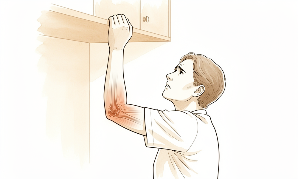
For patients: a plain-language version of this topic is available. See the patient guide.
Overview
Elbow osteoarthritis affects 2% of the population, with an average presentation age of 50 years and a male-to-female ratio of 4:1 [5]. Primary disease is associated with hand dominance and strenuous manual labor, while secondary causes include trauma, osteochondritis dissecans, and synovial osteochondromatosis [5]. The condition is characterized by periarticular hypertrophic osteophytes, capsular contracture, and loose bodies, which act as mechanical blocks at end ranges of motion [5]. Pathologically, the radiocapitellar joint is preferentially involved with relative preservation of the ulnohumeral surfaces and joint space [5]. Clinically, patients present with painful catching, clicking, or locking and loss of terminal extension and flexion, typically without night pain [5]. Ulnar neuropathy is present in up to 50% of cases [5]. Radiographs show osteophytes at the coronoid, radial head, and olecranon with preserved ulnohumeral joint spaces, though they underestimate loose bodies; CT is useful for detailed surgical planning [5].
Nonoperative treatment is the first step in early management [3]. Surgical intervention is indicated for failure of nonsurgical measures, loss of motion interfering with activities of daily living, or painful locking and catching [5]. The goal of treatment is to obtain low pain levels with sufficient motion for function while preserving future surgical options and delaying arthroplasty [1]. Management must be individualized based on etiology, severity, patient age, and functional demands [6, 11]. Joint-sparing procedures, including arthroscopic or open débridement, osteophyte excision, capsular release, and loose body removal, are preferred for most patients [5]. Arthroscopic osteocapsular arthroplasty is safe and efficacious for mild to moderate disease, providing good short-term outcomes with low complication rates [2, 7, 41]. Open procedures, such as the Outerbridge-Kashiwagi arthroplasty, are also effective and safe for both posttraumatic and primary osteoarthritis [16, 19].
Total elbow arthroplasty is rarely indicated for primary osteoarthritis and is generally avoided in patients younger than 65 years or those who are physically active due to concerns about implant longevity [5, 77]. It remains associated with substantial complication and reoperation rates, with survival rates that remain low [42, 70]. Arthroscopic procedures carry risks of deep infection (0.8% to 2.2%) and transient nerve palsies (1% to 3%), primarily involving the radial and ulnar nerves [5]. For patients with less than 90° to 100° of flexion, ulnar nerve transposition and release of the posterior bundle of the medial collateral ligament should be considered regardless of the procedure type [5].
Anatomy & Pathophysiology
Epidemiology and Demographics
Symptomatic primary osteoarthritis of the elbow affects 2% of the population [5]. The average age of presentation is 50 years, with a range of 20 to 70 years [5]. Men are affected more often than women at a 4:1 ratio [5, 66]. Hand dominance and strenuous manual labor are associated with primary elbow osteoarthritis [5, 66]. Primary osteoarthritis most commonly affects middle-aged males, manual laborers, throwing athletes, and heavy weight lifters [71]. Secondary causes of elbow osteoarthritis include trauma, osteochondritis dissecans, and synovial osteochondromatosis [5]. Posttraumatic arthritis is a common complication following traumatic soft-tissue injury, fracture, or surgery [71].
Bony Anatomy and Joint Structure
The elbow is a trocho-ginglymoid joint consisting of the ulnohumeral, radiocapitellar, and proximal radioulnar joints [36, 37]. The articular surface of the distal humerus is angled 30 degrees anterior to the humeral shaft axis [37, 38]. The distal humerus consists of medial and lateral columns [37, 38]. The medial column diverges from the humeral shaft at a 45-degree angle, and the lateral column diverges at a 20-degree angle [56]. The ulnohumeral joint allows for flexion and extension of the elbow [56]. The radiocapitellar joint allows for forearm rotation [56]. The trochlea has a 300-degree arc of cartilage [56]. The ulnohumeral joint is highly congruous and nearly hingelike [123]. The radiohumeral and proximal radioulnar joints allow rotation [123]. The radial head is an elliptical structure covered with articular cartilage along the radiocapitellar joint and approximately 270 degrees of the articular margin [88]. The olecranon fossa is an oval structure that is wider in the medial to lateral dimension [5]. The sublime tubercle provides the attachment site for the anterior bundle of the medial collateral ligament [88, 123].
Ligamentous Anatomy and Stability
Elbow stability is determined by primary stabilizers (ulnohumeral articulation, medial ulnar collateral ligament, lateral ulnar collateral ligament) and secondary stabilizers (radiocapitellar articulation, common flexor tendon, common extensor tendon, joint capsule) [36]. The medial collateral ligament complex comprises the anterior oblique, posterior oblique, and transverse ligaments [60]. The anterior oblique ligament is the strongest component and the primary stabilizer to valgus stress [60]. The anterior band of the medial collateral ligament is taut in extension, and the posterior band is tight in flexion [60]. The posterior bundle of the medial collateral ligament is the primary restraint to valgus stress with the elbow in maximal flexion [37, 38]. Stability in full extension is provided by the medial collateral ligament, joint capsule, and ulnohumeral articulation [37, 38]. The lateral ulnar collateral ligament acts as the primary stabilizer to posterolateral rotatory instability [123]. The radial head functions as an important secondary stabilizer to valgus stress, particularly in medial collateral ligament-deficient elbows [123]. In full extension, 60% of axial load is transmitted through the radiocapitellar joint [37, 38].
Neurovascular Anatomy
The ulnar nerve passes through the cubital tunnel at the medial column of the elbow [56]. The posterior bundle of the medial collateral ligament forms the floor of the cubital tunnel along the course of the ulnar nerve [39]. The radial nerve courses superficial to the elbow joint capsule, anterior to the midpoint of the radiocapitellar joint, where it is vulnerable to injury during arthroscopic or open anterior capsular release [123]. The brachial artery branches into the radial and ulnar arteries at the level of the elbow joint [123]. The inferior ulnar collateral artery provides the only direct supply of oxygenated blood to the ulnar nerve proximal to the cubital tunnel [123].
Pathophysiology and Disease Progression
Osteoarthritis of the elbow is characterized by osteophyte formation, capsular contracture, and loose bodies, often with relative preservation of the joint space [5]. Periarticular hypertrophic osteophytes act as a mechanical block at the end ranges of flexion and extension [5]. Advanced elbow osteoarthritis rarely presents with joint space narrowing [5]. Primary elbow osteoarthritis typically involves the radiocapitellar joint articular cartilage preferentially, with relative preservation of the ulnohumeral articular surfaces [5]. Radiocapitellar degeneration is more common with increasing age [17]. The posteromedial aspect of the radial head appears to be consistently involved in primary elbow osteoarthritis, with reciprocal changes seen on the capitellum [17]. Ulnohumeral osteophytes were found in 95% of patients with symptomatic elbow osteoarthritis, while radiohumeral osteophytes were found in 59% [84]. Osteophytic change occurs predominantly in the ulnohumeral compartment of the elbow, whereas joint space narrowing more frequently affects the radiocapitellar articulation [53]. The primary pathology of elbow osteoarthritis is loss of articular cartilage with resulting osteophyte formation on the olecranon process, coronoid process, and their respective fossae [71]. Secondary resultant changes in elbow osteoarthritis involve osteophyte formation along the margin of the radial head and formation of loose bodies that may result in mechanical block or crepitance [71]. Most patients with elbow osteoarthritis initially complain of pain at terminal limits of motion as a result of capsular stretch and osteophyte impingement [71]. Later in the disease process, pain through the mid arc of motion develops as the extent of cartilage loss progresses [71]. Normal kinematics were preserved in the osteoarthritic elbow with a normal radiocapitellar joint [21]. Three-dimensional computational models identified unique regions of bony impingement in elbow osteoarthritis, such as between the radial head and a posterior capitellar osteophyte in extension [54]. Posttraumatic arthritis can develop in response to the initial cartilage insult or secondary to residual articular incongruities from injuries [10]. Postinjury malunions, nonunions, or residual instability can alter kinematics and load across the elbow, predisposing it to degenerative changes [10]. Posttraumatic arthritis of the elbow is frequently associated with bone loss, making surgical treatment difficult [10]. The mechanisms responsible for the development of post-traumatic osteoarthritis following injury are complex and remain incompletely understood [154]. There is an association between the development of post-traumatic osteoarthritis and the injury pattern and amount of energy absorbed within the joint [154]. Radial head resection in the case of ulnohumeral degeneration increases stress on the ulnohumeral compartment and is suggested to lead to aggravation of pre-existing degeneration [154]. Intrinsic contracture in elbow osteoarthritis leads to the loss of articular cartilage and the formation of marginal osteophytes at the coronoid and olecranon tips and fossae that can limit motion [117].
Classification
Radiographic Classification Systems
Broberg and Morrey: This system classifies elbow osteoarthritis based on osteophyte formation and joint space narrowing [17]. It defines four grades: grade 0 represents a normal joint; grade 1 indicates slight joint-space narrowing with minimum osteophyte formation; grade 2 denotes moderate joint-space narrowing with moderate osteophyte formation; and grade 3 signifies severe degenerative change with gross destruction of the joint [127]. While the system demonstrated substantial intraobserver and interobserver reliability for evaluating radiographic severity in post-traumatic arthritis and primary osteoarthritis [55], the four grades exhibit only fair interobserver reliability that is influenced by subspecialty and experience [127].
Hastings and Retting: This classification focuses on radiocapitellar wear and subluxation [17]. It demonstrated substantial intraobserver and interobserver reliability for evaluating radiographic severity of post-traumatic arthritis and primary osteoarthritis of the elbow [55].
Rettig et al.: This system rates the severity of primary osteoarthritis of the elbow based on joint space narrowing and marginal osteophytes [25]. Clinical and radiographic outcomes following debridement were best in patients classified as class I preoperatively and worst in those classified as class III [25].
Simplified binary rating systems, such as “none or mild” versus “moderate or severe” arthrosis, resulted in moderate agreement among observers [127].
CT-Based Classification Systems
Kwak et al.: This CT-based method quantifies elbow arthritis [17]. The system demonstrated high correlation with the visual analog scale and the Mayo Elbow Performance Score, and moderate correlation with range of motion [17]. A CT-based staging system for elbow osteoarthritis was highly reproducible and clinically feasible compared with previous plain radiograph-based staging systems [40]. Preoperative computed tomography can be used to evaluate the radiologic severity of primary elbow osteoarthritis using the Kwak classification [140].
Other Classification Systems
Bony Landmarks: A bony landmarks classification system effectively delineated osteophyte distribution in elbow patients [59].
Clinical Presentation
Epidemiology and Demographics
Elbow osteoarthritis (OA) predominantly affects middle-aged men, with a male-to-female ratio of 4:1 [5]. Primary elbow OA is most common in males around 50 years of age who are manual laborers, throwing athletes, or heavy weight lifters [71]. The prevalence of symptomatic elbow OA is 2% in the general study population, rising to 3.5% in at-risk individuals older than 40 years [15]. In a Japanese cohort aged 50–89 years, the prevalence of primary elbow OA was 25.2%, with the majority of cases remaining asymptomatic [14]. Among respondents aged 40 years or older, the overall prevalence of elbow OA was 55.0%, with a symptomatic prevalence of 22.6% [23]. Significant risk factors for developing elbow OA include older age, male sex, and a history of elbow trauma [23].
Clinical Symptoms
Patients typically present with pain at the terminal limits of motion, resulting from capsular stretch and osteophyte impingement at the extremes of movement [71]. This pain is usually noted at the end ranges of motion rather than through the midrange [5]. Mechanical symptoms, including painful catching, clicking, or locking, are common [5]. As the disease progresses and cartilage loss extends, pain through the mid-arc of motion develops [71]. Night pain is not typical; its presence should prompt consideration of an inflammatory cause of arthritis [5]. Primary elbow OA may also present with limited movement and potential ulnar nerve symptoms [27]. Clinically, the condition is characterized by stiffness, pain, mechanical symptoms, and weakness [17].
Physical Examination
Inspection should assess for prior surgical incisions and joint effusion at the lateral soft spot [5]. During range-of-motion assessment, pain is usually elicited at the end ranges of flexion and extension rather than throughout the arc [5]. Forearm rotation remains relatively preserved until later stages of the disease process [5]. The degree of disability caused by osteoarthritis depends on the patient’s vocation and physical disability [5].
Pathoanatomy and Radiographic Findings
The primary pathology of elbow OA is loss of articular cartilage, which results in osteophyte formation on the olecranon process, coronoid process, and their respective fossae [71]. Secondary changes involve osteophyte formation along the margin of the radial head and the formation of loose bodies that may cause mechanical block or crepitance [71]. Elbow OA is characterized by osteophyte formation, capsular contracture, and loose bodies, often with relative preservation of the joint space [5]. The disease preferentially involves the radiocapitellar joint articular cartilage, with relative preservation of the ulnohumeral articular surfaces [5]. Primary elbow OA is unique due to this relative preservation of articular cartilage and maintenance of joint space alongside hypertrophic osteophyte formation [18]. Radiographs typically show osteophyte formation at the coronoid process (anterior and medial), coronoid fossa, radial fossa, radial head, olecranon tip, and olecranon fossa [5]. Joint spaces at the ulnohumeral joint are usually preserved, while those at the radiocapitellar joint are mildly narrowed [5]. Loose bodies may be evident on radiographs, which typically underestimate the number present [5]. Radiographically, primary elbow OA is highlighted predominantly by osteophyte formation and progresses with cartilage loss and joint space narrowing [17]. Rarely, advanced disease presents with joint space narrowing [5]. Normal kinematics were preserved in the osteoarthritic elbow with a normal radiocapitellar joint [21].
Imaging
Standard AP and lateral radiographs should be obtained for initial evaluation [5]. CT may be useful for surgical planning, allowing detailed assessment of osteophytes and the presence of loose bodies [5]. CT scans with 3D reconstructions may be useful for evaluating the extent and location of disease and for surgical planning [107]. MRI may be useful to evaluate the status of the soft tissues, including the medial and lateral collateral ligamentous complexes [107]. Electromyography and nerve conduction studies (EMG/NCS) may be useful to evaluate the degree of nerve compression and its contribution to elbow pain and/or dysfunction [107]. A CT-based staging system was highly reproducible and clinically feasible compared with previous plain radiograph-based staging systems for elbow osteoarthritis [40].
Investigations
History and Physical Examination
Pain in elbow osteoarthritis is typically noted at the end ranges of motion rather than through the midrange [5]. Night pain is not typical for this condition; if present, an inflammatory cause of arthritis should be considered [5]. Primary elbow osteoarthritis predominantly affects middle-aged men undertaking heavy manual work, presenting with pain, limited movement, and potential ulnar nerve symptoms [27]. Physical examination includes inspection for prior surgical incisions and joint effusion at the lateral soft spot [5].
Imaging
Plain radiography: Standard AP and lateral radiographs are the initial imaging modality for evaluating elbow osteoarthritis [5]. Radiographs typically show osteophyte formation at the coronoid process (anterior and medial), coronoid fossa, radial fossa, radial head, olecranon tip, and olecranon fossa [5]. Joint spaces at the ulnohumeral joint are usually preserved, while those at the radiocapitellar joint are mildly narrowed [5]. Osteophytic change occurs predominantly in the ulnohumeral compartment, whereas joint space narrowing more frequently affects the radiocapitellar articulation [53]. On imaging assessment, signs of impingement (osteophytes and filling of fossae) are more common than cartilage lesions (joint narrowing) [162]. The Broberg and Morrey and Hastings and Retting classification systems demonstrated substantial intraobserver and interobserver reliability for evaluating radiographic severity of post-traumatic arthritis and primary osteoarthritis of the elbow [55]. The bony landmarks classification system effectively delineated osteophyte distribution in elbow patients [59].
CT: CT may be useful for surgical planning by allowing a detailed assessment of osteophytes and the presence of loose bodies [5]. CT has greater sensitivity than radiographs for the detection of osteophytes and loose bodies in primary elbow osteoarthritis [153].
Other Considerations: Three-dimensional computational models identified the locations and volumes of bony impingement in patients with osteoarthritis of the elbow, including between the radial head and a posterior capitellar osteophyte in extension [54]. Pre-operative simulation results can be used as an index to determine the osteophytes to be removed during arthroscopic surgery for elbow osteoarthritis [51].
Treatment
Non-Operative
Rest, NSAIDs, corticosteroid injections, and activity modification constitute the mainstays of nonsurgical treatment for elbow osteoarthritis [5]. While these interventions can effectively reduce symptoms, they have limited effectiveness for improving range of motion limitations and pain related to impinging osteophytes [66].
Operative
Indications: Surgical treatment is indicated for failure to respond to nonsurgical interventions, loss of motion that interferes with activities of daily living, and painful locking or catching of the elbow [5]. Treatment decisions must be individualized based on disease etiology, severity of degeneration, patient age, and functional demands [13, 28].
Surgical Approach / Technique: Joint-sparing surgical procedures are preferred for the treatment of elbow osteoarthritis [66]. Osteocapsular debridement is an effective option for patients with symptomatic primary elbow osteoarthritis who have failed conservative management [45]. Surgical debridement is effective for disabling symptoms of primary elbow OA with an acceptable complication rate [103]. Open or arthroscopic débridement may be effective in the treatment of early arthritis [106]. Both open and arthroscopic debridement procedures seem to be safe and effective in the treatment of elbow OA [62]. Elbow AOA is a safe, efficacious treatment for patients with mild to moderate osteoarthritis [2]. This minimally invasive technique provides good short-term outcomes in primary elbow osteoarthritis and is associated with a low complication rate [7]. The study recommends this technique in the surgical management of patients with osteoarthritis of the elbow [8]. Patients with either posttraumatic or primary degenerative osteoarthritis can expect satisfactory elbow function and an improvement in pain with little chance of reoperation at the midterm of the follow-up duration [9]. Arthroscopic treatment of elbow osteoarthritis significantly improved 6-month clinical results for functional scores, pain, strength and range of motion [43]. Arthroscopic debridement for elbow osteoarthritis provides satisfactory pain relief, improvement of elbow motion, and good functional outcome [64, 104]. The medial approach is effective for the treatment of advanced primary osteoarthritis of the elbow, especially in patients with ulnar nerve symptoms as well as medial osteophytes [29]. This approach is associated with a low rate of complications and is safe and effective for the treatment of primary osteoarthritis of the elbow [4]. The OK procedure is an effective and safe way of treating both posttraumatic arthritis and osteoarthritis of the elbow [16]. Both open elbow debridement and the OK procedure had excellent survivorship until conversion to total elbow arthroplasty and are viable options in the treatment of primary elbow osteoarthritis and post-traumatic cases [19]. The modified Outerbridge-Kashiwagi procedure combining mini-open and arthroscopic techniques achieved short-term clinical success in pain relief and function restoration in patients with elbow osteoarthritis [112]. Most patients had an uneventful postoperative course and have shown a painless elbow joint, with satisfactory functional recovery at short-term follow-up after open debridement and radiocapitellar replacement [34]. In doing so, we present a reproducible technique that can restore adequate joint space, mobility, and function to the patient with degenerative elbow osteoarthritis [20]. The study evaluated outcomes at an average of ten years after débridement arthroplasties performed through a posteromedial approach for primary osteoarthritis of the elbow [33].
Implant Selection: Total elbow arthroplasty is rarely indicated, and it is not indicated for patients younger than 65 years or physically active patients because of concerns about implant longevity [5]. Total elbow arthroplasty outcomes differ based on etiology and indication for surgery, and TEA is best reserved for low demand, elderly patients who will be able to comply with the 5-lb weightlifting restriction imposed postoperatively to protect the implants from bearing wear, hardware loosening, or failure [106]. Interposition arthroplasty or total elbow arthroplasty is best reserved for more advanced cases of elbow arthritis [106]. It provides favorable surgical outcomes with high satisfaction rates among young patients with elbow osteoarthritis treated with interposition elbow arthroplasty using fascia lata graft [26]. Total elbow arthroplasty has become a reliable treatment option for elbows severely affected by rheumatoid arthritis, with recent reports showing decreased prevalence of complications, maintenance of excellent pain relief, and functional restoration [32]. Elbow arthrodesis is reserved for patients with painful arthritis who are not candidates for total elbow arthroplasty, especially individuals who place high demands on the upper extremities, such as manual laborers [105]. Elbow fusion is also indicated for persistent infection, including tuberculosis [105]. Elbow arthrodesis has also become recognized as the optimal treatment for massive upper extremity trauma seen on the battlefield [105]. For unilateral arthrodesis of the elbow, a position of 90 to 100 degrees of flexion is desirable to provide the most powerful grip strength [105]. If bilateral elbow arthrodesis is indicated, one elbow should be placed in 110 to 120 degrees of flexion to permit the patient to reach the mouth, and the other should be placed in 45 to 65 degrees to aid in personal hygiene [105].
Other Considerations: Regardless of the type of procedure used, ulnar nerve transposition and release of the posterior bundle of the medial collateral ligament (MCL) should be considered for patients who have less than 90° to 100° of elbow flexion [5, 66]. Contraindications for arthroscopic procedures include severe contracture and periarticular heterotopic ossification [5]. Relative contraindications for arthroscopic procedures include prior ulnar nerve transposition and prior extensive open procedures [5]. Osteocapsular arthroplasty refers to the arthroscopic technique for elbow joint débridement; it involves capsular release, loose body removal, and excision of osteophytes [5]. Either a medial or lateral column approach can be used for open débridement, loose body removal, osteophyte resection, and capsulectomy depending upon the location of the pathology and concomitant procedures to be performed [5]. The Outerbridge-Kashiwagi arthroplasty is the classic open procedure in which the olecranon fossa is trephinated and osteophytes are removed [5]. Limitations of the Outerbridge-Kashiwagi procedure are incomplete anterior release and incomplete osteophyte removal anteriorly [5]. During arthroscopic surgery, joint distention moves the capsule away from bone, but the distance between the neurovascular structures and the capsule remain unchanged; therefore, the nerves remain at risk with capsular work [5]. Neurovascular structures at risk during portal placement, débridement, and capsular release include median nerve (anteromedial), the ulnar nerve (posteromedial), and the radial nerve (lies adjacent to the anterolateral capsule) [5]. The brachialis muscle protects the median nerve and brachial artery during capsular procedures [5]. The olecranon fossa is an oval structure that is wider in the medial to lateral dimension [5]. The olecranon osteophytosis extends medially and laterally and not just at the tip, so resection needs to be extended along the medial and lateral aspects of the olecranon to allow maximal extension and prevent impingement along the medial and lateral posterior columns [5]. Coronoid osteophytosis extends medially and not just at the tip, so resection should be extended medially if necessary to maximize the restoration of flexion range of motion [5]. Elbow arthroscopy is technically demanding, and several neurovascular structures that are at risk during the procedure include the radial, ulnar, and median nerves [66]. Unlike other joints, arthroscopic débridement in the elbow can be very effective at reducing symptoms and improving range of motion [66].
Complications
Arthroscopic and Joint-Sparing Procedures
Infection: Deep infections in the elbow are more common than in other joints treated arthroscopically, with a reported rate of 0.8% to 2.2% [5]. Infection can manifest as superficial minor wound complications or deep infection [5]. Infection is associated with intraoperative corticosteroid injections [5].
Nerve palsy: Transient nerve palsies complicate 1% to 3% of cases [5]. The radial and ulnar nerves are the most commonly affected by transient nerve palsies [5].
Stiffness / Arthrofibrosis: Heterotopic ossification is a recognized complication associated with stiffness [5].
Wound complications: Hematoma formation is a recognized complication [5]. Synovial ganglion formation is a recognized complication [5].
Other Considerations: Arthroscopic debridement for primary degenerative osteoarthritis is associated with low complication rates [12]. Arthroscopic treatment of elbow osteoarthritis is associated with a low complication rate [7]. The medial trans-flexor approach for capsulectomy and debridement is associated with a low rate of complications [4]. Elbow arthroscopic osteocapsular arthroplasty is considered a safe treatment for patients with mild to moderate osteoarthritis [2]. Arthroscopic osteophyte resection and capsulectomy were safe and effective in the reported series [49]. Patients undergoing arthroscopic elbow debridement for primary or posttraumatic osteoarthritis have a little chance of reoperation at midterm follow-up [9].
Total Elbow Arthroplasty
Other Considerations: Total elbow replacement is associated with a high complication rate [122]. Complication rates for the Latitude primary total elbow arthroplasty remain high, though they are comparable to those of other elbow arthroplasties [70]. Wear of the hinge mechanism is a risk during follow-up for semiconstrained elbow arthroplasties [79]. Total elbow arthroplasty for rheumatoid arthritis has shown decreased prevalence of complications in recent reports [32].
Recovery
Non-Operative Management: The provided evidence base does not contain specific recovery timelines or rehabilitation protocols for non-operative management of elbow osteoarthritis.
Operative Management: Joint-Sparing Procedures: Arthroscopic debridement for primary degenerative osteoarthritis yields statistically significant and clinically relevant improvements in elbow range of motion and clinical outcomes, with low complication and reoperation rates [12]. Serial assessment of patients undergoing arthroscopic osteocapsular arthroplasty for primary elbow osteoarthritis demonstrates that clinical outcomes improve from preoperative assessment to short- and medium-term follow-up [31]; however, range of motion decreases between short- and medium-term follow-up in this cohort [31]. Open elbow debridement and the Outerbridge-Kashiwagi procedure exhibit excellent survivorship until conversion to total elbow arthroplasty, representing viable options for primary elbow osteoarthritis and post-traumatic cases [19]. Most patients undergoing open debridement and radiocapitellar replacement for primary and post-traumatic arthritis experience an uneventful postoperative course, resulting in a painless elbow joint with satisfactory functional recovery at short-term follow-up [34]. A tendency of advancing roentgenological osteoarthritic changes is recognized in most patients 10 years after the Outerbridge-Kashiwagi method for arthroplasty of osteoarthritis of the elbow [86].
Operative Management: Arthroplasty: Total elbow arthroplasty is rarely indicated for elbow osteoarthritis and is not indicated for patients younger than 65 years or physically active patients due to concerns about implant longevity [5]. After 15 years, elbows treated with radial head arthroplasty present signs of arthritis in the majority of patients [72]. Open arthrolysis combined with radial head arthroplasty for post-traumatic elbow stiffness yields satisfactory short-term outcomes at 3 years, with substantial improvements in elbow mobility and function, and these results are durable over the long term (8 years) [74].
Other Considerations: The evidence provided reports outcome trajectories at specific follow-up intervals (short-term, medium-term, 3 years, 8 years, 10 years, and 15 years) but does not define specific recovery phases such as light activity, full activity, or complete recovery timelines. Consequently, no specific week or month ranges for return to desk work, driving, manual labor, or sport can be derived from this text. Rehabilitation protocols, immobilisation durations, and functional milestone benchmarks (e.g., Constant, ASES scores) are not detailed in the provided evidence.
Key Evidence
- [L4] The goal of treatment is to obtain a low level of pain with sufficient motion range to ensure good function, while preserving future surgical options and delaying elbow arthroplasty to the extent possible. [1] (10.1016/j.otsr.2013.11.004)
- [L4] Elbow AOA is a safe, efficacious treatment for patients with mild to moderate osteoarthritis. [2] (10.1016/j.jhsa.2015.11.018)
- [L5] Nonoperative treatment remains the first step in the early management of elbow osteoarthritis. [3] (10.2106/jbjs.e.00568)
- [L4] This approach is associated with a low rate of complications and is safe and effective for the treatment of primary osteoarthritis of the elbow. [4] (10.1016/j.jhsa.2011.07.018)
- [L5] Treatment of posttraumatic arthritis and osteoarthritis of the elbow must be individualized on a case-by-case basis depending on location of disease, age/demand level of the patient, and stage of disease. [6] (10.1016/j.hcl.2011.02.001)
- [L4] This minimally invasive technique provides good short-term outcomes in primary elbow osteoarthritis and is associated with a low complication rate. [7] (10.1016/j.otsr.2019.09.003)
- [L4] The study recommends this technique in the surgical management of patients with osteoarthritis of the elbow. [8] (10.1302/0301-620x.96b2.30714)
- [L3] Patients with either pathology can expect satisfactory elbow function and an improvement in pain with little chance of reoperation at the midterm of the follow-up duration. [9] (10.1016/j.jseint.2021.07.018)
- [L5] [10] (10.1016/j.jhsa.2022.12.014)
- [L5] The appropriate treatment for elbow arthritis depends on the etiology, severity, patient age, and functional demands. [11] (10.1016/j.jhsa.2009.02.019)
- [L1] Elbow arthroscopic debridement for primary degenerative osteoarthritis results in statistically significant and clinically relevant improvement in elbow range of motion and clinical outcomes with low complication and reoperation rates. [12] (10.1016/j.arthro.2017.08.247)
- [L5] Surgical treatment for elbow arthritis is based on disease etiology, severity of degeneration, and patient age. [13] (10.1016/j.jhsa.2007.12.022)
- [L3] The prevalence of primary elbow osteoarthritis in Japanese subjects aged 50-89 years was 25.2%, with most cases being asymptomatic. [14] (10.1016/j.jse.2021.07.015)
- [L4] The prevalence of symptomatic elbow osteoarthritis was 2% in the study population, with a prevalence of 3.5% in the at-risk population older than 40 years. [15] (10.1016/s1058-2746(09)80024-4)
- [L4] The OK procedure is an effective and safe way of treating both posttraumatic arthritis and osteoarthritis of the elbow. [16] (10.1016/j.jse.2015.11.052)
- [L3] [17] (10.1177/17585732251327183)
- [L4] Primary osteoarthritis of the elbow is unique due to relative preservation of articular cartilage and maintenance of joint space with hypertrophic osteophyte formation. [18] (10.5435/00124635-200802000-00005)
- [L4] Both open elbow debridement and the OK procedure had excellent survivorship until conversion to total elbow arthroplasty and are viable options in the treatment of primary elbow osteoarthritis and post-traumatic cases. [19] (10.1016/j.jse.2022.01.138)
- [L5] In doing so, we present a reproducible technique that can restore adequate joint space, mobility, and function to the patient with degenerative elbow osteoarthritis. [20] (10.1016/j.eats.2024.103332)
- [L4] Normal kinematics was preserved in the osteoarthritic elbow with a normal radiocapitellar joint (OAN group). [21] (10.1016/j.jhsa.2013.02.006)
- [L3] The prevalence of elbow OA was 55.0% in respondents aged 40 years or older, with a symptomatic prevalence of 22.6%; older age, male sex, and a history of elbow trauma were identified as significant risk factors. [23] (10.1016/j.jse.2018.02.049)
- [L4] [25] (10.1016/j.jse.2007.03.014)
- [L4] It provides favorable surgical outcomes with high satisfaction rates among young patients with elbow osteoarthritis. [26] (10.1016/j.jhsg.2024.05.002)
- [L4] Primary elbow osteoarthritis predominantly affects middle-aged men undertaking heavy manual work, presenting with pain, limited movement, and potential ulnar nerve symptoms. [27] (10.1111/j.1758-5740.2010.00089.x)
- [L5] Treatment of elbow arthritis must be individualized based on etiology, severity, patient age, and functional demands; nonsurgical management may provide relief in early stages, while surgical options range from arthroscopic debridement for pain at motion extremes to total elbow arthroplasty for pain throughout the arc of motion. [28] (10.1016/j.jhsa.2012.12.037)
- [L4] The medial approach is effective for the treatment of advanced primary osteoarthritis of the elbow, especially in patients with ulnar nerve symptoms as well as medial osteophytes. [29] (10.2106/jbjs.d.02684)
- [L4] Serial assessment of patients with primary elbow OA who underwent arthroscopic OCA showed that the clinical outcomes improved from preoperative assessment to short- and medium-term follow-up, although ROM decreased between short- and medium-term follow-up. [31] (10.1177/23259671231162398)
- [L4] Total elbow arthroplasty has become a reliable treatment option for elbows severely affected by rheumatoid arthritis, with recent reports showing decreased prevalence of complications, maintenance of excellent pain relief, and functional restoration. [32] (10.2106/00004623-199805000-00008)
- [L4] Most patients had an uneventful postoperative course and have shown a painless elbow joint, with satisfactory functional recovery at short-term follow-up. [34] (10.1016/j.jse.2011.08.071)
- [L4] CT-based staging system was highly reproducible and clinically feasible, compared with previous plain radiograph-based staging systems, for elbow osteoarthritis. [40] (10.1016/j.joca.2019.03.004)
- [L4] Arthroscopic osteocapsular arthroplasty can be recommended for its favorable overall treatment outcomes for elbow osteoarthritis. [41] (10.1016/j.jse.2019.09.036)
- [L4] Total elbow arthroplasty remains associated with substantial complication and reoperation rates. [42] (10.1016/j.jhsg.2026.100981)
- [L3] Arthroscopic treatment of elbow osteoarthritis significantly improved 6-month clinical results for functional scores, pain, strength and range of motion. [43] (10.1016/j.otsr.2019.09.002)
- [L4] The ulno-humeral arthroplasty is a simple and safe procedure for the treatment of elbow osteoarthritis. [44] (10.1016/s1058-2746(96)80501-5)
- [L2] Osteocapsular debridement is an effective surgical treatment option for patients with symptomatic primary elbow osteoarthritis who have failed conservative management. [45] (10.1016/j.jse.2020.01.060)
- [L4] This procedure addresses the pathologic processes associated with arthritis of the elbow and was safe and effective in this series. [49] (10.1016/j.jse.2007.04.005)
- [L4] These results can be used as an index to determine the osteophytes to be removed during arthroscopic surgery for elbow osteoarthritis. [51] (10.1016/j.jseint.2026.101667)
- [L4] Elbow hemiarthroplasty is an option for young or active patients with end-stage posttraumatic arthritis who are unwilling to accept activity limitations, though high rates of revision surgery and revision to total elbow arthroplasty occur. [52] (10.5435/jaaos-d-18-00055)
- [L3] Osteophytic change occurs predominantly in the ulnohumeral compartment of the elbow, whereas joint space narrowing more frequently affects the radiocapitellar articulation. [53] (10.1016/j.jse.2006.08.005)
- [L4] Three-dimensional computational models identified the locations and volumes of bony impingement in patients with osteoarthritis of the elbow and highlighted unique regions of impingement, such as between the radial head and a posterior capitellar osteophyte in extension. [54] (10.1016/j.jhsa.2013.03.035)
- [L3] Both the BM and HR classification systems demonstrated substantial intraobserver and interobserver reliability for evaluating radiographic severity of post-traumatic arthritis and primary osteoarthritis of the elbow. [55] (10.1016/j.jse.2014.10.015)
- [L3] The bony landmarks classification system effectively delineated osteophyte distribution in elbow patients. [59] (10.1186/s13018-025-06145-9)
- [L5] However, from the data we obtained the open and arthroscopic debridement procedures seem to be safe and effective in the treatment of elbow OA. [62] (10.1186/s12891-018-2318-x)
- [L4] Arthroscopic debridement for elbow osteoarthritis provides satisfactory pain relief, improvement of elbow motion, and good functional outcome. [64] (10.1016/j.jse.2014.01.009)
- [L4] Survival rates nonetheless remain low and complication rates remain high yet are comparable to those of other elbow arthroplasties. [70] (10.1016/j.jse.2021.08.028)
- [L4] After 15 years, elbows treated with RHA presented signs of arthritis in the majority of patients. [72] (10.1016/j.jse.2025.06.026)
- [L4] OEA with RHA yielded satisfactory short-term outcomes for PTES at 3 years, with substantial improvements in elbow mobility and function, and the results were durable over the long term (8 years). [74] (10.1016/j.jse.2021.10.028)
- [L4] Surgical options must be tailored to cartilage integrity and bone structure, with total elbow arthroplasty generally avoided in young, active patients due to poor durability. [77] (10.1016/j.jhsg.2025.100736)
- [Case_report] Long-term survival of semiconstrained elbow arthroplasties is favorable, but wear of the hinge mechanism is a risk during follow-up. [79] (10.1016/j.otsr.2014.07.013)
- [L4] [84] (10.1016/j.jse.2008.03.012)
- [L4] However, a tendency of advancing roentgenological osteoarthritic changes was recognized in most patients 10 years after OKM. [86] (10.1007/bf01234111)
- [L1] Surgical debridement is an effective treatment for the disabling symptoms of primary elbow OA with an acceptable complication rate. [103] (10.1302/2058-5241.5.190095)
- [L4] Arthroscopic debridement for the elbow osteoarthritis provided satisfactory pain relief, improvement of elbow motion, and good functional outcome. [104] (10.1016/s0363-5023(11)60056-7)
- [L4] The modified Outerbridge-Kashiwagi procedure combining mini-open and arthroscopic techniques achieved short-term clinical success in pain relief and function restoration in patients with elbow osteoarthritis. [112] (10.1016/j.jse.2020.05.007)
- [L3] Arthroscopic osteocapsular arthroplasty is comparable to the OPEN procedure in managing primary osteoarthritis of the elbow; however, the OPEN procedure shows the better outcome in improvement of flexion limitation. [113] (10.1016/j.arthro.2018.11.057)
- [L3] Total elbow replacement is associated with a high complication rate and therefore may be warranted only for seriously disabled patients. [122] (10.2106/jbjs.d.02734)
- [L3] [127] (10.1016/j.jhsa.2011.12.043)
- [L3] [140] (10.1016/j.jse.2023.05.041)
- [L1] CT has greater sensitivity than radiographs for the detection of osteophytes and loose bodies in primary elbow osteoarthritis. [153] (10.1016/j.jse.2021.04.001)
- [L5] [154] (10.1007/s00167-015-3518-7)
- [L3] On the imaging assessment, signs of impingement (osteophytes and filling of fossae) are more common than cartilage lesions (joint narrowing). [162] (10.1016/j.otsr.2019.09.012)
See Also
References
[1] Post-traumatic osteoarthritis of the elbow. Orthopaedics & Traumatology: Surgery & Research. 2014. DOI: 10.1016/j.otsr.2013.11.004
[2] Outcomes of Elbow Arthroscopic Osteocapsular Arthroplasty. The Journal of Hand Surgery. 2016. DOI: 10.1016/j.jhsa.2015.11.018
[3] Management of Elbow Osteoarthritis. The Journal of Bone & Joint Surgery. 2006. DOI: 10.2106/jbjs.e.00568
[4] Capsulectomy and Debridement for Primary Osteoarthritis of the Elbow Through a Medial Trans-Flexor Approach. The Journal of Hand Surgery. 2011. DOI: 10.1016/j.jhsa.2011.07.018
[5] Aaos Comprehensive Orthopaedic Review 3. Arthritis and Arthroplasty of the Elbow > I. Osteoarthritis.
[6] Primary Osteoarthritis and Posttraumatic Arthritis of the Elbow. Hand Clinics. 2011. DOI: 10.1016/j.hcl.2011.02.001
[7] Arthroscopic treatment of elbow osteoarthritis. Orthopaedics & Traumatology: Surgery & Research. 2019. DOI: 10.1016/j.otsr.2019.09.003
[8] Arthroscopic debridement in the treatment of patients with osteoarthritis of the elbow, based on computer simulation. The Bone & Joint Journal. 2014. DOI: 10.1302/0301-620x.96b2.30714
[9] Midterm outcomes and survivorship of arthroscopic elbow debridement: a comparison of posttraumatic versus primary degenerative osteoarthritis. JSES International. 2022. DOI: 10.1016/j.jseint.2021.07.018
[10] Elbow Arthritis. The Journal of Hand Surgery. 2023. DOI: 10.1016/j.jhsa.2022.12.014
[11] Elbow Arthritis: Current Concepts. The Journal of Hand Surgery. 2009. DOI: 10.1016/j.jhsa.2009.02.019
[12] Arthroscopic Debridement for Primary Degenerative Osteoarthritis of the Elbow Leads to Significant Improvement in Range of Motion and Clinical Outcomes: A Systematic Review. Arthroscopy. 2017. DOI: 10.1016/j.arthro.2017.08.247
[13] Surgical Options for the Arthritic Elbow. The Journal of Hand Surgery. 2008. DOI: 10.1016/j.jhsa.2007.12.022
[14] Prevalence and associated factors of primary elbow osteoarthritis in the Japanese general elderly population: a Japanese cohort survey randomly sampled from a basic resident registry. Journal of Shoulder and Elbow Surgery. 2022. DOI: 10.1016/j.jse.2021.07.015
[15] Prevalence and etiology of symptomatic elbow osteoarthritis. Journal of Shoulder and Elbow Surgery. 1994. DOI: 10.1016/s1058-2746(09)80024-4
[16] The outerbridge-kashiwagi procedure for primary degenerative arthritis of the elbow vs. post traumatic arthritis. Journal of Shoulder and Elbow Surgery. 2016. DOI: 10.1016/j.jse.2015.11.052
[17] Sex-related differences in wear patterns in primary elbow osteoarthritis. Shoulder & Elbow. 2025. DOI: 10.1177/17585732251327183
[18] Primary Osteoarthritis of the Elbow: Current Treatment Options. Journal of the American Academy of Orthopaedic Surgeons. 2008. DOI: 10.5435/00124635-200802000-00005
[19] Long-term survivorship of open débridement and débridement arthroplasty for elbow arthritis: a retrospective chart review. Journal of Shoulder and Elbow Surgery. 2022. DOI: 10.1016/j.jse.2022.01.138
[20] Triceps Tendon‐Guided Arthroscopic Olecranon Debridement for Posterior Impingement Caused by Degenerative Elbow Osteoarthritis: A Technical Note. Arthroscopy Techniques. 2025. DOI: 10.1016/j.eats.2024.103332
[21] Kinematic Changes in Elbow Osteoarthritis: In Vivo and 3-Dimensional Analysis Using Computed Tomographic Data. The Journal of Hand Surgery. 2013. DOI: 10.1016/j.jhsa.2013.02.006
[23] The prevalence of elbow osteoarthritis in Japanese middle-aged and elderly populations: the relationship between risk factors and function. Journal of Shoulder and Elbow Surgery. 2018. DOI: 10.1016/j.jse.2018.02.049
[25] Primary osteoarthritis of the elbow: Lack of radiographic evidence for morphologic predisposition, results of operative debridement at intermediate follow-up, and basis for a new radiographic classification system. Journal of Shoulder and Elbow Surgery. 2008. DOI: 10.1016/j.jse.2007.03.014
[26] Functional Outcomes Following Interposition Elbow Arthroplasty Using Fascia Lata Graft for Post-Traumatic Elbow Osteoarthritis Without Ligament Reconstruction: A Minimum 3-Year Follow-Up Study. Journal of Hand Surgery Global Online. 2024. DOI: 10.1016/j.jhsg.2024.05.002
[27] Primary Elbow Osteoarthritis: An Updated Review. Shoulder & Elbow. 2011. DOI: 10.1111/j.1758-5740.2010.00089.x
[28] Elbow Arthritis: Current Concepts. The Journal of Hand Surgery. 2013. DOI: 10.1016/j.jhsa.2012.12.037
[29] Débridement Arthroplasty for Primary Osteoarthritis of the Elbow. Journal of Bone and Joint Surgery. 2005. DOI: 10.2106/jbjs.d.02684
[31] Serial Changes in Clinical Outcomes After Arthroscopic Osteocapsular Arthroplasty for Primary Elbow Osteoarthritis: A Medium-term Follow-up Study. Orthopaedic Journal of Sports Medicine. 2023. DOI: 10.1177/23259671231162398
[32] Total Elbow Arthroplasty in Patients Who Have Juvenile Rheumatoid Arthritis. The Journal of Bone & Joint Surgery*. 1998. DOI: 10.2106/00004623-199805000-00008
[33] 00004623-200503001-00009. 2005.
[34] Open debridement and radiocapitellar replacement in primary and post-traumatic arthritis of the elbow: a multicenter study. Journal of Shoulder and Elbow Surgery. 2012. DOI: 10.1016/j.jse.2011.08.071
[36] Orthopaedic Knowledge Update 13 Ebook Without Multimedia. Anatomy, Biomechanics, Physical Examination, and Imaging of the Elbow > Summary and Conclusions.
[37] Miller S Review Of Orthopaedics. SECTION 16 PATELLAR TRACKING IN TOTAL KNEE ARTHROPLASTY > ELBOW.
[38] Miller S Review Of Orthopaedics. ELBOW.
[39] Aaos Comprehensive Orthopaedic Review 3. Elbow Stiffness* > IV. Evaluation.
[40] Intraobserver and interobserver reliability of the computed tomography-based radiographic classification of primary elbow osteoarthritis: comparison with plain radiograph-based classification and clinical assessment. Osteoarthritis and Cartilage. 2019. DOI: 10.1016/j.joca.2019.03.004
[41] Arthroscopic osteocapsular arthroplasty for advanced-stage primary osteoarthritis of the elbow using a computed tomography–based classification. Journal of Shoulder and Elbow Surgery. 2020. DOI: 10.1016/j.jse.2019.09.036
[42] Total Elbow Arthroplasty Complication, Reoperation, and Revision Rates: A Comparison Between Arthroplasty for Arthritis Versus Fracture. Journal of Hand Surgery Global Online. 2026. DOI: 10.1016/j.jhsg.2026.100981
[43] Arthroscopic debridement for osteoarthritis of the elbow: Results and analysis of predictive factors. Orthopaedics & Traumatology: Surgery & Research. 2019. DOI: 10.1016/j.otsr.2019.09.002
[44] The Outerbridge-Kashiwagi ulno-humeral arthroplasty. Journal of Shoulder and Elbow Surgery. 1996. DOI: 10.1016/s1058-2746(96)80501-5
[45] The Clinical Impact of Different Approaches to Osteocapsular Debridement for Primary Osteoarthritis of the Elbow: A Systematic Review. Journal of Shoulder and Elbow Surgery. 2020. DOI: 10.1016/j.jse.2020.01.060
[49] Osteoarthritis of the elbow: Results of arthroscopic osteophyte resection and capsulectomy. Journal of Shoulder and Elbow Surgery. 2008. DOI: 10.1016/j.jse.2007.04.005
[51] Predicting impinging osteophytes based on pre-operative simulation results to guide arthroscopic débridement for elbow osteoarthritis. JSES International. 2026. DOI: 10.1016/j.jseint.2026.101667
[52] Outcomes After Hemiarthroplasty of the Elbow for the Management of Posttraumatic Arthritis: Minimum 2-Year Follow-up. Journal of the American Academy of Orthopaedic Surgeons. 2019. DOI: 10.5435/jaaos-d-18-00055
[53] Radiographic changes at the elbow in primary osteoarthritis: A comparison with normal aging of the elbow joint. Journal of Shoulder and Elbow Surgery. 2007. DOI: 10.1016/j.jse.2006.08.005
[54] Identifying the Location and Volume of Bony Impingement in Elbow Osteoarthritis by 3-Dimensional Computational Modeling. The Journal of Hand Surgery. 2013. DOI: 10.1016/j.jhsa.2013.03.035
[55] Reliability testing of two classification systems for osteoarthritis and post-traumatic arthritis of the elbow. Journal of Shoulder and Elbow Surgery. 2015. DOI: 10.1016/j.jse.2014.10.015
[56] A Lange Medical Book Current Diagnosis Treatment In Orthopedics Fifth Edition. 2Musculoskeletal Trauma Surgery > INJURIES AROUND THE ELBOW.
[59] Bony landmarks guided mapping of the osteophytes of the elbow osteoarthritis patients: a three dimensional computed tomograph based study. Journal of Orthopaedic Surgery and Research. 2025. DOI: 10.1186/s13018-025-06145-9
[60] Aaos Comprehensive Orthopaedic Review 3. Elbow Injuries in the Athlete* > II. Medial Collateral Ligament Injuries.
[62] Treatment of osteoarthritis of the elbow with open or arthroscopic debridement: a narrative review. BMC Musculoskeletal Disorders. 2018. DOI: 10.1186/s12891-018-2318-x
[64] Arthroscopic débridement for primary osteoarthritis of the elbow: analysis of preoperative factors affecting outcome. Journal of Shoulder and Elbow Surgery. 2014. DOI: 10.1016/j.jse.2014.01.009
[66] Aaos Comprehensive Orthopaedic Review 3. Arthritis and Arthroplasty of the Elbow > Osteoarthritis.
[70] Mid-term results of the Latitude primary total elbow arthroplasty. Journal of Shoulder and Elbow Surgery. 2022. DOI: 10.1016/j.jse.2021.08.028
[71] Orthopaedic Knowledge Update 13 Ebook Without Multimedia. Elbow Degenerative Conditions and Nerve Disorders > Etiologies.
[72] What happens to the elbow 15 years after a radial head prosthesis? A clinical and imaging long-term follow-up study. Journal of Shoulder and Elbow Surgery. 2026. DOI: 10.1016/j.jse.2025.06.026
[74] Long-term outcomes of open arthrolysis combined with radial head arthroplasty for post-traumatic elbow stiffness: results are durable over 8 years. Journal of Shoulder and Elbow Surgery. 2022. DOI: 10.1016/j.jse.2021.10.028
[77] Management of Severe Elbow Arthritis in a Young Patient. Journal of Hand Surgery Global Online. 2025. DOI: 10.1016/j.jhsg.2025.100736
[79] Mechanical failure of the Coonrad-Morrey linked total elbow arthroplasty: A case report. Orthopaedics & Traumatology: Surgery & Research. 2014. DOI: 10.1016/j.otsr.2014.07.013
[84] Pattern of osteophyte distribution in primary osteoarthritis of the elbow. Journal of Shoulder and Elbow Surgery. 2008. DOI: 10.1016/j.jse.2008.03.012
[86] Outerbridge-Kashiwagi's method for arthroplasty of osteoarthritis of the elbow — 44 elbows followed for 8–16 years. Journal of Orthopaedic Science. 1996. DOI: 10.1007/bf01234111
[88] Orthopaedic Knowledge Update 13 Ebook Without Multimedia. Anatomy, Biomechanics, Physical Examination, and Imaging of the Elbow > Anatomy > Bony Anatomy.
[103] Arthroscopic and open debridement in primary elbow osteoarthritis: a systematic review and meta-analysis. EFORT Open Reviews. 2020. DOI: 10.1302/2058-5241.5.190095
[104] Factors Affecting the Results of Arthroscopic Debridement for the Elbow Osteoarthritis. The Journal of Hand Surgery. 2011. DOI: 10.1016/s0363-5023(11)60056-7
[105] Campbell S Operative Orthopaedics 4 Volume Set. ARTHROSCOPIC SHOULDER ARTHRODESIS FOR BRACHIAL PLEXUS INJURY > ELBOW ARTHRODESIS.
[106] Orthopaedic Knowledge Update 13 Ebook Without Multimedia. Elbow Degenerative Conditions and Nerve Disorders > Summary.
[107] Orthopaedic Knowledge Update 13 Ebook Without Multimedia. Elbow Degenerative Conditions and Nerve Disorders > Evaluation.
[112] Clinical outcomes of a combined arthroscopic and mini-open Outerbridge-Kashiwagi procedure for elbow osteoarthritis. Journal of Shoulder and Elbow Surgery. 2020. DOI: 10.1016/j.jse.2020.05.007
[113] Clinical Outcome of Osteocapsular Arthroplasty for Primary Osteoarthritis of the Elbow: Comparison of Arthroscopic and Open Procedure. Arthroscopy: The Journal of Arthroscopic & Related Surgery. 2019. DOI: 10.1016/j.arthro.2018.11.057
[117] Aaos Comprehensive Orthopaedic Review 3. Elbow Stiffness* > V. Classification.
[122] Primary Souter-Strathclyde Total Elbow Prosthesis in Rheumatoid Arthritis. Journal of Bone and Joint Surgery. 2005. DOI: 10.2106/jbjs.d.02734
[123] Aaos Comprehensive Orthopaedic Review 3. Anatomy of the Shoulder, Arm, and Elbow > II. Arm and Elbow.
[127] Radiographic Arthrosis After Elbow Trauma: Interobserver Reliability. The Journal of Hand Surgery. 2012. DOI: 10.1016/j.jhsa.2011.12.043
[140] Radiologically severe osteoarthritis is related to worse clinical outcomes after arthroscopic osteocapsular arthroplasty in primary elbow osteoarthritis at medium-term follow-up: a retrospective cohort study. Journal of Shoulder and Elbow Surgery. 2023. DOI: 10.1016/j.jse.2023.05.041
[153] Effectiveness of radiographs and computed tomography in evaluating primary elbow osteoarthritis. Journal of Shoulder and Elbow Surgery. 2021. DOI: 10.1016/j.jse.2021.04.001
[154] Biomechanical considerations in the pathogenesis of osteoarthritis of the elbow. Knee Surgery, Sports Traumatology, Arthroscopy. 2015. DOI: 10.1007/s00167-015-3518-7
[162] Can preoperative imaging predict the outcomes after arthroscopic release for elbow arthritis?. Orthopaedics & Traumatology: Surgery & Research. 2019. DOI: 10.1016/j.otsr.2019.09.012