Clinicians › Elbow
Pronator and AIN Release
For patients: a plain-language version of this topic is available. See the patient guide.
Overview
Pronator syndrome is a clinical diagnosis lacking objective pathophysiological confirmation, supported primarily by subjective operative findings and post-surgical relief [3]. The terminology should be refined to distinguish superficialis-pronator syndrome from lacertus syndrome, as these represent distinct surgical targets [13]. Given the paucity of controlled trials demonstrating superiority over sham surgery or other treatments [3], conservative management remains the first-line choice for median nerve compression in the forearm [9]. This initial approach includes rest, avoidance of aggravating activities, and anti-inflammatory medication [6]. A prolonged nonsurgical strategy is warranted in most cases, as successful nonoperative treatment is achievable when the correct diagnosis is established [15, 8]. For idiopathic anterior interosseous nerve palsy, the decision between nonsurgical and surgical management, and the duration of waiting for spontaneous recovery, remains controversial [35].
Surgical decompression of the median or anterior interosseous nerve in the forearm is rarely indicated [15]. Indications for pronator syndrome include persistent symptoms for more than six months despite conservative management [1, 6], or when activities of daily living require frequent strong pronation and electromyography confirms compression [2]. For anterior interosseous nerve syndrome, surgery is considered after a minimum of twelve months with no signs of motor improvement [1]. In these cases, other medical causes should be excluded before proceeding to decompression [2].
Operative options include minimally invasive and endoscopically assisted techniques, which can release all potential entrapment sites and have demonstrated improved DASH scores [14, 16]. A mini-invasive technique resulted in the disappearance of pronator teres symptoms in 93% of cases [4]. In contrast, pronator teres release is more invasive, requiring deeper dissection and more complicated surgical skills [11]. When associated with a persistent median artery, resection of the arterial segment is the recommended treatment [21]. Caution is advised given the high rate of morbidity associated with radial tunnel syndrome and its treatment [73]. For reoperations to be successful, the ulnar nerve must be examined and all potential levels of compression released [18].
Anatomy & Pathophysiology
Median Nerve and AIN Anatomy
The median nerve traverses a series of musculoaponeurotic potential sites of compression in the proximal forearm, including the deep fascia (lacertus fibrosus), the fibrous arch beneath the superficialis head of the pronator teres, the fibrous arch beneath the deep head of the pronator teres, the fibrous arch within the flexor digitorum superficialis, and Gantzer's muscle [145]. The median nerve exits the pronator tunnel and gives rise to the anterior interosseous nerve (AIN) roughly 5 cm distal to the medial epicondyle [54]. The AIN serves as the motor nerve for the flexor pollicis longus, the flexor digitorum profundus of the index and ring fingers, and the pronator quadratus muscle [54]. Additionally, the AIN innervates a portion of the wrist capsule, which may result in vague volar wrist pain [54].
Anatomical variants significantly influence compression risk. In 74% of cadaveric specimens, the AIN branched at or distal to the flexor digitorum superficialis (FDS) arch [26]. Only 8% of cadaveric specimens had an AIN with an ulnar-sided origin off the median nerve [26]. When the AIN takes origin from the radial side of the median nerve, it is more susceptible to compression against a fibrous arch than if it originates on the deep or posterior aspect of the median nerve [145]. Gantzer's muscle, an accessory head of the flexor pollicis longus or flexor digitorum profundus, is observed in 45 to 74% of dissected forearms [134]. The most frequent origin sites for Gantzer's muscle are the coronoid process of the ulna and the medial epicondyle of the humerus [134]. This muscle usually runs anteriorly to the AIN and posteriorly to the median nerve [134]. The majority of Gantzer's muscles are asymptomatic [134].
Compression Sites and Variants
Cadaveric dissections have identified two types of the FDS arch: a distinct fibrous arch and an indistinct fibrous arch with vertical fibers blending into overlying fascia [26]. Only 42% of cadaveric specimens had a distinct FDS arch, which averaged 1.69 cm in length [26]. The majority of cadaveric specimens had an indistinct FDS arch, and 77% of those had overlying muscle requiring an average release of 2.6 cm [26]. The musculoaponeurotic system formed by the multiple heads of the pronator teres and flexor superficialis can be arranged in a variable number of arches across the median nerve, ranging from no arch to two arches [145].
The ligament of Struthers is a variant anatomy arising from the supracondylar process to attach to the medial epicondyle and is a potential site of median nerve compression [78]. This ligament is present in roughly 1% of the population [54].
Pathophysiology and Mechanisms
Pronator syndrome is a median nerve neuropathy due to entrapment usually between the two heads of the pronator teres or less commonly by the arch of the flexor digitorum superficialis [134]. In patients with pronator teres muscle overuse, the median nerve can be dynamically compressed when it passes between the two heads of the pronator teres [134]. The most plausible mechanism for AIN or median nerve syndrome associated with Gantzer's muscle is direct compression by a hypertrophic muscle [134]. Conversely, compression to the anterior interosseous nerve by normal tissues surrounding it may not exist [70].
Anterior interosseous nerve palsy is an uncommon disease of unknown etiology and pathophysiology [22]. AIN palsy creates objective weakness and electrophysiological abnormalities, in contrast to pronator syndrome which involves forearm pain ascribed to median nerve compression with no objective tests that can verify the diagnosis [22].
Supracondylar elbow fractures in children are associated with nerve-related complications in 6.6 to 31% of cases [10]. Since 95% of supracondylar elbow fractures occur in extension, the displacement of the distal fragment is an important factor in the pathophysiology of nerve trunk injury [10]. In a series of 210 supracondylar fractures, the median nerve was involved in 28 cases, with posterolateral displacement present in 86% of these cases [10].
Classification
Terminology and Nomenclature: The term "pronator syndrome" should be replaced with "superficialis-pronator syndrome" to clearly distinguish it from lacertus syndrome as distinct targets for surgical release [13]. The authors recommend that the term "lacertus syndrome" refer to intermittent median nerve compression at the level of the lacertus fibrosus, while the pathology originally described by Bennett should be termed chronic exertional compartment syndrome of the pronator teres or "Bennett's syndrome" [144].
Anterior Interosseous Nerve Syndrome (AINS): Anterior interosseous nerve syndrome (AINS) in the majority of cases is not a surgically treatable entrapment neuropathy but a multifocal mononeuropathy selectively involving the motor fascicles that continue distally to form the anterior interosseous nerve [50]. Anterior interosseous nerve paralysis is a form of neuritis and can safely be treated without operation, with patients achieving complete recovery [41]. In a series of 10 cases of spontaneous partial anterior interosseous nerve paralysis, eight patients treated by observation had signs of recovery in 6 months and full recovery within 1 year [41]. Surgical decompression did not affect recovery time in patients with spontaneous anterior interosseous nerve paralysis who underwent the procedure [41].
Anatomical and Pathological Considerations: The potential for fibrous compression of the median nerve at the pronator tunnel was substantiated from a series of anatomical dissections [28]. The median nerve passes under the pronator teres, which can cause irritation due to repetitive elbow extension and pronation seen with throwing [54]. The pronator teres can become hypertrophied in throwers and cause direct compression on the median nerve [54]. The two clinically distinct syndromes that occur from median nerve compression in the proximal forearm are pronator syndrome and anterior interosseous nerve syndrome [54]. The median nerve can be irritated or compressed by a supracondylar process and the ligament of Struthers, which are present in roughly 1% of the population [54].
Median Nerve Course and AIN Origin: The median nerve enters the "pronator tunnel" as it runs medial to both the biceps tendon and brachial artery in the antecubital fossa [54]. The median nerve exits the pronator tunnel and gives rise to the anterior interosseous nerve roughly 5 cm distal to the medial epicondyle [54]. The anterior interosseous nerve arises from the median nerve five to eight centimeters distal to the level of the lateral epicondyle [158]. The anterior interosseous nerve usually innervates three muscles: the flexor pollicis longus, the radial half of the flexor digitorum profundus, and the pronator quadratus [158]. The anterior interosseous nerve supplies sensory fibers to the radiocarpal, intercarpal, carpometacarpal, and distal radio-ulnar joints [158]. The motor branch of the flexor pollicis longus muscle usually arises from the anterior interosseous nerve approximately four centimeters distal to its origin from the main median-nerve trunk [158].
Flexor Digitorum Superficialis (FDS) Arch Variants: Two types of the flexor digitorum superficialis (FDS) arch were discovered in a cadaveric study: a distinct fibrous arch and an indistinct fibrous arch with vertical fibers blending into overlying fascia [26]. The majority of cadaveric specimens had an indistinct FDS arch, and of those, 77% had overlying muscle, requiring an average release of 2.6 cm [26]. The anterior interosseous nerve branched at or distal to the FDS arch in 74% of cadaveric specimens [26]. Only 8% of cadaveric specimens were found to have an ulnar-sided origin of the anterior interosseous nerve off the median nerve [26]. A longer surgical release is needed with indistinct FDS arches [26]. Overlying muscle during dissection may be indicative of an indistinct FDS arch [26]. Dissection along the ulnar side of the median nerve can possibly decrease the chance of injury to the anterior interosseous nerve during decompression [26].
Other Considerations: Bilateral pronator syndrome can be caused by an anomalous tendinous origin of the ulnar belly of the pronator teres muscle [51]. Pronator syndrome can be associated with a persistent median artery [21].
Clinical Presentation
Pronator Syndrome
Pronator syndrome presents with vague volar forearm pain, median nerve paraesthesia, and few motor findings [48]. Because median nerve entrapment in the forearm without motor paralysis is a clinical diagnosis, evaluation relies on pain drawings, pain quality, and provocative tests [121]. Nerve conduction studies are usually normal and thus unhelpful in diagnosing proximal median nerve entrapment [102]. When carpal tunnel syndrome coexists, electrodiagnostic, ultrasound, and MRI findings are not helpful for diagnosing pronator syndrome if clinical evaluation serves as the reference standard [45]. Electrodiagnostic tests may reveal denervation in muscles and slowed nerve conduction, but results can often be normal [48].
Magnetic resonance imaging plays a central role in diagnosis by demonstrating the typical pattern of muscle denervation from axonal degeneration, including edema and fatty atrophy when chronic, in the pronator teres and muscles innervated by the median nerve [48]. Imaging may occasionally identify an anatomic cause, such as a mass or bony pathology along the median nerve course [48]. The term "pronator syndrome" should be replaced with "superficialis-pronator syndrome," and lacertus syndrome and superficialis-pronator syndrome should be clearly presented as distinct targets for surgical release [13]. Concurrent carpal tunnel syndrome and pronator syndrome are rarely considered, leading to easily overlooked proximal compression sites [124]. Differentiating pronator syndrome from carpal tunnel syndrome remains a challenge due to overlapping symptoms and limited reliable information in the literature [106].
Anterior Interosseous Nerve Syndrome
Anterior interosseous nerve palsy creates objective weakness and electrophysiological abnormalities [22]. Clinical suspicion should arise in the presence of isolated paralysis of the AIN-supplied muscles [49]. The condition manifests as partial or complete paralysis of the flexor pollicis longus, flexor digitorum profundus to the index finger, and often the flexor digitorum profundus to the long finger, as well as the pronator quadratus [41]. Data strongly support that anterior interosseous nerve syndrome in the majority of cases is not a surgically treatable entrapment neuropathy but a multifocal mononeuropathy selectively involving, within the main trunk of the median nerve, the motor fascicles that continue distally to form the anterior interosseous nerve [50].
Electrodiagnostic testing may reveal proximal diffuse abnormalities in instances of brachial plexus neuritis-induced AIN palsy [118]. Anterior interosseous nerve palsy can occur as a complication following open capsular release for elbow stiffness [30, 58] and may recover completely with conservative management [30]. It can also be secondary to supracondylar humerus fractures in children [10]. In a series of 210 supracondylar fractures, the median nerve was involved in 28 cases of nerve-related complications [10].
Investigations
Diagnostic Limitations and Evidence Quality
Reliable diagnosis of pronator syndrome requires a thorough understanding of median nerve anatomy, possible sites of compression, and characteristic clinical findings [25]. When evaluating throwers with medial elbow pain, clinicians must take a complete history and perform a careful examination to avoid confirmation bias [34].
Clinical Examination and History
The clinical evaluation serves as the primary diagnostic tool, necessitating a detailed history and physical examination to identify characteristic findings associated with median nerve compression [25]. In the context of medial elbow pain in throwers, this rigorous approach is essential to prevent confirmation bias during the assessment [34].
Imaging and Electrodiagnostics
MRI: Pre- and post-exercise MRI supplements the clinical picture with reliable objective data without requiring invasive or painful procedures [37]. In one institution’s series, MRI identified the cause of medial elbow pain in 20 (95%) of 21 throwing athletes [164]. Fascicular constrictions above the elbow were identified in all MRN cases and 88% of ultrasound cases, predominantly in the posterior/posteromedial region proximal to the elbow joint line [153].
Dynamic Imaging: In cases of suspected dynamic nerve compression in a high-level athlete, additional diagnostic and imaging modalities are warranted if initial history and physical examination do not reveal an etiology [159]. These modalities include dynamic nerve studies with inching technique and dynamic MRI [159].
Treatment
Non-Operative
Conservative management for pronator syndrome and anterior interosseous nerve syndrome (AIN) involves avoidance of aggravating activities, rest, and anti-inflammatory medication [6]. The treatment of idiopathic AIN palsy remains controversial regarding the choice between nonsurgical and surgical approaches, as well as the duration to wait for spontaneous recovery before recommending surgery [35]. In cases of AIN palsy following open capsular release for elbow stiffness, complete recovery has been achieved with conservative management [30].
Operative
Indications: Surgical decompression for pronator syndrome is indicated when symptoms persist for more than 6 months [1]. For anterior interosseous nerve syndrome, surgery is considered after a minimum of 12 months with no signs of motor improvement [1]. Additionally, decompression may be considered for pronator syndrome if activities of daily living require frequent strong pronation and electromyography confirms the presence of compression [2].
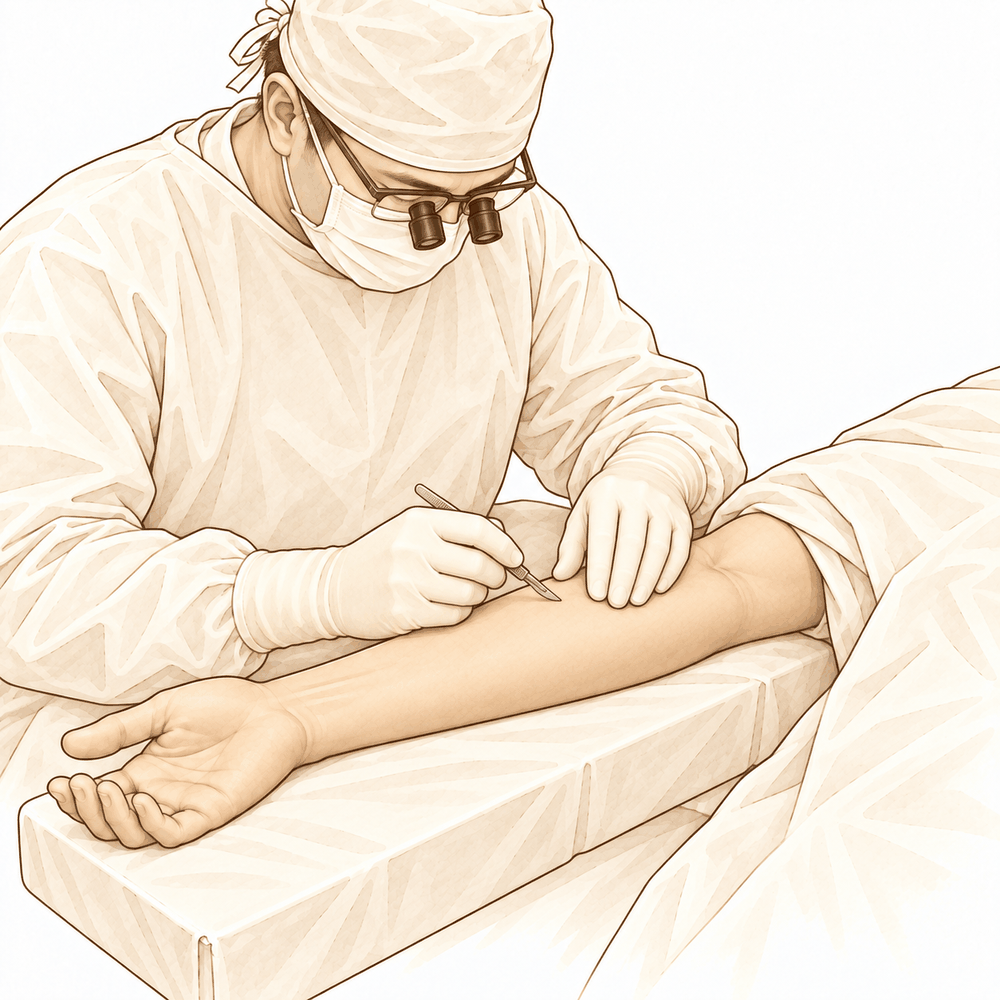
Surgical Approach / Technique: A mini-invasive technique for pronator teres syndrome resulted in symptom disappearance in 93% of cases [4]. An endoscopically assisted, minimally invasive approach decompressed all anatomical points of compression and improved DASH scores [16]. A transverse skin incision approach allows for satisfactory decompression of all structures involved in pronator teres syndrome [117]. Surgical release in local anesthesia for proximal median nerve entrapment at the elbow is a safe, ambulatory, and cost-efficient procedure with low morbidity [43]. The pronator teres release is more invasive than other approaches and requires more complicated surgical skills, deeper dissection, and more invasive anesthetic preparation [11]. Release and resection of a fibrovascular band causing median nerve neuralgia in the distal forearm resulted in complete remission of disabling focal pain and tenderness [147]. There was no correlation between the entrapment site and clinical signs on examination in a surgical series for median nerve entrapment syndrome in the elbow and proximal forearm [60].
Complications
Diagnostic and Preoperative Considerations: Pre- and post-exercise MRI provides reliable objective data to supplement the clinical picture without requiring invasive or painful procedures [37]. Surgical indications for nerve decompression in pronator syndrome include persistent symptoms for >6 months [1]. Treatment recommendations for AIN palsy range from surgical exploration after 6 to 12 weeks to observation for at least 18 months [22]. Patients presenting with paresis in AIN syndrome should be observed, as most will improve spontaneously without surgery [171]. Surgical release in local anesthesia allows for a safe, ambulatory, and cost-efficient procedure with low morbidity [43].
Operative Complications and Risks: The pronator teres release is more invasive than alternative approaches and requires more complicated surgical skills, deeper dissection, and more invasive anesthetic preparation [11]. One patient had failed percutaneous release and required reoperation [7]. The risks associated with decompressing the radial tunnel include partial or transient PIN palsy [94]. Atroshi and colleagues reported 2 cases of partial radial nerve paresis and 3 cases with diminished sensation in the radial nerve distribution following surgical release of the radial tunnel in 37 patients [94]. Lawrence and colleagues reported 1 case of transient PIN palsy, 3 cases of mild chronic regional pain syndrome, and 2 patients with other complications in a study of 30 patients [94]. Understanding the position of the MACN posterior branch during ulnar nerve release surgery at the elbow may help in preventing iatrogenic injury [27]. Recognition of injury to the medial antebrachial cutaneous nerve during cubital tunnel surgery permits more complete preoperative patient informing and appropriate rehabilitation direction [168]. The endoscopically assisted, minimally invasive approach to treat pronator syndrome adequately and safely decompressed all anatomical points of compression [16]. This minimally invasive approach can release all the potential entrapment sites [14]. The double transfer of the PIN and AIN nerves onto the SBUN and DBUN led to functional recovery within the first year after the surgical procedure [36]. Two studies found no deficits in pronation range of motion or strength following supercharged end-to-side anterior interosseous nerve to ulnar nerve transfer [128]. Two studies reported minor issues like infection and hematoma following supercharged end-to-side anterior interosseous nerve to ulnar nerve transfer, which were manageable with standard care [128]. No complications were reported in the study of the lasso en cravate technique for correcting the Wartenberg sign [170]. No adverse events or revisions were reported in the study of protective sensation after hemi-contralateral C7 nerve root transfer to the median nerve [139].
Postoperative Outcomes and Recovery: Pronator teres symptoms disappeared in 93% of cases following the mini-invasive technique [4]. The endoscopically assisted, minimally invasive approach to treat pronator syndrome improved DASH scores [16]. Outcomes of pronator and AIN release surgeries should be evaluated at least months after release to measure true effectiveness [174]. Prognosis for complete recovery is good if irreversible end-organ damage has not occurred owing to long-standing compression [75]. One patient experienced immediate subsidence of clinical signs after median nerve decompression for ectopic calcification [33]. Another patient showed progressive subjective improvement and increased ulnar nerve conduction velocity to 43 m/sec one year after decompression for ectopic calcification, though no motor recovery was observed [33]. The patient underwent release and transposition subcutaneously for ulnar nerve entrapment and has begun to show signs of clinical recovery 17 months after his surgery [38]. An excellent or good functional result was documented for all but three patients in the operative treatment of palsy of the posterior interosseous nerve of the forearm [182]. The success rate of 85% good or excellent results for surgical and therapeutic management of tennis elbow is comparable to other procedures, with a considerable decrease in morbidity and length of disability [179]. The main benefit of using a simultaneous release protocol for ipsilateral pronator teres and carpal tunnel syndromes is to shorten total morbidity time and to avoid exposure of the patient to two operations instead of one [173]. Increasing evidence suggests that supervised rehabilitation after early active, rather than delayed, mobilization protocols is safe and effective [47].
Revision Surgery and Recurrence: It is estimated that as many as 25% of patients treated for cubital tunnel syndrome will experience recurrence [138]. Younger age at presentation (<50 years) as well as greater static 2-point discrimination and history of diabetes have been associated with a greater number of revision surgeries for cubital tunnel syndrome [138]. In a large systematic review of patients undergoing revision cubital tunnel surgery, transposition surgery was the most common procedure for primary surgery (51%) [138]. In a large systematic review of patients undergoing revision cubital tunnel surgery, perineural scarring was the most common intraoperative finding at revision surgery (79%) [138]. In a large systematic review of patients undergoing revision cubital tunnel surgery, the medial intermuscular septum was the most frequent entrapment site (33%) [138]. Novak and Mackinnon found that the most common operative findings in 100 patients who underwent reoperation following cubital tunnel surgery included a medial antebrachial cutaneous nerve neuroma (n = 73) and a distal kink of the ulnar nerve (n = 57) caused by fascial flaps or tendinous bands [138]. Outcomes following revision cubital tunnel surgery are inferior to those following primary surgery, with only 75% to 80% of revision patients reporting symptomatic improvement [138]. Worse outcomes are reported on all measured standardized questionnaires following revision cubital tunnel surgery compared to primary surgery [138]. Recurrent cubital tunnel syndrome has been attributed to inaccurate preoperative diagnosis, incomplete nerve decompression, iatrogenic injury, postsurgical perineural adhesions, irreversible nerve pathology, or conditions associated with secondary nerve compression [138]. In a study of 19 patients treated with nonoperative modalities for radial sensory nerve entrapment after a mean of 28 months from the onset of symptoms or their injury, seven (37%) were improved [19, 20].
Recovery
Non-Operative Management
Nonoperative management yields variable results. In a cohort of 19 patients, seven (37%) demonstrated improvement after a mean of 28 months from the onset of symptoms or their injury [19, 20].
Surgical Outcomes
Surgical intervention generally restores function, with specific outcomes varying by pathology and technique. Following a mini-invasive decompression technique, pronator teres symptoms disappeared in 93% of cases [4]. For median nerve compression in the proximal forearm, all patients returned to normal activities without sequelae, although four developed a keloid scar [167]. In cases of anterior interosseous nerve palsy caused by a nerve tumour, patients were able to return to work 3 months after surgery with increased pinch force and active interphalangeal joint motion of the thumb [148].
Decompression procedures for other nerve entrapments also show positive trajectories. Surgical decompression of a posterior interosseous nerve syndrome caused by a bursa led to complete recovery [160]. Similarly, full recovery followed local excision with decompression of the peripheral nerve involved in synovial chondromatosis [157]. For synovial sarcoma with radial nerve involvement, recovery of wrist and finger extensors was first seen after four months, and both motor and sensory recovery was complete eight months after surgery [166]. In hypoplasia of bilateral humeral trochlea associated with unilateral ulnar nerve palsy, gradual recovery of motor and sensory function was evident after surgery [169].
Outcomes for ectopic calcification and nerve transfers present more complex timelines. One patient experienced immediate subsidence of clinical signs after median nerve decompression for ectopic calcification, while the other showed progressive subjective improvement and increased ulnar nerve conduction velocity to 43 m/sec one year later, though no motor recovery was observed [33]. The double transfer of the posterior interosseous nerve and anterior interosseous nerve onto the superficial and deep branches of the ulnar nerve led to functional recovery within the first year after the surgical procedure [36]. A patient with ulnar nerve entrapment who underwent release and subcutaneous transposition began to show signs of clinical recovery 17 months after surgery [38].
Prognosis and Recovery Timelines
For anterior interosseous nerve paralysis as a complication of supracondylar fracture of the humerus in children, complete recovery is usual within 3 months [113].
Key Evidence
- [Paper] Surgical indications for nerve decompression include persistent symptoms for >6 months in patients with pronator syndrome or for a minimum of 12 months with no signs of motor improvement in those with anterior interosseous nerve syndrome. [1] (10.5435/jaaos-21-05-268)
- [L5] If the activities of daily living of a patient require frequent strong pronation, and if the electromyography findings confirm the presence of compression, surgical decompression can be considered against the background of pronator syndrome, whereas other medical causes should be considered in AIN syndrome. [2] (10.1177/17531934221080018)
- [L5] The diagnosis of pronator syndrome lacks objective pathophysiology and is supported only by subjective operative findings and relief after surgery, with a paucity of controlled trials demonstrating that operative treatment is more effective than other treatments or sham surgery. [3] (10.1016/j.jhsa.2011.02.014)
- [L4] Pronator teres symptoms disappeared in 93% of cases following the mini-invasive technique. [4] (10.1016/j.jhsa.2012.05.033)
- [L5] Conservative management, including avoidance of aggravating activities, rest, and anti-inflammatory medication, is recommended for pronator syndrome and anterior interosseous nerve syndrome; surgery is appropriate if symptoms persist. [6] (10.1016/j.jhsa.2009.10.017)
- [L4] One patient had failed percutaneous release and required reoperation. [7] (10.1097/00132589-200112000-00003)
- [L5] When the correct diagnosis is made, pronator syndrome can be successfully treated nonoperatively or surgically, if necessary. [8] (10.1016/s0278-5919(05)70267-2)
- [L5] Conservative treatment stays the first choice for median nerve compression either in the form of pronator or anterior interosseous nerve syndrome. [9] (10.1016/j.main.2004.10.024)
- [L4] [10] (10.1016/j.otsr.2013.04.002)
- [L4] The pronator teres release was more invasive and required more complicated surgical skills, deeper dissection, and more invasive anesthetic preparation. [11] (10.1051/sicotj/2016006)
- [L5] Lacertus syndrome and superficialis-pronator syndrome should be clearly presented as distinct targets for surgical release, and the term pronator syndrome should be replaced with superficialis-pronator syndrome. [13] (10.1177/17531934211024092)
- [L5] This minimally invasive approach can release all the potential entrapment sites. [14] (10.1016/j.eats.2024.103391)
- [L5] Surgical decompression of the median nerve or the AIN in the forearm is rarely indicated; a prolonged nonsurgical approach is warranted in most cases. [15] (10.5435/jaaos-d-16-00010)
- [L4] The endoscopically assisted, minimally invasive approach to treat pronator syndrome adequately and safely decompressed all anatomical points of compression and improved DASH scores. [16] (10.1016/j.jhsa.2012.02.023)
- [L4] For a reoperation to be successful, the ulnar nerve must be examined and all potential levels of compression must be released. [18] (10.2106/jbjs.24.00493)
- [L4] Seven (37%) of 19 patients treated with nonoperative modalities after a mean of 28 months from the onset of symptoms or their injury were improved. [19] (10.1016/s0363-5023(86)80050-8)
- [L4] Seven (37%) of 19 patients treated with nonoperative modalities after a mean of 28 months from the onset of symptoms or their injury were improved. [20] (10.1016/s0363-5023(86)80051-x)
- [Case_report] Resection of a segment of the persistent median artery is the recommended treatment for pronator syndrome associated with this anomaly. [21] (10.2106/00004623-198769020-00026)
- [L5] [22] (10.1016/j.jhsa.2010.08.018)
- [L5] It is essential for clinicians to have a thorough understanding of median nerve anatomy, possible sites of compression, and characteristic clinical findings of pronator syndrome to provide a reliable diagnosis and treat their patients. [25] (10.1016/j.jhsa.2020.07.006)
- [L5] [26] (10.1007/s11552-014-9639-5)
- [L5] Understanding the position of the MACN posterior branch during ulnar nerve release surgery at the elbow may help in preventing iatrogenic injury. [27] (10.1016/j.otsr.2020.02.006)
- [L4] The potential for fibrous compression of the median nerve at the pronator tunnel was substantiated from a series of anatomical dissections. [28] (10.1016/s0363-5023(79)80104-5)
- [L4] The palsy in these patients recovered completely with conservative management. [30] (10.1016/j.jhsa.2008.10.019)
- [L5] One patient experienced immediate subsidence of clinical signs after median nerve decompression, while the other showed progressive subjective improvement and increased ulnar nerve conduction velocity to 43 m/sec one year later, though no motor recovery was observed. [33] (10.1016/s0363-5023(80)80039-6)
- [L5] It is important to take a complete history and perform a careful examination to avoid confirmation bias when evaluating throwers with medial elbow pain. [34] (10.1016/j.csm.2020.03.004)
- [L5] The treatment of idiopathic AIN palsy is controversial in terms of nonsurgical or surgical approaches and how long to wait for spontaneous recovery before recommending surgery. [35] (10.1177/1753193415596108)
- [L5] The double transfer of the PIN and AIN nerves onto the SBUN and DBUN led to functional recovery within the first year after the surgical procedure. [36] (10.1016/j.main.2014.08.001)
- [L5] Pre- and post-exercise MRI is a way to supplement the patient's clinical picture with reliable objective data that does not require an invasive or painful procedure. [37] (10.1016/j.radcr.2021.02.022)
- [L5] The patient underwent release and transposition subcutaneously and has begun to show signs of clinical recovery 17 months after his surgery. [38] (10.1016/s0363-5023(88)80033-9)
- [L4] [41] (10.1016/0363-5023(90)90069-4)
- [L4] Surgical release in local anesthesia allows for a safe, ambulatory, and cost-efficient procedure with low morbidity. [43] (10.1007/s11552-012-9483-4)
- [L4] With clinical evaluation as the reference standard, electrodiagnostic, ultrasound, and MRI are not helpful in making a diagnosis of pronator syndrome concurrent with carpal tunnel syndrome. [45] (10.1016/j.jhsa.2020.06.006)
- [L5] Increasing evidence suggests that supervised rehabilitation after early active, rather than delayed, mobilization protocols is safe and effective. [47] (10.1016/j.jhsa.2010.05.023)
- [L5] [48] (10.1097/phm.0000000000000973)
- [L5] Clinical suspicion should arise in the presence of isolated paralysis of the AIN-supplied muscles. [49] (10.1016/j.ijscr.2016.02.021)
- [L4] Our data strongly support that AINS in the majority of cases is not a surgically treatable entrapment neuropathy but a multifocal mononeuropathy selectively involving, within the main trunk of the median nerve, the motor fascicles that continue distally to form the anterior interosseous nerve. [50] (10.1212/wnl.0000000000000128)
- [L5] We report the case of a patient with bilateral pronator syndrome caused by an anomalous tendinous origin of the ulnar belly of the pronator teres muscle. [51] (10.1016/0363-5023(93)90373-b)
- [L5] [54] (10.1016/j.csm.2004.04.012)
- [Case_report] [58] (10.5397/cise.2022.00899)
- [Paper] There was no correlation between entrapment site and clinical signs on examination. [60] (10.1016/j.otsr.2021.102825)
- [L5] [70] (10.1177/17531934221074903)
- [L5] The authors believe that a high rate of morbidity is associated with both the disease and its treatment, suggesting that great caution has to be taken before performing radial tunnel release. [73] (10.1097/00130911-200212000-00010)
- [L5] Prognosis for complete recovery is good if irreversible end-organ damage has not occurred owing to long-standing compression. [75] (10.1016/s0278-5919(05)70256-8)
- [L4] [94] (10.1016/j.hcl.2013.04.014)
- [L4] [102] (10.1177/1753193417726214)
- [L4] The diagnostic process to differentiate pronator syndrome from carpal tunnel syndrome remains a challenge due to overlapping symptoms and limited reliable information in the literature; this review provides a comprehensive clinical comparison to aid in establishing appropriate diagnosis and treatment. [106] (10.3390/diagnostics12102433)
- [L4] Complete recovery is usual within 3 months. [113] (10.1016/0020-1383(89)90172-1)
- [L4] The transverse incision and surgical technique allows for satisfactory decompression of all the structures involved in pronator teres syndrome. [117] (10.1016/0266-7681(94)90047-7)
- [L5] Electrodiagnostic testing may reveal proximal diffuse abnormalities; in these instances of branchial plexus neuritis-induced AIN palsy, nonsurgical management is recommended. [118] (10.1016/s0363-5023(97)80025-1)
- [L4] Median nerve entrapment in the forearm without motor paralysis is a clinical diagnosis supported by pain drawings, pain quality, and provocative tests. [121] (10.1177/1558944719874137)
- [L4] Concurrent carpal tunnel syndrome and pronator syndrome are rarely considered and proximal compression sites are easily overlooked. [124] (10.1016/j.otsr.2016.10.009)
- [L3] [128] (10.1016/j.jhsg.2024.06.003)
- [L5] [134] (10.1016/j.jisako.2023.12.007)
- [L4] [138] (10.1016/j.jhsg.2022.07.008)
- [L3] No adverse events or revisions were reported. [139] (10.1016/j.jhsa.2016.07.035)
- [L5] The authors recommend that the term 'lacertus syndrome' refer to intermittent median nerve compression at the level of the lacertus fibrosus, while the pathology originally described by Bennett should be termed chronic exertional compartment syndrome of the pronator teres or 'Bennett's syndrome' to resolve current terminology confusion. [144] (10.1177/17531934231170347)
- [L5] [145] (10.1016/0266-7681_87_90189-6)
- [L5] Release and resection of the band resulted in complete remission of the disabling focal pain and tenderness. [147] (10.1016/s0363-5023(86)80245-3)
- [L5] The patient was able to return to work 3 months after surgery with increased pinch force and active interphalangeal joint motion of the thumb. [148] (10.1177/1753193412454801)
- [L4] These constrictions were identified in all MRN cases and 88% of ultrasound cases, predominantly in the posterior/posteromedial region proximal to the elbow joint line. [153] (10.1002/mus.26768)
- [L4] Full recovery followed local excision with decompression of the peripheral nerve involved. [157] (10.1016/0266-7681(87)90048-9)
- [L4] In cases of suspected dynamic nerve compression in a high-level athlete, additional diagnostic and imaging modalities, such as dynamic nerve studies with inching technique and dynamic MRI, are warranted if initial history and physical examination do not reveal an etiology. [159] (10.1016/j.xrrt.2021.04.005)
- [L5] Surgical decompression led to a complete recovery. [160] (10.1016/0266-7681(87)90047-7)
- [L3] MRI from our institution was able to identify the cause of medial elbow pain in 20 (95%) of 21 throwing athletes. [164] (10.1016/s1058-2746(95)80208-8)
- [L5] Recovery of wrist and finger extensors was first seen after four months, and both motor and sensory recovery was complete eight months after surgery. [166] (10.1016/0266-7681(85)90028-2)
- [L4] All patients returned to their normal activities without sequelae but 4 of them developed a keloid scar. [167] (10.1177/1558944716660555ku)
- [L4] Recognition of this complication permits more complete preoperative patient informing and appropriate rehabilitation direction. [168] (10.1016/s0266-7681(85)80011-5)
- [L5] After surgery gradual recovery of motor and sensory function was evident. [169] (10.1016/s1058-2746(00)90064-8)
- [L4] No complications were reported. [170] (10.1016/s1297-3203(00)73575-x)
- [L4] Patients presenting with paresis should be observed, as most will improve spontaneously without surgery. [171] (10.1016/s0363-5023(85)80240-9)
- [L4] The main benefit of using this protocol in this selected group of patients is to shorten total morbidity time and to avoid exposure of the patient to two operations instead of one. [173] (10.1097/01.prs.0000260703.56453.06)
- [L5] Outcomes of these surgeries should be evaluated at least months after release to measure true effectiveness. [174] (10.1177/17531934231177831)
- [L4] The success rate of 85% good or excellent results is comparable to the results of the other types of procedures reported in the literature, with a considerable decrease in morbidity and length of disability. [179] (10.1016/s0894-1130(12)80105-0)
- [L4] An excellent or good functional result was documented for all but three patients. [182] (10.2106/00004623-199072080-00014)
See Also
- Radial Tunnel Syndrome
- Tennis Elbow
- Cubital Tunnel Syndrome
References
[1] Pronator Syndrome and Anterior Interosseous Nerve Syndrome. Journal of the American Academy of Orthopaedic Surgeons. 2013. DOI: 10.5435/jaaos-21-05-268
[2] Two clinical observations: pronator syndrome in violinists and anterior interosseous nerve syndrome with pure motor loss. Journal of Hand Surgery (European Volume). 2022. DOI: 10.1177/17531934221080018
[3] Pronator Syndrome. The Journal of Hand Surgery. 2011. DOI: 10.1016/j.jhsa.2011.02.014
[4] New Mini-invasive Decompression for Pronator Teres Syndrome. The Journal of Hand Surgery. 2012. DOI: 10.1016/j.jhsa.2012.05.033
[6] Unusual Compression Neuropathies of the Forearm, Part II: Median Nerve. The Journal of Hand Surgery. 2009. DOI: 10.1016/j.jhsa.2009.10.017
[7] Management of Lateral Epicondylitis With Percutaneous Release. Techniques in Shoulder & Elbow Surgery. 2001. DOI: 10.1097/00132589-200112000-00003
[8] PRONATOR SYNDROME. Clinics in Sports Medicine. 2001. DOI: 10.1016/s0278-5919(05)70267-2
[9] Compression du nerf médian au coude. Chirurgie de la Main. 2004. DOI: 10.1016/j.main.2004.10.024
[10] The anatomical basis for anterior interosseous nerve palsy secondary to supracondylar humerus fractures in children. Orthopaedics & Traumatology: Surgery & Research. 2013. DOI: 10.1016/j.otsr.2013.04.002
[11] Role of pronator release in revision carpal tunnel surgery. SICOT-J. 2016. DOI: 10.1051/sicotj/2016006
[13] Median nerve compression: lacertus syndrome versus superficialis-pronator syndrome. Journal of Hand Surgery (European Volume). 2021. DOI: 10.1177/17531934211024092
[14] Endoscopic Radial Tunnel Release. Arthroscopy Techniques. 2024. DOI: 10.1016/j.eats.2024.103391
[15] Ulnar Tunnel Syndrome, Radial Tunnel Syndrome, Anterior Interosseous Nerve Syndrome, and Pronator Syndrome. Journal of the American Academy of Orthopaedic Surgeons. 2017. DOI: 10.5435/jaaos-d-16-00010
[16] Endoscopically Assisted Decompression for Pronator Syndrome. The Journal of Hand Surgery. 2012. DOI: 10.1016/j.jhsa.2012.02.023
[18] Risk Factors for Lumbar Spinal Fusion Reoperation: An Unusual Suspect. Journal of Bone and Joint Surgery. 2024. DOI: 10.2106/jbjs.24.00493
[19] Cheiralgia paresthetica (entrapment of the radial sensory nerve). The Journal of Hand Surgery. 1986. DOI: 10.1016/s0363-5023(86)80050-8
[20] Radial sensory nerve entrapment in the forearm. The Journal of Hand Surgery. 1986. DOI: 10.1016/s0363-5023(86)80051-x
[21] Pronator syndrome associated with a persistent median artery. A case report.. The Journal of Bone & Joint Surgery. 1987. DOI: 10.2106/00004623-198769020-00026
[22] Anterior Interosseous Nerve Syndrome. The Journal of Hand Surgery. 2010. DOI: 10.1016/j.jhsa.2010.08.018
[25] Proximal Median Nerve Compression: Pronator Syndrome. The Journal of Hand Surgery. 2020. DOI: 10.1016/j.jhsa.2020.07.006
[26] Median Nerve Compression at the Fibrous Arch of the Flexor Digitorum Superficialis: An Anatomic Study of the Pronator Syndrome. HAND. 2014. DOI: 10.1007/s11552-014-9639-5
[27] Anatomy of the posterior branch of the medial antebrachial cutaneous nerve: A cadaveric study. Orthopaedics & Traumatology: Surgery & Research. 2020. DOI: 10.1016/j.otsr.2020.02.006
[28] Median nerve entrapment syndrome in the proximal forearm. The Journal of Hand Surgery. 1979. DOI: 10.1016/s0363-5023(79)80104-5
[30] Anterior Interosseous Nerve Palsy After Open Capsular Release for Elbow Stiffness: Report of 2 Cases. The Journal of Hand Surgery. 2009. DOI: 10.1016/j.jhsa.2008.10.019
[33] Median and ulnar nerve entrapment caused by ectopic calcification: Report of two cases. The Journal of Hand Surgery. 1980. DOI: 10.1016/s0363-5023(80)80039-6
[34] The Lacertus Syndrome of the Elbow in Throwing Athletes. Clinics in Sports Medicine. 2020. DOI: 10.1016/j.csm.2020.03.004
[35] Spontaneous recovery of a case with suspected hourglass-like fascicular constriction and anterior interosseous nerve palsy. Journal of Hand Surgery (European Volume). 2015. DOI: 10.1177/1753193415596108
[36] Case report: Double nerve transfer of the anterior and posterior interosseous nerves to treat a high ulnar nerve defect at the elbow. Chirurgie de la Main. 2014. DOI: 10.1016/j.main.2014.08.001
[37] Lacertus syndrome: Use of pre- and post-exercise MRI to aid in diagnosis and treatment. Radiology Case Reports. 2021. DOI: 10.1016/j.radcr.2021.02.022
[38] Ulnar nerve entrapment. The Journal of Hand Surgery. 1988. DOI: 10.1016/s0363-5023(88)80033-9
[41] Nonoperative treatment of anterior interosseous nerve paralysis. The Journal of Hand Surgery. 1990. DOI: 10.1016/0363-5023(90)90069-4
[43] Clinical Diagnosis and Wide-Awake Surgical Treatment of Proximal Median Nerve Entrapment at the Elbow: A Prospective Study. HAND. 2013. DOI: 10.1007/s11552-012-9483-4
[45] Clinical, Radiological, and Electrodiagnostic Diagnosis of Pronator Syndrome Concurrent With Carpal Tunnel Syndrome. The Journal of Hand Surgery. 2020. DOI: 10.1016/j.jhsa.2020.06.006
[47] Update on Tendon Transfers for Peripheral Nerve Injuries. The Journal of Hand Surgery. 2010. DOI: 10.1016/j.jhsa.2010.05.023
[48] Pronator Syndrome: An Uncommon Median Nerve Entrapment Syndrome. American Journal of Physical Medicine & Rehabilitation. 2019. DOI: 10.1097/phm.0000000000000973
[49] Anterior interosseous nerve syndrome diagnosis and intraoperative findings: A case report. International Journal of Surgery Case Reports. 2016. DOI: 10.1016/j.ijscr.2016.02.021
[50] Anterior interosseous nerve syndrome. Neurology. 2014. DOI: 10.1212/wnl.0000000000000128
[51] Bilateral pronator syndrome associated with anomalous heads of the pronator teres muscle: A case report. The Journal of Hand Surgery. 1993. DOI: 10.1016/0363-5023(93)90373-b
[54] Nerve injuries in the throwing elbow. Clinics in Sports Medicine. 2004. DOI: 10.1016/j.csm.2004.04.012
[58] Anterior interosseous nerve palsy in the early postoperative period after open capsular release for elbow stiffness: a case report. Clinics in Shoulder and Elbow. 2023. DOI: 10.5397/cise.2022.00899
[60] Median nerve entrapment syndrome in the elbow and proximal forearm. Anatomic causes and results for a 55-case surgical series at a mean 7 years’ follow-up. Orthopaedics & Traumatology: Surgery & Research. 2021. DOI: 10.1016/j.otsr.2021.102825
[70] Compression to the anterior interosseous nerve is very rare: compression by the normal tissues surrounding it may not exist. Journal of Hand Surgery (European Volume). 2022. DOI: 10.1177/17531934221074903
[73] Radial Tunnel Syndrome. Techniques in Hand and Upper Extremity Surgery. 2002. DOI: 10.1097/00130911-200212000-00010
[75] Nerve Injuries of the Elbow, Wrist, and Hand in Athletes. Clinics in Sports Medicine. 2001. DOI: 10.1016/s0278-5919(05)70256-8
[78] Miller S Review Of Orthopaedics. SECTION 16 PATELLAR TRACKING IN TOTAL KNEE ARTHROPLASTY > TABLE 2.3 Shoulder Spaces.
[94] Uncommon Upper Extremity Compression Neuropathies. Hand Clinics. 2013. DOI: 10.1016/j.hcl.2013.04.014
[102] The scratch collapse test in the diagnosis of compression of the median nerve in the proximal forearm. Journal of Hand Surgery (European Volume). 2017. DOI: 10.1177/1753193417726214
[106] How to Differentiate Pronator Syndrome from Carpal Tunnel Syndrome: A Comprehensive Clinical Comparison. Diagnostics. 2022. DOI: 10.3390/diagnostics12102433
[113] Anterior interosseous nerve paralysis: an underdiagnosed complication of supracondylar fracture of the humerus in children. Injury. 1989. DOI: 10.1016/0020-1383(89)90172-1
[117] A Transverse Skin Incision Approach for Decompression of Pronator Teres Syndrome. Journal of Hand Surgery. 1994. DOI: 10.1016/0266-7681(94)90047-7
[118] Brachial neuritis presenting as anterior interosseous nerve compression—Implications for diagnosis and treatment: A case report. The Journal of Hand Surgery. 1997. DOI: 10.1016/s0363-5023(97)80025-1
[121] Median Nerve Compression in the Forearm: A Clinical Diagnosis. HAND. 2019. DOI: 10.1177/1558944719874137
[124] Concurrent carpal tunnel syndrome and pronator syndrome: A retrospective study of 21 cases. Orthopaedics & Traumatology: Surgery & Research. 2017. DOI: 10.1016/j.otsr.2016.10.009
[128] Exploring Outcomes and Mediating Factors Following Supercharged End-to-Side Anterior Interosseous Nerve to Ulnar Nerve Transfer: A Scoping Review With Expert Insight. Journal of Hand Surgery Global Online. 2024. DOI: 10.1016/j.jhsg.2024.06.003
[134] Causes, symptoms, and treatments of nerve entrapments around the elbow: Current concepts. Journal of ISAKOS. 2024. DOI: 10.1016/j.jisako.2023.12.007
[138] Modern Treatment of Cubital Tunnel Syndrome: Evidence and Controversy. Journal of Hand Surgery Global Online. 2023. DOI: 10.1016/j.jhsg.2022.07.008
[139] Evaluation of Protective Sensation after Hemi-contralateral C7 Nerve Root Transfer to Median Nerve in Total Root Avulsion Brachial Plexus Injury. The Journal of Hand Surgery. 2016. DOI: 10.1016/j.jhsa.2016.07.035
[144] Lacertus syndrome: one term – two different pathologies. Journal of Hand Surgery (European Volume). 2023. DOI: 10.1177/17531934231170347
[145] Musculoaponeurotic Variations along the Course of the Median Nerve in the Proximal Forearm. Journal of Hand Surgery. 1987. DOI: 10.1016/0266-7681_87_90189-6
[147] Median nerve neuralgia caused by a fibrovascular band in the distal forearm. The Journal of Hand Surgery. 1986. DOI: 10.1016/s0363-5023(86)80245-3
[148] Nerve tumour as a rare cause of anterior interosseous nerve palsy. Journal of Hand Surgery (European Volume). 2012. DOI: 10.1177/1753193412454801
[153] Fascicular constrictions above elbow typify anterior interosseous nerve syndrome. Muscle & Nerve. 2019. DOI: 10.1002/mus.26768
[157] Synovial chondromatosis presenting with peripheral nerve compression — A report of two cases. The Journal of Hand Surgery: Journal of the British Society for Surgery of the Hand. 1987. DOI: 10.1016/0266-7681(87)90048-9
[158] The Anterior Interosseous-Nerve Syndrome: WITH SPECIAL ATTENTION TO ITS VARIATIONS.. The Journal of Bone and Joint Surgery. American Volume. 1970.
[159] Dynamic ulnar nerve compression at the elbow in a collegiate baseball player due to aberrant branch of the brachial artery. JSES Reviews, Reports, and Techniques. 2021. DOI: 10.1016/j.xrrt.2021.04.005
[160] Posterior interosseous nerve syndrome caused by a bursa in the vicinity of the elbow. The Journal of Hand Surgery: Journal of the British Society for Surgery of the Hand. 1987. DOI: 10.1016/0266-7681(87)90047-7
[164] Diagnostic utility of MRI in elite throwing athletes with medial elbow pain. Journal of Shoulder and Elbow Surgery. 1995. DOI: 10.1016/s1058-2746(95)80208-8
[166] Synovial sarcoma: with radial nerve involvement. The Journal of Hand Surgery: Journal of the British Society for Surgery of the Hand. 1985. DOI: 10.1016/0266-7681(85)90028-2
[167] Compression of the Median Nerve in the Proximal Forearm. HAND. 2016. DOI: 10.1177/1558944716660555ku
[168] Injury to the Medial Antebrachial Cutaneous Nerve During Cubital Tunnel Surgery. Journal of Hand Surgery. 1985. DOI: 10.1016/s0266-7681(85)80011-5
[169] Hypoplasia of bilateral humeral trochlea associated with unilateral ulnar nerve palsy: A case report. Journal of Shoulder and Elbow Surgery. 2000. DOI: 10.1016/s1058-2746(00)90064-8
[170] 78 Le lasso en cravate : une nouvelle technique pour corriger le signe de Wartenberg des mains cubitales. Chirurgie de la Main. 2000. DOI: 10.1016/s1297-3203(00)73575-x
[171] The incomplete anterior interosseous nerve syndrome. The Journal of Hand Surgery. 1985. DOI: 10.1016/s0363-5023(85)80240-9
[173] Simultaneous Surgical Release of Ipsilateral Pronator Teres and Carpal Tunnel Syndromes. Plastic and Reconstructive Surgery. 2007. DOI: 10.1097/01.prs.0000260703.56453.06
[174] Immediate return to normal after releasing nerve entrapment and rebound phenomenon. Journal of Hand Surgery (European Volume). 2023. DOI: 10.1177/17531934231177831
[179] Surgical and therapeutic management of tennis elbow: An update. Journal of Hand Therapy. 1991. DOI: 10.1016/s0894-1130(12)80105-0
[182] Operative treatment of palsy of the posterior interosseous nerve of the forearm.. The Journal of Bone & Joint Surgery. 1990. DOI: 10.2106/00004623-199072080-00014