Clinicians › Shoulder
Revision shoulder replacement
Surgeon-side topic for revision shoulder replacement. Backed by 422 articles from the corpus, retrieved via combined MeSH + title-text matching.
For patients: a plain-language version of this topic is available. See the patient guide.
Overview
Shoulder arthroplasties are generally designed to last 10-15 years [1], yet revision procedures are increasingly performed at a mean of 3.9 years from the primary procedure [1]. The incidence of both primary and revision shoulder arthroplasties is expected to increase significantly in the coming years [7], with a massive rise projected by 2040 driven largely by fracture-related procedures [10]. This growing volume constitutes a substantial burden for healthcare systems [7]. While revision shoulder arthroplasties lead to notable improvements in pain, motion, and function [2], outcomes are generally inferior to those of primary reverse total shoulder arthroplasty [2]. Complications and further revision surgeries are common in this setting [2], with complication rates for revision of failed shoulder prosthesis approximately 50% [13]. Specifically, complication and reoperation rates for revision reverse shoulder arthroplasty are higher than those for primary reverse shoulder arthroplasty [61].
The clinical outcome of revision shoulder arthroplasty can be predicted on the basis of the indication for the procedure [4]. Revision reverse shoulder arthroplasty demonstrates significant long-term clinical improvements [3], with an implant survival rate of 85% at ten years [3]. For patients below the age of 55, reverse shoulder arthroplasty and stemless anatomic total shoulder arthroplasty both carry a lower short-term revision risk than stemmed anatomic total shoulder arthroplasty [11]. Optimum revision rates vary by gender and implant type for osteoarthritis [17]. Patients with neurologic disorders experience higher reported complication and revision rates after shoulder arthroplasty compared with those without neurologic conditions [23].
Current controversies in shoulder arthroplasty management include glenoid bone loss, the role of patient-specific instrumentation, differing design features of reverse shoulder arthroplasty, and options for humeral fixation in primary and revision procedures [6]. Anatomic total shoulder arthroplasties and reverse shoulder arthroplasties are the most revised implants for humeral loosening [9], with reverse shoulder arthroplasties being the most common implant used for this specific indication [9]. The majority of revision cases are performed for patients unlikely to have a periprosthetic joint infection [8], as less than 10% of revision patients meet ICM criteria for definite periprosthetic joint infection [8].
Anatomy & Pathophysiology
Bony Anatomy
The proximal humerus comprises four main parts: the humeral head, greater tuberosity, lesser tuberosity, and humeral shaft [73]. The articular surface of the humeral head is spherical with a diameter of 37 to 57 mm [73] and an arc of approximately 160 degrees covered by articular cartilage [85]. The radius of curvature of the humeral head is approximately 25 mm [85], with a reported range of 23-28 mm [85]. The most superior portion of the articular surface averages 8 mm above the greater tuberosity [73], while the superior margin is normally superior to the top of the greater tuberosity by 8 to 10 mm [85]. Head height is approximately 5.6 cm above the superior border of the pectoralis major tendon [87].
Humeral version averages 29.8 degrees, with a range of 10 to 55 degrees [73]. Other measurements indicate the humeral head is retroverted 19 degrees [76], 30 degrees relative to the transepicondylar axis [87], and an average of 30 degrees [74]. The humeral head version ranges from 0-55 degrees retroversion [85]. The humeral head is inclined approximately 130 degrees with respect to the humeral shaft [73], with an average inclination of 41° [76] and a range of 30-55 degrees [85]. The anatomic neck is located at the junction of the articular surface and the tuberosities [73], while the surgical neck represents an indistinct region below the tuberosities but above the humeral shaft [73].
The glenoid is a convex structure of shallow depth shaped like an inverted pear [73] and is approximately one third the size of the humeral head [74]. The glenoid is connected with the flat body of the scapula by the scapular neck [75]. The subchondral bone of the glenoid is relatively flat, with the articular concavity augmented by cartilage and a circumferential labrum [76]. The glenoid articular surface radius of curvature is 2 to 3 mm larger than that of the humeral head [85], with a specific range of 22-28 mm [85]. The glenoid diameter ranges from 18-30 mm superior anteroposterior, 21-35 mm inferior anteroposterior, and 30-48 mm superoinferior [85]. The glenoid surface area is 4-6 mm and the humeral head surface area is 11-19 mm [85].
Glenoid version averages 5° of retroversion in relation to the axis of the scapular body [76], approximately 5 degrees relative to the scapular body [87], and 1.5 degrees retroversion with a range of 10.5-9.5 degrees anteversion [85]. The normal position of the glenoid surface in relation to the axis of the scapular body ranges from 2 degrees of anteversion to 7 degrees of retroversion [85]. Glenoid inclination averages 4.2 degrees, with a range of –7 to 20 degrees [85]. The scapula is anteverted on the chest wall approximately 30 degrees relative to the body [87].
The distribution of bony mass in the scapula is highly uneven, with the highest concentration in the glenoid, scapular neck, and lateral border of the scapular body [75]. Two bony pillars extend between the glenoid and the scapular body to transmit compressive forces from the glenoid fossa [75]. The lateral pillar connects the inferior border of the glenoid with the inferior angle [75], while the spinal pillar arises from the central part of the glenoid and continues medially to become part of the base of the scapular spine [75]. The weakest bone in the scapula is located primarily in the central part of the biomechanical body, specifically in the infraspinous fossa [75]. The weakest area of the circumference of the biomechanical body of the scapula is the spinomedial angle [75].
The scapula is attached to the axial skeleton by the acromioclavicular and sternoclavicular joints [75]. The coracoid process curves forwards from the superior surface of the scapular neck [75], and the scapular spine ends in a flattened bony process, the acromion, which curves forwards [75]. The acromion, coracoacromial ligament, and coracoid process form the coracoacromial arch, a rigid bony-ligamentous structure that imparts stability to the shoulder girdle [73]. The distance from the lateral base of the coracoid process to the lateral margin of the greater tuberosity is called the lateral humeral offset [85]. The medial (coronal) humeral offset is 4-14 mm and the posterior (transverse) humeral offset is –2 to 10 mm [85].
The neck-shaft angle averages 45 degrees, with a range of 30 to 50 degrees [85], and measures an average of 135 degrees [74]. Arthritic shoulders have a flatter neck-shaft angle close to 50 degrees [85]. The neck-shaft angle ranges from 30-55 degrees [85]. The glenoid cartilage thickness is 2.16 mm and the humeral head cartilage thickness is 1.44 mm [85].
The proximal humerus has three ossification centers: the humeral head, greater tuberosity, and lesser tuberosity [76]. The humeral head ossification center is usually present at birth [80]. The greater tuberosity ossification center appears by 1 to 3 years of age [80], and the lesser tuberosity ossification center appears by 5 years of age [80]. The proximal humeral ossification centers fuse by 5 to 7 years of age to form the humeral head [80]. The proximal humeral physis closes by 14 to 17 years of age in girls and by 16 to 18 years in boys [80]. Humeral retroversion averages 65 degrees in infants and young children and gradually decreases, approaching adult values by 11 years of age [80]. Eighty percent of subsequent humeral growth comes from the proximal humeral physis [80]. The proximal humeral physis is irregularly shaped, with its apex located on the posteromedial portion of the proximal humerus [80]. The periosteum is thicker and stronger in the posteromedial portion of the proximal humerus compared to the anterolateral portion [80].
The clavicle is the first bone to ossify at the fifth week of gestation [76] and is the only long bone to ossify by intramembranous ossification [76]. The medial (sternal) epiphysis of the clavicle is the last ossification center to fuse, at age 20 to 25 years [76]. The primary blood supply to the clavicle is periosteal, with no nutrient artery present [76]. The scapula has only one true diarthrodial articulation, the acromioclavicular joint [76]. Ossification of the scapular body begins at the eighth week of gestation [76]. The acromion has three ossification centers: the metacromion, mesoacromion, and preacromion [76]. Failure of fusion of acromial ossification centers results in os acromiale [76].
Vascular Supply
The proximal humerus receives its blood supply from the anterior and posterior humeral circumflex branches from the third division of the axillary artery [73]. The anterior and posterior humeral circumflex arteries provide a rich blood supply to the proximal humerus [80]. The anterior humeral circumflex artery arises from the axillary artery at the inferior border of the subscapularis [73]. It provides vascular inflow to the humeral head by way of its terminal anterolateral branch known as the artery of Laing or arcuate artery [73]. The ascending branch of the anterior humeral circumflex artery courses parallel to the lateral aspect of the long head biceps tendon and enters the humeral head at the interface of the bicipital groove and greater tuberosity [73]. The major blood supply to the humeral head is through the ascending branch of the anterior humeral circumflex artery, which penetrates the head at the bicipital groove and becomes the arcuate artery [74]. The anterolateral ascending branch of the anterior humeral circumflex artery provides the primary blood supply to the humeral head [76]. The terminal intraosseous portion of the anterior humeral circumflex artery enters at the proximal aspect of the intertubercular groove as the arcuate artery [76].
The posterior humeral circumflex artery travels with the axillary nerve, enters the quadrilateral space posteriorly, and anastomoses with a branch of the anterior circumflex to supply the posterior cuff [73]. Quantitative assessment has shown that 64% of the humeral head blood supply arises from the posterior humeral circumflex artery [80]. Injury to the arcuate artery may result in osteonecrosis of the humeral head [73]. Additional extraosseous collateral branches can permit humeral head perfusion despite complete ligation of the arcuate artery [73].
Soft Tissue Anatomy
The rotator cuff consists of four muscles: the subscapularis, supraspinatus, infraspinatus, and teres minor [74]. The teres major is not a rotator cuff muscle [74]. The rotator cuff is a sheet of conjoined tendons closely applied over the shoulder capsule and inserting mainly into the greater tuberosity of the humerus [81]. The greater tuberosity serves as the attachment site for the supraspinatus, infraspinatus, and teres minor tendons [73] and provides attachment superiorly and posteriorly for these muscles [80]. The lesser tuberosity serves as the attachment site for the subscapularis tendon [73], which originates from the anterior scapula and inserts anteriorly onto the lesser tuberosity [80] [81].
The rotator cuff muscles serve as depressors of the humeral head to allow the deltoid to efficiently abduct the humerus [74]. The rotator cuff has an important function in stabilizing the head of the humerus by pulling it firmly into the glenoid whenever the deltoid lifts the arm forwards or sideways [81]. The infraspinatus and teres minor are external rotators, while the subscapularis is an internal rotator of the humerus [74]. The deltoid forward flexes and abducts the shoulder and courses from the clavicle and acromion superiorly, coalescing into a common tendinous insertion onto the lateral upper third of the humeral shaft [80]. The pectoralis major powers adduction and internal rotation due to its tendinous insertion anteriorly onto the lateral wall of the bicipital groove [80]. The deltoid and pectoralis major muscles, along with the rotator cuff, cause predictable displacement of fractures around the proximal humerus [74].
The coracoacromial arch is formed by the acromion process posterosuperiorly, the coracoid process anteriorly, and the coracoacromial ligament joining them [81]. The subacromial bursa separates the rotator cuff tendons from the coracoacromial arch, allowing them to glide [81]. The subscapular bursa lies between the subscapularis tendon and the neck of the scapula [77]. It communicates with the joint cavity between the superior and middle glenohumeral ligaments [77] and protects the tendon of the subscapularis at the point where it passes under the base of the coracoid process and over the neck of the scapula [77]. The subscapular bursa is linked to the coracoid process by a suspensory ligament [77]. In 28% of specimens dissected by Colas and colleagues, the subscapular bursae merged with the subcoracoid bursae, forming a unique wide bursa [77]. The subscapular bursa often houses loose bodies in the shoulder [77] and is a region in which synovitis of the shoulder may be most intense, where small fringes or villi can project into the joint cavity [77].
A soft tissue sheath consistently covers the long head of the biceps tendon to the level of the proximal margin of the pectoralis major tendon [77]. The pectoralis major forms the roof of the distal continuation of the bicipital tunnel, which is a closed space that extends proximally to the glenohumeral joint [80]. The fibro-osseous bicipital tunnel consists of three distinct anatomic zones [77]. Zone 1 represents the traditional bony bicipital groove beginning at the articular margin and ending at the distal margin of the subscapularis tendon [77]. Zone 2 extends from the distal margin of the subscapularis tendon to the proximal margin of the pectoralis major tendon and is not viewable from arthroscopy above or from subpectoral exposure below [77]. Zone 3 is distal to the proximal margin of the pectoralis major tendon and represents the subpectoral region [77].
The rotator interval is defined medially by the base of the coracoid, superiorly by the supraspinatus tendon, and inferiorly by the subscapularis tendon [76]. It contains the coracohumeral ligament, the superior glenohumeral ligament, and the intra-articular portion of the long head of the biceps tendon [76]. Laxity of the rotator interval results in inferior laxity (the sulcus sign) [76], while contracture of the rotator interval is seen with adhesive capsulitis [76].
The coracohumeral ligament restricts external rotation in adduction and is a static restraint to inferior and posterior translation in adduction and external rotation [76]. The superior glenohumeral ligament is a primary static restraint against anterior translation with the arm at the side [76]. With the coracohumeral ligament, the superior glenohumeral ligament forms a pulley that provides restraint against medial subluxation of the long head of the biceps tendon [76]. The middle glenohumeral ligament is a primary static restraint against anterior translation with the arm in external rotation and 45° of abduction [76]. The anterior band of the inferior glenohumeral ligament is a primary static restraint against anterior-inferior dislocation of the glenohumeral joint in 90° of abduction and external rotation [76]. The posterior band of the inferior glenohumeral ligament is a primary static restraint against posterior-inferior translation in internal rotation and adduction [76].
The superior transverse scapular ligament arises from the medial base of the coracoid overlying the suprascapular notch [76]. The suprascapular artery runs superior to the superior transverse scapular ligament, and the nerve runs deep to the ligament [76]. Entrapment of the suprascapular nerve at the superior transverse scapular ligament causes denervation of both the supraspinatus and the infraspinatus [76]. The spinoglenoid ligament overlies the suprascapular nerve at the spinoglenoid notch [76]. Entrapment, traction, or compression of the suprascapular nerve at the spinoglenoid notch causes denervation of [76].
Normal shoulder motion is approximately two-thirds glenohumeral and one-third scapulothoracic [76]. The coracobrachialis muscle and the short head of the biceps tendon originate from the coracoid process [76]. The pectoralis minor muscle inserts onto the medial coracoid process [76].
Classification
Walch: The Walch classification is used to categorize glenoid morphology and has demonstrated fair to substantial agreement in previous publications [185]. In a study of glenoid deficiency, preoperative imaging identified four type A2 glenoids, which were classified as severe examples per the Walch system, and two type C glenoids [24]. Two shoulders in that same cohort were deemed unclassifiable due to prior revision total shoulder arthroplasty [24].
Proximal Humeral Bone Loss: This classification system defines bone loss based on the extent of defect and deltoid integrity. Type I consists of less than 5 cm of bone loss, often involving the tuberosities [65]. Type IB is characterized by less than 5 cm of medial bone loss but significantly more lateral bone loss [65]. Type II includes greater than 5 cm but less than 10 cm of bone loss, where the majority of the deltoid insertion remains intact [65]. Type III involves greater than 10 cm of bone loss with significant compromise of the deltoid insertion [65].
Graft Incorporation: Jones et al. defined and graded graft incorporation based on the amount of graft remaining on the latest radiographs [42]. Graft retention of >75% of the initial size is defined as fully incorporated, while 25–75% retention is classified as partial incorporation [42].
Glenoid Component Stability: Components are judged to be at-risk if a 1.5-mm circumferential radiolucent line is noted or if there is a shift in component position, or both [36]. Radiolucent lines are measured for width in millimeters and graded by zones using a modification of the systems described by Mileti et al and Sperling et al [36]. Zones 1 and 5 are confined to the area under the faceplate, zones 2 and 4 involve the superior and inferior pegs, and zone 3 involves the central post [36].
Other Considerations: A clear standardised set of shoulder arthroplasty complication definitions is lacking [15]. Current classifications exhibit poor reliability in categorizing glenoid defects post-reverse shoulder arthroplasty removal [109]. Commonly used classification schemes for periprosthetic humeral fractures are not comprehensive enough to capture fracture types encountered in current practice [68]. The Unified Classification of periprosthetic fractures needs to be substantially expanded to provide guidance into the management of periprosthetic humeral fractures after shoulder arthroplasty [68]. The decision to revise a total shoulder arthroplasty requires a rigorous assessment to answer five key questions regarding infection, instability, wear, loosening, and rotator cuff status [22]. The most common causes of revision were different between anatomic and reverse shoulder arthroplasty patients, with rotator cuff tears in TSA patients and glenoid component loosening in RTSA patients [176]. The optimum shoulder arthroplasty revision rates vary for both the gender and implant type for the diagnosis of OA [17]. In the predominantly male patient population below the age of 55, reverse shoulder arthroplasty and stemless anatomic total shoulder arthroplasty have a lower short-term revision risk than stemmed anatomic total shoulder arthroplasty [11]. Based on data from 2 national shoulder arthroplasty registries, there was no significant difference in risk of revision between stemmed and stemless total shoulder arthroplasties after adjusting for the type of glenoid component [140]. The most revised implants for humeral loosening were anatomic total shoulder arthroplasties and reverse shoulder arthroplasties, with reverse shoulder arthroplasties being the most common implant used for revision [9]. Component loosening is a common failure mode after anatomic total shoulder arthroplasty [170]. Fifty-two percent of revision shoulder arthroplasty cases with humeral loosening were aseptic [14]. The majority of revision shoulder arthroplasties are performed for patients who are unlikely to have a PJI, with less than 10% meeting ICM criteria for definite PJI [8]. Significant differences in the skin Cutibacterium subtype distributions were found between shoulders undergoing revision shoulder arthroplasty and those undergoing primary shoulder arthroplasty [158]. A hemi shoulder arthroplasty and a previous shoulder surgery were both found to independently increase the risk of humeral stem revision [39]. The amount of unforeseen stem-revision (excluding the periprosthetic joint infections and stem loosening) in revisions from anatomic/hemi to reverse was 27.5% [39]. In total, including the obligatory stem revisions, the stem was revised in 42.0% of cases during conversion from anatomic/hemi to reverse shoulder arthroplasty [39]. After correction for confounders, survival of the humeral stem remained similar for convertible vs non-convertible designs [39]. All 4 types of revision arthroplasty had a similar reoperation rate (20%) at the latest follow-up, but conversion to RSA provided a higher proportion of patients satisfied with the procedure [155]. Single-stage revision demonstrated infection control and complication rates comparable with 2-stage revision for shoulder PJI [62]. Patients undergoing treatment of shoulder PJI with DAIR did not have an increased rate of reinfection compared with single-stage and 2-stage revision procedures [20]. The number of revision arthroplasties involving the reverse component increased from 51% in the first 6 years (2005-2010) to 78% over the most recent 6 years (2011-2016) of the “reverse era” [34]. In the most recent 2 years (2015 through 2016), there were 55 RSAs in the revision setting compared with only 4 TSAs [34]. The average duration between the primary and secondary procedure from 2005 to 2010 was 20 months (range, 0-140 months) [34]. For the period of 2011 to 2016, the average duration between the procedures was 40 months (range, 0-184 months) [34]. There was no significant difference between the primary diagnoses (osteoarthritis, rotator cuff insufficiency, fracture, avascular necrosis, instability, or other) for the two periods of revision arthroplasty [34]. In patients with humeral loosening, signs of humeral loosening developed, on average, after 3.9 years (range, 0.6-8.9 years) of follow-up [195]. In patients with humeral loosening, grade 3 proximal humeral bone loss was present in 4 of 8 cases (50%) [195]. In patients without humeral loosening, grade 2 proximal humeral bone loss was present in 34 of 90 cases (38%) [195]. In patients with humeral loosening, 5 of 8 patients (63%) had an allograft-prosthetic composite (APC) used in the revision [195]. In patients without humeral loosening, only 28 of 90 shoulders (31%) required use of an APC in the revision [195]. A second revision surgical procedure was performed in 12 of 98 patients (12%) who underwent cement-within-cement humeral component revision [195]. The causes of second revision after cement-within-cement humeral component revision included dissociation of the glenosphere (n=3), infection (n=3), instability (n=2), humeral loosening (n=2), periprosthetic fracture (n=1), and a broken baseplate (n=1) [195]. Heterotopic ossification developed in 28 of 41 shoulders after revision shoulder arthroplasty (68%) [188]. Heterotopic ossification developed in 137 of 505 shoulders after reverse total shoulder arthroplasty (27%) [188]. Heterotopic ossification developed in 113 of 434 shoulders after total shoulder arthroplasty (26%) [188]. Heterotopic ossification developed in nine of 58 shoulders after hemiarthroplasty (16%) [188]. Most studies on periprosthetic postoperative humeral fractures after shoulder arthroplasty have reported occurrence rates between 0.5% and 3% [68]. A substantial number of periprosthetic humeral fractures can be successfully treated nonsurgically [68]. Excessively displaced fractures and those associated with a loose humeral implant are typically addressed surgically [68]. Internal fixation is likely to be successful provided the humeral implant is well fixed and adequate fracture stability can be achieved [68]. Revision procedures for the management of periprosthetic fractures with a loose humeral implant requires mastery of several reconstructive techniques, including implant revision, strut augmentation, supplemental internal fixation, modular segmental prostheses, and allograft prosthetic composites [68]. The mean preoperative glenoid retroversion was 18.5° (range, 9.5°-30°) for 7 of the shoulders in a study of glenoid deficiency [24]. The mean postoperative retroversion was 17° (range, 0°-26°; 10 of 11 shoulders), which was not significantly different from preoperative measurements (P = .758) [24]. The humeral subluxation index showed a mean preoperative measurement of 53.0% (7 of 9 shoulders), which was not significantly different from the postoperative measurement of 53.1% (7 of 9 shoulders) (P = .982) [24]. A well-centered glenoid has a humeral subluxation index value of 45% to 55% [24]. The incidence of shoulder replacement and revision is expected to increase in the following years, constituting a burden for the healthcare systems [7]. A massive increase of primary and revision shoulder arthroplasties is expected by 2040, mainly due to a rising number of fracture-related procedures [10]. Generally, shoulder arthroplasties are designed to last 10-15 years; however, revisions are being performed at a mean 3.9 years from the primary procedure [1]. Although the outcomes are generally inferior to primary reverse total shoulder arthroplasty and complications and revision surgeries are common, revision procedures still lead to notable improvements in pain, motion, and function [2]. Revision reverse shoulder arthroplasty demonstrates significant long-term clinical improvements and an implant survival rate of 85% at ten years [3]. We reviewed 39 consecutive revision shoulder arthroplasties performed on 38 patients at our institution between 1977 and 1993 [5]. Long-term studies are necessary to determine if the results of anatomic shoulder arthroplasty with the use of newer humeral components can match those of anatomic shoulder arthroplasty with the use of older humeral components [33]. Cooperation among registries may allow for earlier identification of risk factors for failure in shoulder arthroplasty [41]. Modular shoulder arthroplasty is a suitable procedure for conversion RSA in elderly patients [161].
Clinical Presentation
Shoulder arthroplasties are generally designed to last 10-15 years, yet revisions are being performed at a mean of 3.9 years from the primary procedure [1]. Severe pain and stiffness are the chief complaints and main criteria for revision surgery in patients with failed hemiarthroplasty [16]. Most patients with painful glenoid arthrosis after a hemiarthroplasty have marked pain relief and improvement in motion after revision to a total shoulder replacement [51]. Patients with a diagnosis of depression should be counseled that they will experience a significant clinical improvement from baseline after total shoulder arthroplasty [128].
The majority of revision shoulder arthroplasties are performed for patients who are unlikely to have a periprosthetic joint infection, with less than 10% meeting ICM criteria for definite PJI [8]. In a multicenter study of 157 patients undergoing revision to reverse total shoulder arthroplasty for failed hemiarthroplasty, the most common indications were superior subluxation of the humeral component with a massive rotator cuff tear (n = 127) and glenoid wear (n = 30) [16]. In the same cohort of 157 patients, prosthesis subluxation and glenoid wear were associated in 38 cases [16]. Among patients with glenoid wear in the revision cohort, there was associated thinning of the rotator cuff tendons and computed tomography evidence of fatty infiltration [16]. Eight patients in the revision cohort had a diagnosis of infection, requiring a 2-stage reimplantation [16].
Glenoid component failure remains a major cause of poor patient outcomes after total shoulder arthroplasty [47]. In cohorts of total shoulder arthroplasty, loosening, not rotator cuff failure, was the most common diagnosis for revision [45]. Revisions for humeral loosening yield modest clinical improvements, but patients are more likely to experience complications resulting in rerevision [28]. Instability after shoulder arthroplasty remains a complication with limited salvage options, and reoperation for instability with anatomic designs has led to high rates of persistent instability [37]. Patients with neurologic disorders demonstrate improvements in pain and function after shoulder arthroplasty but have higher reported complication and revision rates when compared with patients without neurologic conditions [23].
Investigations
Plain radiography: Standardized plain films are almost always sufficient to garner the information needed for shoulder arthroplasty planning [32]. The purpose of shoulder imaging is to help establish the diagnosis, determine the severity of the pathoanatomy, assist in surgical planning, and enable the surgeon to illustrate the condition of the shoulder to the patient [32]. At least two X-ray views should be obtained: an anteroposterior (AP) view in the plane of the scapula and an axillary projection with the arm in abduction [91]. The AP view is taken so that the x-ray beam passes through the glenohumeral joint [32]. This view shows the superoinferior position of the humeral head relative to the glenoid, the presence of osteophytes, narrowing of the joint space, and the degree of medial displacement of the humerus in relation to the lateral acromial line [32].
The axillary view is taken with the arm in the functional position of elevation in the plane of the scapula [32]. It is referred to as the "truth view" because it demonstrates the glenohumeral relationships in the functional position of elevation [32]. Unlike CT scans, which are taken with the arm in the adducted position, the standardized axillary view enables measurement of posterior subluxation or "functional decentering" that is not evident in images taken with the arm at the side [32]. The degree of posterior subluxation can be measured by the position of the center of the humeral head in relation to the plane of the scapula, the position of the center of the humeral head in relation to the glenoid face, or the point of contact of the humeral articular surface on the glenoid articular surface [32]. The point of contact reflects the degree of centering of the net humeral joint reaction force on the glenoid [32]. Malcentering of the joint reaction force leads to posterior instability, posterior glenoid wear, and "rocking horse" loosening of prosthetic glenoid components [32]. Anteroposterior abduction radiographies up to 90 degrees are superior to conventional radiographies for detecting radiolucent zones and loosening of the glenoid component [246]. Radiographic measurements are generally valid for evaluating postoperative parameters in reverse total shoulder arthroplasty [216].
CT: CT is helpful for planning fracture surgery and shoulder joint replacement [91]. CT imaging is frequently used to evaluate fractures of the shoulder, to assess for bony lesions in recurrent instability cases, or for preoperative templating for shoulder arthritis [99]. CT with three-dimensional reconstructions is the advanced imaging study of choice for determining the extent of glenoid bone loss in the setting of shoulder instability [100]. CT scans may offer a few degrees of increased precision in the measurement of glenoid version, but this precision does not necessarily improve the quality of the surgery or the clinical outcome [32].
MRI: MRI is useful to identify osteonecrosis of the humeral head, or a bone tumour [91]. MRI is the modality of choice for evaluating the rotator cuff, biceps, and subacromial/subdeltoid bursa [99]. Traditional magnetic resonance imaging (MRI) is utilized for evaluation of soft tissues with high contrast and spatial resolution [96]. T1-weighted MRI can reveal Hill-Sachs lesions and is often used with magnetic resonance (MR) arthrograms to provide a more detailed picture of the joint surfaces [99]. T2-weighted MRI provides better visualization of full thickness rotator cuff tears [99]. MR accuracy in identifying labral and rotator cuff tears in the literature ranges from 70% to 100% [96].
MR arthrography (MRA) refers to MRI of a joint that has been injected with an intra-articular contrast agent such as diluted gadolinium or saline solution [96]. MRA has proven utility by increasing both sensitivity and specificity in detecting injuries to the capsulolabral–ligamentous complex as compared to traditional MRI [96]. In a meta-analysis of 4,667 shoulders, MRA had greater diagnostic test accuracy for the detection of glenoid labral lesions than MRI, with MRA sensitivity of 88% and specificity of 93% versus MRI sensitivity of 76% and specificity of 87% [96]. Abduction and external rotation (ABER) of the arm is an alternative position utilized to increase the sensitivity and specificity for detecting anteroinferior labroligamentous injury [96]. MR arthrography is considered the benchmark for evaluation for labral tears and rarely is indicated for evaluation of rotator cuff pathology [99]. When MRI or MR arthrography is contraindicated, CT arthrography is indicated [99].
Ultrasonography: Ultrasonography is a simple and accurate test for identifying rotator cuff tears and calcific tendinitis [91]. It is a low-cost alternative to MRI and arthrography for evaluating both skeletal and soft-tissue structures of the shoulder [99]. Ultrasonography can provide immediate, real-time visualization of the rotator cuff, biceps tendon, and calcific deposits [99]. It can be used to measure the subacromial space and detect atrophy of rotator cuff muscles [99]. Ultrasonography can evaluate impingement in various positions and motions due to real-time imaging [99]. It can also be useful in guiding injections or barbotage (aspirating calcific deposits in the rotator cuff) [91].
Ultrasonography is highly operator dependent and is not as useful for evaluating labral tears or rotator cuff tears that are very small or larger than 3 cm [99]. The sensitivity of ultrasonography for the detection of full-thickness rotator cuff tears is 98% [100]. The specificity of ultrasonography for the detection of full-thickness rotator cuff tears is 80% [100]. The positive predictive value of ultrasonography for the detection of full-thickness rotator cuff tears is 90% [100]. The negative predictive value of ultrasonography for the detection of full-thickness rotator cuff tears is 95% [100]. The accuracy of ultrasonography for the detection of full-thickness rotator cuff tears is 94% [100].
Other Considerations: Standardized assessment tools for clinical evaluation include the Constant-Murley score, Disabilities of the Arm, Shoulder and Hand (DASH) questionnaire, Shoulder Pain and Disability Index, and American Shoulder and Elbow Surgeons Standardized Shoulder Assessment questionnaires [43]. Radiographs are taken at 6 weeks and 3 years postoperatively in standardized follow-up protocols [43]. The diagnosis of a stiff shoulder depends on awareness of the problem, with history and physical examination being paramount [31].
Treatment
Non-Operative
The provided evidence base does not detail specific conservative management protocols such as weight loss, physical therapy, or pharmacologic interventions for revision shoulder replacement.
Operative
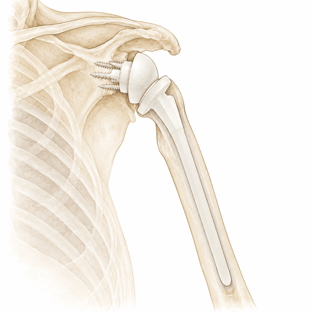
Indications: Revision shoulder arthroplasty is indicated when primary implants fail, with revisions currently being performed at a mean of 3.9 years from the primary procedure despite designs intended to last 10-15 years [1]. The outcome of revision surgery can be predicted based on the specific indication for the procedure [4]. For patients with unstable shoulder prostheses, revision to reverse total shoulder arthroplasty (RTSA) restores stability [37]. Similarly, revision of hemiarthroplasty or anatomical total shoulder replacement (ATSA) for instability using a reverse design prosthesis yields good short-term results [64]. In cases of fracture sequelae, shoulder arthroplasty is an effective treatment that provides significant improvement in pain and function [225].
Surgical Approach / Technique: Key operative techniques include the creation of an anterior cortical window, which is an excellent technique for selecting cases of revision total shoulder arthroplasty [112]. A unicortical humeral osteotomy is a useful tool that greatly facilitates component removal and lessens the overall risk of humeral fracture [121]. When glenoid bone loss is present, glenoid bone grafting can produce good short-term outcomes, and glenoid component reinsertion should be attempted whenever possible [19]. The glenoid-to-humeral head cut distance should be studied further as a potential indication for humeral stem revision, as it correlates with the space available for a revision implant [174].
Implant Selection: Revision RTSA outcomes are generally inferior to primary RTSA, with complications and revision surgeries being common, yet the procedure leads to notable improvements in pain, motion, and function [2]. Cement-within-cement fixation of the humeral component in revision RTSA is associated with reasonable operative time, good medium-term survival rates, and good pain relief and functional outcomes with low complications [40]. Humeral stem fixation with or without cement during primary shoulder arthroplasty demonstrated similar operative time, need for intraoperative humeral osteotomy, and postoperative complication rates in the setting of revision arthroplasty [66]. Both cemented and press-fit humeral fixation techniques yield durable and significant improvements in shoulder function with similar rates of survival at 10 years of follow-up [192]. Convertible humeral and glenoid platforms address challenges in minimizing complexity and complications during revisions from anatomic to reverse total shoulder arthroplasty, but these implants are not always convertible and can introduce additional challenges [127]. For Walch B2 and B3 glenoids, anatomic total shoulder arthroplasty with a posterior augmented glenoid is a viable alternative to RTSA [126]. Findings suggest limited utility of hemiarthroplasty in the long term, supporting the need for careful patient selection and consideration of alternative surgical options such as reverse shoulder arthroplasty [50].
Infection Management: The majority of revision shoulder arthroplasties are performed for patients who are unlikely to have a periprosthetic joint infection (PJI), with less than 10% meeting ICM criteria for definite PJI [8]. Patients undergoing treatment of shoulder PJI with debridement, antibiotics, and implant retention (DAIR) did not have an increased rate of reinfection compared with single-stage and 2-stage revision procedures [20]. Functional improvement was obtained after reimplantation of a reverse total shoulder prosthesis but was not seen after hemiarthroplasty and cement spacer in staged revision with antibiotic spacers [189].
Complications and Risk Factors: Complication rates for revision of failed shoulder prostheses are high, approximately 50% [13]. Nearly 45% of RTSA patients returned to the hospital within one year, most often for shoulder or non-shoulder complications [218]. Similarly, nearly 42% of ATSA patients returned to the hospital within one year, most often for shoulder or non-shoulder complications [218]. Shoulder arthroplasty for inflammatory arthritis is associated with higher rates of medical and surgical complications [71]. Surgical treatment of humeral fractures occurring on reverse total shoulder arthroplasties showed a substantially higher risk of complications without functional benefit at final follow-up when compared with nonoperative care [201]. The revision risk with constrained reverse total shoulder implants for proximal humerus tumors is higher than reported for other proximal humerus prostheses, likely due to increased stresses at the humeral component-bone interface causing aseptic loosening [169]. Shoulder arthroplasty patients from distressed communities use more opioids within 90 days before and after their surgery and are more likely to become prolonged opioid users, placing them at risk for readmission and revision surgery [221].
Other Considerations: Patients achieve maximum medical improvement at 1 postoperative year following reverse total shoulder arthroplasty [21]. Anatomic total shoulder arthroplasty provides pain relief and improved quality of life with a 10-year survival rate of 96% [223]. Pain relief after shoulder fracture arthroplasty appears reliable and predictable in nearly all patients, while functional outcome is dependent on age and time from injury to surgery [226]. Anti-osteoporotic treatment is associated with reduced rates of 2-year revision following total shoulder arthroplasty in patients with osteoporosis [222].
Complications
Infection (PJI): Infection remains a rare but devastating complication of shoulder arthroplasty [253]. Less than 10% of revision shoulder arthroplasties meet ICM criteria for definite periprosthetic joint infection (PJI) [8], yet nearly one-quarter of these procedures yield unexpected positive cultures [255]. Consequently, a high index of suspicion for infection is warranted in cases of apparently aseptic shoulder arthroplasty failure [256]. Risk factors include preoperative malnutrition, which increases the 2-year risk of PJI and revision total shoulder arthroplasty [259], and patient-reported penicillin allergy, which elevates PJI risk likely due to second-line antibiotic use [257]. Prior ipsilateral shoulder arthroscopy, particularly within 2 years of arthroplasty, is associated with greater infection risk [258, 260]. Patients with previous rotator cuff repair face an increased risk of revision owing to PJI after reverse shoulder arthroplasty [261]. Aseptic reoperation within 90 days of primary total or reverse shoulder arthroplasty also notably increases subsequent PJI risk [232]. When implant exchange is not feasible, permanent antibiotic spacers and resection arthroplasty provide similar rates of infection eradication [252]. Complications associated with antibiotic administration are more common when the initial protocol involves intravenous antibiotics [54].
Aseptic loosening: Fifty-two percent of revision shoulder arthroplasty cases presenting with humeral loosening were aseptic [14]. A higher critical shoulder angle correlates with increased revision rates, primarily due to prosthetic loosening [231]. In patients aged 60 or under, glenoid loosening occurred in 19.3% of total shoulder arthroplasty shoulders [104]. Glenospheres smaller than 38 mm are expected to increase revision rates in primary reverse total shoulder arthroplasties [72]. Although cement-within-cement fixation of the humeral component is associated with low complications [40], higher rates of complications and re-revision surgery were observed over time secondary to aseptic glenoid component loosening and instability [177]. In stemless shoulder arthroplasty cohorts, two patients were revised for aseptic glenoid component loosening [108]. Metallic lateralized baseplates for glenoid bone loss are associated with post-operative complications including humeral aseptic loosening and subsidence of the glenoid implant [166].
Instability: Instability is a leading cause of multiple reinterventions, accounting for 26% of cases in patients undergoing revision of failed shoulder arthroplasty [175]. Patients with Parkinson disease have a higher risk of prosthetic shoulder dislocation compared with non-Parkinson patients (6.1% vs. 2.6%) [105]. In stemless shoulder arthroplasty cohorts, three patients developed secondary rotator cuff insufficiency and were revised to a reverse shoulder arthroplasty [108].
Periprosthetic fracture: Routine immediate postoperative radiographs identified a complication in 0.2% of shoulder arthroplasties, with periprosthetic fractures representing 90% of these findings [107]. Of these complications, only 3 were newly identified and unknown to the surgeon at the time the patient left the operating room [107]. None of the complications identified on immediate postoperative radiographs resulted in a change in postoperative care prior to the first follow-up visit [107]. Sustaining a fragility fracture before shoulder arthroplasty portends substantial postoperative risk of periprosthetic fractures and all-cause revision at the 2-year postoperative period [262].
Thromboembolism: Among various procedures, venous thromboembolism was the most frequently reported after shoulder arthroplasty compared to open reduction and internal fixation, with reverse shoulder arthroplasty having the highest rate [241]. Patients with staged bilateral shoulder arthroplasty who have the second arthroplasty within 3 months have significantly higher rates of venous thromboembolism [206].
Wound complications: Wound complications and revision rates in patients undergoing shoulder arthroplasty who require postoperative therapeutic anticoagulation are significantly elevated compared with controls [234]. Patients with staged bilateral shoulder arthroplasty who have the second arthroplasty within 3 months also have significantly higher rates of wound complications [206].
Polyethylene wear: Polyethylene dissociation is a rare complication after reverse shoulder arthroplasty, with an incidence of 0.7% in a series of 549 patients [178]. In a cohort of patients aged 60 or under, progressive glenoid wear was the most common complication in shoulders treated with hemiarthroplasty [104].
Other Considerations: Although outcomes are generally inferior to primary reverse total shoulder arthroplasty, complications and revision surgeries are common in revision procedures [2]. The rate of re-revision after revision reverse total shoulder arthroplasty is 13% in the first 2 years postoperatively and increases to 35% at 5 years [235]. Complications and reoperation rates for revision reverse shoulder arthroplasty for baseplate failure were higher than those for primary reverse shoulder arthroplasty [61]. Overall, 21% of patients undergoing revision of failed shoulder arthroplasty required multiple reinterventions, mainly for instability (26%) and/or infection (25%) [175]. In a study of stemless anatomic and reverse shoulder arthroplasty, the overall complication rate during follow-up was 13.7%, with the rate significantly higher in the nonanatomic positioning group (35%) than the anatomic group (6%) [108]. Two patients in a stemless shoulder arthroplasty cohort developed periprosthetic joint infection, requiring a 2-stage revision surgery to a reverse shoulder arthroplasty [108]. In a study of metallic lateralized baseplates for glenoid bone loss, 12% of shoulders had post-operative complications, including complete brachial plexus palsy [166]. In a cohort of patients aged 60 or under, postoperative complications occurred in 29% of shoulders treated with hemiarthroplasty [104]. Patients with Parkinson disease were more likely to experience one or more postoperative complications following shoulder arthroplasty (14.1%) compared with non-Parkinson patients (10.5%) [105]. Patients with staged bilateral shoulder arthroplasty who have the second arthroplasty within 3 months have significantly higher rates of revision surgery, loosening/lysis, periprosthetic fracture, and blood transfusions [206]. Prior bariatric surgery is associated with an increased rate of complications after primary shoulder arthroplasty independent of body mass index, with risks more pronounced when surgery is performed within 2 years of bariatric surgery [244]. Preoperative malnutrition is associated with a higher rate of 90-day complications, including sepsis and myocardial infarction, following shoulder replacement [259]. Surgeons should consider potential complications and employ a multidisciplinary approach in preoperative risk stratification of patients with inflammatory arthritis undergoing shoulder replacement [71]. The incidence of shoulder replacement and revision is expected to increase in the following years, constituting a burden for healthcare systems [7]. The incidence of shoulder arthroplasty per 100,000 members increased from 6.1 in 2005 to 13.4 in 2013 [102]. Age below 65 years and previous shoulder surgery are significantly associated with an increased risk of revision [248]. A hemi shoulder arthroplasty and a previous shoulder surgery independently increase the risk of humeral stem revision [39]. Considerable between-hospital variation was observed for 1- and 3-year revision rates following primary shoulder arthroplasty [60]. Patient race and ethnicity are associated with higher unplanned 90-day emergency department visits and readmissions but not 10-year all-cause complications or reoperations in primary shoulder arthroplasties [230].
Recovery
Light activity (weeks): A majority of patients return to work after shoulder arthroplasty at an average of 2.3 months postoperatively [25]. While specific week-based milestones for desk work or driving are not detailed in the provided evidence, this average return-to-work timeline serves as the primary benchmark for resuming light occupational activities.
Full activity (months): Most patients are able to return to one or more sports following shoulder arthroplasty [173]. Anatomic total shoulder arthroplasty demonstrates the highest rate of return to sport among shoulder arthroplasty types [173], and a high return to sport can be expected after total shoulder arthroplasty [183].
Complete recovery / outcome plateau (months): Patients undergoing reverse total shoulder arthroplasty achieve maximum medical improvement at 1 postoperative year [21]. This 12-month mark represents the point at which pain, strength, and final functional outcomes stabilize for this specific implant type.
Rehabilitation protocol: Early, active rehabilitation after reverse total shoulder arthroplasty is safe and effective [167]. This approach may have early clinical benefits over a conservative, delayed mobilisation programme [167].
Functional milestones: Six distinct early pain recovery trajectories were identified after total shoulder arthroplasty [171]. In the 'Faster group', 83.7% of patients experienced very low pain scores after only 2 weeks following total shoulder arthroplasty [171].
Other Considerations: Workers' compensation patients have a higher reoperation rate after shoulder arthroplasty compared to non-workers' compensation patients [148]. This cohort also has inferior patient-reported outcomes [148] and a higher rate of persistent pain after shoulder arthroplasty compared to non-workers' compensation patients [148]. Regarding revision procedures, revisions for humeral loosening yield modest clinical improvements [28], and patients undergoing revision for humeral loosening are more likely to experience complications resulting in rerevision [28]. Revision reverse shoulder arthroplasty has an implant survival rate of 85% at ten years [3]. While revision procedures for failed shoulder prostheses lead to notable improvements in pain, motion, and function [2], outcomes of revision reverse shoulder arthroplasty are generally inferior to primary reverse total shoulder arthroplasty [2]. Complications and revision surgeries are common following revision reverse shoulder arthroplasty [2]. Specifically, patients undergoing revision of a failed anatomic total shoulder arthroplasty to reverse total shoulder arthroplasty have worse clinical outcomes compared with those undergoing primary reverse total shoulder arthroplasty [57]. This conversion is associated with higher complication rates [57] and higher revision rates compared to primary reverse total shoulder arthroplasty [57].
Key Evidence
- [L4] Generally, shoulder arthroplasties are designed to last 10-15 years; however, revisions are being performed at a mean 3.9 years from the primary procedure. [1] (10.1016/j.jse.2019.12.015)
- [L5] Although the outcomes are generally inferior to primary reverse total shoulder arthroplasty and complications and revision surgeries are common, revision procedures still lead to notable improvements in pain, motion, and function. [2] (10.5435/jaaos-d-17-00535)
- [L3] Revision reverse shoulder arthroplasty demonstrates significant long-term clinical improvements and an implant survival rate of 85% at ten years. [3] (10.1302/0301-620x.107b11.bjj-2025-0436.r1)
- [L4] We reviewed 39 consecutive revision shoulder arthroplasties performed on 38 patients at our institution between 1977 and 1993. [5] (10.1016/s1058-2746(95)80058-1)
- [Paper] Areas of current controversy concerning shoulder arthroplasty include management of glenoid bone loss, the role of patient-specific instrumentation, differing design features of reverse shoulder arthroplasty, and options for humeral fixation in primary and revision procedures. [6] (10.1097/bte.0000000000000061)
- [L3] The incidence of shoulder replacement and revision is expected to increase in the following years, constituting a burden for the healthcare systems. [7] (10.1186/s12891-022-05849-x)
- [L3] The majority of revision shoulder arthroplasties are performed for patients who are unlikely to have a PJI, with less than 10% meeting ICM criteria for definite PJI. [8] (10.1016/j.jse.2025.01.040)
- [L4] The most revised implants for humeral loosening were anatomic total shoulder arthroplasties and reverse shoulder arthroplasties, with reverse shoulder arthroplasties being the most common implant used for revision. [9] (10.1016/j.jse.2024.08.053)
- [L3] A massive increase of primary and revision shoulder arthroplasties is expected by 2040, mainly due to a rising number of fracture-related procedures. [10] (10.3390/jcm10215123)
- [L3] In the predominantly male patient population below the age of 55, reverse shoulder arthroplasty and stemless anatomic total shoulder arthroplasty have a lower short-term revision risk than stemmed anatomic total shoulder arthroplasty. [11] (10.1016/j.jse.2024.07.032)
- [Paper] Although the prosthesis offers some hope of pain relief and restoration of function in patients who require revision of failed shoulder prosthesis, complication rates are high (approximately 50%). [13] (10.1097/01.bte.0000169730.36840.4b)
- [L4] Fifty-two percent of revision shoulder arthroplasty cases with humeral loosening were aseptic. [14] (10.1016/j.jse.2024.03.004)
- [L1] A clear standardised set of shoulder arthroplasty complication definitions is lacking. [15] (10.1007/s00402-017-2635-9)
- [L4] [16] (10.1016/j.jse.2017.06.038)
- [L3] The optimum shoulder arthroplasty revision rates vary for both the gender and implant type for the diagnosis of OA. [17] (10.1016/j.jse.2024.08.033)
- [L3] Two-stage revisions using a reverse total shoulder arthroplasty at the time of reimplantation generate superior range of motion and functional outcome scores. [18] (10.1016/j.jse.2019.03.001)
- [L4] Revision shoulder arthroplasty with glenoid bone grafting can produce good short-term outcome and glenoid component reinsertion should be attempted whenever possible. [19] (10.1007/s11999-007-0108-0)
- [L3] Patients undergoing treatment of shoulder PJI with DAIR did not have an increased rate of reinfection compared with single-stage and 2-stage revision procedures. [20] (10.1016/j.jse.2023.06.012)
- [L2] Patients achieved maximum medical improvement at 1 postoperative year following reverse total shoulder arthroplasty. [21] (10.1016/j.jse.2018.05.029)
- [Paper] The decision to revise a total shoulder arthroplasty requires a rigorous assessment to answer five key questions regarding infection, instability, wear, loosening, and rotator cuff status. [22] (10.1016/j.otsr.2012.11.010)
- [L4] Patients with neurologic disorders demonstrate improvements in pain and function after shoulder arthroplasty but have higher reported complication and revision rates when compared with patients without neurologic conditions. [23] (10.1016/j.jse.2024.05.023)
- [L4] [24] (10.1016/j.jse.2015.12.010)
- [L1] A majority of patients return to work after shoulder arthroplasty at an average of 2.3 months postoperatively. [25] (10.1016/j.jse.2018.12.011)
- [L4] Revision shoulder arthroplasty for failed HHRH improves functional outcome. [27] (10.1177/1758573217715256)
- [L4] Revisions for humeral loosening yield modest clinical improvements, but patients are more likely to experience complications resulting in rerevision. [28] (10.1016/j.jse.2023.02.006)
- [L4] It has a higher early revision rate compared to conventional shoulder arthroplasties, which may be a result of both the ease of revision and implant-related factors. [30] (10.1177/1758573217704818)
- [L5] Long-term studies are necessary to determine if the results of anatomic shoulder arthroplasty with the use of newer humeral components can match those of anatomic shoulder arthroplasty with the use of older humeral components. [33] (10.5435/jaaos-d-17-00088)
- [L4] [34] (10.1016/j.jse.2018.08.002)
- [L4] [36] (10.1016/j.jse.2018.01.012)
- [L4] [37] (10.1007/s11999-017-5429-z)
- [L3] [39] (10.1186/s12891-021-04247-z)
- [L4] Cement-within-cement fixation of the humeral component in revision reverse shoulder arthroplasty is associated with a reasonable operative time, good medium-term survival rates, and good pain relief and functional outcomes with low complications. [40] (10.1016/j.jse.2017.01.013)
- [L4] Cooperation among registries may allow for earlier identification of risk factors for failure in shoulder arthroplasty. [41] (10.1177/1758573219842168)
- [L5] [42] (10.1530/eor-22-0062)
- [L2] [43] (10.1016/j.jse.2014.02.024)
- [L3] In all these cohorts, loosening, not rotator cuff failure, was the most common diagnosis for revision. [45] (10.1016/j.jse.2022.04.015)
- [L4] Glenoid component failure remains a major cause of poor patient outcomes after total shoulder arthroplasty. [47] (10.1016/j.jse.2017.09.029)
- [L5] These findings suggest limited utility of hemiarthroplasty in the long term, supporting the need for careful patient selection and consideration of alternative surgical options such as reverse shoulder arthroplasty. [50] (10.1016/j.xrrt.2025.100616)
- [L4] Most patients with painful glenoid arthrosis after a hemiarthroplasty have marked pain relief and improvement in motion after revision to a total shoulder replacement. [51] (10.2106/00004623-199806000-00010)
- [L4] Complications associated with antibiotic administration after revision shoulder arthroplasty are not infrequent and are more common in patients whose initial protocol is IV antibiotics. [54] (10.2106/jbjs.19.00846)
- [L3] Patients undergoing revision of a failed anatomic total shoulder arthroplasty to reverse total shoulder arthroplasty have worse clinical outcomes compared with those undergoing primary rTSA, including all PROMs, abduction, elevation, pain relief, and patient satisfaction, with higher complication and revision rates. [57] (10.1016/j.jse.2024.09.019)
- [L3] Deciding on a primary repair does not seem to impact outcomes such as cost, length of stay, and readmission when revision by reverse shoulder arthroplasty is needed. [58] (10.1016/j.jse.2021.03.094)
- [L3] Considerable between-hospital variation was observed for 1- and 3-year revision rates following primary shoulder arthroplasty, where outlier hospitals could be identified based on large differences in revision for specific indications to direct quality improvement initiatives. [60] (10.1016/j.jse.2022.06.006)
- [L4] Complications and reoperation rates were higher than those for primary RSA but outcomes were comparable for revision of failed anatomic shoulder arthroplasty. [61] (10.1016/j.jse.2023.06.039)
- [L3] Single-stage revision demonstrated infection control and complication rates comparable with 2-stage revision for shoulder PJI. [62] (10.2106/jbjs.rvw.26.00055)
- [L4] Revision of hemiarthroplasty or anatomical total shoulder replacement for instability using a reverse design prosthesis gives good short-term results. [64] (10.1302/0301-620x.95b5.30964)
- [L4] [65] (10.1016/j.jse.2018.10.023)
- [L3] Humeral stem fixation with or without cement during primary shoulder arthroplasty demonstrated similar operative time, need for intraoperative humeral osteotomy, and postoperative complication rates in the setting of revision arthroplasty. [66] (10.1016/j.jse.2017.11.010)
- [L4] [68] (10.5435/jaaos-d-21-01001)
- [L3] Surgeons should consider these potential complications and employ a multidisciplinary approach in preoperative risk stratification of IA undergoing shoulder replacement. [71] (10.1016/j.jse.2023.09.014)
- [L3] Glenospheres <38 mm can be expected to increase revision rates in primary RTSAs. [72] (10.1016/j.jse.2021.11.013)
- [L3] The incidence of shoulder arthroplasty per 100,000 members increased from 6.1 in 2005 to 13.4 in 2013. [102] (10.1002/acr.23167)
- [L3] [104] (10.1016/j.jse.2019.03.006)
- [L3] [105] (10.1016/j.jse.2023.04.003)
- [L4] [107] (10.1016/j.jse.2022.10.027)
- [L3] [108] (10.1016/j.jse.2024.01.051)
- [Paper] Current classifications exhibit poor reliability in categorizing glenoid defects post-reverse shoulder arthroplasty removal. [109] (10.1016/j.jseint.2024.08.170)
- [L4] The creation of the anterior cortical window is an excellent technique to implement in selecting cases of revision total shoulder arthroplasty. [112] (10.1097/00132589-200606000-00008)
- [L5] The use of a unicortical humeral osteotomy is a useful tool in revision shoulder arthroplasty that greatly facilitates component removal and lessens the overall risk of humeral fracture. [121] (10.1097/00132589-200606000-00007)
- [L3] The study supports the use of anatomic total shoulder arthroplasty with a posterior augmented glenoid as a viable alternative to reverse total shoulder arthroplasty for this patient population. [126] (10.1016/j.jse.2026.08.001)
- [L5] Convertible humeral and glenoid platforms address some challenges in minimizing complexity and complications during revisions from anatomic to reverse total shoulder arthroplasty, but these implants are not always convertible and can introduce additional challenges. [127] (10.5435/jaaos-d-23-01134)
- [L3] Patients with a diagnosis of depression should be counseled that they will experience a significant clinical improvement from baseline after total shoulder arthroplasty. [128] (10.2106/jbjs.16.00541)
- [L3] Based on data from 2 national shoulder arthroplasty registries, we found no significant difference in risk of revision between stemmed and stemless total shoulder arthroplasties after adjusting for the type of glenoid component. [140] (10.1016/j.jse.2024.03.022)
- [L3] Although both workers' compensation and non-workers' compensation patients experienced significant clinical improvements after shoulder arthroplasty, workers' compensation patients had a higher reoperation rate, inferior patient-reported outcomes, and a higher rate of persistent pain. [148] (10.1016/j.jse.2018.10.007)
- [L1] All 4 types of revision arthroplasty had a similar reoperation rate (20%) at the latest follow-up, but conversion to RSA provided a higher proportion of patients satisfied with the procedure. [155] (10.1016/j.jse.2022.10.006)
- [L3] Significant differences in the skin Cutibacterium subtype distributions were found between shoulders undergoing revision shoulder arthroplasty and those undergoing primary shoulder arthroplasty. [158] (10.1016/j.jse.2020.02.007)
- [L4] Modular shoulder arthroplasty is a suitable procedure for conversion RSA in elderly patients. [161] (10.3390/jcm11030834)
- [Paper] [166] (10.1007/s00264-018-4249-4)
- [L1] Early, active rehabilitation after reverse total shoulder arthroplasty is safe and effective, and may have early clinical benefits over a conservative, delayed mobilisation programme. [167] (10.1177/1758573220937394)
- [L4] The revision risk with this constrained reverse total shoulder implant was higher than reported for other proximal humerus prostheses, likely due to increased stresses at the humeral component-bone interface causing aseptic loosening. [169] (10.1097/corr.0000000000001245)
- [L4] Component loosening is a common failure mode after anatomic total shoulder arthroplasty. [170] (10.1016/j.jse.2022.10.004)
- [L2] Six distinct early recovery trajectories were identified after total shoulder arthroplasty, with 83.7% of patients (the 'Faster group') experiencing very low pain scores after only 2 weeks. [171] (10.1016/j.jse.2025.06.016)
- [L1] Most patients are able to return to one or more sports following shoulder arthroplasty, with anatomic total shoulder arthroplasty having the highest rate of return. [173] (10.1007/s00167-017-4547-1)
- [L3] Glenoid to the humeral head cut distance should be studied further as a potential indication for humeral stem revision, as it correlates with the space available for a revision implant. [174] (10.1016/j.jse.2023.01.030)
- [L4] [175] (10.1016/j.jse.2019.07.034)
- [L3] Although revision risk was similar, the most common causes of revision were different, with rotator cuff tears in TSA patients and glenoid component loosening in RTSA patients. [176] (10.1016/j.jse.2023.03.021)
- [L4] Although a low rate of humeral component loosening was observed, higher rates of complications and re-revision surgery were observed over time secondary to aseptic glenoid component loosening and instability. [177] (10.1016/j.xrrt.2024.08.006)
- [L4] [178] (10.1016/j.jse.2017.05.004)
- [L1] A high return to sport can be expected after total shoulder arthroplasty. [183] (10.1016/j.jseint.2025.05.028)
- [L4] [185] (10.1016/j.jse.2024.11.039)
- [L3] Patients treated with mismatched components demonstrated similar clinical outcomes and revision rates to those treated with matched components in revision shoulder arthroplasty, though results are limited by small sample size and short-term follow-up. [187] (10.1016/j.jse.2025.02.003)
- [L3] [188] (10.5435/jaaos-d-24-00063)
- [L3] Functional improvement was obtained after reimplantation of a reverse total shoulder prosthesis but was not seen after hemiarthroplasty and cement spacer. [189] (10.1007/s11999.0000000000000049)
- [L3] Both cemented and press-fit humeral fixation techniques yield durable and significant improvements in shoulder function with similar rates of survival at 10 years of follow-up. [192] (10.1016/j.jse.2023.11.029)
- [L3] [195] (10.1016/j.jse.2020.01.094)
- [L4] Surgical treatment of humeral fractures occurring on reverse total shoulder arthroplasties showed a substantially higher risk of complications without functional benefit at final follow-up when compared with nonoperative care. [201] (10.1016/j.otsr.2026.104876)
- [L3] Patients with staged bilateral shoulder arthroplasty who have the second arthroplasty within 3 months have significantly higher rates of revision surgery, loosening/lysis, periprosthetic fracture, VTE, and blood transfusions. [206] (10.1016/j.jse.2020.06.010)
- [L3] The study confirms that radiographic measurements are generally valid for evaluating postoperative parameters in reverse total shoulder arthroplasty. [216] (10.1016/j.jse.2024.10.016)
- [L4] Nearly 45% of RTSA and 42% of ATSA patients returned to the hospital within one year, most often for shoulder or non-shoulder complications. [218] (10.1016/j.jse.2024.05.009)
- [L3] Shoulder arthroplasty patients from distressed communities use more opioids within 90 days before and after their surgery and are more likely to become prolonged opioid users, placing them at risk for readmission and revision surgery. [221] (10.1016/j.jse.2024.04.016)
- [L3] This study demonstrates a significant association between anti-osteoporotic therapy and reduced rates of 2-year revision following total shoulder arthroplasty. [222] (10.1016/j.jse.2024.09.020)
- [L4] Anatomic total shoulder arthroplasty provides pain relief and improved quality of life with a 10-year survival rate of 96%. [223] (10.5435/jaaos-d-21-00302)
- [L3] Shoulder arthroplasty is an effective treatment for fracture sequelae with significant improvement in pain and function. [225] (10.1007/s00402-014-2074-9)
- [L4] Pain relief after shoulder fracture arthroplasty appears reliable and predictable in nearly all patients, while functional outcome is dependent on age and time from injury to surgery. [226] (10.1097/01.bte.0000156561.97672.ea)
- [L3] Patient race and ethnicity are associated with higher unplanned 90-day emergency department visits and readmissions but not 10-year all-cause complications or reoperations in primary shoulder arthroplasties. [230] (10.1016/j.xrrt.2024.12.012)
- [L4] A higher CSA correlates with an increased revision rate following shoulder arthroplasty, primarily due to complications such as prosthetic loosening. [231] (10.1186/s13018-025-06655-6)
- [L3] Aseptic reoperation within 90 days of primary TSA or primary RSA was associated with a notably increased risk of subsequent PJI. [232] (10.1016/j.jseint.2021.06.002)
- [L3] Wound complications and revision rates in patients undergoing shoulder arthroplasty who require postoperative therapeutic anticoagulation are significantly elevated compared with controls. [234] (10.1016/j.jse.2019.11.029)
- [L4] The rate of re-revision after revision RTSA is low in the first 2 years postoperatively (13%) but increases to 35% at 5 years. [235] (10.1016/j.jse.2022.11.024)
- [L4] Among the various procedures, VTE was the most frequently reported after SA when compared to ORIF, with RSA having the highest VTE rate. [241] (10.1016/j.xrrt.2023.06.003)
- [L3] These risks were more pronounced when shoulder arthroplasty was performed within 2 years of bariatric surgery. [244] (10.1016/j.jse.2023.02.120)
- [L4] Anteroposterior abduction radiographies up to 90 degrees are superior to conventional radiographies for detecting both radiolucent zones (78 per cent) and loosening of the glenoid component (15.5 per cent). [246] (10.1016/s1058-2746(96)80401-0)
- [L3] Age below 65 years (HR 2.63) and previous shoulder surgery (HR 2.00) were also significantly associated with an increased risk of revision. [248] (10.1016/j.jseint.2025.101482)
- [L1] When implant exchange after shoulder PJI is not feasible, permanent antibiotic spacers and resection arthroplasty are both salvage procedures that provide similar rates of infection eradication. [252] (10.1016/j.jse.2021.10.016)
- [L4] Infection is a rare but devastating complication of shoulder arthroplasty. [253] (10.1016/s1058-2746(96)80085-1)
- [L3] Nearly one-quarter of revision shoulder arthroplasties had unexpected positive cultures, but patients without these cultures had a nonsignificantly higher risk of reoperation compared with those with unexpected positive cultures. [255] (10.1016/j.jse.2016.10.023)
- [L4] A high index of suspicion for infection is warranted in cases of apparently aseptic shoulder arthroplasty failure. [256] (10.1016/j.jse.2015.12.026)
- [L3] Patients with patient-reported penicillin allergy were at an increased risk for prosthetic joint infection after total knee arthroplasty and total shoulder arthroplasty, likely due to receiving second-line antibiotics. [257] (10.1097/corr.0000000000001497)
- [L3] Undergoing an arthroscopic procedure of the ipsilateral shoulder before undergoing an arthroplasty was associated with greater risk of prosthetic joint infection. [258] (10.1016/j.arthro.2021.01.013)
- [L3] Preoperative malnutrition is associated with a higher rate of 90-day complications, including sepsis and myocardial infarction following shoulder replacement and a higher 2-year risk of PJI and revision TSA. [259] (10.1016/j.jse.2025.03.005)
- [L3] Shoulder arthroscopy performed within 2 years before shoulder arthroplasty is associated with a higher infection rate in the first year after shoulder arthroplasty. [260] (10.1016/j.jse.2020.09.019)
- [L3] There is an increased risk of revision owing to periprosthetic joint infection after reverse shoulder arthroplasty for patients with previous rotator cuff repair. [261] (10.1016/j.jse.2022.07.001)
- [L3] Sustaining a fragility fracture before shoulder arthroplasty portends substantial postoperative risk of periprosthetic fractures, infection, subsequent fragility fractures, and all-cause revision at the 2-year postoperative period. [262] (10.5435/jaaos-d-22-00752)
See Also
- Total shoulder arthroplasty
- Reverse Shoulder Arthroplasty
- Shoulder Arthroplasty
- Os Acromiale
- Rotator Cuff
- Fractures
- Internal Fixation
- Shoulder Arthritis
- Shoulder Instability
- Calcific Tendinitis
- Shoulder Arthroscopy
- Rotator Cuff Repair
References
[1] Revision shoulder arthroplasty: a systematic review and comparison of North American vs. European outcomes and complications. Journal of Shoulder and Elbow Surgery. 2020. DOI: 10.1016/j.jse.2019.12.015
[2] Revision Reverse Shoulder Arthroplasty. Journal of the American Academy of Orthopaedic Surgeons. 2019. DOI: 10.5435/jaaos-d-17-00535
[3] Long-term results and implant survival of revision reverse shoulder arthroplasty after a mean follow-up of ten years. The Bone & Joint Journal. 2025. DOI: 10.1302/0301-620x.107b11.bjj-2025-0436.r1
[4] Retrospective, single-centre analysis comparing clinical outcomes of reverse total shoulder replacement for fracture, degenerative changes and revision procedure. 2025.
[5] An analysis of failed humeral head and total shoulder arthroplasty. Journal of Shoulder and Elbow Surgery. 1995. DOI: 10.1016/s1058-2746(95)80058-1
[6] Controversies in Shoulder Arthroplasty. Techniques in Shoulder & Elbow Surgery. 2015. DOI: 10.1097/bte.0000000000000061
[7] Shoulder replacement: an epidemiological nationwide study from 2009 to 2019. BMC Musculoskeletal Disorders. 2022. DOI: 10.1186/s12891-022-05849-x
[8] Assessment of periprosthetic joint infection in revision shoulder arthroplasty. Journal of Shoulder and Elbow Surgery. 2025. DOI: 10.1016/j.jse.2025.01.040
[9] Revision for humeral stem loosening: a systematic review. Journal of Shoulder and Elbow Surgery. 2025. DOI: 10.1016/j.jse.2024.08.053
[10] Projections of Primary and Revision Shoulder Arthroplasty until 2040: Facing a Massive Rise in Fracture-Related Procedures. Journal of Clinical Medicine. 2021. DOI: 10.3390/jcm10215123
[11] Stemless anatomic and reverse shoulder arthroplasty in patients under 55 years of age with primary glenohumeral osteoarthritis: an analysis of the Australian Orthopedic Association National Joint Replacement Registry at 5 years. Journal of Shoulder and Elbow Surgery. 2025. DOI: 10.1016/j.jse.2024.07.032
[13] Reverse Shoulder Arthroplasty Indications, Technique, and Results. Techniques in Shoulder & Elbow Surgery. 2005. DOI: 10.1097/01.bte.0000169730.36840.4b
[14] Humeral stem loosening is not always prosthetic joint infection. Journal of Shoulder and Elbow Surgery. 2024. DOI: 10.1016/j.jse.2024.03.004
[15] Towards standardised definitions of shoulder arthroplasty complications: a systematic review of terms and definitions. Archives of Orthopaedic and Trauma Surgery. 2017. DOI: 10.1007/s00402-017-2635-9
[16] Revision of failed shoulder hemiarthroplasty to reverse total arthroplasty: analysis of 157 revision implants. Journal of Shoulder and Elbow Surgery. 2018. DOI: 10.1016/j.jse.2017.06.038
[17] Comparing optimum prosthesis combinations of total stemmed, stemless and reverse shoulder arthroplasty revision rates for men and women with glenohumeral osteoarthritis. Journal of Shoulder and Elbow Surgery. 2025. DOI: 10.1016/j.jse.2024.08.033
[18] Management of infected shoulder arthroplasty: a comparison of treatment strategies. Journal of Shoulder and Elbow Surgery. 2019. DOI: 10.1016/j.jse.2019.03.001
[19] Glenoid Reconstruction in Revision Shoulder Arthroplasty. Clinical Orthopaedics & Related Research. 2008. DOI: 10.1007/s11999-007-0108-0
[20] Outcomes after débridement, antibiotics, and implant retention for prosthetic joint infection in shoulder arthroplasty. Journal of Shoulder and Elbow Surgery. 2024. DOI: 10.1016/j.jse.2023.06.012
[21] Establishing maximum medical improvement following reverse total shoulder arthroplasty for rotator cuff deficiency. Journal of Shoulder and Elbow Surgery. 2018. DOI: 10.1016/j.jse.2018.05.029
[22] Revision of total shoulder arthroplasty. Orthopaedics & Traumatology: Surgery & Research. 2013. DOI: 10.1016/j.otsr.2012.11.010
[23] The impact of neurologic disorders on clinical and functional outcomes after shoulder arthroplasty: a systematic review. Journal of Shoulder and Elbow Surgery. 2024. DOI: 10.1016/j.jse.2024.05.023
[24] Total shoulder arthroplasty using an inlay mini-glenoid component for glenoid deficiency: a 2-year follow-up of 9 shoulders in 7 patients. Journal of Shoulder and Elbow Surgery. 2016. DOI: 10.1016/j.jse.2015.12.010
[25] Return to work after shoulder arthroplasty: a systematic review and meta-analysis. Journal of Shoulder and Elbow Surgery. 2019. DOI: 10.1016/j.jse.2018.12.011
[27] Revision shoulder arthroplasty for failed humeral head resurfacing hemiarthroplasty. Shoulder & Elbow. 2017. DOI: 10.1177/1758573217715256
[28] Humeral loosening in reverse shoulder arthroplasty: an analysis of 2,342 cases. Journal of Shoulder and Elbow Surgery. 2023. DOI: 10.1016/j.jse.2023.02.006
[30] Seven-year survival analysis of the Global® CAP® (Conservative Anatomic Prosthesis) shoulder resurfacing. Shoulder & Elbow. 2017. DOI: 10.1177/1758573217704818
[31] Rockwood And Matsen S The Shoulder. Arthroscopic Management of Prearthritic and Arthritic Conditions of the Shoulder and the Postarthroplasty Shoulder > SUMMARY.
[32] Rockwood And Matsen S The Shoulder. Arthroscopic Management of Prearthritic and Arthritic Conditions of the Shoulder and the Postarthroplasty Shoulder > Radiographic Evaluation.
[33] Stemless Prosthesis for Total Shoulder Arthroplasty. Journal of the American Academy of Orthopaedic Surgeons. 2017. DOI: 10.5435/jaaos-d-17-00088
[34] The impact of the reverse prosthesis on revision shoulder arthroplasty: analysis of a high-volume shoulder practice. Journal of Shoulder and Elbow Surgery. 2019. DOI: 10.1016/j.jse.2018.08.002
[36] Five-year minimum clinical and radiographic outcomes of total shoulder arthroplasty using a hybrid glenoid component with a central porous titanium post. Journal of Shoulder and Elbow Surgery. 2018. DOI: 10.1016/j.jse.2018.01.012
[37] Revision to Reverse Total Shoulder Arthroplasty Restores Stability for Patients With Unstable Shoulder Prostheses. Clinical Orthopaedics & Related Research. 2017. DOI: 10.1007/s11999-017-5429-z
[39] Stem retention and survival in revision of anatomical convertible shoulder arthroplasty to reverse arthroplasty: a Dutch registry study. BMC Musculoskeletal Disorders. 2021. DOI: 10.1186/s12891-021-04247-z
[40] Cement-within-cement technique in revision reverse shoulder arthroplasty. Journal of Shoulder and Elbow Surgery. 2017. DOI: 10.1016/j.jse.2017.01.013
[41] Early revision in anatomic total shoulder arthroplasty in osteoarthritis: a cross-registry comparison. Shoulder & Elbow. 2019. DOI: 10.1177/1758573219842168
[42] A narrative review of treatment strategies for major glenoid defects during primary reverse shoulder arthroplasty, with a focus on the use of structural bone graft. EFORT Open Reviews. 2023. DOI: 10.1530/eor-22-0062
[43] Patterns of proximal humeral bone resorption after total shoulder arthroplasty with an uncemented rectangular stem. Journal of Shoulder and Elbow Surgery. 2014. DOI: 10.1016/j.jse.2014.02.024
[45] Total shoulder replacement stems in osteoarthritis—short, long, or reverse? An analysis of the impact of crosslinked polyethylene. Journal of Shoulder and Elbow Surgery. 2022. DOI: 10.1016/j.jse.2022.04.015
[47] What can be learned from an analysis of 215 glenoid component failures?. Journal of Shoulder and Elbow Surgery. 2018. DOI: 10.1016/j.jse.2017.09.029
[50] Long-term outcomes of hemiarthroplasty for complex proximal humerus fractures: a systematic review of clinical studies with minimum 10-year follow-up. JSES Reviews, Reports, and Techniques. 2026. DOI: 10.1016/j.xrrt.2025.100616
[51] Revision Total Shoulder Arthroplasty for the Treatment of Glenoid Arthrosis. The Journal of Bone & Joint Surgery*. 1998. DOI: 10.2106/00004623-199806000-00010
[54] The Use and Adverse Effects of Oral and Intravenous Antibiotic Administration for Suspected Infection After Revision Shoulder Arthroplasty. Journal of Bone and Joint Surgery. 2020. DOI: 10.2106/jbjs.19.00846
[57] Anatomic total shoulder arthroplasty revised to reverse total shoulder arthroplasty: clinical and radiographic outcomes compared to primary reverse total shoulder arthroplasty. Journal of Shoulder and Elbow Surgery. 2025. DOI: 10.1016/j.jse.2024.09.019
[58] Reverse Shoulder Arthroplasty for Rotator Cuff Tears with and Without Prior Failed Repair: A Comparative Analysis. Journal of Shoulder and Elbow Surgery. 2021. DOI: 10.1016/j.jse.2021.03.094
[60] The reliability of revision rates following primary shoulder arthroplasty as a quality indicator to rank hospital performance: a national registry analysis including 13,104 shoulders and 87 hospitals. Journal of Shoulder and Elbow Surgery. 2023. DOI: 10.1016/j.jse.2022.06.006
[61] Revision reverse shoulder arthroplasty for the management of baseplate failure: an analysis of 676 revision reverse shoulder arthroplasty procedures. Journal of Shoulder and Elbow Surgery. 2024. DOI: 10.1016/j.jse.2023.06.039
[62] One-Stage or Two-Stage Revision for Infected Shoulder Arthroplasty. JBJS Reviews. 2026. DOI: 10.2106/jbjs.rvw.26.00055
[64] Revision of an unstable hemiarthroplasty or anatomical total shoulder replacement using a reverse design prosthesis. The Bone & Joint Journal. 2013. DOI: 10.1302/0301-620x.95b5.30964
[65] Clinical outcomes following reverse shoulder arthroplasty–allograft composite for revision of failed arthroplasty associated with proximal humeral bone deficiency: 2- to 15-year follow-up. Journal of Shoulder and Elbow Surgery. 2019. DOI: 10.1016/j.jse.2018.10.023
[66] Effects of cemented versus press-fit primary humeral stem fixation in the setting of revision shoulder arthroplasty. Journal of Shoulder and Elbow Surgery. 2018. DOI: 10.1016/j.jse.2017.11.010
[68] Periprosthetic Postoperative Humeral Fractures After Shoulder Arthroplasty. Journal of the American Academy of Orthopaedic Surgeons. 2022. DOI: 10.5435/jaaos-d-21-01001
[71] Shoulder arthroplasty for inflammatory arthritis is associated with higher rates of medical and surgical complications: a nationwide matched cohort analysis from 2016-2020. Journal of Shoulder and Elbow Surgery. 2024. DOI: 10.1016/j.jse.2023.09.014
[72] Effect of glenosphere size on reverse shoulder arthroplasty revision rate: an analysis from the Australian Orthopaedic Association National Joint Replacement Registry (AOANJRR). Journal of Shoulder and Elbow Surgery. 2022. DOI: 10.1016/j.jse.2021.11.013
[73] Rockwood And Matsen S The Shoulder. Shoulder and Elbow Specialty Clinic Workers’ Survey > ANATOMY.
[74] A Lange Medical Book Current Diagnosis Treatment In Orthopedics Fifth Edition. 2Musculoskeletal Trauma Surgery > SHOULDER AND ARM INJURIES.
[75] Rockwood And Green S Fractures In Adults. 29: Principles of Nonunion and Bone Defect Treatment > Applied Anatomy Related to Scapular Fractures.
[76] Aaos Comprehensive Orthopaedic Review 3. Anatomy of the Shoulder, Arm, and Elbow > I. Shoulder.
[77] Rockwood And Matsen S The Shoulder. Developmental Anatomy of the Shoulder and Anatomy of the Glenohumeral Joint > Bursae.
[80] Rockwood And Matsen S The Shoulder. Fractures, Dislocations, and Acquired Problems of the Shoulder in Children > FRACTURES OF THE PROXIMAL HUMERUS.
[81] Apley And Solomon S Concise System Of Orthopaedics And Trauma. DISORDERS OF THE ROTATOR CUFF.
[85] Campbell S Operative Orthopaedics 4 Volume Set. RECONSTRUCTIVE PROCEDURES OF THE SHOULDER AND ELBOW IN ADULTS > ANATOMY AND BIOMECHANICS.
[87] Miller S Review Of Orthopaedics. Genetics of musculoskeletal conditions and abnormalities are summarized in Table 1.27 > UPPER EXTREMITY > SHOULDER.
[91] Apley And Solomon S Concise System Of Orthopaedics And Trauma. INVESTIGATION.
[96] Rockwood And Green S Fractures In Adults. 29: Principles of Nonunion and Bone Defect Treatment > Magnetic Resonance Imaging and Arthrography.
[99] Orthopaedic Knowledge Update 13 Ebook Without Multimedia. Shoulder Anatomy and Biomechanics, Clinical Evaluation, Imaging > Clinical Evaluation > Imaging.
[100] Aaos Comprehensive Orthopaedic Review 3. Imaging of the Shoulder and Elbow > I. Shoulder.
[102] Yearly Trends in Elective Shoulder Arthroplasty, 2005–2013. Arthritis Care & Research. 2017. DOI: 10.1002/acr.23167
[104] Mid- to long-term follow-up of shoulder arthroplasty for primary glenohumeral osteoarthritis in patients aged 60 or under. Journal of Shoulder and Elbow Surgery. 2019. DOI: 10.1016/j.jse.2019.03.006
[105] Inpatient charges, complication, and revision rates for shoulder arthroplasty in Parkinson disease: a regional database study. Journal of Shoulder and Elbow Surgery. 2023. DOI: 10.1016/j.jse.2023.04.003
[107] Routine immediate postoperative radiographs rarely identify unknown complications after shoulder arthroplasty. Journal of Shoulder and Elbow Surgery. 2023. DOI: 10.1016/j.jse.2022.10.027
[108] Influence of humeral position of the Affinis Short stemless shoulder arthroplasty system on long-term survival and clinical outcome. Journal of Shoulder and Elbow Surgery. 2024. DOI: 10.1016/j.jse.2024.01.051
[109] Glenoid Defects In Revision Reverse Shoulder Arthroplasty: Are Current Classification Systems Appropriate?. JSES International. 2024. DOI: 10.1016/j.jseint.2024.08.170
[112] Anterior Humeral Window for Revision Shoulder Arthroplasty. Techniques in Shoulder and Elbow Surgery. 2006. DOI: 10.1097/00132589-200606000-00008
[121] Humeral Osteotomy in Revision Shoulder Arthroplasty. Techniques in Shoulder and Elbow Surgery. 2006. DOI: 10.1097/00132589-200606000-00007
[126] Anatomic Total Shoulder Arthroplasty with the Tornier Perform Posterior Augmented Glenoid versus Reverse Total Shoulder Arthroplasty for the Treatment of Walch B2 and B3 Glenoids: A Propensity Score Matched Analysis. Journal of Shoulder and Elbow Surgery. 2026. DOI: 10.1016/j.jse.2026.08.001
[127] Convertible Humeral and Glenoid Components for Anatomic Shoulder Arthroplasty. Journal of the American Academy of Orthopaedic Surgeons. 2025. DOI: 10.5435/jaaos-d-23-01134
[128] Depression and Patient-Reported Outcomes Following Total Shoulder Arthroplasty. Journal of Bone and Joint Surgery. 2017. DOI: 10.2106/jbjs.16.00541
[140] Comparable low revision rates of stemmed and stemless total anatomic shoulder arthroplasties after exclusion of metal-backed glenoid components: a collaboration between the Australian and Danish national shoulder arthroplasty registries. Journal of Shoulder and Elbow Surgery. 2024. DOI: 10.1016/j.jse.2024.03.022
[148] Inferior outcomes and higher complication rates after shoulder arthroplasty in workers' compensation patients. Journal of Shoulder and Elbow Surgery. 2019. DOI: 10.1016/j.jse.2018.10.007
[155] What is the optimal management of a loose glenoid component after anatomic total shoulder arthroplasty: a systematic review. Journal of Shoulder and Elbow Surgery. 2023. DOI: 10.1016/j.jse.2022.10.006
[158] Cutibacterium subtype distribution on the skin of primary and revision shoulder arthroplasty patients. Journal of Shoulder and Elbow Surgery. 2020. DOI: 10.1016/j.jse.2020.02.007
[161] Conversion of Hemiarthroplasty to Reverse Shoulder Arthroplasty with Humeral Stem Retention. Journal of Clinical Medicine. 2022. DOI: 10.3390/jcm11030834
[166] Benefits of a metallic lateralized baseplate prolonged by a long metallic post in reverse shoulder arthroplasty to address glenoid bone loss. International Orthopaedics. 2018. DOI: 10.1007/s00264-018-4249-4
[167] A randomised trial comparing two rehabilitation approaches following reverse total shoulder arthroplasty. Shoulder & Elbow. 2020. DOI: 10.1177/1758573220937394
[169] Proximal Humerus Tumors: Higher-than-Expected Risk of Revision With Constrained Reverse Shoulder Arthroplasty. Clinical Orthopaedics & Related Research. 2020. DOI: 10.1097/corr.0000000000001245
[170] Revision of total shoulder arthroplasty to hemiarthroplasty: results at mean 5-year follow-up. Journal of Shoulder and Elbow Surgery. 2023. DOI: 10.1016/j.jse.2022.10.004
[171] Trajectories of pain recovery during the first 8 weeks after shoulder arthroplasty: results from the shoulder diary study using latent growth curve modeling. Journal of Shoulder and Elbow Surgery. 2026. DOI: 10.1016/j.jse.2025.06.016
[173] Return to sport after shoulder arthroplasty: a systematic review and meta-analysis. Knee Surgery, Sports Traumatology, Arthroscopy. 2017. DOI: 10.1007/s00167-017-4547-1
[174] Factors associated with humeral stem revision in anatomic to reverse total shoulder arthroplasty revision. Journal of Shoulder and Elbow Surgery. 2023. DOI: 10.1016/j.jse.2023.01.030
[175] Revision of failed shoulder arthroplasty: epidemiology, etiology, and surgical options. Journal of Shoulder and Elbow Surgery. 2020. DOI: 10.1016/j.jse.2019.07.034
[176] Similar rates of revision surgery following primary anatomic compared with reverse shoulder arthroplasty in patients aged 70 years or older with glenohumeral osteoarthritis: a cohort study of 3791 patients. Journal of Shoulder and Elbow Surgery. 2023. DOI: 10.1016/j.jse.2023.03.021
[177] Cement-within-cement technique in revision reverse total shoulder arthroplasty: complications, reoperations, and revision rates at 5-year mean follow-up. JSES Reviews, Reports, and Techniques. 2025. DOI: 10.1016/j.xrrt.2024.08.006
[178] Polyethylene dissociation from humeral stem status after reverse total shoulder arthroplasty. Journal of Shoulder and Elbow Surgery. 2017. DOI: 10.1016/j.jse.2017.05.004
[183] Why do patients not return to sports or work after anatomical or reverse total shoulder arthroplasty? A systematic review and meta-analysis. JSES International. 2025. DOI: 10.1016/j.jseint.2025.05.028
[185] Anatomic total shoulder arthroplasty with keeled glenoids in patients younger than 60 years at 10 years minimum: which risk factors of failure are still valid at long-term follow-up?. Journal of Shoulder and Elbow Surgery. 2025. DOI: 10.1016/j.jse.2024.11.039
[187] Mismatched implants yield comparable outcomes in revision shoulder arthroplasty. Journal of Shoulder and Elbow Surgery. 2025. DOI: 10.1016/j.jse.2025.02.003
[188] Heterotopic Ossification After Shoulder Arthroplasty: A Systematic Review. Journal of the American Academy of Orthopaedic Surgeons. 2024. DOI: 10.5435/jaaos-d-24-00063
[189] Staged Revision With Antibiotic Spacers for Shoulder Prosthetic Joint Infections Yields High Infection Control. Clinical Orthopaedics & Related Research. 2018. DOI: 10.1007/s11999.0000000000000049
[192] Cemented vs. press-fit humeral stems: a matched cohort analysis at a mean follow-up of 10 years. Journal of Shoulder and Elbow Surgery. 2024. DOI: 10.1016/j.jse.2023.11.029
[195] Optimizing humeral stem fixation in revision reverse shoulder arthroplasty with the cement-within-cement technique. Journal of Shoulder and Elbow Surgery. 2020. DOI: 10.1016/j.jse.2020.01.094
[201] Humeral Fractures After Reverse Total Shoulder Arthroplasty: Functional Outcomes and Complications. Orthopaedics & Traumatology: Surgery & Research. 2026. DOI: 10.1016/j.otsr.2026.104876
[206] The relationship of bilateral shoulder arthroplasty timing and postoperative complications. Journal of Shoulder and Elbow Surgery. 2021. DOI: 10.1016/j.jse.2020.06.010
[216] The validity of radiographic measurements compared to 3-dimensional-surface model-based measurements in shoulders with reverse total shoulder arthroplasty. Journal of Shoulder and Elbow Surgery. 2025. DOI: 10.1016/j.jse.2024.10.016
[218] Postoperative revision, complication and economic outcomes of patients with reverse or anatomic total shoulder arthroplasty at one year: a retrospective, United States hospital billing database analysis. Journal of Shoulder and Elbow Surgery. 2025. DOI: 10.1016/j.jse.2024.05.009
[221] Effects of socioeconomic burden on opioid use following total shoulder arthroplasty. Journal of Shoulder and Elbow Surgery. 2024. DOI: 10.1016/j.jse.2024.04.016
[222] Anti-osteoporotic treatment reduces risk of revision following total shoulder arthroplasty in patients with osteoporosis. Journal of Shoulder and Elbow Surgery. 2025. DOI: 10.1016/j.jse.2024.09.020
[223] Survivorship of Anatomic Total Shoulder Arthroplasty. Journal of the American Academy of Orthopaedic Surgeons. 2022. DOI: 10.5435/jaaos-d-21-00302
[225] Treatment of fracture sequelae of the proximal humerus: comparison of hemiarthroplasty and reverse total shoulder arthroplasty. Archives of Orthopaedic and Trauma Surgery. 2014. DOI: 10.1007/s00402-014-2074-9
[226] Shoulder Arthroplasty for Fracture: Restoration of the “Gothic Arch”. Techniques in Shoulder & Elbow Surgery. 2005. DOI: 10.1097/01.bte.0000156561.97672.ea
[230] Patient race and ethnicity are associated with higher unplanned 90-day emergency department visits and readmissions but not 10-year all-cause complications or reoperations: a matched cohort analysis of primary shoulder arthroplasties. JSES Reviews, Reports, and Techniques. 2025. DOI: 10.1016/j.xrrt.2024.12.012
[231] Impact of the critical shoulder angle on shoulder arthroplasty outcomes: a systematic review. Journal of Orthopaedic Surgery and Research. 2026. DOI: 10.1186/s13018-025-06655-6
[232] Early aseptic reoperation after shoulder arthroplasty increases risk of subsequent prosthetic joint infection. JSES International. 2021. DOI: 10.1016/j.jseint.2021.06.002
[234] Therapeutic postoperative anticoagulation is a risk factor for wound complications, infection, and revision after shoulder arthroplasty. Journal of Shoulder and Elbow Surgery. 2020. DOI: 10.1016/j.jse.2019.11.029
[235] Survivorship analysis of revision reverse total shoulder arthroplasty. Journal of Shoulder and Elbow Surgery. 2023. DOI: 10.1016/j.jse.2022.11.024
[241] Venous thromboembolism following surgical management of proximal humerus fractures: a systematic review. JSES Reviews, Reports, and Techniques. 2023. DOI: 10.1016/j.xrrt.2023.06.003
[244] Prior bariatric surgery is associated with an increased rate of complications after primary shoulder arthroplasty independent of body mass index. Journal of Shoulder and Elbow Surgery. 2023. DOI: 10.1016/j.jse.2023.02.120
[246] Clinical and radiological results with the neer shoulder prosthesis. Journal of Shoulder and Elbow Surgery. 1996. DOI: 10.1016/s1058-2746(96)80401-0
[248] Survival Analysis of Total Shoulder Arthroplasty for Glenohumeral Osteoarthritis. Comparison of anatomical and reversed implants a registry study. JSES International. 2026. DOI: 10.1016/j.jseint.2025.101482
[252] Outcomes after resection arthroplasty versus permanent antibiotic spacer for salvage treatment of shoulder periprosthetic joint infections: a systematic review and meta-analysis. Journal of Shoulder and Elbow Surgery. 2022. DOI: 10.1016/j.jse.2021.10.016
[253] Infected shoulder arthroplasties: Treatment with staged reimplantations vs. resection arthroplasty. Journal of Shoulder and Elbow Surgery. 1996. DOI: 10.1016/s1058-2746(96)80085-1
[255] Future surgery after revision shoulder arthroplasty: the impact of unexpected positive cultures. Journal of Shoulder and Elbow Surgery. 2017. DOI: 10.1016/j.jse.2016.10.023
[256] Loose glenoid components in revision shoulder arthroplasty: is there an association with positive cultures?. Journal of Shoulder and Elbow Surgery. 2016. DOI: 10.1016/j.jse.2015.12.026
[257] Is Patient-reported Penicillin Allergy Independently Associated with Increased Risk of Prosthetic Joint Infection After Total Joint Arthroplasty of the Hip, Knee, and Shoulder?. Clinical Orthopaedics & Related Research. 2020. DOI: 10.1097/corr.0000000000001497
[258] Undergoing an Arthroscopic Procedure Prior to Shoulder Arthroplasty is Associated With Greater Risk of Prosthetic Joint Infection. Arthroscopy. 2021. DOI: 10.1016/j.arthro.2021.01.013
[259] Preoperative malnutrition is associated with increased risk of 90-day major medical complications and increased 2-year revision rates following total shoulder arthroplasty. Journal of Shoulder and Elbow Surgery. 2025. DOI: 10.1016/j.jse.2025.03.005
[260] The impact of prior ipsilateral arthroscopy on infection rates after shoulder arthroplasty. Journal of Shoulder and Elbow Surgery. 2021. DOI: 10.1016/j.jse.2020.09.019
[261] Previous rotator cuff repair increases the risk of revision surgery for periprosthetic joint infection after reverse shoulder arthroplasty. Journal of Shoulder and Elbow Surgery. 2023. DOI: 10.1016/j.jse.2022.07.001
[262] Increased Risk of Periprosthetic Fractures and Revision Arthroplasty in Patients Undergoing Shoulder Arthroplasty With a History of Prior Fragility Fractures: A Matched Cohort Analysis. Journal of the American Academy of Orthopaedic Surgeons. 2023. DOI: 10.5435/jaaos-d-22-00752