Clinicians › Elbow
Elbow arthroscopy
For patients: a plain-language version of this topic is available. See the patient guide.
Overview
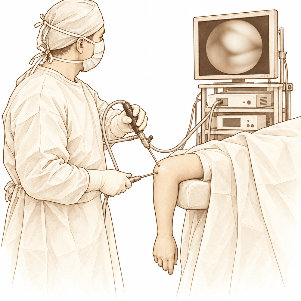
Elbow arthroscopy has evolved from a diagnostic tool into an accepted therapeutic modality for numerous elbow conditions, driven by advances in instrumentation, surgical technique, and anatomical understanding [5, 6, 7, 8, 14]. The procedure offers distinct advantages, including decreased surgical morbidity, improved joint visualization, and the ability to address coexisting intra-articular pathologies with minimal soft tissue injury [7, 9]. It is most successful for the removal of loose bodies [8, 26]. Indications are restricted to patients with clear, arthroscopically treatable pathologies managed by an experienced surgeon; it is not indicated for unclear diagnoses or progressive degenerative joint disease [19]. The procedure requires careful attention to surgical anatomy, patient selection, and standardized technique to avoid neurovascular complications [14, 27].
In experienced hands, elbow arthroscopy is a safe and effective treatment with a low complication rate [10, 12, 26, 27, 65]. When performed in a standardized fashion, the rate of major complications is approximately 0.5% [11]. However, overall complications occur in approximately 14% of cases, with most being minor and major complications occurring in 5% of cases, often requiring repeat surgery [3]. Predominantly low-level evidence studies report varying complication rates with a median of 3% (range 0%-71%) and reoperation rates with a median of 2% (range 0%-59%) [4]. While risks cannot be reduced to zero, the available evidence supports the use of elbow arthroscopy for the majority of conditions where it is currently applied [1, 2, 27].
Elbow arthroscopy is appropriate and safe in children and adolescents, yielding good and excellent postoperative results with an acceptable safety profile similar to adult populations [31, 34, 39]. A significant proportion of pediatric patients may require subsequent surgery in following years [31]. For athletes, the procedure appears safe and effective, allowing most patients a full return to sports participation [33]. As the number of procedures rises, a corresponding rise in complications is foreseen, necessitating continued adherence to safe setup and technique protocols [25, 30].
Anatomy & Pathophysiology
Bony Anatomy
The elbow is a trocho-ginglymoid joint consisting of medial and lateral articulations that provide bony stability [92]. The ulnohumeral joint is formed by the articulation of the trochlea with the ulna within the greater sigmoid notch, providing highly congruent anatomy through almost 180° of articular contact, except for a bare area of the greater sigmoid notch devoid of cartilage [92]. The radiocapitellar joint is formed by the capitellum and the radial head, which is a concave elliptical structure covered with articular cartilage along the radiocapitellar joint and approximately 270° of the articular margin [92]. The proximal radioulnar joint holds the radius in close approximation to the ulna via the annular ligament [92].
The distal humeral articulation is angled 30° from the longitudinal axis, and the axis of rotation is angulated 5° to 7° in the coronal plane relative to the epicondylar axis, with the medial side more distal than the lateral side [92]. The ulna bends approximately 8° medially at 8 cm from the tip of the olecranon, and the articulation to the tip of the coronoid is approximately 30° from the long axis of the ulna in the sagittal plane [92]. The normal range of motion for elbow flexion and extension is 0° to 140°, while forearm pronation and supination are 75° and 85° respectively [36]. A functional arc of motion for the elbow is 100° for flexion and extension and forearm rotation [36]. In full extension, 60% of axial load is transmitted through the radiocapitellar joint [95]. The normal valgus carrying angle of the elbow is 5 to 10 degrees for men and 10 to 15 degrees for women [95].
Ligamentous Anatomy
Elbow stability is determined by primary stabilizers, including the ulnohumeral articulation, medial ulnar collateral ligament, and lateral ulnar collateral ligament, and secondary stabilizers, including the radiocapitellar articulation, common flexor tendon, common extensor tendon, and joint capsule [36]. The medial collateral ligament complex comprises the anterior oblique, posterior oblique, and transverse ligaments [56]. The anterior oblique ligament is the strongest component and the primary stabilizer to valgus stress [56]. It originates on the anterior-inferior edge of the medial epicondyle and inserts on the sublime tubercle of the ulna [56]. The anterior band of the medial collateral ligament is taut in extension, while the posterior band is tight in flexion [56]. The posterior bundle of the medial collateral ligament is the primary restraint to valgus stress with the elbow in maximal flexion [95].
The lateral ulnar collateral ligament origin center is 10.7 mm from the lateral epicondyle and its insertion is 3.3 mm from the apex of the supinator crest [91]. The ulnar nerve passes through the cubital tunnel at the medial column of the elbow [49]. The posterior bundle of the medial collateral ligament forms the floor of the cubital tunnel along the course of the ulnar nerve [55].
Pathophysiology
Elbow arthroscopy is not without complications and morbidity despite being a minimally invasive procedure [1]. Complication rates after elbow arthroscopy vary with a median of 3% and a range of 0%-71%, while reoperation rates vary with a median of 2% and a range of 0%-59% [4]. Elbow arthroscopy has become a safer and more effective treatment modality due to advances in equipment and surgical technique [6]. Improved instrumentation, advanced surgical technique, and better understanding of anatomy offer advantages such as decreased surgical morbidity and improved joint visualization [7]. Elbow arthroscopy can be performed safely with appropriate knowledge of articular and periarticular anatomy, precise surgical technique, and understanding of the procedure's limitations [15]. It is a reliable procedure that requires a clear understanding of anatomy to safely access the joint [16]. Elbow arthroscopy is a safe procedure with a low complication rate, but risks cannot be reduced to zero and require careful attention to anatomy, technique, and surgeon experience [27].
Elbow arthroscopy poses greater technical challenges and neurological risks than knee or shoulder arthroscopy due to the proximity of neurovascular structures to established portals [69]. Complications after elbow arthroscopy have been reported to be 10%, compared with a rate of approximately 3% after knee arthroscopy [69]. The most common complication of elbow arthroscopy is neurovascular injury resulting from surgeon inexperience, poor technique, and lack of knowledge of elbow anatomy [80]. Most neurovascular injuries from elbow arthroscopy are transient and resolve without residual deficit [80]. Transient nerve injury rates following elbow arthroscopy range from 1.7% to 2.0% [80]. No substantial association has been found between complication rate and surgical complexity in elbow arthroscopy [80].
During arthroscopic surgery, joint distention moves the capsule away from bone, but the distance between neurovascular structures and the capsule remains unchanged [38]. Neurovascular structures at risk during portal placement, débridement, and capsular release include the median nerve (anteromedial), ulnar nerve (posteromedial), and radial nerve (adjacent to the anterolateral capsule) [38]. The brachialis muscle protects the median nerve and brachial artery during capsular procedures [38]. The distance between the route of the scope and neurovascular structures might diminish significantly during elbow motion [118]. Joint distention and positioning of the elbow in 90° of flexion provides an increase in safety margin for proximal anterolateral portals [119]. Elbows in 90° flexion with joint distension carry a lower risk for neurovascular injury during portal placement [122].
Valgus extension overload syndrome involves the olecranon being repeatedly and forcefully driven into the olecranon fossa during throwing, exerting shear forces on the medial aspect of the olecranon tip and fossa [37]. The pathoanatomy of valgus extension overload syndrome includes chondrosis, osteophyte development on the posteromedial olecranon and humerus, and loose bodies [37]. Osteoarthritis of the elbow is characterized by osteophyte formation, capsular contracture, and loose bodies, often with relative preservation of the joint space [38]. Periarticular hypertrophic osteophytes act as a mechanical block at the end ranges of flexion and extension in elbow osteoarthritis [38]. Osteoarthritis typically involves the radiocapitellar joint articular cartilage preferentially, with relative preservation of the ulnohumeral articular surfaces [38].
Lateral epicondylitis is an angiofibroblastic hyperplasia of the extensor carpi radialis brevis tendon, a noninflammatory, dysvascular degenerative process caused by repetitive microtrauma [142]. Osteochondritis dissecans of the capitellum is caused by repetitive microtrauma to the vulnerable epiphysis, which has a tenuous blood supply [142]. Repetitive loading of the lateral compartment of the elbow results in subchondral bone degeneration causing cartilage fragmentation in osteochondritis dissecans [142]. Elbow valgus torque increases contact pressure in the radiocapitellar joint [120]. Ulnar collateral ligament injury increases radiocapitellar contact pressures and reduces resistance of the elbow to valgus loading [131].
Intrinsic causes of elbow stiffness include articular damage or malunion, intraarticular hardware, and loose bodies [139]. Extrinsic causes of elbow stiffness include a contracted joint capsule and ligaments, heterotopic ossification, prominent hardware, and skin contracture [139]. Posttraumatic or postsurgical contractures often present with a damaged, abnormally thickened joint capsule and altered anatomy [139]. In posttraumatic or postsurgical stiffness, the joint space is usually contracted and the capsule is scarred down to the bones, making it difficult to enter the elbow joint [139]. Adhesions and scarring can make anatomy visually confusing once the joint is entered in posttraumatic or postsurgical stiffness [139]. The locations of the median and radial nerves may be altered in posttraumatic or postsurgical stiffness due to contracted anterior soft tissues [139].
Classification
Classification of osteochondral defects (OCD) of the elbow is critical because current management guidelines depend on the specific type of lesion and its stability [42]. Most existing classifications are based on standard radiographs, CT, MRI, or arthroscopy [42]. While OCD lesions can be classified during the arthroscopic procedure [42], the value of grading capitellar OCD appears limited [42].
Minami Classification: This system grades lesions based on radiographic appearance. Grade 1 describes a stable lesion with a translucent cystic shadow in the capitellum [42]. Grade 2 describes a clear zone between the OCD and adjacent subchondral bone [42]. Grade 3 describes loose bodies [42]. A recent study identified the Minami Classification as the most reliable for classifying different stages of capitellar OCD [42]. However, it remains unclear whether radiographic evidence categorized by this system guides treatment in clinical practice due to fair agreement [42].
Itsubo et al Classification: Based on MRI, this system distinguishes five stages of capitellar OCD [42]. Stage 1 is characterized by a normally shaped capitellum with several spotted areas of high signal intensity [42]. Stage 2 is characterized by a normally shaped capitellum with several spotted areas of higher intensity than that of cartilage [42]. Stage 3 is characterized by discontinuity and noncircularity of the chondral surface signal of the capitellum and no high signal interface apparent between the lesion and the bottom of the lesion [42]. Stage 4 is characterized by a lesion separated by a high-intensity line in comparison with the cartilage [42]. Stage 5 is characterized by a capitellar lesion displaced from the floor or a defect of the capitellar lesion [42].
Other Considerations: The Kwak classification was used to categorize patients into stage I, II, and III groups in a study of primary elbow osteoarthritis [171]. In a study of simple elbow dislocations, osteochondral lesions were staged from I to IV based on MRI findings ranging from subchondral edema to loose bodies [88].
Clinical Presentation
General Evaluation Principles
Accurate diagnosis and treatment initiation for the elbow require an intimate understanding of the joint's anatomy, biomechanics, and diagnostic tests, as isolated elbow injuries are rare and fractures serve as proxies for associated soft tissue injuries [67]. A thorough history and physical examination are invaluable for understanding the disease process and its impact on the patient [144]. The history must establish whether pain occurs throughout the arc of motion or only at terminal limits, and identify any associated mechanical symptoms or instability [144]. Clinicians must document the location, quality, context, duration, and severity of pain to focus the physical examination [158]. Prior treatments, including surgical interventions and injections, aid in diagnosis [158]. Determining the symptom trajectory—whether pain is improving, worsening, or constant—is extremely helpful [158]. A working knowledge of pathologic conditions affecting different locations around the elbow is paramount to correct diagnosis [158].
The physical examination is directed by the history and the location of pain in the anterior, posterior, medial, or lateral aspect of the elbow [36]. Pathologic entities associated with these discrete compartments assist the examiner in detecting conditions [36].
Imaging
Plain radiographs remain the hallmark and best screening test for elbow evaluation [36]. Standard and specialized views provide valuable clinical information when used appropriately [45]. CT scans with 3D reconstructions may be useful for evaluating the extent and location of disease and for surgical planning [144]. MRI may be useful to evaluate the status of soft tissues, including the medial and lateral collateral ligamentous complexes [144]. Electromyography and nerve conduction studies (EMG/NCS) may be useful to evaluate the degree of nerve compression and its contribution to elbow pain or dysfunction [144].
Osteoarthritis
Patients with primary osteoarthritis of the elbow typically present with loss of terminal extension and flexion, along with painful catching, clicking, or locking [38]. Pain is typically noted at the end ranges of motion rather than through the midrange [38]. Night pain is not typical; if present, an inflammatory cause should be considered [38]. On physical examination, pain is usually felt at the end ranges of flexion and extension [38]. Forearm rotation is relatively preserved until later in the disease process [38]. Ulnar neuropathy is present in up to 50% of patients with primary osteoarthritis [38].
Radiographs typically show osteophyte formation at the coronoid process (anterior and medial), coronoid fossa, radial fossa, radial head, olecranon tip, and olecranon fossa [38]. Joint spaces at the ulnohumeral joint are usually preserved, while those at the radiocapitellar joint are mildly narrowed [38]. Loose bodies may be evident on radiographs, which typically underestimate the number present [38]. CT may be useful for surgical planning, allowing detailed assessment of osteophytes and loose bodies [38].
Valgus Extension Overload Syndrome
Patients with valgus extension overload syndrome report posteromedial elbow pain during the deceleration phase of throwing as the elbow reaches terminal extension [37]. Pain may also occur during acceleration [37]. Loss of terminal elbow extension may occur [37]. Physical examination may reveal crepitus and tenderness over the posteromedial olecranon [37]. Pain is reproduced when the elbow is forced into extension [37]. Elbow flexion contracture may be seen [37].
AP, lateral, oblique, and axillary views may reveal posteromedial olecranon osteophytes and/or loose bodies [37]. CT with two-dimensional reconstruction and three-dimensional surface rendering best visualizes the pathology [37]. MRI may be most helpful in evaluating associated injuries, including partial or complete tears of the MCL [37].
Medial Collateral Ligament Injuries
Patients with MCL injuries report medial elbow pain during the acceleration phase of throwing [56]. Pain may occur only when throwing at more than 50% to 75% of maximal effort [56]. Acute injuries may present suddenly with a pop, sharp pain, and inability to continue throwing [56]. Point tenderness can be noted at the MCL or toward its insertion sites [56].
Valgus instability is tested with the elbow flexed between 20° and 30° to unlock the olecranon from its fossa as valgus stress is applied [56]. The milking maneuver is performed by pulling on the patient’s thumb to create valgus stress while the forearm is supinated and the elbow is flexed beyond 90° [56]. A subjective feeling of apprehension, instability, or localized pain at the MCL indicates injury during this maneuver [56]. The moving valgus stress test is a modification where valgus stress is applied while the elbow moves through an arc of flexion or extension [56]. A subjective feeling of apprehension, instability, or localized pain at the MCL indicates injury during this test [56].
AP, lateral, and axillary views should be obtained to assess for joint space narrowing, osteophytes, and loose bodies [56]. Valgus stress radiographs with the elbow in 20° to 30° of flexion and the forearm in full supination may be used to measure medial joint line opening [56]. Greater than 3 mm medial joint line opening on valgus stress radiographs has been considered diagnostic for valgus instability [56]. Conventional MRI can help identify thickening within the ligament from chronic injury or more obvious full-thickness tears [56]. Magnetic resonance arthrography enhanced with intra-articular gadolinium improves the diagnosis of partial undersurface tears [56]. Dynamic ultrasonography can help detect increased laxity with valgus stress; however, the diagnostic quality of the results is operator dependent [56].
Osteochondritis Dissecans (OCD)
Physical examination findings in capitellum OCD include lateral elbow tenderness, crepitus, and often a 15° to 20° flexion contracture [37]. Classification of the lesion is critical because current management guidelines depend on the type of lesion present and its stability [42]. Most classifications are based on standard radiographs, as in the Minami Classification, CT, MRI, or arthroscopy [42].
Minami Classification: * Grade 1: Stable lesion with a translucent cystic shadow in the capitellum [42]. * Grade 2: Clear zone between the OCD and adjacent subchondral bone [42]. * Grade 3: Loose bodies [42].
Itsubo MRI Classification: * Stage 1: Normally shaped capitellum with several spotted areas of high signal intensity [42]. * Stage 2: Capitellum with several spotted areas of higher intensity than that of cartilage [42]. * Stage 3: Discontinuity and noncircularity of the chondral surface signal of the capitellum and no high signal interface apparent between the lesion and the bottom of the lesion [42]. * Stage 4: Lesion separated by a high-intensity line in comparison to the cartilage [42]. * Stage 5: Capitellar lesion displaced from the floor or a defect of the capitellar lesion [42].
The Minami Classification was found to be the most reliable for classifying different stages of OCD of the capitellum in a recent study [42]. It was unclear whether radiographic evidence of OCD of the humeral capitellum, as categorised by the Minami Classification, guides treatment in clinical practice as a result of this fair agreement [42].
General Elbow Pathology and Instability
The presence and extent of elbow instability in the athlete can be a difficult diagnosis to determine [126]. Subtle instabilities can be a source of elbow pain and yet not be clear on physical examination [126]. Arthroscopy has provided the benefit of assessing articular changes related to these instabilities and allows direct assessment of the capsule and ligaments [126]. The majority of instabilities are based upon a history such as medial elbow pain that occurs in the acceleration phase of the throwing motion [126]. Less common lateral instabilities are often confused with more common conditions such as lateral epicondylitis [126]. Provocative physical exam maneuvers are necessary to assist with the diagnosis of elbow instability, but it is important to confirm these with imaging studies with a diagnostic arthroscopy in many cases [126]. Elbow arthroscopy is not indicated for patients with an unclear diagnosis or progressive degenerative joint disease [19].
Investigations
Plain radiography: Plain radiographs remain the hallmark and best screening test for elbow evaluation [36]. Standard and specialized views provide valuable clinical information during patient evaluation [45]. In the context of elbow osteoarthritis, radiographs typically underestimate the number of loose bodies present [38]. For Little Leaguer's elbow, plain radiographs can potentially show widening of the apophysis or fragmentation of the medial epicondyle [110].
CT: CT is useful for identifying associated osseous injury in acute dislocations [107]. In elbow osteoarthritis, CT may be useful for surgical planning by allowing detailed assessment of osteophytes and the presence of loose bodies [38].
MRI: MRI is the imaging modality best suited for evaluating soft-tissue structures in the elbow, including ligaments, tendons, cartilage, and nerves [101]. Magnetic resonance arthrography is particularly beneficial in the evaluation of osteochondral lesions, loose bodies, and ulnar collateral ligament injury in a throwing athlete [101]. MRI may be most helpful in evaluating associated injuries including partial or complete tears of the medial collateral ligament in valgus extension overload syndrome [37]. While MRI can be used to evaluate ligaments and tendons in elbow stiffness, it is rarely indicated [55]. In symptomatic youth athletes with Little Leaguer's elbow, MRI studies often show edema at the distal humerus or medial epicondyle apophysis [110]. MRI of the elbow and the upper extremity athlete may be employed to identify partial or periosteal sleeve avulsions of the medial epicondyle [111]. One institution reported that MRI was able to identify the cause of medial elbow pain in 20 (95%) of 21 throwing athletes [150].
Ultrasonography: Ultrasonography allows dynamic imaging, which may be useful in evaluating for ulnar nerve subluxation or a snapping triceps [101]. Ultrasonographic soft-tissue evaluation in the elbow is most useful in evaluating the distal biceps and the common flexor and extensor tendons [101].
Arthroscopic Diagnosis: Patients treated arthroscopically benefit from additional diagnostic techniques and improved visualization of the elbow joint [9]. Elbow arthroscopy has become a valid and safe option for the diagnosis of both acute and chronic elbow instability [43]. Three reproducible arthroscopic techniques are valuable in identifying and quantifying elbow instability [13]. To perform a relatively thorough diagnostic arthroscopy of the elbow, at least three portals are necessary [64]. The combination of the low postero-lateral, the direct lateral, and the high anteromedial portals is recommended for diagnostic elbow arthroscopy [64]. Elbow arthroscopy is indicated for patients with clear and arthroscopically treatable pathologies but is not indicated for patients with an unclear diagnosis [19]. Elbow arthroscopy has significant diagnostic value in radial head fractures when compared to standard radiological imaging and revealed concomitant injuries even in patients with uneventful MRI/CT [134].
Other Considerations: The Minami Classification was the most reliable for classifying different stages of osteochondritis dissecans of the capitellum [42].
Treatment
Non-Operative
Arthroscopic management of the arthritic elbow serves as an intermediate step between nonoperative management and elbow replacement surgery [29].
Operative
Indications: Diagnostic and surgical arthroscopy of the elbow is most successful for removing loose bodies [8]. Elbow arthroscopy has expanded indications for diagnosing and treating acute trauma, including radial head fractures and instability, provided patients are carefully selected and neurovascular risks are managed with proper technique [44]. Recent advances in arthroscopic instrumentation and techniques have led to growing interest in arthroscopic treatment of elbow osteoarthritis [41]. Elbow arthroscopy is an appropriate and safe treatment option in children and adolescents with good and excellent postoperative results [39].
Surgical Approach / Technique: Elbow arthroscopy can be performed in a supine, prone, or lateral decubitus position, which is chosen at the discretion of the surgeon [82]. The supine position allows for regional anesthesia and easier access to the anterior compartment but requires a traction device to suspend the arm [82]. The supine position also more easily allows for conversion to an open approach [82]. The lateral decubitus and prone positions may require general anesthesia to secure the airway and require a special arm holder but provide better access to the posterior compartment of the elbow [82]. There are over seven portals that are commonly used for elbow arthroscopy [82]. For each procedure, at least two portals are required anteriorly and two posteriorly [82].
One of the greatest effects on maximizing the distance from the portal to the neurovascular structures is achieved when the joint is insufflated with saline prior to developing the portal [82]. Current inflation of the elbow initial to introduction of the arthroscope merely leads to a minimal extent of the desired transpositioning of anteriorly positioned nerves at risk [162]. The operative extremity landmarks should be mapped out before insufflation, as distension can alter position [166]. Approximately 25 to 30 mL of arthroscopy fluid is injected into the joint space until resistance is met [166]. The joint is insufflated with sterile isotonic fluid, typically with a spinal needle directed into the lateral soft spot of the elbow [79]. In most cases of elbow contracture, the volume of the joint is decreased and the capsule is noncompliant, which minimizes the potential benefit of joint insufflation in protecting the surrounding neurovascular structures [79].
The proximal lateral and proximal medial portals are the safest portals used for the anterior compartment [82]. The proximal lateral portal is developed on the superior and anterior surface of the supracondylar humerus 3 cm proximal and 1 cm anterior to the epicondyles in a relative safe zone [82]. The proximal lateral portal is approximately 1 cm from the radial nerve with insufflation of the joint and allows for medial joint visualization [82]. The proximal anterolateral portal is placed approximately 2 cm proximal to the lateral epicondyle and directly anterior to the humerus [166]. The proximal anterolateral portal is safe, out of the direct pathway of the radial nerve and approximately 6 mm from the posterior antebrachial cutaneous nerve [166].
The location of the proximal anteromedial portal is approximately 2 cm proximal and 1 cm anterior to the medial epicondyle [79]. Only the skin is incised for the proximal anteromedial portal in order to protect branches of the medial antebrachial cutaneous nerve [79]. The cannula for the proximal anteromedial portal is directed anterior to the intermuscular septum into the elbow joint [79]. The superomedial viewing portal is placed 3 cm proximal to the medial epicondyle and anterior to the intermuscular septum [166]. When creating the superomedial portal, the trochar should penetrate the capsule while aiming toward the coronoid fossa [166]. Care should be taken to avoid the median nerve and brachial artery, which are approximately 12.4 and 18.0 mm away, respectively [166]. In the case of subluxation, the nerve should be reduced into the cubital tunnel and stabilized externally while an incision is made for the superomedial viewing portal [166].
Elbow arthroscopy is a standardized technique where a comfortable installation and knowledge of five portals allow for complete joint exploration [165]. A mechanical forearm holder provides an effective means to temporarily or permanently maintain the elbow joint in any desired degree of extension or flexion during elbow arthroscopy without the need for an assistant [75]. An inexpensive support device provides excellent support and stability to the elbow during arthroscopy and open procedures with excellent proximal and distal access [143]. A new setup for elbow arthroscopy reduces operative time, facilitates easy training and triangulation, and has no additional risks compared with conventional elbow arthroscopy setup [102]. The described technique for arthroscopic ulnar nerve identification translates the open surgery philosophy of identifying neurovasculature to arthroscopy, allowing for safer performance of procedures in the posteromedial region of the elbow [99]. The authors describe an approach for diagnostic and therapeutic procedures in the elbow using needle arthroscopy, detailing portal locations for direct in-line visualization and the importance of a dedicated inflow cannula to control flow and decrease fluid utilization [83].
Specific Procedures and Outcomes: Patients treated arthroscopically benefit from additional diagnostic techniques, improved visualization of the elbow joint, the ability to address coexisting intra-articular pathologic conditions, and minimal soft tissue injury with no clinical consequences in outcomes [9]. The authors present 3 reproducible arthroscopic techniques that are valuable in identifying and quantifying elbow instability [13]. Patients with stiff elbows who underwent arthroscopic arthrolysis achieved satisfactory clinical outcomes very early postoperatively [24]. Overall, patients saw improvement in elbow ROM, but many still had residual symptoms from their underlying disease after arthroscopic elbow capsular release [46]. This minimally invasive method allows effective management of elbow instability while promoting quicker patient recovery and long-term functional restoration [47]. The presented arthroscopic technique is reproducible and achieves the reconstruction of the lateral ulnar collateral ligament of the elbow as well as avoids residual instability [164].
Arthroscopic excision of the radial head is a safe and effective technique, and it has several potential advantages over open excision [72]. Arthroscopic evaluation of the anterior and posterior compartments allows for identification of loose bodies, effective bony debridement, osteophyte excision, and synovectomy at the time of radial head excision [72]. This arthroscopic procedure, performed with the elbow in the maximum flexed position using posterolateral and lateral portals and absorbable pins, is effective for fragment fixation of osteochondritis dissecans of the humeral capitellum [154]. Indications, surgical pearls and pitfalls, and postoperative care are outlined for arthroscopic autologous minced cartilage implantation for focal lesions of the capitellum humeri of the elbow [129].
Arthroscopic extensor carpi radialis brevis muscle resection for chronic elbow lateral epicondylitis requires familiarity with arthroscopic surgery and experience in elbow arthroscopy [59]. Upon completion of arthroscopic biportal visualization and precise resection for recalcitrant lateral epicondylitis, portal sites are sutured, and a soft dressing is applied [169]. Elbow range-of-motion exercises are initiated on the first postoperative day following arthroscopic biportal visualization and precise resection for recalcitrant lateral epicondylitis with a manageable level of discomfort [169]. Patients may resume daily activities provided that their pain levels remain within acceptable limits following arthroscopic biportal visualization and precise resection for recalcitrant lateral epicondylitis [169]. A return to work can be expected within 6 weeks following arthroscopic biportal visualization and precise resection for recalcitrant lateral epicondylitis [169].
Other Considerations: Elbow arthroscopy is not necessarily contraindicated in patients with a subluxating or transposed ulnar nerve [123]. Neither elbow arthroscopy nor specifically the use of the proximal anteromedial portal is contraindicated in patients with prior transposition or subluxation of the ulnar nerve [124].
Safety and Complications: Complications of elbow arthroscopy are seen in approximately 14% of cases, with most being minor and major complications occurring in 5% of cases, often requiring repeat surgery [3]. Predominantly low-level evidence studies demonstrate varying complication rates (median 3%, range 0%-71%) and reoperation rates (median 2%, range 0%-59%) after elbow arthroscopy [4]. The majority of neurologic complications following elbow arthroscopy are transient [23]. Pediatric patients who underwent elbow arthroscopy had an 86% return-to-sport rate, a 12% reoperation rate, and a 3.7% complication rate [31]. Bilateral pulmonary emboli are a rare but potentially serious complication of elective elbow arthroscopy [21]. With the rising number of elbow arthroscopies being performed, a rise in complications is foreseen [25].
Elbow arthroscopy can be performed safely with appropriate knowledge of the articular and periarticular anatomy, precise surgical technique, and understanding of the procedure's limitations [15]. Elbow arthroscopy is a reliable procedure that requires a clear understanding of the anatomy to be able to safely access the joint [16]. Elbow arthroscopy has become a safe and effective treatment for elbow pathology due to improved instrumentation, advanced surgical technique, and better understanding of anatomy, offering advantages such as decreased surgical morbidity and improved joint visualization [7]. When properly executed, elbow arthroscopy can be performed in a safe and efficient manner with minimal risk to patients [105].
Complications
Overall Safety and Rates: Complications of elbow arthroscopy occur in approximately 14% of cases, with most being minor [3]. Major complications arise in 5% of cases, often necessitating repeat surgery [3]. While the procedure is generally safe and effective, life-changing events such as deep infection and permanent nerve injury do occur [186]. Historically, complication rates have been reported as high as 20% [80], and novice portal placement specifically carries a surgical complication rate of 30% [184]. In comparative cohorts, overall complication rates were lower following arthroscopic approaches than open approaches [181].
Nerve Palsy: Neurologic injury prevalence ranges from 1.7% to 10.4%, with the vast majority being transient [116]. Current reported rates for transient nerve injury are lower, ranging from 1.7% to 2.0% [80]. Iatrogenic nerve injury is considered one of the most common and serious complications, alongside prolonged wound drainage and elbow stiffness [116]. The potential for neurologic injury remains significant despite few reported cases in the literature [160]. Anatomical proximity is a key risk factor, as distances from the capsule to the median and radial nerves may be as little as 6 mm [160]. The ulnar nerve’s proximity to the capsule requires especially cautious and limited work in this area [160]. If capsular penetration or excision occurs, arthroscopy should be aborted and an open procedure considered [160]. Elbow flexion and preinsufflation are highly desirable, though they do little to change capsule-to-nerve distances [160]. Common insults include compression from cannulas, fluid extravasation, local anesthesia, and laceration with the scalpel or cannula [80]. Most neurovascular injuries are transient and resolve without residual deficit [80]. In one 13-year series, a specific surgeon group reported no neurovascular complications [89].
Infection (PJI): The overall infection rate within 6 months after elbow arthroscopy was 1.55% in a cohort of 2,704 Medicare patients [103]. In a 30-day postoperative profile, deep infection was the most common complication, occurring in 3 of 530 cases (0.57%) [121]. None of the 9 patients with a postoperative infectious complication in that 30-day profile had a preoperative diagnosis of infection [121]. Risk factors include intraoperative intra-articular corticosteroid injection or preoperative corticosteroid injection within 4 weeks of surgery [191]. An increased risk was also noted with intra-articular steroid injection at the end of the procedure, which is not recommended [80]. Additional significant risk factors include BMI >30, diabetes, and smoking tobacco [191].
Stiffness / Arthrofibrosis: Obesity is associated with significantly increased rates of all assessed complications after elbow arthroscopy in Medicare patients, including stiffness [130].
Other Considerations: Other known complications include articular cartilage injury, synovial fistula formation, instrument breakage, and tissue injury secondary to tourniquet use [80]. Heterotopic ossification was a minor complication with a prevalence of 6.3% among 205 patients, usually located in the medial compartment [185]. A significant proportion of patients from a large cohort visited the emergency department at least once in the 90 days following surgery [17]. No substantial association has been found between complication rate and surgical complexity for procedures including complete synovectomy, radial head resection, osteocapsular arthroplasty, and medial epicondylectomy [80]. Pediatric elbow arthroscopy performed by an experienced surgeon using a standardized technique has an acceptable complication rate similar to published rates in pediatric and adult populations [31].
Recovery
Light activity (weeks): The evidence provided does not specify a typical week range for desk work, driving, or light activities of daily living following elbow arthroscopy.
Full activity (months): The evidence provided does not specify a month range for the return to manual work, sport, or full range of motion and strength.
Complete recovery / outcome plateau (months): The evidence provided does not specify a month range for the stabilization of pain, strength, or final functional outcomes.
Rehabilitation protocol: The evidence provided does not detail specific physical therapy phasing, immobilisation duration, weight-bearing or range-of-motion progression schedules, or sling and brace removal timing.
Functional milestones: Clinical outcomes for patients with primary elbow osteoarthritis who underwent arthroscopic osteocapsular arthroplasty improved from preoperative assessment to short- and medium-term follow-up [159], although range of motion decreased between short- and medium-term follow-up in this cohort [159]. Arthroscopic circumferential release provides safe and effective restoration of range of motion, pain relief, and functional improvement in patients with posttraumatic elbow stiffness regardless of preoperative severity [187]. Patients saw improvement in elbow range of motion after arthroscopic elbow capsular release [46], though many still had residual symptoms from their underlying disease [46]. A surgical protocol for advanced articular lesions of osteochondritis dissecans of the elbow serves good functional recovery to some extent for damaged young athletes [71]. Surgical reconstruction of the medial ulnar collateral ligament gets athletes back to their previous level of play more than 80% of the time [176] and has more favorable outcomes compared with primary repair [176]. After elbow arthroscopy, professional baseball players saw an improvement in several performance metrics [178]. Arthroscopic treatment for elbow lateral epicondylitis leads to optimized long-term outcomes [180]. Arthroscopic mosaicplasty for elbow osteochondritis dissecans resulted in good-to-excellent patient satisfaction [190] and a return to activity in 6 of 7 patients [190]. Satisfactory outcomes were obtained with lateral collateral ligament repair for acute posterolateral rotatory instability of the elbow [189].
Other Considerations: Elbow arthroscopy is associated with complications and morbidity despite being a minimally invasive procedure [1]. Complications of elbow arthroscopy occur in approximately 14% of cases [3], with most complications following the procedure being minor [3]. Major complications following elbow arthroscopy occur in 5% of cases [3] and often require repeat surgery [3]. Systematic review data demonstrate a median complication rate of 3% after elbow arthroscopy, with a range of 0%-71% [4], and a median reoperation rate of 2%, with a range of 0%-59% [4]. A significant proportion of patients visited the emergency department at least once in the 90 days following elbow arthroscopy [17]. Open arthrolysis for elbow stiffness has no impact on elbow functional recovery [68]. Arthroscopic treatment for elbow lateral epicondylitis is recommended for surgeons with elbow arthroscopy training [180].
Key Evidence
- [L3] Elbow arthroscopy is not without complications and morbidity despite being a minimally invasive procedure and advances made in surgical technique. [1] (10.1177/17585732241249393)
- [L4] The available evidence supports the use of elbow arthroscopy in the management of the majority of conditions where it is currently used. [2] (10.1016/j.arthro.2011.10.007)
- [L4] Complications of elbow arthroscopy are seen in approximately 14% of cases, with most being minor and major complications occurring in 5% of cases, often requiring repeat surgery. [3] (10.1016/j.jse.2013.09.026)
- [L4] Predominantly low-level evidence studies demonstrate varying complication rates (median 3%, range 0%-71%) and reoperation rates (median 2%, range 0%-59%) after elbow arthroscopy. [4] (10.1016/j.arthro.2023.04.015)
- [L5] Elbow arthroscopy has become an accepted surgical option for treating numerous conditions of the elbow. [5] (10.1016/j.jhsa.2012.10.023)
- [L5] Elbow arthroscopy has become a safer and more effective treatment modality for several elbow pathologies due to advances in equipment and surgical technique. [6] (10.5435/00124635-200810000-00003)
- [L5] Diagnostic and surgical arthroscopy of the elbow has become an accepted treatment modality for numerous conditions, most successful for removing loose bodies. [8] (10.5435/00124635-200605000-00007)
- [L4] Patients treated arthroscopically benefit from additional diagnostic techniques, improved visualization of the elbow joint, the ability to address coexisting intra-articular pathologic conditions, and minimal soft tissue injury with no clinical consequences in outcomes. [9] (10.1016/j.arthro.2013.08.016)
- [L4] In experienced hands, elbow arthroscopy is a safe modality of treatment for a variety of pathologies. [10] (10.1016/j.arthro.2007.03.081)
- [L4] Elbow arthroscopy is a relatively safe procedure with a 0.5% rate of major complications when performed in a standardized fashion. [11] (10.1016/j.jse.2013.01.032)
- [L4] Based on these findings, we conclude that elbow arthroscopy is a relatively safe procedure. [12] (10.1016/j.arthro.2017.12.004)
- [Paper] The authors present 3 reproducible arthroscopic techniques that are valuable in identifying and quantifying elbow instability. [13] (10.1016/j.eats.2023.04.029)
- [L5] Elbow arthroscopy has evolved from a diagnostic tool to a therapeutic procedure with expanded indications, though it requires careful attention to surgical anatomy and patient selection to avoid neurovascular complications. [14] (10.1177/03635465990270022401)
- [L5] Elbow arthroscopy can be performed safely with appropriate knowledge of the articular and periarticular anatomy, precise surgical technique, and understanding of the procedure's limitations. [15] (10.1136/jisakos-2016-000089)
- [L5] Elbow arthroscopy is a reliable procedure that requires a clear understanding of the anatomy to be able to safely access the joint. [16] (10.1016/j.arthro.2019.05.014)
- [L3] A significant proportion of patients from a large cohort of elbow arthroscopy patients visited the ED at least once in the 90 days following surgery. [17] (10.1016/j.jseint.2024.03.015)
- [L4] Elbow arthroscopy is indicated for patients with clear and arthroscopically treatable pathologies performed by an experienced surgeon, but is not indicated for patients with an unclear diagnosis or progressive degenerative joint disease. [19] (10.1007/s004020050237)
- [Case_report] This report highlights a rare but potentially serious complication of elbow surgery, specifically symptomatic bilateral pulmonary emboli after elective elbow arthroscopy. [21] (10.1016/j.jse.2015.01.002)
- [L2] Furthermore, the majority of neurologic complications following elbow arthroscopy are transient. [23] (10.1016/j.arthro.2020.12.134)
- [L1] Patients with stiff elbows who underwent arthroscopic arthrolysis achieved satisfactory clinical outcomes very early postoperatively. [24] (10.1016/j.jse.2024.06.009)
- [L5] The authors suggest that with the rising number of elbow arthroscopies being performed, a rise in complications is foreseen. [25] (10.5312/wjo.v8.i2.99)
- [L4] Elbow arthroscopy has become a safe and effective treatment option for a number of elbow disorders, with the most rewarding and successful indication being the removal of loose bodies. [26] (10.2152/jmi.61.233)
- [L5] Elbow arthroscopy is a safe procedure with a low complication rate, but risks cannot be reduced to zero and require careful attention to anatomy, technique, and surgeon experience. [27] (10.1016/j.arthro.2020.03.030)
- [L4] This procedure is a valuable adjunct in the management of the arthritic elbow, serving as an intermediate step between nonoperative management and elbow replacement surgery. [29] (10.1016/s1058-2746(99)90131-3)
- [L5] The purpose of this review is to enable orthopedic surgeons initiating elbow arthroscopy to approach it more safely and easily by describing history, setup, and indications. [30] (10.5397/cise.2023.01032)
- [L4] Pediatric elbow arthroscopy performed by an experienced surgeon using a standardized technique for a wide variety of elbow conditions has an acceptable complication rate that is similar to rates in the previously published literature on elbow arthroscopy in the pediatric and adult populations; however, a significant proportion of patients needed subsequent surgery in the following years. [31] (10.1016/j.asmr.2024.100952)
- [L4] Elbow arthroscopy appears to be a safe and effective treatment for athletes, allowing most a full return to participation in sports. [33] (10.1016/0363-5023(93)90350-c)
- [L4] Elbow arthroscopy has applications in the pediatric population with an acceptable safety profile. [34] (10.1016/j.jse.2017.07.005)
- [L4] Elbow arthroscopy is an appropriate and safe treatment option in children and adolescents with good and excellent postoperative results. [39] (10.1186/s40001-018-0338-5)
- [L5] Recent advances in arthroscopic instrumentation and techniques have led to growing interest in arthroscopic treatment of elbow osteoarthritis. [41] (10.1016/j.jhsa.2017.05.023)
- [L5] [42] (10.1136/jisakos-2015-000008)
- [L5] Elbow arthroscopy has become a valid and safe option for the diagnosis and treatment of both acute and chronic elbow instability, allowing for the management of soft tissue lesions and associated intra-articular bone or cartilage lesions with minimal disruption. [43] (10.1016/j.jseint.2022.12.001)
- [L5] Elbow arthroscopy has expanded indications for diagnosing and treating acute trauma, including radial head fractures and instability, provided patients are carefully selected and neurovascular risks are managed with proper technique. [44] (10.1016/j.hcl.2004.07.003)
- [L5] Standard and specialized views of the elbow can be very beneficial and, when used appropriately, will provide valuable clinical information during the evaluation of patients. [45] (10.1016/j.jhsa.2014.04.035)
- [L4] Overall, patients saw improvement in elbow ROM, but many still had residual symptoms from their underlying disease after arthroscopic elbow capsular release. [46] (10.1177/23259671231190381)
- [L5] This minimally invasive method allows effective management of elbow instability while promoting quicker patient recovery and long-term functional restoration. [47] (10.1016/j.eats.2025.103529)
- [L5] However, this method requires familiarity with arthroscopic surgery and experience in elbow arthroscopy. [59] (10.1016/j.eats.2023.07.017)
- [L5] To perform a relatively thorough diagnostic arthroscopy of the elbow, at least three portals are necessary, and the combination of the low postero-lateral, the direct lateral, and the high anteromedial portals is recommended. [64] (10.1016/s1058-2746(09)80110-9)
- [L4] Elbow arthroscopy is a safe procedure with low complication rates. [65] (10.1016/j.arthro.2019.11.108)
- [L5] Adequate elbow assessment is essential for accurate diagnosis and initiating proper treatment, as isolated elbow injuries are rare and fractures should be interpreted as proxies for associated soft tissue injuries. [67] (10.1016/j.jhsa.2014.04.028)
- [L4] However, this had no impact on elbow functional recovery. [68] (10.1186/s12891-016-1205-6)
- [Paper] [69] (10.1016/j.eats.2018.03.010)
- [L4] This protocol will serve good functional recovery to some extent for the damaged young athletes’ elbow. [71] (10.1177/1558944716660555cl)
- [Paper] [72] (10.1097/bte.0b013e3181bb0ff4)
- [L5] This technique simply and reproducibly provides an effective means to temporarily or permanently maintain the elbow joint in any desired degree of extension or flexion during elbow arthroscopy without the need for an assistant. [75] (10.1016/j.eats.2024.102991)
- [L5] [79] (10.5435/00124635-201105000-00004)
- [L5] [82] (10.1197/j.jht.2006.02.013)
- [Paper] The authors describe an approach for diagnostic and therapeutic procedures in the elbow using needle arthroscopy, detailing portal locations for direct in-line visualization and the importance of a dedicated inflow cannula to control flow and decrease fluid utilization. [83] (10.1016/j.eats.2020.07.013)
- [L3] [88] (10.1177/2325967120946269)
- [L5] They note that in 13 years of elbow arthroscopy, they have yet to experience any neurovascular complication. [89] (10.1097/bte.0b013e3181fe44dc)
- [Paper] The described technique translates the open surgery philosophy of identifying neurovasculature to arthroscopy, allowing for safer performance of procedures in the posteromedial region of the elbow. [99] (10.1016/j.eats.2012.05.002)
- [Paper] The technique reduces operative time, facilitates easy training and triangulation, and has no additional risks compared with conventional elbow arthroscopy setup. [102] (10.1016/j.eats.2012.11.003)
- [L3] [103] (10.1016/j.arthro.2017.02.004)
- [Paper] When properly executed, elbow arthroscopy can be performed in a safe and efficient manner with minimal risk to patients. [105] (10.1016/j.eats.2016.08.022)
- [L4] [116] (10.1007/s00167-017-4719-z)
- [L5] The distance between the route of the scope and neurovascular structures might diminish significantly during elbow motion. [118] (10.1016/j.jse.2005.09.012)
- [L4] Joint distention and positioning of the elbow in 90° of flexion provides an increase in safety margin. [119] (10.1136/jisakos-2018-000205)
- [L5] Elbow valgus torque increases contact pressure in the radiocapitellar joint. [120] (10.1177/0363546513490652)
- [L4] [121] (10.1016/j.arthro.2017.08.286)
- [L4] Elbows in 90° flexion with joint distension carry a lower risk for neurovascular injury during portal placement. [122] (10.1016/j.arthro.2019.02.046)
- [L4] Elbow arthroscopy is not necessarily contraindicated in patients with a subluxating or transposed ulnar nerve. [123] (10.1016/j.arthro.2009.04.024)
- [L4] Neither elbow arthroscopy nor specifically the use of the proximal anteromedial portal is contraindicated in patients with prior transposition or subluxation of the ulnar nerve. [124] (10.1016/j.arthro.2009.12.029)
- [L5] [126] (10.1197/j.jht.2006.02.006)
- [L5] Indications, surgical pearls and pitfalls, and postoperative care are outlined, and the potential advantages of this technique in the treatment of elbow cartilage lesions are discussed. [129] (10.1002/atn2.70230)
- [L3] [130] (10.1016/j.arthro.2015.08.025)
- [L5] UCL injury increases radiocapitellar contact pressures and reduces resistance of the elbow to valgus loading. [131] (10.1016/j.jse.2010.09.011)
- [L4] Elbow arthroscopy has a significant diagnostic value in radial head fractures when compared to standard radiological imaging and revealed concomitant injuries even in patients with uneventful MRI/CT. [134] (10.1186/s12891-019-2726-6)
- [Paper] This device provides excellent support and stability to the elbow during arthroscopy and open procedures with excellent proximal and distal access. [143] (10.1016/j.eats.2017.05.022)
- [L3] MRI from our institution was able to identify the cause of medial elbow pain in 20 (95%) of 21 throwing athletes. [150] (10.1016/s1058-2746(95)80208-8)
- [L4] This arthroscopic procedure, performed with the elbow in the maximum flexed position using posterolateral and lateral portals and absorbable pins, is effective for fragment fixation. [154] (10.1007/s00167-009-0945-3)
- [L4] Serial assessment of patients with primary elbow OA who underwent arthroscopic OCA showed that the clinical outcomes improved from preoperative assessment to short- and medium-term follow-up, although ROM decreased between short- and medium-term follow-up. [159] (10.1177/23259671231162398)
- [L5] [160] (10.1016/s1058-2746(05)80047-3)
- [L5] Current inflation of the elbow initial to introduction of the arthroscope merely leads to a minimal extent of the desired transpositioning of anteriorly positioned nerves at risk. [162] (10.1016/s1058-2746(95)80170-7)
- [L5] The presented arthroscopic technique is reproducible and achieves the reconstruction of the LUCL of the elbow as well as avoids residual instability. [164] (10.1016/j.eats.2024.103096)
- [L5] Elbow arthroscopy is a standardized technique where a comfortable installation and knowledge of five portals allow for complete joint exploration. [165] (10.1016/j.main.2006.07.020)
- [L5] [166] (10.1016/j.eats.2025.103463)
- [L5] [169] (10.1016/j.eats.2025.103862)
- [L3] [171] (10.1016/j.jse.2023.05.041)
- [L5] Surgical reconstruction of the medial ulnar collateral ligament gets athletes back to their previous level of play more than 80% of the time and has more favorable outcomes compared with primary repair. [176] (10.1016/j.hcl.2015.07.002)
- [L3] After elbow arthroscopy, professional baseball players saw an improvement in several performance metrics. [178] (10.1177/03635465261424876)
- [L5] The authors favor arthroscopic treatment for surgeons with elbow arthroscopy training due to the ability to address concomitant intra-articular pathologies and minimal disruption of superficial extensors, leading to optimized long-term outcomes. [180] (10.1016/j.arthro.2022.10.004)
- [L3] Overall rates of complication were lower following arthroscopic approaches in this cohort of surgeons. [181] (10.1177/23259671261425647)
- [L5] A surgical complication rate of 30% was found with novice portal placement during elbow arthroscopy. [184] (10.1007/s00167-016-4186-y)
- [L4] Among 205 patients who underwent elbow arthroscopy, HO was a minor complication of elbow arthroscopy, with a prevalence rate of 6.3%, and was usually located on the medial compartment of the elbow. [185] (10.1177/03635465231198862)
- [L5] Elbow arthroscopy is mostly safe and effective, but life-changing complications such as deep infection and permanent nerve injury do occur; surgeons must strive to minimize patient exposure to these events by mastering indications, anatomy, and technique. [186] (10.1016/j.arthro.2023.06.011)
- [L3] Arthroscopic circumferential release provides safe and effective restoration of ROM, pain relief, and functional improvement in patients with posttraumatic elbow stiffness, regardless of preoperative severity. [187] (10.1177/23259671261450556)
- [L4] We obtained satisfactory outcomes with lateral collateral ligament repair for acute posterolateral rotatory instability of the elbow. [189] (10.1016/j.jse.2013.06.018)
- [L4] The authors performed 7 cases of arthroscopic mosaicplasty for elbow OCD with good-to-excellent patient satisfaction and reported a return to activity in 6 patients. [190] (10.1097/01.bte.0000235401.59599.31)
- [L3] Significant increases in postoperative infection risk in patients who underwent arthroscopy of the ankle, knee, hip, shoulder, and elbow were noted with intraoperative intra-articular CSI administration or preoperative CSI administration within 4 weeks of surgery, BMI >30, diabetes, and smoking tobacco. [191] (10.1177/03635465261429468)
See Also
References
[1] 30-Day complications, operative time, and overnight admission following elective elbow arthroscopy. Shoulder & Elbow. 2024. DOI: 10.1177/17585732241249393
[2] Evidence‐Based Indications for Elbow Arthroscopy. Arthroscopy. 2012. DOI: 10.1016/j.arthro.2011.10.007
[3] Elbow arthroscopy: early complications and associated risk factors. Journal of Shoulder and Elbow Surgery. 2014. DOI: 10.1016/j.jse.2013.09.026
[4] Wide Range in Complication Rates Following Elbow Arthroscopy in Adult and Pediatric Patients: A Systematic Review. Arthroscopy. 2023. DOI: 10.1016/j.arthro.2023.04.015
[5] Elbow Arthroscopy: The Basics. The Journal of Hand Surgery. 2013. DOI: 10.1016/j.jhsa.2012.10.023
[6] Elbow Arthroscopy. Journal of the American Academy of Orthopaedic Surgeons. 2008. DOI: 10.5435/00124635-200810000-00003
[7] Chapter 6 Elbow Arthroscopy and the Thrower’s Elbow. 2019.
[8] Elbow Arthroscopy: Basic Setup and Portal Placement. Journal of the American Academy of Orthopaedic Surgeons. 2006. DOI: 10.5435/00124635-200605000-00007
[9] The Role of Arthroscopy in Chronic Elbow Instability. Arthroscopy. 2013. DOI: 10.1016/j.arthro.2013.08.016
[10] Complications of Elbow Arthroscopy (SS‐67). Arthroscopy. 2007. DOI: 10.1016/j.arthro.2007.03.081
[11] Arthroscopic elbow surgery, is it safe?. Journal of Shoulder and Elbow Surgery. 2013. DOI: 10.1016/j.jse.2013.01.032
[12] Peripheral Nerve Injury After Elbow Arthroscopy: An Analysis of Risk Factors. Arthroscopy. 2018. DOI: 10.1016/j.arthro.2017.12.004
[13] Arthroscopic Techniques to Recognize and Quantify Subtle Medial and Lateral Elbow Instability. Arthroscopy Techniques. 2023. DOI: 10.1016/j.eats.2023.04.029
[14] Arthroscopy of the Elbow. The American Journal of Sports Medicine. 1999. DOI: 10.1177/03635465990270022401
[15] Elbow arthroscopy: state of the art. Journal of ISAKOS. 2017. DOI: 10.1136/jisakos-2016-000089
[16] Elbow Arthroscopy Made Simple: Indications and Techniques. Arthroscopy. 2019. DOI: 10.1016/j.arthro.2019.05.014
[17] Emergency department utilization after elbow arthroscopy. JSES International. 2024. DOI: 10.1016/j.jseint.2024.03.015
[19] Good and relative indications for elbow arthroscopy. Archives of Orthopaedic and Trauma Surgery. 2005. DOI: 10.1007/s004020050237
[21] Bilateral pulmonary emboli after elective elbow arthroscopy: a case report. Journal of Shoulder and Elbow Surgery. 2015. DOI: 10.1016/j.jse.2015.01.002
[23] Neurovascular Complications in Elbow Arthroscopy: A Systematic Review of 5,767 Cases. Arthroscopy: The Journal of Arthroscopic & Related Surgery. 2021. DOI: 10.1016/j.arthro.2020.12.134
[24] Does tranexamic acid reduce elbow swelling and improve early function following arthroscopic arthrolysis? A double-blind randomized controlled trial. Journal of Shoulder and Elbow Surgery. 2024. DOI: 10.1016/j.jse.2024.06.009
[25] Tips to avoid nerve injury in elbow arthroscopy. World Journal of Orthopedics. 2017. DOI: 10.5312/wjo.v8.i2.99
[26] State of the Art: Elbow Arthroscopy: Review of the Literature and Application for Osteochondritis Dissecans of the Capitellum. The Journal of Medical Investigation. 2014. DOI: 10.2152/jmi.61.233
[27] Editorial Commentary: Elbow Arthroscopy Is a Safe Procedure. Sure.. Arthroscopy. 2020. DOI: 10.1016/j.arthro.2020.03.030
[29] Arthroscopic management of the arthritic elbow: Indications, technique, and results. Journal of Shoulder and Elbow Surgery. 1999. DOI: 10.1016/s1058-2746(99)90131-3
[30] Basic to advanced elbow arthroscopy: the history, basic set up, and indications. Clinics in Shoulder and Elbow. 2024. DOI: 10.5397/cise.2023.01032
[31] Pediatric Patients Who Underwent Elbow Arthroscopy Had an 86% Return‐to‐Sport Rate, a 12% Reoperation Rate, and a 3.7% Complication Rate. Arthroscopy, Sports Medicine, and Rehabilitation. 2024. DOI: 10.1016/j.asmr.2024.100952
[33] Elbow arthroscopy in a mostly athletic population. The Journal of Hand Surgery. 1993. DOI: 10.1016/0363-5023(93)90350-c
[34] Pediatric elbow arthroscopy: indications and safety. Journal of Shoulder and Elbow Surgery. 2017. DOI: 10.1016/j.jse.2017.07.005
[36] Orthopaedic Knowledge Update 13 Ebook Without Multimedia. Anatomy, Biomechanics, Physical Examination, and Imaging of the Elbow > Summary and Conclusions.
[37] Aaos Comprehensive Orthopaedic Review 3. Elbow Injuries in the Athlete* > III. Valgus Extension Overload Syndrome and Posterior Impingement.
[38] Aaos Comprehensive Orthopaedic Review 3. Arthritis and Arthroplasty of the Elbow > I. Osteoarthritis.
[39] Elbow arthroscopy in children and adolescents: analysis of outcome and complications. European Journal of Medical Research. 2018. DOI: 10.1186/s40001-018-0338-5
[41] Arthroscopic Management of Elbow Osteoarthritis. The Journal of Hand Surgery. 2017. DOI: 10.1016/j.jhsa.2017.05.023
[42] Osteochondritis dissecans of the elbow: state of the art. Journal of ISAKOS. 2017. DOI: 10.1136/jisakos-2015-000008
[43] The role of arthroscopy in instability of the elbow. JSES International. 2023. DOI: 10.1016/j.jseint.2022.12.001
[44] Arthroscopic management of elbow trauma. Hand Clinics. 2004. DOI: 10.1016/j.hcl.2004.07.003
[45] Radiographic Evaluation of the Elbow. The Journal of Hand Surgery. 2014. DOI: 10.1016/j.jhsa.2014.04.035
[46] Incidence of Repeat Elbow Capsular Release After Arthroscopic Elbow Capsular Release. Orthopaedic Journal of Sports Medicine. 2023. DOI: 10.1177/23259671231190381
[47] Arthroscopic Lateral Ulnar Collateral Ligament Plication/Reconstruction With Augmented Lateral Collateral Ligament Imbrication. Arthroscopy Techniques. 2025. DOI: 10.1016/j.eats.2025.103529
[49] A Lange Medical Book Current Diagnosis Treatment In Orthopedics Fifth Edition. 2Musculoskeletal Trauma Surgery > INJURIES AROUND THE ELBOW.
[55] Aaos Comprehensive Orthopaedic Review 3. Elbow Stiffness* > IV. Evaluation.
[56] Aaos Comprehensive Orthopaedic Review 3. Elbow Injuries in the Athlete* > II. Medial Collateral Ligament Injuries.
[59] Arthroscopic Extensor Carpi Radialis Brevis Muscle Resection for Chronic Elbow Lateral Epicondylitis. Arthroscopy Techniques. 2023. DOI: 10.1016/j.eats.2023.07.017
[64] Arthroscopy of the elbow joint: A cadaveric study of portal placement. Journal of Shoulder and Elbow Surgery. 1994. DOI: 10.1016/s1058-2746(09)80110-9
[65] Complications of Elbow Arthroscopy in a Community‐Based Practice. Arthroscopy. 2019. DOI: 10.1016/j.arthro.2019.11.108
[67] Examination of the Elbow: Current Concepts. The Journal of Hand Surgery. 2014. DOI: 10.1016/j.jhsa.2014.04.028
[68] Open arthrolysis for elbow stiffness increases carrying angle but has no impact on functional recovery. BMC Musculoskeletal Disorders. 2016. DOI: 10.1186/s12891-016-1205-6
[69] Avoiding Neurological Complications of Elbow Arthroscopy. Arthroscopy Techniques. 2018. DOI: 10.1016/j.eats.2018.03.010
[71] Surgical Treatment Protocol for the Advanced Articular Lesion of Osteochondritis Dissecans of the Elbow. HAND. 2016. DOI: 10.1177/1558944716660555cl
[72] Arthroscopic Radial Head Resection and the Arthroscopic Management of Degenerative Diseases of the Elbow. Techniques in Shoulder & Elbow Surgery. 2009. DOI: 10.1097/bte.0b013e3181bb0ff4
[75] Use of a Mechanical Forearm Holder During Elbow Arthroscopy in Lateral Decubitus Position. Arthroscopy Techniques. 2024. DOI: 10.1016/j.eats.2024.102991
[79] Arthroscopic Management of the Stiff Elbow. American Academy of Orthopaedic Surgeon. 2011. DOI: 10.5435/00124635-201105000-00004
[80] Orthopaedic Knowledge Update Sports Medicine 6. Elbow Arthroscopy and the Thrower’s Elbow > Complications.
[82] Elbow Arthroscopy: Surgical Techniques and Rehabilitation. Journal of Hand Therapy. 2006. DOI: 10.1197/j.jht.2006.02.013
[83] Diagnostic and Therapeutic Elbow Arthroscopy Using Small‐Bore Needle Arthroscopy. Arthroscopy Techniques. 2020. DOI: 10.1016/j.eats.2020.07.013
[88] Distribution of Osteochondral Lesions in Patients With Simple Elbow Dislocations Based on MRI Analysis. Orthopaedic Journal of Sports Medicine. 2020. DOI: 10.1177/2325967120946269
[89] Miller D, et al. Arthroscopy of the Elbow: The Accessory Lateral Portal. September 2010, Vol. 11. Techniques in Shoulder & Elbow Surgery. 2010. DOI: 10.1097/bte.0b013e3181fe44dc
[91] Orthopaedic Knowledge Update 13 Ebook Without Multimedia. Anatomy, Biomechanics, Physical Examination, and Imaging of the Elbow > Annotated References.
[92] Orthopaedic Knowledge Update 13 Ebook Without Multimedia. Anatomy, Biomechanics, Physical Examination, and Imaging of the Elbow > Anatomy > Bony Anatomy.
[95] Miller S Review Of Orthopaedics. SECTION 16 PATELLAR TRACKING IN TOTAL KNEE ARTHROPLASTY > ELBOW.
[99] Arthroscopic Ulnar Nerve Identification During Posterior Elbow Arthroscopy. Arthroscopy Techniques. 2012. DOI: 10.1016/j.eats.2012.05.002
[101] Orthopaedic Knowledge Update Sports Medicine 6. Magnetic Resonance Imaging of the Elbow > Introduction.
[102] Elbow Arthroscopy: A New Setup to Avoid Visual Paradox and Improve Triangulation. Arthroscopy Techniques. 2013. DOI: 10.1016/j.eats.2012.11.003
[103] Factors That Increase the Risk of Infection After Elbow Arthroscopy: Analysis of Patient Demographics, Medical Comorbidities, and Steroid Injections in 2,704 Medicare Patients. Arthroscopy. 2017. DOI: 10.1016/j.arthro.2017.02.004
[105] Basics of Elbow Arthroscopy Part III: Positioning and Diagnostic Arthroscopy in the Lateral Decubitus Position. Arthroscopy Techniques. 2016. DOI: 10.1016/j.eats.2016.08.022
[107] Aaos Comprehensive Orthopaedic Review 3. Acute and Recurrent Elbow Instability* > III. Acute Dislocation.
[110] Orthopaedic Knowledge Update 13 Ebook Without Multimedia. Pediatric Athletic Injuries > Little Leaguer’s Elbow (Medial Epicondyle Apophysitis).
[111] Tachdjian S Pediatric Orthopaedics From The Texas Scottish Rite Hospital For Children E Book. Plate 35.2 Scapulocostal Stabilization for Scapular Winging (Ketenjian Technique) > Medial Elbow (Epicondyle Apophysitis and Avulsion) > Medial Epicondyle Avulsion.
[116] Nerve injuries do occur in elbow arthroscopy. Knee Surgery, Sports Traumatology, Arthroscopy. 2017. DOI: 10.1007/s00167-017-4719-z
[118] Anatomic relationship between elbow arthroscopy portals and neurovascular structures in different elbow and forearm positions. Journal of Shoulder and Elbow Surgery. 2006. DOI: 10.1016/j.jse.2005.09.012
[119] Proximal anterolateral portals in elbow arthroscopy are safer for use relative to the radial nerve: a systematic review. Journal of ISAKOS. 2018. DOI: 10.1136/jisakos-2018-000205
[120] Biomechanical Characteristics of Osteochondral Defects of the Humeral Capitellum. The American Journal of Sports Medicine. 2013. DOI: 10.1177/0363546513490652
[121] Elbow Arthroscopy: 30‐Day Postoperative Complication Profile and Associated Risk Factors. Arthroscopy. 2017. DOI: 10.1016/j.arthro.2017.08.286
[122] Safety of Anteromedial Portals in Elbow Arthroscopy: A Systematic Review of Cadaveric Studies. Arthroscopy. 2019. DOI: 10.1016/j.arthro.2019.02.046
[123] Is Elbow Arthroscopy Safe in Patients with a Subluxating ulnar nerve or Previous Ulnar Nerve Transposition? (SS‐24). Arthroscopy. 2009. DOI: 10.1016/j.arthro.2009.04.024
[124] Anteromedial Elbow Arthroscopy Portals in Patients With Prior Ulnar Nerve Transposition or Subluxation. Arthroscopy. 2010. DOI: 10.1016/j.arthro.2009.12.029
[126] Sports-related Injuries of the Elbow. Journal of Hand Therapy. 2006. DOI: 10.1197/j.jht.2006.02.006
[129] Arthroscopic Technique for Autologous Minced Cartilage Implantation for Focal Lesions of the Capitellum Humeri of the Elbow: A Technical Note. Arthroscopy Techniques. 2026. DOI: 10.1002/atn2.70230
[130] Effect of Obesity on Complication Rate After Elbow Arthroscopy in a Medicare Population. Arthroscopy. 2015. DOI: 10.1016/j.arthro.2015.08.025
[131] The impact of ulnar collateral ligament tear and reconstruction on contact pressures in the lateral compartment of the elbow. Journal of Shoulder and Elbow Surgery. 2011. DOI: 10.1016/j.jse.2010.09.011
[134] The value of elbow arthroscopy in diagnosing and treatment of radial head fractures. BMC Musculoskeletal Disorders. 2019. DOI: 10.1186/s12891-019-2726-6
[139] Green S Operative Hand Surgery. Arthroscopic Release of the Stiff Posttraumatic or Postsurgical Elbow.
[142] Orthopaedic Knowledge Update Sports Medicine 6. Elbow Arthroscopy and the Thrower’s Elbow > Specific Procedures.
[143] An Inexpensive Support for Prone or Lateral Decubitus Elbow Arthroscopy and Open Elbow Surgery. Arthroscopy Techniques. 2017. DOI: 10.1016/j.eats.2017.05.022
[144] Orthopaedic Knowledge Update 13 Ebook Without Multimedia. Elbow Degenerative Conditions and Nerve Disorders > Evaluation.
[150] Diagnostic utility of MRI in elite throwing athletes with medial elbow pain. Journal of Shoulder and Elbow Surgery. 1995. DOI: 10.1016/s1058-2746(95)80208-8
[154] Arthroscopic technique for fragment fixation using absorbable pins for osteochondritis dissecans of the humeral capitellum: a report of 4 cases. Knee Surgery, Sports Traumatology, Arthroscopy. 2009. DOI: 10.1007/s00167-009-0945-3
[158] Orthopaedic Knowledge Update 13 Ebook Without Multimedia. Anatomy, Biomechanics, Physical Examination, and Imaging of the Elbow > Biomechanics > Clinical Examination.
[159] Serial Changes in Clinical Outcomes After Arthroscopic Osteocapsular Arthroplasty for Primary Elbow Osteoarthritis: A Medium-term Follow-up Study. Orthopaedic Journal of Sports Medicine. 2023. DOI: 10.1177/23259671231162398
[160] Neuroanatomy in elbow arthroscopy. Journal of Shoulder and Elbow Surgery. 1995. DOI: 10.1016/s1058-2746(05)80047-3
[162] Topographic anatomy of peripheral nerves of the elbow and their positional changes during arthroscopy — An experimental study. Journal of Shoulder and Elbow Surgery. 1995. DOI: 10.1016/s1058-2746(95)80170-7
[164] Posterolateral Elbow Dislocation: An All‐Arthroscopic Reconstruction of the Lateral Ulnar Collateral Ligament. Arthroscopy Techniques. 2024. DOI: 10.1016/j.eats.2024.103096
[165] Installations, voies d'abord et exploration normale. Chirurgie de la Main. 2006. DOI: 10.1016/j.main.2006.07.020
[166] Basics of Elbow Arthroscopy. Arthroscopy Techniques. 2025. DOI: 10.1016/j.eats.2025.103463
[169] Arthroscopic Biportal Visualization and “Precise Resection” for Recalcitrant Lateral Epicondylitis Lesion. Arthroscopy Techniques. 2025. DOI: 10.1016/j.eats.2025.103862
[171] Radiologically severe osteoarthritis is related to worse clinical outcomes after arthroscopic osteocapsular arthroplasty in primary elbow osteoarthritis at medium-term follow-up: a retrospective cohort study. Journal of Shoulder and Elbow Surgery. 2023. DOI: 10.1016/j.jse.2023.05.041
[176] Elbow Trauma in the Athlete. Hand Clinics. 2015. DOI: 10.1016/j.hcl.2015.07.002
[178] Elbow Arthroscopy and the Risk of Future Ulnar Collateral Ligament Surgery in Professional Baseball Pitchers. The American Journal of Sports Medicine. 2026. DOI: 10.1177/03635465261424876
[180] Editorial Commentary: For Surgical Treatment of Elbow Lateral Epicondylitis, Arthroscopic Treatment Is Recommended for Surgeons With Elbow Arthroscopy Training. Arthroscopy. 2023. DOI: 10.1016/j.arthro.2022.10.004
[181] Arthroscopic Versus Open Elbow Debridements Among ABOS Part II Candidates: A Decline in Arthroscopic Volume yet Fewer Complications After Arthroscopic Procedures. Orthopaedic Journal of Sports Medicine. 2026. DOI: 10.1177/23259671261425647
[184] Portal placement in elbow arthroscopy by novice surgeons: cadaver study. Knee Surgery, Sports Traumatology, Arthroscopy. 2016. DOI: 10.1007/s00167-016-4186-y
[185] Prevalence, Timing, Locational Distribution, and Risk Factors for Heterotopic Ossification After Elbow Arthroscopy. The American Journal of Sports Medicine. 2023. DOI: 10.1177/03635465231198862
[186] Editorial Commentary: Avoiding Complications in Elbow Arthroscopy: Know the Indications, Learn the Anatomy, and Master a Safe Technique. Arthroscopy. 2023. DOI: 10.1016/j.arthro.2023.06.011
[187] Serial Comparison of Clinical Outcomes After Arthroscopic Circumferential Release for Severe and Nonsevere Posttraumatic Elbow Stiffness. Orthopaedic Journal of Sports Medicine. 2026. DOI: 10.1177/23259671261450556
[189] Ligamentous repair of acute lateral collateral ligament rupture of the elbow. Journal of Shoulder and Elbow Surgery. 2013. DOI: 10.1016/j.jse.2013.06.018
[190] Treatment of Capitellar Osteochondritis Dissecans. Techniques in Shoulder & Elbow Surgery. 2006. DOI: 10.1097/01.bte.0000235401.59599.31
[191] Risk Factors for Infection After Ankle, Knee, Hip, Shoulder, or Elbow Arthroscopy: A Systematic Review and Meta-analysis. The American Journal of Sports Medicine. 2026. DOI: 10.1177/03635465261429468