Clinicians › Elbow
Bone Anatomy
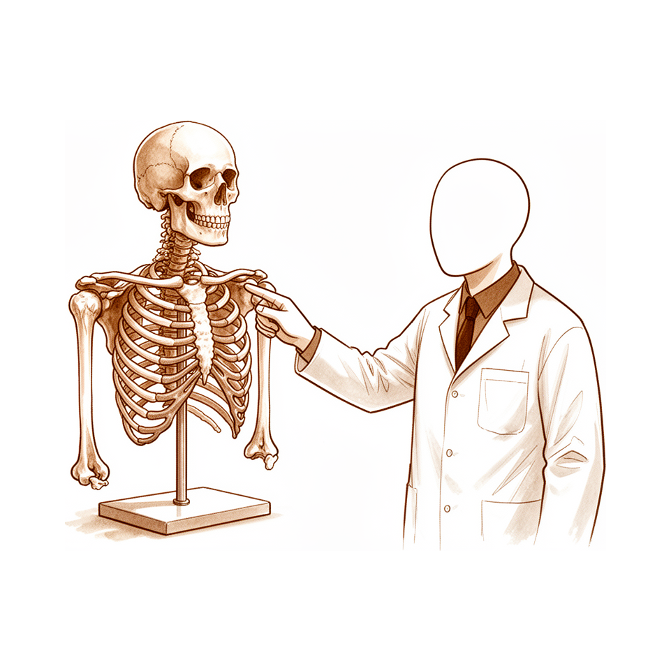
Elbow osseous anatomy: humerus, ulna, radius—critical landmarks for fracture classification & surgical approaches.
Overview
The upper limb skeleton presents distinct anatomical variations that influence surgical planning and biomechanical stability. An anatomical variation of the humerus, characterized by a process arising 3.2–6.4 cm from the internal condyle, was observed in 14 human subjects and 4 museum preparations [1]. In the forearm, intraindividual parameter variations in the proximal radius must be accounted for in the design of anatomically precontoured plates [6]. The ulnar greater sigmoid notch coverage angle involves contributions from both bone and cartilage, and anatomic variations of this structure require biomechanical and clinical studies to determine their true clinical relevance [2]. Additionally, the arcade of Struthers serves as a site of compression and tethering where hourglass indentation is found in all nerves at this point, with its anatomical features and clinical implications discussed in the literature [13, 19].
Bone composition and mechanics are defined by constituents deposited as a crystalline structure [70]. A rational approach to studying pathological changes involves analyzing factors that influence the fundamental processes of bone deposition and demineralization, allowing bone diseases to be classified based on sclerosis and lysis [70]. However, data regarding the mechanical properties of bone tissue can support or contradict hypotheses due to the absence of standardization or unification of experimental methods, approaches, and goals [9]. Understanding nuances of developmental processes, cellular interactions, and signaling pathways is essential for advancing fundamental knowledge and developing novel therapies for genetic bone diseases and tissue regeneration [7].
Surgical exposure of the radius and ulna requires a thorough understanding of forearm anatomy, including the locations of cutaneous nerves, muscular intervals, and deep neurovascular structures [23]. Detailed knowledge of the anatomical situation in areas of pin implantation is of great importance for unilateral external fixation in the upper limb [4]. The anatomy of the shoulder, arm, and elbow encompasses ossification patterns, joint mechanics, ligamentous stabilizers, musculature, neurovascular structures, and surgical approaches [14]. Preparation and detailed anatomic knowledge are important in surgical procedures for upper extremity trauma [3]. Regarding injury management, functional bracing produces beneficial effects of movement at the fracture site, supported by research relating to the biology and biomechanics of fracture healing [12]. Current techniques for proximal radio-ulnar joint repair and reconstruction are classified according to patient age, timing, and the presence of associated bony or ligamentous injuries [69].
Osseous Anatomy
Humerus
The humeral anatomical variation process arises 3.2–6.4 cm from the internal condyle [1]. The anatomical neck possesses significantly higher trabecular density than other regions of the proximal humerus [16]. Removal of the anatomical neck leads to a substantial increase in displacement and strain concentration, particularly on the medial and posterior surfaces [16]. The micro-architecture of the posterolateral area of the lateral condyle is weaker, especially in the elderly [34]. This structural weakness may lead to mechanical failure or loosening of screws [34]. Cartilage thickness is not uniform and modifies the morphologic shape and diameters of the humeral articular surface [27]. In growing children, the positions of the radial and axillary nerves maintain linear relationships with arm lengths [30]. The locations of these nerves in relation to palpable osseous landmarks are predictable in this population [30].
Radius
Intraindividual parameter variations in the proximal radius have to be taken into account in the design of anatomically precontoured plates [6]. Cartilage thickness varies between subjects and does not correlate with bone parameters of the radial head [28]. Consequently, the diameters of the radial head cannot be inferred from indirect measurements of dry bones or radiographs [28].
Ulna
The volume and articular surface area of the coronoid can be estimated based on anatomical measurements and gender [29]. The Coronoid height index (CHI) is a reliable CT-based technique to assess coronoid height and bone loss [31]. This index is independent of patient size [31].
General Osseous Properties and Imaging
Data from the literature concerning the mechanical properties of bone tissue can be extracted to support or contradict many hypotheses [9]. The ability to extract such data is limited by the absence of standardization or unification of experimental methods, approaches, and goals [9]. Most commonly utilized radiographic measures for the elbow were consistent between sexes [32]. These measures were also consistent across the adolescent age group [32] and between adolescents and young adults [32].
Ligaments and Joint Capsule
The central band of the interosseous membrane functions as a ligament and serves as a secondary restraint to proximal migration of the radius when the radial head is intact [40]. Sequential transection studies of the proximal radioulnar joint identify the annular ligament and the central band of the interosseous membrane as the main stabilizers in pronation [20]. In supination, the central band acts as the significant stabilizer [20].
Surgical Approaches: Correct placement of dual direct lateral portals does not disrupt the lateral ligamentous complex and allows access to a large portion of the capitellum [57]. A combination of two ligament-preserving approaches enables viewing the entirety of the joint surface and may represent an alternative to ligament-releasing approaches [11].
Anatomical Landmarks: The anatomy of the arcade of Struthers is clinically relevant because the site is recognized as a point of compression and tethering, with hourglass indentation found in all nerves at this point [13]. Detailed descriptions of the insertional anatomy of the anterior medial collateral ligament at the sublime tubercle are available [53].
Surgical Technique: Ligament orientation angles from 9° to 38° produced significantly less suture excursion than angles of 39°, with minimal suture excursion noted at angles of 20° [49].
Muscles and Tendons
Shoulder
The infraspinatus bare area of the proximal humerus broadens as the humeral head is evaluated in a superior to inferior fashion [68]. In the deltopectoral approach for revision shoulder arthroplasty and periprosthetic fracture fixation, the proximal spiral groove is located distal to the inferior edge of the latissimus dorsi tendon in all specimens [71]. The minimum distance between the proximal spiral groove and the inferior edge of the latissimus dorsi tendon is 25.5 mm [71].
Elbow and Forearm
The ulnar interosseous crest coincides with the forearm axis of rotation [72], while the radial interosseous crest apex coincides with the proximal central band footprint of the interosseous membrane [72]. The footprints of the posterolateral ligament of the elbow have been described [73]. This ligament has clinical significance in cases of elbow instability caused by acute ligament tears [73] and in cases of elbow pain due to ligament enthesopathy [73]. A distally based tendon graft reconstruction of the annular ligament of the elbow using the tendon of the superficial head of the brachialis muscle is feasible in most patients [78].
Anatomical variations in the musculofascial structures about the region of the medial humeral epicondyle were evaluated in 104 extremities in sixty-four cadavers [79, 80]. Measurements of tendon origins on the medial epicondyle have allowed the creation of a map of the specific common flexor tendon origins and their sizes [76]. This map includes their position relative to the MCL [76].
Hand and Wrist
A longitudinal groove runs the length of the phalangeal shaft in the volar anatomy of the proximal phalanx [81]. Dorsally placed bicortical screws for fixation of proximal phalanx shaft fractures could protrude into this longitudinal groove unnoticed on intraoperative imaging [81]. Such protrusion can potentially cause impingement on the flexor tendon [81].
Lower Extremity
A cadaveric study maps the anatomic landscape encountered endoscopically for proximal hamstring repair [74]. This anatomic mapping supports the efficacy and safety of endoscopic proximal hamstring repair [74].
Neurovascular Anatomy
Humeral and Elbow Neurovascular Anatomy
Anatomical variations of the humerus and humeral artery have been documented in 14 human subjects and 4 museum preparations [1]. In these cases, the process arises 3.2–6.4 cm from the internal condyle [1]. Understanding the extraosseous and intraosseous arterial anatomy of the adult elbow is critical for avoiding iatrogenic injury to the intraosseous circulation during trauma or extensive dissection [8]. The course of the radial nerve in the distal part of the upper arm exhibits significant variety [21]. A rare anatomic variation exists in which the median nerve travels in a bony canal of the distal humerus, separating from the brachial artery [42]. Dissection has also demonstrated a notch or foramen in the medial epicondyle through which the posterior inferior ulnar collateral artery and accompanying veins and nerves pass [47]. The inferior ulnar collateral artery provides the only direct vascularization to the ulnar nerve in the region just proximal to the cubital tunnel [48].
The "arcade of Struthers" is a clinically relevant site recognized for compression and tethering [13]. Hourglass indentation is present in all nerves at this location [13].
Forearm and Wrist Neurovascular Anatomy
Accurate diagnosis and surgical decompression of the ulnar nerve require recognition of its anatomical details and variations to prevent iatrogenic injury [10]. The periosteal network supplies an extensive and diffuse vascular tree to the anterior surface of the radius or ulna [22]. Consequently, areas suitable for bone graft harvest from the anterior surface of the radius or ulna are virtually without bounds due to this vascular distribution [22].
Shoulder and Graft Neurovascular Anatomy
Neurovascular distribution on the bony en-face view of the humeral greater tuberosity is organized into 15 zones, highlighting both safe and dangerous areas [15]. Vascular anatomy for vascularized olecranon bone grafts is consistent, and flap harvest is simple and straightforward in all cadaveric specimens [45]. The laterovolar group of vessels is the most important contributor to the intraosseous blood supply of the scaphoid [52].
Biomechanics and Function
Elbow
Biomechanical and clinical studies are required to determine the true clinical relevance of anatomic variations in the ulnar greater sigmoid notch coverage angle [2]. Surgical visualization of the capitellum can be achieved by combining two ligament-preserving approaches, which enables viewing the entirety of the capitellar joint surface [11]. This combination may represent an alternative to ligament-releasing approaches for the capitellum [11]. Three-dimensional analysis of elbow soft tissue footprints provides information that may aid in restoring elbow biomechanics and preserving range of motion [18]. In fractures of both bones of the forearm, a loss in the range of supination-pronation rotation can be expected with residual angular malalignment of 20 degrees or more [60].
Regarding the Souter-Strathclyde total elbow prosthesis, slight malpositioning of a congruous articulation results in an imbalance in periarticular soft tissues [61]. This imbalance causes either maltracking or increased transfer of stresses to the bone-cement interface [61]. Biomechanical alterations in the Souter-Strathclyde total elbow prosthesis are predicted to explain reported relatively high rates of mechanical loosening [50].
Shoulder
The anatomical neck of the proximal humerus possesses significantly higher trabecular density than other regions [16]. Removal of the anatomical neck leads to a substantial increase in displacement and strain concentration, particularly on the medial and posterior surfaces of the proximal humerus [16]. Mechanical strength and subchondral mineralization in the humeral head are significantly associated with a P-value of less than .01 [58]. Increasing the size of the humeral stem had no significant effects on bone-to-implant contact during loading [54]. Extremes in variables lead to a drop in range of motion, and more precise range of motion such as internal rotation has to be investigated separately [59].
Strength at the proximal subchondral level of the glenoid averaged 66.9 MPa [66]. Higher peak values of strength at the proximal subchondral level of the glenoid were measured posteriorly, superiorly, and anteriorly to the bare area [66]. Density data for Type E3 glenoids may help optimize future implant designs that leverage high-density regions to improve fixation and reduce micromotion [65].
General Bone Properties and Healing
The ability to extract data to support or contradict hypotheses is limited by the absence of standardization or unification of experimental methods, approaches, and goals [9]. Research relating to the biology and biomechanics of fracture healing supports the argument for the beneficial effects of movement at the fracture site produced by functional bracing [12].
Hand and Forearm
A biomechanical model successfully created a reproducible and clinically relevant palmar beak fracture in a biomechanical setting for Bennett fractures [56]. A thorough understanding of biomechanical principles and neurological pathways is necessary for the hand surgeon in managing upper limb spasticity [67].
Common Sites of Injury
Elbow
The ulnar greater sigmoid notch exhibits anatomic variations in bone and cartilage contribution to the coverage angle [2]. Soft tissue stability of the proximal radioulnar joint relies on specific structures depending on the position of the forearm: the annular ligament and the central band of the interosseous membrane are the main stabilizers in pronation [20], while the central band of the interosseous membrane serves as the significant stabilizer in supination [20]. Three-dimensional analysis of elbow soft tissue footprints and anatomy provides information that may aid in restoring elbow biomechanics and preserving range of motion [18].
Functional outcomes in the elbow region are influenced by multiple factors. Surgical results for posttraumatic stiff elbow vary depending upon the initial injury, preoperative motion, presence of heterotopic ossification, and etiologic factors such as brain injury [37]. In Monteggia fracture-dislocations, severe osseous, soft tissue, and neural trauma affect the functional results of the elbow region [41].
Forearm
Impinging exostoses of the proximal radius can cause atypical symptoms arising from impingement on soft-tissue structures [5]. Anatomic considerations of these exostoses may provide guidance in assessment and planning for surgical resection [5]. Traumatic bowing of the forearm is a distinct clinical entity caused by plastic deformation from longitudinal forces [36]. This condition often occurs with a fracture of the other bone [36]. In pediatric Monteggia fracture–dislocation with ipsilateral distal radius fracture, maintaining heightened suspicion for associated injuries and implementing accurate radiography with frequent follow-up are essential [35].
Wrist
Open anatomical reduction with stabilization of all injured structures, followed by six to eight weeks of immobilization, is probably optimum treatment for fracture-dislocation of the radiocarpal joint [17].
Upper Arm
Observable nerve trauma is relatively common with brachial gunshot wounds. In patients with clinical nerve deficits, directly traumatized nerves are present in at least 39% and nerve transection occurs in at least 16% [38]. The anatomical features and clinical implications of variants of the Arcade of Struthers are discussed [19].
Pediatric Specific
Five distinct fracture patterns of the humeral medial epicondyle are identifiable on plain radiographs and are associated with specific injury mechanisms [25]. Accurate identification of partial avulsion patterns in the pediatric humeral medial epicondyle is likely important for understanding the natural history of these injuries and the outcomes of different treatment strategies [24]. MRI has a distinct advantage over plain radiography and ultrasound scanning for diagnosing distal humeral epiphysis fracture separation in neonates as it provides direct visualisation of the bony, cartilaginous and soft tissue components of the injury [26]. Injuries to the long bones are relatively common complications of breech deliveries, especially in babies born to primiparas [39].
Surgical Anatomy
Detailed anatomic knowledge is essential for surgical procedures involving upper extremity trauma [3]. Specific considerations regarding impinging exostoses of the proximal radius provide guidance for assessing atypical symptoms arising from soft-tissue impingement and aid in planning surgical resection [5].
Elbow and Shoulder
A combination of two ligament-preserving approaches enables viewing the entirety of the capitellum joint surface and may represent an alternative to ligament-releasing approaches for the capitellum [11]. The neurovascular distribution in 15 zones on the bony-en-face view of the humeral greater tuberosity highlights both safe and dangerous zones [15].
Forearm and Wrist
The periosteal network provides an extensive and diffuse vascular tree to the distal radius and ulna [22]. Consequently, the areas from which bone grafts can be harvested from the anterior surface of the radius or ulna are virtually without bounds [22].
Operative Techniques and Safety
Allowing easy egress from an open bone-biopsy site may prevent distant spread but may increase local spread [43]. A specific technique can be utilized to safely identify the entry point of the ulnar canal [44]. This technique avoids complications associated with perforation of the ulnar cortex and allows for accurate placement of the ulnar component [44].
Using the narrowest edge lacking cartilage as a reference to locate the bare area of the proximal ulna, a designed chevron osteotomy entered the joint in the bare area in most specimens [46]. This approach decreased associated damage to the joint cartilage [46].
Key Evidence
- [L5] This anatomical variation was observed in 14 instances in human subjects and 4 in museum preparations, with the process arising 3.2–6.4 cm from the internal condyle. [1] (10.1016/j.jhsb.2006.09.001)
- [L5] Biomechanical and clinical studies are needed to understand the true clinical relevance of these anatomic variations. [2] (10.1016/j.jse.2015.06.006)
- [L5] The issue emphasizes the importance of preparation and detailed anatomic knowledge in surgical procedures for upper extremity trauma, providing comprehensive descriptions of exposures and coverage with clinical examples. [3] (10.1016/j.hcl.2014.08.004)
- [L5] Detailed knowledge about the anatomical situation in the areas of pin implantation is of great importance. [4] (10.1016/s0020-1383(99)00258-2)
- [L4] The anatomic considerations illustrated by these two cases may provide guidance in assessment of atypical symptoms arising from impingement on soft-tissue structures and aid in planning for surgical resection. [5] (10.1016/j.xrrt.2021.01.001)
- [L5] Besides the wide range in size, intraindividual parameter variations have to be taken into account in the design of anatomically precontoured plates. [6] (10.1016/j.jse.2011.11.008)
- [L5] Understanding this anatomy may help avoid iatrogenic injury to the intraosseous circulation during trauma or extensive dissection. [8] (10.2106/00004623-199711000-00007)
- [L4] Data from the literature concerning the mechanical properties of bone tissue can be extracted to support or contradict many hypotheses, not because of any inherent ambiguities in the data but rather because of the absence of standardization or unification of experimental methods, approaches, and goals. [9] (10.2106/00004623-197456050-00012)
- [L5] Recognition of these anatomical details and variations is essential for accurate diagnosis and surgical decompression to avoid iatrogenic injury. [10] (10.1016/j.hcl.2007.05.001)
- [L5] A combination of these two ligament-preserving approaches enables viewing the entirety of the joint surface and may represent an alternative to ligament-releasing approaches. [11] (10.1016/j.jse.2022.01.013)
- [Paper] The authors present an excellent overview of research relating to the biology and biomechanics of fracture healing to support their argument for the beneficial effects of movement at the fracture site produced by functional bracing. [12] (10.1016/0020-1383(96)89814-7)
- [L5] The anatomy is clinically relevant because the site is recognized as a point of compression and tethering, with hourglass indentation found in all nerves at this point. [13] (10.1016/j.jhsa.2003.12.014)
- [L5] The study demonstrates the neurovascular distribution in 15 zones on the bony-en-face view of the HGT, clearly highlighting both safe and dangerous zones. [15] (10.1016/j.jse.2025.02.061)
- [L5] The anatomical neck possesses significantly higher trabecular density than other regions, and its removal leads to a substantial increase in displacement and strain concentration, particularly on the medial and posterior surfaces. [16] (10.1186/s13018-025-06486-5)
- [L4] Open anatomical reduction with stabilization of all injured structures, followed by six to eight weeks of immobilization, is probably optimum treatment. [17] (10.2106/00004623-197759020-00011)
- [L5] This study provides the upper extremity surgeon with information that may aid in restoring elbow biomechanics and preserving range of motion in these patients. [18] (10.1016/j.jse.2014.05.003)
- [L5] The anatomical features and clinical implications of the variants are discussed. [19] (10.1016/0266-7681(91)90059-w)
- [L5] Sequential transection of the soft tissue constraints of the proximal radioulnar joint demonstrated the annular ligament and the central band of the interosseous membrane to be the main stabilizers in pronation and the central band to be the significant stabilizer in supination. [20] (10.1016/1058-2746(92)90013-s)
- [L5] The course of the radial nerve in the distal part of the upper arm has great variety. [21] (10.1371/journal.pone.0186890)
- [L5] The areas from which bone grafts can be harvested from the anterior surface of the radius or ulna are virtually without bounds as the periosteal network provides an extensive and diffuse vascular tree to these bones. [22] (10.1016/s0266-7681(02)00279-6)
- [L5] A thorough understanding of forearm anatomy, including the locations of cutaneous nerves, muscular intervals, and deep neurovascular structures, is essential to adequately perform surgical exposure of the radius and ulna. [23] (10.5435/00124635-201107000-00006)
- [L4] Accurate identification of these injury patterns and their underlying pathology is likely important for understanding the natural history of these injuries and the outcomes of different treatment strategies. [24] (10.1177/03635465241310407)
- [L4] This large cohort confirmed five distinct fracture patterns that are identifiable on plain radiographs and are associated with specific injury mechanisms. [25] (10.1177/2325967126s00493)
- [L5] It has a distinct advantage over plain radiography and ultrasound scanning as it provides direct visualisation of the bony, cartilaginous and soft tissue components of the injury. [26] (10.1016/s0020-1383(01)00102-4)
- [L4] Cartilage thickness is not uniform and modifies the morphologic shape and diameters of the humeral articular surface. [27] (10.1016/j.jse.2016.10.012)
- [L5] The observation that cartilage thickness varies between subjects and does not correlate with bone parameters suggests that the diameters of the radial head cannot be inferred from indirect measurements of dry bones or radiographs. [28] (10.1016/j.jse.2015.07.003)
- [L4] The volume and articular surface area of the coronoid can be estimated based on anatomical measurements and gender. [29] (10.1016/j.jhsa.2010.11.002)
- [L4] The positions of the radial and axillary nerves maintain linear relationships with arm lengths in growing children, and their locations in relation to palpable osseous landmarks are predictable. [30] (10.2106/jbjs.19.00019)
- [L4] The Coronoid height index (CHI) is a reliable CT-based technique to assess coronoid height and bone loss that is independent of patient size and can be used for clinical and research purposes. [31] (10.1016/j.jseint.2024.07.004)
- [L4] Most commonly utilized radiographic measures were consistent between sexes, across the adolescent age group, and between adolescents and young adults. [32] (10.1016/j.jse.2011.10.026)
- [L5] This study evaluated the differences in cortical and trabecular bone parameters in each region of the distal humerus, finding that the micro-architecture of the posterolateral area of the lateral condyle is weaker, especially in the elderly, which may lead to mechanical failure or loosening of screws. [34] (10.1016/j.jse.2009.08.005)
- [Case_report] Maintaining heightened suspicion for associated injuries and implementing accurate radiography with frequent follow-up are essential. [35] (10.1016/j.xrrt.2024.06.002)
- [L4] Traumatic bowing of the forearm is a distinct clinical entity caused by plastic deformation from longitudinal forces, often occurring with a fracture of the other bone. [36] (10.2106/00004623-197456030-00019)
- [L5] Results are generally favorable but do vary depending upon the initial injury, preoperative motion, presence of heterotopic ossification, and etiologic factors such as brain injury. [37] (10.1097/00132589-200412000-00007)
- [L4] Observable nerve trauma is relatively common with directly traumatized nerves in at least 39% and nerve transection in at least 16% of patients with clinical nerve deficits. [38] (10.1177/15589447231221170)
- [L5] The central band functions as a ligament and serves as a secondary restraint to proximal migration of the radius when the radial head is intact. [40] (10.1016/s0363-5023(97)80036-6)
- [L4] Severe osseous, soft tissue, and neural trauma affect the functional results of the elbow region. [41] (10.1186/1749-799x-1-12)
- [L5] We report a rare anatomic variation in which the median nerve travels in a bony canal of the distal humerus, separating from the brachial artery. [42] (10.1016/j.jhsa.2004.05.018)
- [L5] Allowing easy egress from an open bone-biopsy site may prevent distant spread but may increase local spread. [43] (10.2106/00004623-198466080-00012)
- [L5] We feel this technique can be utilized to safely identify the entry point of the canal and avoid complications associated with perforation of the ulnar cortex and allow for accurate placement of the ulnar component. [44] (10.1097/bte.0000000000000042)
- [L5] Vascular anatomy is consistent and flap harvest is simple and straightforward in all cadaveric specimens. [45] (10.1016/j.jhsa.2019.05.014)
- [L5] Using the narrowest edge lacking cartilage as a reference to locate the bare area, the designed chevron osteotomy entered the joint in the bare area in most specimens and decreased associated damage to the joint cartilage. [46] (10.1016/j.jhsa.2017.02.012)
- [L5] Our anatomic dissection demonstrated a notch or foramen through which the posterior inferior ulnar collateral artery and accompanying veins and nerves passed. [47] (10.1016/s1058-2746(99)90084-8)
- [L5] The inferior ulnar collateral artery provided the only direct vascularization to the nerve in the region just proximal to the cubital tunnel. [48] (10.1016/s1058-2746(99)90048-4)
- [L5] Ligament orientation angles from 9° to 38° produced significantly less suture excursion than angles of 39°, with minimal suture excursion noted at angles of 20°. [49] (10.1053/jhsu.1999.0538)
- [L5] These biomechanical alterations are predicted to explain the reported relatively high rates of mechanical loosening with this prosthesis. [50] (10.1016/s1058-2746(00)90041-7)
- [L4] The laterovolar group of vessels is the most important contributor to the intraosseous blood supply of the scaphoid. [52] (10.2106/00004623-196648060-00010)
- [L5] The study presents a detailed description of the insertional anatomy of the anterior medial collateral ligament at the sublime tubercle. [53] (10.1016/j.jse.2018.08.006)
- [L5] Increasing the size of the humeral stem had no significant effects on bone-to-implant contact during loading. [54] (10.1016/j.jseint.2021.05.006)
- [L5] This model successfully created a reproducible and clinically relevant palmar beak fracture in a biomechanical setting. [56] (10.1016/j.jhsa.2018.04.024)
- [L5] Correct placement of dual direct lateral portals does not disrupt the lateral ligamentous complex and allows access to a large portion of the capitellum. [57] (10.1016/j.arthro.2007.01.029)
- [L5] Mechanical strength and subchondral mineralization in the humeral head are significantly associated (P < .01). [58] (10.1016/j.jse.2011.05.018)
- [Abstract] Extremes variables lead to a drop in ROM, and more precise ROM such as internal rotation has to be investigated separately. [59] (10.1016/j.jse.2022.01.082)
- [L5] Loss in the range of rotation can be expected with residual angles of 20 degrees or more. [60] (10.2106/00004623-198264010-00003)
- [L5] These differences, which seem small from a clinical perspective, are probably clinically important because slight malpositioning of a congruous articulation results in an imbalance in the periarticular soft tissues, causing either maltracking or increased transfer of stresses to the bone-cement interface. [61] (10.1067/mse.2000.103616)
- [L4] The density data may help optimize future implant designs that leverage these high-density regions to improve fixation and reduce micromotion. [65] (10.1016/j.jse.2026.03.022)
- [L5] Strength at the proximal subchondral level of the glenoid averaged 66.9 MPa, with higher peak values measured posteriorly, superiorly, and anteriorly to the bare area. [66] (10.1016/s1058-2746(97)90029-x)
- [L5] A thorough understanding of biomechanical principles and neurological pathways is necessary for the hand surgeon. [67] (10.1177/17531934261434453)
- [L5] The infraspinatus bare area broadens as one evaluates the humeral head in a superior to inferior fashion. [68] (10.1177/17585732241229068)
- [L5] This review summarizes current techniques for PRUJ repair and reconstruction, classifying treatment according to patient age (paediatric or adult), timing (acute or chronic), and the presence of associated bony or ligamentous injuries. [69] (10.1016/j.jisako.2026.101159)
- [L5] The proximal spiral groove was found to be distal to the inferior edge of the latissimus dorsi tendon in all specimens, with a minimum distance of 25.5 mm. [71] (10.1016/j.jse.2017.07.021)
- [L5] The ulnar interosseous crest coincides with the forearm axis of rotation, and the radial interosseous crest apex coincides with the proximal central band footprint. [72] (10.5397/cise.2021.00451)
- [L4] Its footprints were described, and its clinical significance was demonstrated in cases of elbow instability caused by acute ligament tears and elbow pain due to ligament enthesopathy. [73] (10.1016/j.jse.2023.08.033)
- [L5] This cadaveric study maps the anatomic landscape encountered endoscopically and supports the efficacy and safety of endoscopic proximal hamstring repair. [74] (10.1177/2325967118s00147)
- [L5] The measurements found have allowed the creation of a map of the specific common flexor tendon origins and their sizes on the medial epicondyle, as well as their position relative to the MCL. [76] (10.1016/j.jhsa.2024.04.008)
- [L5] A distally based tendon graft reconstruction of the annular ligament of the elbow using the tendon of the superficial head of the brachialis muscle would be feasible in most patients, based on this anatomic study. [78] (10.1016/j.jhsa.2013.04.008)
- [L5] Anatomical variations in the musculofascial structures about the region of the medial humeral epicondyle were evaluated in 104 extremities in sixty-four cadavers. [79] (10.1016/0266-7681_86_90254-8)
- [L5] Anatomical variations in the musculofascial structures about the region of the medial humeral epicondyle were evaluated in 104 extremities in sixty-four cadavers. [80] (10.1016/0266-7681(86)90254-8)
- [L5] A longitudinal groove runs the length of the phalangeal shaft, and dorsally placed bicortical screws could protrude into this groove unnoticed on intraoperative imaging, potentially causing impingement on the flexor tendon. [81] (10.1016/j.jhsa.2016.12.009)
See Also
References
[1] On a Peculiarity of the Humerus and Humeral Artery. Journal of Hand Surgery (European Volume). 2007. DOI: 10.1016/j.jhsb.2006.09.001
[2] The ulnar greater sigmoid notch “coverage angle”: bone and cartilage contribution. Magnetic resonance imaging anatomic study on 78 elbows. Journal of Shoulder and Elbow Surgery. 2015. DOI: 10.1016/j.jse.2015.06.006
[3] Options for Surgical Exposure and Soft Tissue Coverage in Upper Extremity Trauma. Hand Clinics. 2014. DOI: 10.1016/j.hcl.2014.08.004
[4] The anatomical base of unilateral external fixation in the upper limb. Injury. 2000. DOI: 10.1016/s0020-1383(99)00258-2
[5] Impinging exostoses of the proximal radius: a report of two cases with distinct clinical features. JSES Reviews, Reports, and Techniques. 2021. DOI: 10.1016/j.xrrt.2021.01.001
[6] The anatomy of the proximal radius: implications on fracture implant design. Journal of Shoulder and Elbow Surgery. 2012. DOI: 10.1016/j.jse.2011.11.008
[7] Chapter 11 Developmental Biology. 2020.
[8] The Extraosseous and Intraosseous Arterial Anatomy of the Adult Elbow (dagger). The Journal of Bone and Joint Surgery (American Volume)*. 1997. DOI: 10.2106/00004623-199711000-00007
[9] The Mechanical Properties of Cortical Bone. The Journal of Bone & Joint Surgery. 1974. DOI: 10.2106/00004623-197456050-00012
[10] Ulnar Nerve Anatomy. Hand Clinics. 2007. DOI: 10.1016/j.hcl.2007.05.001
[11] Surgical Approaches To The Capitellum: A Comparative Anatomic Study. Journal of Shoulder and Elbow Surgery. 2022. DOI: 10.1016/j.jse.2022.01.013
[12] Functional fracture bracing. Injury. 1996. DOI: 10.1016/0020-1383(96)89814-7
[13] Redefining the “arcade of Struthers”. The Journal of Hand Surgery. 2004. DOI: 10.1016/j.jhsa.2003.12.014
[14] Chapter 69 Anatomy of the Shoulder, Arm, and Elbow. 2019.
[15] Novel surgical safety zones on bony-en-face view of humeral greater tuberosity: a fresh cadaveric dissection study. Journal of Shoulder and Elbow Surgery. 2025. DOI: 10.1016/j.jse.2025.02.061
[16] A study on the biomechanical properties of cancellous bone across different regions of the proximal humerus. Journal of Orthopaedic Surgery and Research. 2025. DOI: 10.1186/s13018-025-06486-5
[17] Fracture-dislocation of the radiocarpal joint. The Journal of Bone & Joint Surgery. 1977. DOI: 10.2106/00004623-197759020-00011
[18] Three-dimensional analysis of elbow soft tissue footprints and anatomy. Journal of Shoulder and Elbow Surgery. 2014. DOI: 10.1016/j.jse.2014.05.003
[19] The Arcade of Struthers: An Anatomical Study. Journal of Hand Surgery. 1991. DOI: 10.1016/0266-7681(91)90059-w
[20] The anatomy of the proximal radioulnar joint. Journal of Shoulder and Elbow Surgery. 1992. DOI: 10.1016/1058-2746(92)90013-s
[21] The course of the radial nerve in the distal humerus: A novel, anatomy based, radiographic assessment. PLOS ONE. 2017. DOI: 10.1371/journal.pone.0186890
[22] Vascular Anatomy of the Palmar Surfaces of the Distal Radius And Ulna: Its Relevance to Pedicled Bone Grafts at the Distal Palmar Forearm. Journal of Hand Surgery. 2003. DOI: 10.1016/s0266-7681(02)00279-6
[23] Surgical Exposures of the Radius and Ulna. Journal of the American Academy of Orthopaedic Surgeons. 2011. DOI: 10.5435/00124635-201107000-00006
[24] Partial Avulsion Patterns in the Pediatric Humeral Medial Epicondyle. The American Journal of Sports Medicine. 2025. DOI: 10.1177/03635465241310407
[25] Poster 196. Fracture of the Humeral Medial Epicondyle in Children and Adolescents: Injury Mechanism Differs by Fracture Pattern. Orthopaedic Journal of Sports Medicine. 2026. DOI: 10.1177/2325967126s00493
[26] Distal humeral epiphysis fracture separation in neonates — diagnosis using MRI scan. Injury. 2002. DOI: 10.1016/s0020-1383(01)00102-4
[27] Cartilage thickness of distal humerus and its relationships with bone dimensions: magnetic resonance imaging bilateral study in healthy elbows. Journal of Shoulder and Elbow Surgery. 2017. DOI: 10.1016/j.jse.2016.10.012
[28] Contribution of cartilage to size and shape of radial head circumference: magnetic resonance imaging analysis of 78 elbows. Journal of Shoulder and Elbow Surgery. 2016. DOI: 10.1016/j.jse.2015.07.003
[29] Quantitative Measurements of the Coronoid in Healthy Adult Patients. The Journal of Hand Surgery. 2011. DOI: 10.1016/j.jhsa.2010.11.002
[30] Establishing Safe Zones to Avoid Nerve Injury in the Approach to the Humerus in Pediatric Patients. Journal of Bone and Joint Surgery. 2019. DOI: 10.2106/jbjs.19.00019
[31] Coronoid height index: a reliable and reproducible technique for quantifying coronoid bone loss in elbow instability. JSES International. 2024. DOI: 10.1016/j.jseint.2024.07.004
[32] Elbow radiographic anatomy: measurement techniques and normative data. Journal of Shoulder and Elbow Surgery. 2012. DOI: 10.1016/j.jse.2011.10.026
[34] Three-dimensional osseous micro-architecture of the distal humerus: Implications for internal fixation of osteoporotic fracture. Journal of Shoulder and Elbow Surgery. 2010. DOI: 10.1016/j.jse.2009.08.005
[35] A case of pediatric Monteggia fracture–dislocation with ipsilateral distal radius fracture. JSES Reviews, Reports, and Techniques. 2024. DOI: 10.1016/j.xrrt.2024.06.002
[36] Traumatic Bowing of the Forearm in Children. The Journal of Bone & Joint Surgery. 1974. DOI: 10.2106/00004623-197456030-00019
[37] Surgical Approach to the Posttraumatic Stiff Elbow. Techniques in Shoulder and Elbow Surgery. 2004. DOI: 10.1097/00132589-200412000-00007
[38] Brachial Gunshot Wounds: Injury Patterns and Considerations for Managing the Abnormal Neurological Examination. HAND. 2024. DOI: 10.1177/15589447231221170
[39] liv SI'EXCER T. SNEDECOR, M.D., AND HARRISON B. WILSON, M.D., HACKENSACK, NEW JERSEY. 1949.
[40] The interosseous membrane of the forearm: Anatomy and function. The Journal of Hand Surgery. 1997. DOI: 10.1016/s0363-5023(97)80036-6
[41] Unusual patterns of Monteggia fracture-dislocation. Journal of Orthopaedic Surgery and Research. 2006. DOI: 10.1186/1749-799x-1-12
[42] Anatomic variation—A bony canal for the median nerve at the distal humerus: A case report. The Journal of Hand Surgery. 2004. DOI: 10.1016/j.jhsa.2004.05.018
[43] Refracture of bones of the forearm after plate removal.. The Journal of Bone & Joint Surgery. 1984. DOI: 10.2106/00004623-198466080-00012
[44] Finding the Canal. Techniques in Shoulder & Elbow Surgery. 2015. DOI: 10.1097/bte.0000000000000042
[45] Vascularized Olecranon Bone Graft: An Anatomical Study and Novel Technique. The Journal of Hand Surgery. 2020. DOI: 10.1016/j.jhsa.2019.05.014
[46] The Bare Area of the Proximal Ulna: An Anatomic Study With Relevance to Chevron Osteotomy. The Journal of Hand Surgery. 2017. DOI: 10.1016/j.jhsa.2017.02.012
[47] An aberrant osseous tunnel in the medial epicondyle. Journal of Shoulder and Elbow Surgery. 1999. DOI: 10.1016/s1058-2746(99)90084-8
[48] The extraneural and intraneural arterial anatomy of the ulnar nerve at the elbow. Journal of Shoulder and Elbow Surgery. 1999. DOI: 10.1016/s1058-2746(99)90048-4
[49] Forearm interosseous ligament isometry. The Journal of Hand Surgery. 1999. DOI: 10.1053/jhsu.1999.0538
[50] Kinematics and laxity of the Souter-Strathclyde total elbow prosthesis. Journal of Shoulder and Elbow Surgery. 2000. DOI: 10.1016/s1058-2746(00)90041-7
[52] The Extraosseous and Intraosseous Blood Supply of the Scaphoid Bone. The Journal of Bone & Joint Surgery. 1966. DOI: 10.2106/00004623-196648060-00010
[53] Insertional anatomy of the anterior medial collateral ligament on the sublime tubercle of the elbow. Journal of Shoulder and Elbow Surgery. 2019. DOI: 10.1016/j.jse.2018.08.006
[54] The effect of humeral implant thickness and canal fill on interface contact and bone stresses in the proximal humerus. JSES International. 2021. DOI: 10.1016/j.jseint.2021.05.006
[56] Bennett Fractures: A Biomechanical Model and Relevant Ligamentous Anatomy. The Journal of Hand Surgery. 2019. DOI: 10.1016/j.jhsa.2018.04.024
[57] Dual Direct Lateral Portals for Treatment of Osteochondritis Dissecans of the Capitellum: An Anatomic Study. Arthroscopy. 2007. DOI: 10.1016/j.arthro.2007.01.029
[58] Correlation between mineralization and mechanical strength of the subchondral bone plate of the humeral head. Journal of Shoulder and Elbow Surgery. 2012. DOI: 10.1016/j.jse.2011.05.018
[59] Virtual ROM RSA: An Analysis Based On 10’000 Scapula. Journal of Shoulder and Elbow Surgery. 2022. DOI: 10.1016/j.jse.2022.01.082
[60] The effect on supination-pronation of angular malalignment of fractures of both bones of the forearm.. The Journal of Bone & Joint Surgery. 1982. DOI: 10.2106/00004623-198264010-00003
[61] Kinematics and laxity of the Souter-Strathclyde total elbow prosthesis. Journal of Shoulder and Elbow Surgery. 2000. DOI: 10.1067/mse.2000.103616
[65] Bone Densities in Type E3 Glenoids: Quantitative Assessments and Topographical Distributions. Journal of Shoulder and Elbow Surgery. 2026. DOI: 10.1016/j.jse.2026.03.022
[66] Bone strength and material properties of the glenoid. Journal of Shoulder and Elbow Surgery. 1997. DOI: 10.1016/s1058-2746(97)90029-x
[67] Achieving balance in upper limb spasticity management. Journal of Hand Surgery (European Volume). 2026. DOI: 10.1177/17531934261434453
[68] Describing the infraspinatus bare area of the proximal humerus: An anatomic cadaveric study. Shoulder & Elbow. 2024. DOI: 10.1177/17585732241229068
[69] Acute and chronic proximal radio-ulnar joint injuries (PRUJ): current concepts. Journal of ISAKOS. 2026. DOI: 10.1016/j.jisako.2026.101159
[70] The constituents of bone, as determined by analysis, are best illustrated diagrammatically (Fig. 1). The elements of which bone is composed are deposited as a crystalline. 1950.
[71] Surgical anatomy of the radial nerve in the deltopectoral approach for revision shoulder arthroplasty and periprosthetic fracture fixation: a cadaveric study. Journal of Shoulder and Elbow Surgery. 2017. DOI: 10.1016/j.jse.2017.07.021
[72] Topographical measurement of the attachments of the central band of the interosseous membrane on interosseous crests of the radius and ulna. Clinics in Shoulder and Elbow. 2021. DOI: 10.5397/cise.2021.00451
[73] The posterolateral ligament of the elbow: anatomy and clinical relevance. Journal of Shoulder and Elbow Surgery. 2024. DOI: 10.1016/j.jse.2023.08.033
[74] Endoscopic Proximal Hamstring Repair: Portal Site Anatomy - A Cadaveric Study. Orthopaedic Journal of Sports Medicine. 2018. DOI: 10.1177/2325967118s00147
[76] Mapping Origins of Tendons on the Medial Epicondyle to Improve Treatment of Medial Epicondylitis: Anatomical Study. The Journal of Hand Surgery. 2025. DOI: 10.1016/j.jhsa.2024.04.008
[78] Annular Ligament Reconstruction Using the Distal Tendon of the Superficial Head of the Brachialis Muscle: An Anatomical Feasibility Study. The Journal of Hand Surgery. 2013. DOI: 10.1016/j.jhsa.2013.04.008
[79] Musculotendinous Variations about the Medial Humeral Epicondyle. Journal of Hand Surgery. 1986. DOI: 10.1016/0266-7681_86_90254-8
[80] Musculotendinous variations about the medial humeral epicondyle. The Journal of Hand Surgery: Journal of the British Society for Surgery of the Hand. 1986. DOI: 10.1016/0266-7681(86)90254-8
[81] Volar Anatomy of the Proximal Phalanx: Implications for Screw Length Selection for Fixation of Shaft Fractures. The Journal of Hand Surgery. 2017. DOI: 10.1016/j.jhsa.2016.12.009