Clinicians › General-Health
Cartilage Biology and Repair
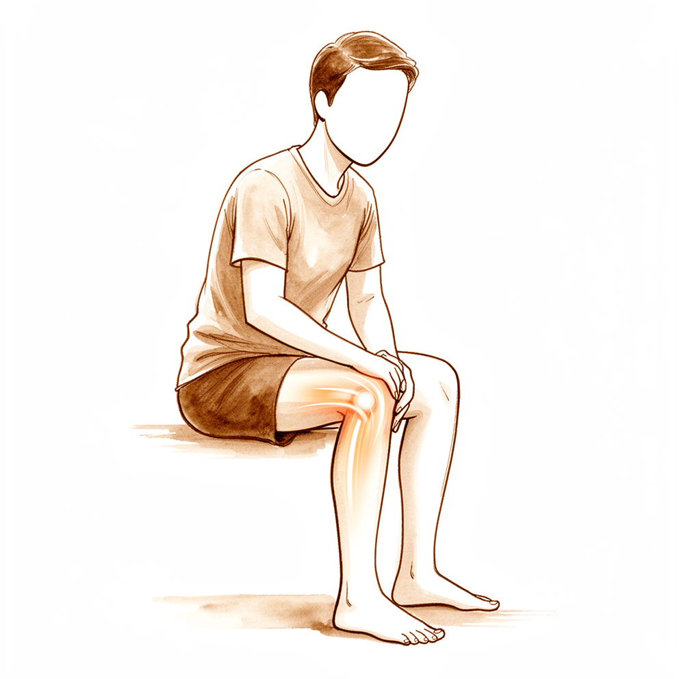
Overview
Articular cartilage defects present a complex therapeutic challenge, as current clinical treatments often result in mechanically inferior tissue and lack a single technology that fully meets essential healing requirements while remaining surgically applicable [6, 7]. The pathophysiology of osteoarthritic cartilage degradation resembles the hypertrophic zone of the fetal growth plate during endochondral ossification, suggesting common regulatory mechanisms that may offer new treatment approaches by targeting chondrocyte phenotype reparation [13]. Success in cell-based orthopaedic strategies for cartilage trauma and early degeneration depends on leveraging known mechanisms of chondrocyte regulation [1]. A profound understanding of subchondral bone anatomy and pathophysiology is key to developing targeted strategies for osteochondral defects [23], yet various problems remain unresolved regarding the formation of hyaline cartilage in vivo [10].
Diagnostic evaluation relies heavily on magnetic resonance imaging, which provides reproducible, noninvasive, and objective monitoring for trauma, degenerative arthritides, and surgical outcomes [22]. New advances in noninvasive detection of cartilage biochemistry offer insight into repair tissue ultrastructure and may eventually obviate the need for surgical biopsy by providing objective assessment of treatment outcomes [5]. Regarding emerging regenerative strategies, the application of growth factors appears promising but requires further basic and clinical research before routine use [9]. While some efficacy has been shown for mesenchymal stem cells in osteoarthritis, evidence for intra-articular MSCs on clinical outcomes remains limited [3]. Similarly, despite promising results from cell therapies and platelet-rich plasma, additional studies are needed for definitive conclusions [35]. Systematic reviews have highlighted difficulties in understanding the real need for cells to increase scaffold-based healing potential due to product heterogeneity and study design issues [8].
Surgical indications and management remain controversial, with the vast majority of cartilage repair procedures in Germany applied to degenerative, non-traumatic defects [4, 11]. Autologous chondrocyte implantation is indicated for medium to large, full-thickness cartilage defects greater than 2 cm² [21]. Patient and defect characteristics must be assessed to determine the best technique for each individual [20]. Clinical outcomes after combined meniscal allograft transplantation and cartilage repair are similar to those after either procedure in isolation [19]. A central tenet of cartilage restoration is to leave future treatment options available should they become necessary [36]. It is noted that a variety of definitions of failure are used to evaluate knee chondral restorative surgery outcomes in the orthopaedic literature [62].
How It Works
Chondrocyte Regulation and Matrix Homeostasis
Articular cartilage function relies on specific cell-and-matrix composition and structure, where interactions between chondrocytes and their matrix are necessary to maintain tissue integrity [43]. Molecular studies elucidate the mechanisms of collagen fibril assembly and the role of proteolytic remodeling in joint disease [46]. Novel compounds that selectively inhibit matrix metalloproteinases or their gene expression offer opportunities for developing new therapeutic approaches to prevent cartilage destruction [45]. In diabetic osteoarthritis, elevated expression of MMP8 and MMP9 contributes to progression through mechanisms that inhibit cartilage differentiation and promote chondrocyte apoptosis [50]. Apoptosis activation via the extrinsic pathway occurs in osteoarthritis cartilage, suggesting that apoptosis-positive cells may act as a protection mechanism after sublethal injury to facilitate repair [42].
Mechanobiology and Injury Pathways
Advances in osteoarthritis research require understanding interactions between mechanics and biology at the organ, tissue, cell, and molecular level [41]. Improved understanding of biophysical and molecular pathways involved in chondrocyte mechanotransduction can provide insight into the development of novel therapeutic approaches [24]. Knowledge of the interaction between inflammatory and biomechanical factors in regulating cartilage metabolism is beneficial for understanding the etiopathogenesis of posttraumatic osteoarthritis and improving therapies for joint injury [49]. Mechanical degradation may underlie the onset of microcracks within cartilage, leading to physiological loading that the cartilage is unable to repair [48]. Evidence showing the potential importance of chondrocyte apoptosis in osteoarthritis, combined with the observation that apoptosis results from articular impacts, suggests a possible link between impact injury and late joint degeneration [47]. While in vitro cartilage injury models have made it possible to investigate the effects of impact load on cartilage, considerably more information is necessary before improvements in the prevention and treatment of posttraumatic arthrosis can be developed [52].
Osteoarthritis Pathophysiology and Developmental Mechanisms
Extracellular matrix destruction in osteoarthritic articular cartilage resembles that in the hypertrophic zone of the fetal growth plate during endochondral ossification, suggesting common regulatory mechanisms that could provide new approaches for treatment by targeting chondrocyte phenotype reparation [13]. Increased bone resorption occurs at an early stage in the development of osteoarthritis, and blocking bone-resorbing cytokines prevents cartilage damage [27]. A profound understanding of the basic anatomic aspects of the subchondral bone and the pathophysiology of diseases affecting it is key to developing targeted and effective therapeutic strategies to treat osteochondral defects [23]. Subchondral bone cysts are described in the context of articular cartilage repair to improve investigations of these pathological changes [16]. As understanding of the mechanisms of inflammation and tissue destruction increases, new therapeutic modalities offer the opportunity to medically treat not simply the symptoms of osteoarthritis but the disease itself [38]. Understanding the basic science of cartilage and the changes that occur in osteoarthritis is imperative to develop novel strategies to diagnose and treat this disorder [40]. Furthermore, understanding the mechanisms underlying developmental skeletogenesis should facilitate the development of regenerative approaches to cartilage repair by harnessing the inherent regenerative potential of skeletal tissues [37].
Regenerative Strategies and Cell-Based Therapies
No single technology fully meets the essential requirements for effective cartilage healing while remaining easily applicable during surgical procedures [7]. Regenerating cartilage through cell culture remains challenging with respect to sourcing cells, their availability, chondrogenic capabilities, and the lasting viability of the graft [18]. Various problems remain unresolved for a successful repair associated with the formation of hyaline cartilage in vivo [10]. The application of growth factors in the treatment of local cartilage defects and osteoarthritis appears promising, but further research is needed at both the basic science and clinical levels before routine application [9]. Growth factors with anabolic effects promoting chondrogenesis and maintenance of the chondrocyte phenotype could be useful in the treatment of injured cartilage or meniscus [15]. The chondrogenic effects of Sprifermin (rhFGF18) are dependent on the inflammatory status of the cartilage prior to treatment [26]. Mesenchymal stem cell-derived miR-125b-1-3p-abundant exosomes alleviate osteoarthritis by modulating the KDM6B-H3K27me3-FOXM1 axis, providing a theoretical rationale for exosome-based therapeutic strategies [39]. Delivery of cells in the form of a cell sheet in conjunction with cartilage particles provides a novel approach for cell-based cartilage regeneration [44]. Cartilage constructs have the potential to provide mechanical functionality immediately upon implantation [28]. The vast majority of cartilage repair procedures were applied in degenerative, non-traumatic cartilage defects [11].
Evidence Limitations and Assessment
The evidence of efficacy of intra-articular mesenchymal stem cells on both clinical outcomes and cartilage repair remains limited [3]. Systematic reviews have underlined difficulties in understanding the real need for cells to increase scaffold-based cartilage healing potential due to the heterogeneity of products used and the design of published studies [8]. Optimal management of cartilage defects is controversial, and future rigorous research methods could minimize common biases through strict study design, patient selection criteria, larger patient enrollment, more extended follow-up, and standardization of clinical treatment pathways [4]. New advances in noninvasive detection of cartilage biochemistry provide insight into the ultrastructure of cartilage repair tissue, potentially obviating the need for surgical biopsy and providing an objective assessment of treatment outcome [5].
What the Evidence Shows
Cartilage Biology and Pathophysiology
Success in cell-based orthopaedic treatment for cartilage trauma and early osteoarthritis depends on strategies that rely on known mechanisms of chondrocyte regulation [1]. Understanding interactions in articular cartilage mechanobiology is a step toward developing tissue engineering approaches and therapeutic interventions for cartilage pathologies such as osteoarthritis [14]. An improved understanding of biophysical and molecular pathways in chondrocyte mechanotransduction provides insight into novel therapeutic approaches for osteoarthritis [24]. The chondrogenic effects of Sprifermin (rhFGF18) on extracellular matrix turnover are dependent on the inflammatory status of the cartilage prior to treatment [26]. The role of vitamin D supplementation in the treatment or prevention of osteoarthritis remains uncertain [58].
Diagnostic Imaging and Assessment
Magnetic resonance imaging provides a reproducible, noninvasive, and objective method for evaluating and monitoring cartilage in trauma, degenerative arthritides, and surgical treatment [22]. Advances in noninvasive detection of cartilage biochemistry provide insight into the ultrastructure of cartilage repair tissue and may obviate the need for surgical biopsy [5].
Current Treatment Limitations and Challenges
Regenerating cartilage through cell culture remains challenging regarding cell sourcing, availability, chondrogenic capabilities, and lasting graft viability [18]. The literature on existing cartilage treatment options is limited by heterogeneity in surgical procedures and reporting of non-standardised outcome measures [56]. Optimal management of cartilage defects is controversial, and future research should minimize biases through strict study design, larger enrollment, extended follow-up, and standardized treatment pathways [4]. Standardised evaluation protocols are needed for biological and mechanical assessment of scaffolds, along with long-term randomised independent clinical trials [65].
Marrow Stimulation and Microfracture
Shortcomings of the microfracture technique include limited hyaline repair tissue, variable repair cartilage volume, and possible functional deterioration [25]. A meta-analysis shows no significant difference between microfracture alone and microfracture with scaffold in treating knee cartilage defects, though some long-term RCTs demonstrate statistically significant differences [61]. Blocking TGF-β1 with losartan during biologically regulated marrow stimulation results in repair cartilage tissue that is superior to other groups and consists primarily of hyaline cartilage in a rabbit model [64].
Cell-Based and Stem Cell Therapies
There is insufficient evidence to determine whether cell-based therapy is superior to other treatment strategies in articular cartilage lesions of the knee [66]. Most studies reported successful cartilage repair with synovium-derived mesenchymal stem cell (sMSC) transplantation despite variability in animals, harvesting techniques, delivery methods, and outcome measures [54]. Despite advances in stem cell and tissue engineering therapies, additional high-powered and well-designed clinical trials are needed to confirm safety and efficacy [34]. Additional studies are needed to provide definitive conclusions on the use of cell therapies and platelet-rich plasma for treating degenerative joint conditions [35].
Scaffolds and Biomaterials
An aragonite-based scaffold was safe and effective for chondral and osteochondral lesions in the knee, including patients with mild to moderate osteoarthritis, and provided superior outcomes compared with the control group [59]. Regenerated cartilage over stiff scaffolds at 3 months had better mechanical properties than soft scaffolds, but properties in both groups were inferior to native articular cartilage at 6 months [67]. The difficulty in understanding the need for cells to increase scaffold-based cartilage healing potential is due to the heterogeneity of products and study designs [8]. Bi-phasic and multi-phasic scaffold-based approaches for osteochondral tissue regeneration highlight the importance of an interface layer between bone and cartilage [74].
Osteochondral Allografts and Transplantation
Basic science and clinical studies support the safety and efficacy of fresh osteochondral allograft transplantation for managing a wide spectrum of chondral and osteochondral knee disorders [31]. Refrigerated and frozen osteochondral allograft technology shows significant promise in the clinical treatment of relatively small chondral and osteochondral defects [63].
Autologous Chondrocyte Implantation (ACI)
Autologous chondrocyte implantation, matrix-induced autologous chondrocyte implantation, osteochondral autograft transplantation, and osteochondral allograft improve knee function and pain, with selection dependent on patient and cartilage defect characteristics [20].
Outcomes and Long-Term Effects
Cartilage repair surgery prevents progression of knee degeneration over 6 years compared to non-operated control subjects with initially identical defects [53]. Overall good to excellent outcomes were observed for restorative treatment of isolated patellar cartilage lesions, with mean estimated failure rates less than 10% and patient-reported outcome measures ranging from 70% to 85% of the maximum score [73]. Articular cartilage repair techniques appear to be safe in children and adolescents, with no differences in complication rates reported when compared with adult patients [30].
Adjunctive and Non-Operative Therapies
Current research focuses on improving outcome scores, reducing reoperation rates, and preventing the progression of defects to osteoarthritis through the use of biologics [32]. The recent literature contains limited evidence on the efficacy, potential toxicity, and long-term safety of glucosamine and chondroitin sulfate for the treatment of osteoarthritis [33]. Nonoperative treatment remains the mainstay of management for patients with articular cartilage injury of the hip, with heterogeneous support in the literature regarding the efficacy of biologic injections [60]. Results from in vitro studies on platelet-rich plasma should be interpreted with caution, and subsequent prospective randomized double-blind clinical trials are needed to compare autogenous PRP to hyaluronic acid for osteoarthritis [70]. Preserving subchondral bone integrity while accessing reparative elements via calcified cartilage zone debridement with a hyaluronic acid-based scaffold and bone marrow aspirate concentrate offers a biologically sound alternative that may improve short-term recovery and long-term joint preservation [68].
Practical Considerations
Clinical Outcomes and Efficacy
Autologous chondrocyte implantation, matrix-induced autologous chondrocyte implantation, osteochondral autograft transplantation, and osteochondral allograft improve knee function and pain [20]. In the United States, currently employed treatments for knee cartilage defects are cost-effective in most clinically acceptable applications [55]. Mesenchymal stem cells have shown some efficacy for cartilage repair in osteoarthritis [3]. The application of growth factors appears promising for treating local cartilage defects and osteoarthritis [9]. Specifically, growth factors with anabolic effects that promote chondrogenesis and maintain the chondrocyte phenotype could be useful in treating injured cartilage or meniscus [15]. Results from autologous chondrocyte transplantation for thumb CMC joint osteoarthritis are encouraging, with implanted cartilage lasting up to 5 to 9 years in the majority of cases [71].
Surgical Techniques and Indications
Autologous chondrocyte implantation is indicated for the treatment of medium to large, full-thickness cartilage defects (>2 cm2) [21]. When selecting a procedure, patient and cartilage defect characteristics should be assessed to determine the best technique for each individual patient [20]. In the United States, there is substantial variation in the procedures performed for cartilage restoration in children and adolescents [69].
Biological and Regenerative Strategies
Success in current efforts towards cell-based orthopaedic treatment options for cartilage trauma and early stages of osteoarthritic degeneration strictly depends on strategies that rely on known mechanisms of a chondrocyte's regulation [1]. Regenerating cartilage through cell culture remains challenging, particularly regarding sourcing cells, their availability, chondrogenic capabilities, and the lasting viability of the graft [18]. Despite significant advances in stem cell and tissue engineering therapies for sports medicine, additional high-powered and well-designed clinical trials are needed to confirm the safety and efficacy of treatment [34]. Difficulties in understanding the real need for cells to increase scaffold-based cartilage healing potential are attributed to the heterogeneity of products used and the design of published studies [8].
Imaging and Assessment
Magnetic resonance imaging has created an undeniably important role for reproducible, noninvasive, and objective evaluation and monitoring of cartilage in the setting of trauma, degenerative arthritides, and surgical treatment for cartilage injury [22]. New advances in noninvasive detection of cartilage biochemistry provide insight into the ultrastructure of cartilage repair tissue, eventually obviating the need for surgical biopsy and providing an objective assessment of treatment outcome [5].
Anatomical and Pathological Considerations
Subchondral bone continues to provide important information about the development of osteoarthritis, and its structural integrity is critical for improving reconstructive therapies for focal cartilage defects [51]. In clinical practice, a thorough analysis of pre-existing defects on the opposing cartilage is recommended when focal metallic implants are considered [17].
Research Methodology and Limitations
Optimal management of cartilage defects is controversial, and future rigorous research methods could minimize common biases through strict study design and patient selection criteria, larger patient enrollment, more extended follow-up, and standardization of clinical treatment pathways [4]. The recent literature contains some limited evidence on the efficacy, potential toxicity, and long-term safety of glucosamine and chondroitin sulfate for the treatment of patients with osteoarthritis [33].
Key Evidence
- [L5] Success in current efforts towards cell-based orthopaedic treatment options in cases of cartilage trauma and early stages of osteoarthritic degeneration will strictly depend on strategies that rely on known mechanisms of a chondrocyte's regulation. [1] (10.1016/j.injury.2008.01.044)
- [L2] Some efficacy has been shown of MSCs for cartilage repair in osteoarthritis; however, the evidence of efficacy of intra-articular MSCs on both clinical outcomes and cartilage repair remains limited. [3] (10.1016/j.arthro.2018.07.028)
- [L1] Optimal management of cartilage defects is controversial, and future rigorous research methods could minimize common biases through strict study design and patient selection criteria, larger patient enrollment, more extended follow-up, and standardization of clinical treatment pathways. [4] (10.1016/j.arthro.2012.02.022)
- [L5] New advances in noninvasive detection of cartilage biochemistry provide insight into the ultrastructure of cartilage repair tissue, eventually obviating the need for surgical biopsy and providing an objective assessment of treatment outcome. [5] (10.1016/j.csm.2008.08.004)
- [L4] Current clinical treatments for articular cartilage defects have limited ability to repair tissue and often result in mechanically inferior cartilage; emerging regenerative approaches and strategies informing future treatment options are discussed to address these limitations. [6] (10.3389/fbioe.2021.770655)
- [L4] Currently, no single technology fully meets the essential requirements for effective cartilage healing while remaining easily applicable during surgical procedures. [7] (10.3390/jcm12206434)
- [L4] This systematic review underlined the difficulties in understanding the real need for cells to increase the scaffold-based cartilage healing potential because of the heterogeneity of products used as well as the design of the published studies. [8] (10.1016/j.arthro.2014.11.017)
- [L4] The application of growth factors in the treatment of local cartilage defects as well as osteoarthritis appears promising; however, further research is needed at both the basic science and clinical levels before routine application. [9] (10.1007/s11999-011-1857-3)
- [L4] Various problems remain unresolved for a successful repair associated with the formation of hyaline cartilage in vivo. [10] (10.1155/2012/168385)
- [L4] The vast majority of cartilage repair procedures were applied in degenerative, non-traumatic cartilage defects. [11] (10.1007/s00402-016-2453-5)
- [L5] The paper concludes that extracellular matrix destruction in osteoarthritic articular cartilage resembles that in the hypertrophic zone of fetal growth plate during endochondral ossification, suggesting common regulatory mechanisms that could provide new approaches for treatment by targeting chondrocyte phenotype reparation. [13] (10.1155/2011/683970)
- [L5] Understanding these interactions is an important step toward developing tissue engineering approaches and therapeutic interventions for cartilage pathologies, such as osteoarthritis. [14] (10.1007/s11926-014-0451-6)
- [Paper] Growth factors with anabolic effects promoting chondrogenesis and maintenance of the phenotype of the chondrocyte could be useful in the treatment of injured cartilage or meniscus. [15] (10.1016/s0020-1383(09)70005-1)
- [L4] This review describes subchondral bone cysts in the context of articular cartilage repair to improve investigations of these pathological changes. [16] (10.1002/ctm2.248)
- [L5] In clinical practice, a thorough analysis of pre-existing defects on the opposing cartilage is recommended when FMI is considered. [17] (10.1186/s12891-020-03292-4)
- [L4] Regenerating cartilage through cell culture remains challenging, particularly with respect to sourcing cells, their availability, chondrogenic capabilities and the lasting viability of the graft. [18] (10.1530/eor-2024-0083)
- [L4] Clinical outcomes after combined MAT and cartilage repair/restoration are similar to those after either procedure in isolation. [19] (10.1016/j.arthro.2010.08.007)
- [L1] When selecting a procedure, patient and cartilage defect characteristics should be assessed to determine the best technique for each individual patient. [20] (10.1002/ksa.12525)
- [Paper] Autologous chondrocyte implantation is indicated for the treatment of medium to large, full-thickness cartilage defects (>2 cm2). [21] (10.1016/j.csm.2017.02.008)
- [L5] Magnetic resonance imaging has created an undeniably important role for reproducible, noninvasive, and objective evaluation and monitoring of cartilage in the setting of trauma, degenerative arthritides, and surgical treatment for cartilage injury. [22] (10.1177/0363546505281938)
- [L5] A profound understanding of the basic anatomic aspects of the subchondral bone, together with the pathophysiology of diseases affecting it, is the key to develop targeted and effective therapeutic strategies to treat osteochondral defects. [23] (10.1007/s00167-010-1054-z)
- [Paper] An improved understanding of the biophysical and molecular pathways involved in chondrocyte mechanotransduction can provide insight into the development of novel therapeutic approaches for osteoarthritis. [24] (10.1016/j.berh.2011.11.013)
- [L1] Shortcomings of the technique include limited hyaline repair tissue, variable repair cartilage volume, and possible functional deterioration. [25] (10.1177/0363546508328414)
- [Paper] In addition, it was observed that these chondrogenic effects are dependent on the inflammatory status of the cartilage prior to treatment. [26] (10.1186/s12967-017-1356-8)
- [L4] Accumulative in vivo evidence shows that increased bone resorption occurs at an early stage in the development of osteoarthritis and that blocking bone-resorbing cytokines prevents cartilage damage. [27] (10.1016/j.cytogfr.2011.04.003)
- [Paper] These cartilage constructs have the potential to provide mechanical functionality immediately upon implantation. [28] (10.1002/jor.25223)
- [L5] Articular cartilage repair techniques appear to be safe in children and adolescents, with no differences in complication rates reported when compared with adult patients. [30] (10.1177/2325967118760190)
- [L5] Basic science and clinical studies support the safety and efficacy of fresh osteochondral allograft transplantation for managing a wide spectrum of chondral and osteochondral knee disorders. [31] (10.5435/jaaos-22-03-199)
- [L5] Current research focuses on improving outcome scores, reducing reoperation rates, and preventing the progression of defects to osteoarthritis through the use of biologics. [32] (10.1016/j.csm.2018.09.001)
- [L5] The recent literature contains some limited evidence on the efficacy, potential toxicity, and long-term safety of glucosamine and chondroitin sulfate for the treatment of patients with osteoarthritis. [33] (10.5435/00124635-200103000-00001)
- [L5] Despite significant advances in stem cell and tissue engineering therapies for sports medicine, there remains a need for additional high-powered and well-designed clinical trials to confirm the safety and efficacy of treatment. [34] (10.1016/j.arthro.2014.11.033)
- [L4] Despite promising results shown by cell therapies and PRP for treating degenerative joint conditions, additional studies are needed to provide more definitive conclusions. [35] (10.2106/jbjs.rvw.19.00075)
- [L5] A central tenet of cartilage restoration is to leave future treatment options available should they become necessary. [36] (10.1177/0363546504273510)
- [Paper] Understanding the mechanisms underlying developmental skeletogenesis should greatly facilitate the development of regenerative approaches to cartilage repair by harnessing the inherent regenerative potential of skeletal tissues. [37] (10.1097/01.blo.0000143560.41767.ee)
- [L5] As our understanding of the mechanisms of inflammation and tissue destruction increases, new therapeutic modalities offer the opportunity to medically treat not simply the symptoms of osteoarthritis but the disease itself. [38] (10.1016/s0894-1130(00)80032-0)
- [L5] These findings provide a theoretical rationale and identify promising therapeutic targets for the development of exosome-based therapeutic strategies against OA. [39] (10.1186/s13018-026-06765-9)
- [L5] Understanding the basic science of cartilage and the changes that occur in osteoarthritis is imperative to develop novel strategies to diagnose and treat this disorder. [40] (10.1016/j.csm.2004.08.007)
- [L5] Future advances in osteoarthritis research will be possible if research is conducted within an ontogenetic framework that recognizes and interprets the interactions of mechanics and biology on the organ, tissue, cell, and molecular level. [41] (10.1097/01.blo.0000144970.05107.7e)
- [L4] The study demonstrated apoptosis activation by the extrinsic pathway in OA cartilage, suggesting that apoptosis-positive cells may act as a protection mechanism after sublethal injury to facilitate repair. [42] (10.1007/s00167-010-1215-0)
- [L5] This review covers the current understanding of the design of articular cartilage (the cell-and-matrix composition and the structure that make normal function of the cartilage possible) as well as the interactions between chondrocytes and their matrix that are necessary to maintain the tissue. [43] (10.2106/00004623-199704000-00021)
- [L5] Delivery of cells in the form of a cell sheet in conjunction with cartilage particles provides a novel approach for cell-based cartilage regeneration. [44] (10.1177/0363546519897912)
- [L5] Novel compounds that selectively inhibit these enzymes or their gene expression offer opportunities for developing new therapeutic approaches to prevent cartilage destruction. [45] (10.1097/01.blo.0000144483.62033.8b)
- [Paper] Molecular studies continue to reveal the mechanisms of assembly of cartilage collagen fibrils and the role of proteolytic remodeling in joint disease. [46] (10.1097/01.blo.0000144855.48640.b9)
- [L5] The long-term effects of articular impact injury remain unknown, but evidence showing the potential importance of chondrocyte apoptosis in osteoarthritis and the observation that apoptosis results from articular impacts suggests a possible link between impact injury and late joint degeneration. [47] (10.1097/01.blo.0000133567.28491.7d)
- [L5] This mechanical degradation may underlie onset of microcracks within the cartilage, leading to physiological loading that the cartilage by its nature is unable to repair. [48] (10.1016/j.otsr.2021.103116)
- [Paper] Knowledge of the interaction of inflammatory and biomechanical factors in regulating cartilage metabolism would be beneficial to an understanding of the etiopathogenesis of posttraumatic osteoarthritis and in the improvement of therapies for joint injury. [49] (10.1097/01.blo.0000131233.83640.91)
- [L5] The underlying mechanism may be related to inhibiting cartilage differentiation and promoting chondrocyte apoptosis. [50] (10.1186/s13018-021-02208-9)
- [L5] Subchondral bone continues to provide important information about the development of OA, and its structural integrity is critical for improving reconstructive therapies for focal cartilage defects. [51] (10.5435/jaaos-d-16-00096)
- [L5] The development of in vitro cartilage injury models has made it possible to investigate some of the effects of impact load (acute injury) on cartilage, and in vivo models have investigated changes in matrix components and chondrocytes over time, but considerably more information is necessary before improvements in the prevention and treatment of posttraumatic arthrosis can be developed. [52] (10.1097/01.blo.0000132627.13539.02)
- [L3] Cartilage repair surgery prevents progression of knee degeneration over 6 years compared to non-operated control subjects with initially identical defects. [53] (10.1007/s00167-018-5321-8)
- [L2] Most studies reported successful cartilage repair with sMSC transplantation despite the variability of animals, cell harvesting techniques, methods of delivery, and outcome measures. sMSC transplantation holds promise as a treatment option for focal cartilage defects. [54] (10.3389/fbioe.2019.00314)
- [L2] Currently employed treatments for knee cartilage defects in the United States are cost-effective in most clinically acceptable applications. [55] (10.1177/0363546519834557)
- [L5] Existing cartilage treatment options include marrow stimulation, osteochondral tissue transfer or transplantation, cell-free synthetic scaffolds and cell-based repair strategies, but the literature is limited by heterogeneity in surgical procedures and reporting of non-standardised outcome measures. [56] (10.1136/jisakos-2015-000037)
- [L4] The role of vitamin D supplementation in the treatment or prevention of OA remains uncertain. [58] (10.1177/2325967117711376)
- [L1] This aragonite-based scaffold was safe and effective in the treatment of chondral and osteochondral lesions in the knee, including patients with mild to moderate osteoarthritis, and provided superior outcomes as compared with the control group. [59] (10.1177/03635465231151252)
- [Paper] Nonoperative treatment remains the mainstay of management for patients with articular cartilage injury of the hip, and there is a heterogeneity of support in the scientific literature regarding the efficacy of biologic injections for cartilage disease of the hip. [60] (10.1016/j.csm.2017.02.010)
- [L1] The meta-analysis shows no significant difference between MF and MF with scaffold in treating knee cartilage defects, though some long-term RCTs demonstrate statistically significant differences. [61] (10.1002/ksa.12495)
- [L1] A variety of definitions of failure are used to evaluate knee chondral restorative surgery outcomes in the orthopaedic literature. [62] (10.1016/j.asmr.2024.101044)
- [L4] This technology shows significant promise in the clinical treatment of relatively small chondral and osteochondral defects. [63] (10.1016/j.arthro.2007.03.058)
- [L5] By blocking TGF-b1 with losartan, the repair cartilage tissue after BMS was superior to the other groups and consisted primarily of hyaline cartilage. [64] (10.1177/0363546519898681)
- [L4] Standardised evaluation protocols are needed for biological and mechanical assessment and comparison between different scaffolds, and long-term randomised independent clinical trials with large study numbers are needed to provide more insight into the use of these biomaterials. [65] (10.1136/jisakos-2020-000600)
- [L1] There is insufficient evidence from the studies included in this review to say whether cell-based therapy is superior to other treatment strategies in articular cartilage lesions of the knee. [66] (10.1016/j.arthro.2009.02.007)
- [L5] Although regenerated cartilage over stiff scaffolds at 3 months had better mechanical properties than the soft scaffold group, properties in both treated groups were the same at 6 months and inferior to native articular cartilage. [67] (10.1177/0363546508322899)
- [L5] By preserving subchondral bone integrity while still accessing reparative elements, this approach offers a biologically sound alternative that may improve both short-term recovery and long-term joint preservation. [68] (10.1016/j.eats.2025.103733)
- [L3] In the United States, there is substantial variation in the procedures performed for cartilage restoration in children and adolescents. [69] (10.1177/2325967120s00248)
- [L5] The authors agree that results from an in vitro study should be interpreted with caution and mention a subsequent prospective randomized double-blind clinical trial comparing autogenous PRP to HA for the treatment of OA. [70] (10.1177/0363546514537991)
- [L4] The results obtained are encouraging and implanted cartilage has lasted in the majority of cases up to 5 to 9 years. [71] (10.1177/1558944716660555q)
- [Paper] In this systematic review with meta-analysis, overall good to excellent outcomes were observed for all identified treatments, with mean estimated failure rates less than 10% and PROMs ranging from 70% to 85% of the maximum score. [73] (10.1177/23259671261443877)
- [L4] This review evaluates bi-phasic and multi-phasic scaffold-based approaches for osteochondral tissue regeneration, highlighting the importance of an interface layer between bone and cartilage. [74] (10.3390/ijms19061755)
See Also
- Osteoarthritis
References
[1] Perspectives on articular cartilage biology and osteoarthritis. Injury. 2008. DOI: 10.1016/j.injury.2008.01.044
[3] Intra-articular Mesenchymal Stem Cells in Osteoarthritis of the Knee: A Systematic Review of Clinical Outcomes and Evidence of Cartilage Repair. Arthroscopy: The Journal of Arthroscopic & Related Surgery. 2019. DOI: 10.1016/j.arthro.2018.07.028
[4] Limitations and Sources of Bias in Clinical Knee Cartilage Research. Arthroscopy. 2012. DOI: 10.1016/j.arthro.2012.02.022
[5] New Techniques in Articular Cartilage Imaging. Clinics in Sports Medicine. 2009. DOI: 10.1016/j.csm.2008.08.004
[6] Strategies for Articular Cartilage Repair and Regeneration. Frontiers in Bioengineering and Biotechnology. 2021. DOI: 10.3389/fbioe.2021.770655
[7] Knee Cartilage Lesion Management—Current Trends in Clinical Practice. Journal of Clinical Medicine. 2023. DOI: 10.3390/jcm12206434
[8] Scaffold‐Based Cartilage Treatments: With or Without Cells? A Systematic Review of Preclinical and Clinical Evidence. Arthroscopy. 2015. DOI: 10.1016/j.arthro.2014.11.017
[9] The Role of Growth Factors in Cartilage Repair. Clinical Orthopaedics & Related Research. 2011. DOI: 10.1007/s11999-011-1857-3
[10] Stem Cells and Gene Therapy for Cartilage Repair. Stem Cells International. 2012. DOI: 10.1155/2012/168385
[11] Cartilage repair surgery for full-thickness defects of the knee in Germany: indications and epidemiological data from the German Cartilage Registry (KnorpelRegister DGOU). Archives of Orthopaedic and Trauma Surgery. 2016. DOI: 10.1007/s00402-016-2453-5
[13] Developmental Mechanisms in Articular Cartilage Degradation in Osteoarthritis. Arthritis. 2011. DOI: 10.1155/2011/683970
[14] The Mechanobiology of Articular Cartilage: Bearing the Burden of Osteoarthritis. Current Rheumatology Reports. 2014. DOI: 10.1007/s11926-014-0451-6
[15] Growth factors in cartilage and meniscus repair. Injury. 2009. DOI: 10.1016/s0020-1383(09)70005-1
[16] Cyst formation in the subchondral bone following cartilage repair. Clinical and Translational Medicine. 2020. DOI: 10.1002/ctm2.248
[17] Effects of focal metallic implants on opposing cartilage – an in-vitro study with an abrasion test machine. BMC Musculoskeletal Disorders. 2020. DOI: 10.1186/s12891-020-03292-4
[18] Current trends in the treatment of focal cartilage lesions: a comprehensive review. EFORT Open Reviews. 2025. DOI: 10.1530/eor-2024-0083
[19] Biological Knee Reconstruction: A Systematic Review of Combined Meniscal Allograft Transplantation and Cartilage Repair or Restoration. Arthroscopy. 2010. DOI: 10.1016/j.arthro.2010.08.007
[20] Autologous chondrocyte implantation, matrix‐induced autologous chondrocyte implantation, osteochondral autograft transplantation and osteochondral allograft improve knee function and pain with considerations for patient and cartilage defects characteristics: A systematic review and meta‐analysis. Knee Surgery, Sports Traumatology, Arthroscopy. 2024. DOI: 10.1002/ksa.12525
[21] Autologous Chondrocytes and Next-Generation Matrix-Based Autologous Chondrocyte Implantation. Clinics in Sports Medicine. 2017. DOI: 10.1016/j.csm.2017.02.008
[22] Magnetic Resonance Imaging of Articular Cartilage. The American Journal of Sports Medicine. 2006. DOI: 10.1177/0363546505281938
[23] The basic science of the subchondral bone. Knee Surgery, Sports Traumatology, Arthroscopy. 2010. DOI: 10.1007/s00167-010-1054-z
[24] Biomechanical factors in osteoarthritis. Best Practice & Research Clinical Rheumatology. 2011. DOI: 10.1016/j.berh.2011.11.013
[25] Clinical Efficacy of the Microfracture Technique for Articular Cartilage Repair in the Knee. The American Journal of Sports Medicine. 2009. DOI: 10.1177/0363546508328414
[26] Sprifermin (rhFGF18) modulates extracellular matrix turnover in cartilage explants ex vivo. Journal of Translational Medicine. 2017. DOI: 10.1186/s12967-017-1356-8
[27] Crosstalk between cartilage and bone: When bone cytokines matter. Cytokine & Growth Factor Reviews. 2011. DOI: 10.1016/j.cytogfr.2011.04.003
[28] Functional tissue engineering of articular cartilage for biological joint resurfacing—The 2021 Elizabeth Winston Lanier Kappa Delta Award. Journal of Orthopaedic Research. 2021. DOI: 10.1002/jor.25223
[30] Articular Cartilage Repair of the Knee in Children and Adolescents. Orthopaedic Journal of Sports Medicine. 2018. DOI: 10.1177/2325967118760190
[31] Fresh Osteochondral Allograft Transplantation for the Knee. Journal of the American Academy of Orthopaedic Surgeons. 2014. DOI: 10.5435/jaaos-22-03-199
[32] Orthobiologics for Focal Articular Cartilage Defects. Clinics in Sports Medicine. 2019. DOI: 10.1016/j.csm.2018.09.001
[33] Use of Glucosamine and Chondroitin Sulfatein the Management of Osteoarthritis. Journal of the American Academy of Orthopaedic Surgeons. 2001. DOI: 10.5435/00124635-200103000-00001
[34] The Role of Stem Cells and Tissue Engineering in Orthopaedic Sports Medicine: Current Evidence and Future Directions. Arthroscopy. 2015. DOI: 10.1016/j.arthro.2014.11.033
[35] Glenohumeral Osteoarthritis: The Role for Orthobiologic Therapies. JBJS Reviews. 2020. DOI: 10.2106/jbjs.rvw.19.00075
[36] Cartilage Restoration, Part 1. The American Journal of Sports Medicine. 2005. DOI: 10.1177/0363546504273510
[37] Biology of Developmental and Regenerative Skeletogenesis. Clinical Orthopaedics & Related Research. 2004. DOI: 10.1097/01.blo.0000143560.41767.ee
[38] Osteoarthritis of the hand: A modifiable disease. Journal of Hand Therapy. 2000. DOI: 10.1016/s0894-1130(00)80032-0
[39] Mesenchymal stem cell-derived miR-125b-1-3p-abundant exosomes alleviate osteoarthritis by modulating the KDM6B-H3K27me3-FOXM1 axis. Journal of Orthopaedic Surgery and Research. 2026. DOI: 10.1186/s13018-026-06765-9
[40] Basic Science of Articular Cartilage and Osteoarthritis. Clinics in Sports Medicine. 2005. DOI: 10.1016/j.csm.2004.08.007
[41] The Mechanobiology of Articular Cartilage Development and Degeneration. Clinical Orthopaedics & Related Research. 2004. DOI: 10.1097/01.blo.0000144970.05107.7e
[42] Characterization of apoptosis in articular cartilage derived from the knee joints of patients with osteoarthritis. Knee Surgery, Sports Traumatology, Arthroscopy. 2010. DOI: 10.1007/s00167-010-1215-0
[43] Instructional Course Lectures, The American Academy of Orthopaedic Surgeons - Articular Cartilage. Part I. The Journal of Bone & Joint Surgery. 1997. DOI: 10.2106/00004623-199704000-00021
[44] Human Amniotic Mesenchymal Stem Cell Sheets Encapsulating Cartilage Particles Facilitate Repair of Rabbit Osteochondral Defects. The American Journal of Sports Medicine. 2020. DOI: 10.1177/0363546519897912
[45] Novel Inhibitors of Matrix Metalloproteinase Gene Expression as Potential Therapies for Arthritis. Clinical Orthopaedics & Related Research. 2004. DOI: 10.1097/01.blo.0000144483.62033.8b
[46] Collagens and Cartilage Matrix Homeostasis. Clinical Orthopaedics & Related Research. 2004. DOI: 10.1097/01.blo.0000144855.48640.b9
[47] The Long-Term Effects of Articular Impaction. Clinical Orthopaedics & Related Research. 2004. DOI: 10.1097/01.blo.0000133567.28491.7d
[48] Early impairment of cartilage poroelastic properties in an animal model of ACL tear. Orthopaedics & Traumatology: Surgery & Research. 2022. DOI: 10.1016/j.otsr.2021.103116
[49] The Role of Biomechanics and Inflammation in Cartilage Injury and Repair. Clinical Orthopaedics & Related Research. 2004. DOI: 10.1097/01.blo.0000131233.83640.91
[50] Elevated expression of MMP8 and MMP9 contributes to diabetic osteoarthritis progression in a rat model. Journal of Orthopaedic Surgery and Research. 2021. DOI: 10.1186/s13018-021-02208-9
[51] Role of the Subchondral Bone in Articular Cartilage Degeneration and Repair. Journal of the American Academy of Orthopaedic Surgeons. 2016. DOI: 10.5435/jaaos-d-16-00096
[52] Acute Effects of Cartilage Impact. Clinical Orthopaedics & Related Research. 2004. DOI: 10.1097/01.blo.0000132627.13539.02
[53] Cartilage repair surgery prevents progression of knee degeneration. Knee Surgery, Sports Traumatology, Arthroscopy. 2018. DOI: 10.1007/s00167-018-5321-8
[54] Synovium-Derived Mesenchymal Stem Cell Transplantation in Cartilage Regeneration: A PRISMA Review of in vivo Studies. Frontiers in Bioengineering and Biotechnology. 2019. DOI: 10.3389/fbioe.2019.00314
[55] Cost-efficacy of Knee Cartilage Defect Treatments in the United States. The American Journal of Sports Medicine. 2019. DOI: 10.1177/0363546519834557
[56] Articular cartilage solutions for the knee: present challenges and future direction. Journal of ISAKOS. 2016. DOI: 10.1136/jisakos-2015-000037
[58] Vitamin D and Its Effects on Articular Cartilage and Osteoarthritis. Orthopaedic Journal of Sports Medicine. 2017. DOI: 10.1177/2325967117711376
[59] Aragonite-Based Scaffold Versus Microfracture and Debridement for the Treatment of Knee Chondral and Osteochondral Lesions: Results of a Multicenter Randomized Controlled Trial. The American Journal of Sports Medicine. 2023. DOI: 10.1177/03635465231151252
[60] A Critical Review. Clinics in Sports Medicine. 2017. DOI: 10.1016/j.csm.2017.02.010
[61] Does scaffold enhancement show significant superiority over microfracture alone for treating knee chondral defects? A systematic review and meta‐analysis of randomised clinical trials. Knee Surgery, Sports Traumatology, Arthroscopy. 2024. DOI: 10.1002/ksa.12495
[62] Revision Surgery Is the Most Common Definition of Failure in Studies Evaluating Knee Cartilage Restoration Outcomes: A Systematic Review. Arthroscopy, Sports Medicine, and Rehabilitation. 2024. DOI: 10.1016/j.asmr.2024.101044
[63] Clinical and Microscopic Evaluation of Implanted Refrigerated and Frozen Osteochondral Allografts (SS‐46). Arthroscopy. 2007. DOI: 10.1016/j.arthro.2007.03.058
[64] Biologically Regulated Marrow Stimulation by Blocking TGF-β1 With Losartan Oral Administration Results in Hyaline-like Cartilage Repair: A Rabbit Osteochondral Defect Model. The American Journal of Sports Medicine. 2020. DOI: 10.1177/0363546519898681
[65] Biomaterials for meniscus and cartilage in knee surgery: state of the art. Journal of ISAKOS. 2022. DOI: 10.1136/jisakos-2020-000600
[66] Cell‐Based Therapy in Articular Cartilage Lesions of the Knee. Arthroscopy. 2009. DOI: 10.1016/j.arthro.2009.02.007
[67] Influence of Scaffold Stiffness on Subchondral Bone and Subsequent Cartilage Regeneration in an Ovine Model of Osteochondral Defect Healing. The American Journal of Sports Medicine. 2008. DOI: 10.1177/0363546508322899
[68] Calcified Cartilage Zone Debridement With Hyaluronic Acid–Based Scaffold and Bone Marrow Aspirate Concentrate. Arthroscopy Techniques. 2025. DOI: 10.1016/j.eats.2025.103733
[69] EPIDEMIOLOGY OF PEDIATRIC CARTILAGE RESTORATION PROCEDURES IN THE UNITED STATES: INSURANCE AND GEOGRAPHY PLAY A ROLE. Orthopaedic Journal of Sports Medicine. 2020. DOI: 10.1177/2325967120s00248
[70] The Anti-inflammatory and Matrix Restorative Mechanisms of Platelet-Rich Plasma in Osteoarthritis: Response to Patel and Dhillon. The American Journal of Sports Medicine. 2014. DOI: 10.1177/0363546514537991
[71] Autologous Chondrocyte Transplantation in the Treatment of Thumb CMC Joint Osteoarthritis. HAND. 2016. DOI: 10.1177/1558944716660555q
[73] Outcomes of Restorative Treatment for Isolated Patellar Cartilage Lesions: A Systematic Review and Meta-analysis. Orthopaedic Journal of Sports Medicine. 2026. DOI: 10.1177/23259671261443877
[74] Recent Approaches to the Manufacturing of Biomimetic Multi-Phasic Scaffolds for Osteochondral Regeneration. International Journal of Molecular Sciences. 2018. DOI: 10.3390/ijms19061755