Clinicians › Hand
Finger Fractures
Phalangeal and metacarpal fractures of the hand — non-operative care and indications for fixation.
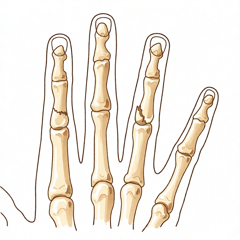
For patients: a plain-language version of this topic is available. See the patient guide.
Overview
Finger fractures account for 19% of all fractures, predominantly affecting men aged 15 to 35 years [2]. While the majority of these injuries are managed nonoperatively with conservative functional techniques [3, 7], surgical intervention offers distinct advantages in properly selected cases [3]. Recent proximal interphalangeal joint fractures present specific diagnostic and therapeutic challenges, with final outcomes significantly impacting global finger and hand function [1]. It is recommended that all finger fractures be assessed and treated by surgeons with training in the management of hand injuries [11]. Indications for surgical treatment are clearly defined, though operative results vary according to fracture type, surgeon experience, and patient compliance [10].
Headline outcomes demonstrate that good or excellent function is achieved in 94% of metacarpal fractures and 85% of phalangeal fractures by nine weeks following external fixation [4]. For pediatric patients, most hand fractures are treated nonoperatively with good results, although a subset of phalangeal fractures requires prompt recognition and surgical intervention to minimize complications [6, 36]. In open finger fractures, approximately one-quarter will likely require more than one surgical procedure, particularly in severely injured fingers with crush injuries or vascular impairment [9]. Injuries sustained in the thumb and index finger were more likely to undergo unplanned reoperation, which may guide initial treatment decision-making and postoperative follow-up [41].
Surgical options continue to evolve, with techniques ranging from screw fixation for closed, isolated proximal phalanx fractures of the long fingers [20] to intramedullary fixation reviewed for its biomechanics and outcomes in metacarpal and phalangeal fractures [55]. No differences in outcome were observed between dorsal and lateral plate fixation for finger proximal phalangeal fractures [5]. Specific surgical techniques have been shown to be safe and effective for extra-articular metacarpal and proximal phalangeal fractures [26], while traction splinting has been shown to be successful in the treatment of closed proximal phalangeal fractures [51]. Results from external fixation of closed metacarpal and phalangeal fractures were similar to those obtained in the management of stable and undisplaced fractures [18].
Anatomy & Pathophysiology
Epidemiology and Demographics
Hand fractures constitute 19% of all fractures, typically affecting men aged 15 to 35 years [2]. Finger phalangeal fractures represent approximately 10% of all fractures and are the second most common fracture in males, exhibiting a type B distribution due to high incidence in young males [39]. The overall incidence is 3.7 per 1000 per year for men and 1.3 per 1000 per year for women [172]. While phalangeal fractures may occur at any age, the majority occur in men in the fourth decade of life [175]. In males aged 80 years and older, the incidence is higher than in males aged 65 and older, whereas in females, incidence remains fairly constant across ages with a slight rise in the 80-and-over group [39].
The little and ring fingers are most frequently affected. Prevalence is 32.5% for the little finger, 25.8% for the ring finger, 14.3% for the middle finger, 18.4% for the thumb, and 9.0% for the index finger [39]. Average age at fracture is 42.4 years (little), 38.9 years (ring), 39.3 years (middle), 44.3 years (thumb), and 38.9 years (index) [39]. Male-to-female ratios are 56/44 (little), 60/40 (ring), 64/36 (middle), 63/37 (thumb), and 58/42 (index) [39]. The little finger is particularly vulnerable due to its unprotected outer location, while the length of the middle finger predisposes it to a higher proportion of fractures [175]. In the series reported by Kremer et al. (2022), the ring finger was the most frequently fractured [175].
Injury mechanisms vary by digit and age. Direct blow or assault is the most common mode, accounting for 39.1% of cases [39]. Falls from standing height account for 29.5% of cases, with an average age of 52.7 years and female predominance (37/63 male/female) [39]. Sports injuries account for 23.8% of cases, with an average age of 29.1 years and strong male predominance (82/18 male/female) [39]. Direct blows or assaults are more prevalent on the radial side, causing 47.4% of thumb, 50.0% of index, and 43.4% of middle finger fractures [39]. Conversely, little and ring finger fractures are more commonly caused by falls and sports injuries [39].
Associated injuries are frequent. Approximately 55.8% of patients have other finger fractures, 13.9% have distal radius or ulna fractures, and 9.3% have metacarpus fractures [39]. Fractures of the little finger metacarpal were common, representing 27% of the total in a study of hand fractures [172].
In the pediatric population, hand and finger fractures are the second most common fracture presenting to emergency departments [22]. These injuries exhibit a bimodal age distribution with peaks at 0 to 2 years and 12 to 16 years [22]. The most commonly injured locations are the base of the proximal phalanx (67%) of the border rays, specifically the little finger (52.2%) and thumb (23.5%) [22]. Salter-Harris II fractures are extremely common in children, with the little finger proximal phalanx being the most frequently injured site [22].
Bony Anatomy and Joint Mechanics
All phalanges consist of a proximal base, a central diaphysis, and a distal head [53]. Unlike metacarpals, the bases of all phalanges develop as metaphyses rather than the heads [53]. The distal portion of the distal phalanx is referred to as the tuft [53]. Relative finger lengths follow a specific pattern: the tip of the index finger extends to the base of the middle finger nail, the tip of the ring finger extends to the mid-aspect of the middle finger nail, and the tip of the small finger extends to a specific point relative to the middle finger nail [53].
The articulations of the fingers form a triarticular chain that flexes toward the thumb and palm to allow grasp [78]. The encompassing movement from extension to full flexion forms an “equiangular spiral” [78]. The length of the metacarpal and phalanges of the same finger resembles the Fibonacci series [78]. Digit articulations function in flexion, supported by two firm collateral ligaments and a thick reinforced anterior capsule, the volar plate [78]. The fibrous dorsal capsule is thin and lax [78].
Normal ranges of motion include approximately 85 degrees of flexion at the metacarpophalangeal (MCP) joint, 115 degrees at the proximal interphalangeal (PIP) joint, and 80 degrees at the distal interphalangeal (DIP) joint [78]. The index finger is capable of less flexion than other fingers because it opposes the thumb [78]. Interphalangeal articulations function uniquely in flexion–extension, with trochlear-shaped articulations that remain closely congruent throughout excursion [78].
Normal ulnar inclination of the fingers occurs at the MCP joints [83]. This inclination is most marked in the index finger, less in the middle and little fingers, and almost non-existent in the ring finger [83]. It results from articular effects due to asymmetry of metacarpal heads and collateral ligaments [83]. Tendon factors influence inclination as extrinsic tendons cross into the hand on the ulnar side of the longitudinal axis [83]. Muscle factors contribute because intrinsic muscles with a predominantly ulnar inclination predominate over those with a radial inclination [83]. Additionally, forward displacement of the two ulnar metacarpals pulls ulnarward the extensor tendons united by intertendinous connections [83]. This inclination is normally limited by capsuloligamentous resistance at the MCP joints and the action of interosseous muscles [83].
Functional roles vary by digit. The small finger plays an important role in palmar grip due to the mobility of its CMC joint and the action of hypothenar muscles, and it increases hand span for grasp owing to its abduction moment [27]. The ring finger forms the keystone of the palmar arch and participates in power grip, though it plays a minimal role in precision pinch [47]. The third and fourth metacarpal heads help stabilize the metacarpal arch by providing attachments for the transverse metacarpal ligament [13]. Absence of the proximal phalanx of either the middle or ring finger creates a hole through which small objects can pass, affects the hand when used as a cup or in scooping maneuvers, and causes remaining fingers to deviate toward the midline [13].
Soft Tissue Anatomy and Biomechanics
Intrinsic and extrinsic tendon insertions act as deforming forces that create typical angulation patterns in phalangeal fractures [53]. Proximal and middle phalangeal shaft fractures typically collapse into apex volar angulation due to the proximal flexion moment of intrinsics and the distal extensor moment of the extensor mechanism [53].
The interosseous muscles produce lateral movements of fingers through insertions on the lateral aspect of the base of the proximal phalanges [88]. When the MCP joint is in extension, interosseous muscles flex the proximal phalanx and extend the two distal phalanges [88]. When the MCP joint is in flexion, interosseous muscles reinforce flexion of the proximal phalanx and lose their extensor action on the distal phalanges [88]. Lumbrical muscles extend the two distal phalanges whether the MCP joint is in extension or flexion [88]. Lumbricals appear primarily to be interphalangeal extensors, following which they may act as metacarpophalangeal flexors [88].
The forces of the extensor digitorum communis and flexor superficialis on the middle phalanx have a component that produces extension of the proximal phalanx [88]. Intrinsic muscles (lumbrical and interossei) normally oppose this force [88]. When intrinsic muscles are paralyzed, no force exists to prevent the proximal phalanx from swinging into hyperextension if the extensor digitorum communis and flexor superficialis are active [88].
Extensor tendons pass from the forearm onto the dorsum of the hand through six compartments beneath the extensor retinaculum [82]. Compartment 1: Contains the extensor pollicis brevis and abductor pollicis longus [82]. Compartment 2: Contains the extensors carpi radialis longus and brevis [82]. Compartment 3: Contains the extensor pollicis longus [82]. Compartment 4: Contains the four tendons of the extensor digitorum communis plus the extensor indicis proprius [82]. Compartment 5: Contains the extensor digiti quinti [82]. Compartment 6: Contains the extensor carpi ulnaris [82].
Soft tissue anatomy varies by region. Fingertip soft tissue is tightly anchored [80]. The thin covering layer above the triangular center of the palm is relatively fixed, corresponding to the pretendinous portion of the aponeurosis [80]. Soft tissue between the fingertips and the aponeurosis area is mobile and flexible, overlying fibrous tendon sheaths from distal phalanges to MCP joints [80]. This compartment undergoes considerable length changes, to which incompressible fibrofatty tissue responds by folding and unfolding [80].
Palmar skin is anchored to underlying fascial planes by fibrous tracts [79]. In the mid-palmar area, a triangle bounded by the thumb opposition crease, lateral border of the hypothenar eminence, and distally by the two transverse palmar creases, skin adheres closely to the aponeurosis [79]. In other areas, deep skin is separated from superficial palmar fascia by fatty tissue divided into compartments by fibrous septa [79]. Three pads are recognizable: the thenar pad, hypothenar pad, and metacarpophalangeal pad [79]. The thenar pad lines only the part of the thenar eminence nearest the palm, while proximal and lateral parts are almost fat-free [79]. The hypothenar pad is broader and thicker, lining hypothenar muscles and spilling over the ulnar border [79]. The metacarpophalangeal pad sits transversely over the base of the fingers from ulnar to radial border, is thicker over interdigital spaces than flexor tendons, and is bounded proximally by transverse palmar creases [79]. Each phalanx presents a similar system of fat pads between digital flexion creases [79].
The pulp has a lobulated palmar pad where fibrous septa join the periosteum of the distal phalanx to the deep dermis [79]. Cutaneous striations forming fingerprints reflect the arrangement of papillary ridges in the underlying dermis [79]. Overall orientation of palmar skin striations is predominantly transverse, forming a concentric pattern at pulps [79]. These striae prevent object sliding during gripping and ensure a group of striae perpendicular to exerted force regardless of direction [79]. Striae also have a tactile function due to the distribution of Meissner’s corpuscles [79]. Palmar creases do not overlie joints whose flexion induces them to fold, except for middle digit creases which coincide with PIP joints [79]. The distal crease lies just distal to the DIP joint, and the proximal crease lies almost halfway down the proximal phalanx [79].
Pathophysiology of Injury and Deformity
Fracture hematoma permeates the zone of injury in proximal phalangeal fractures, inciting a proliferative fibroblastic response of surrounding collagenous structures [30]. Scar tissue tends to involve all structures within the zone of injury, described as “one wound–one scar” [30]. Resulting scar produces adherence between bone and adjacent tendons, joint capsule, and ligaments, especially when tendons remain dormant during healing [30]. Adhesions between the extensor mechanism and the fracture may result in loss of extensor glide [30]. Simple fractures from low-energy injuries may be complicated by permanent stiffness due to this process [30].
Fractures of the proximal phalangeal shaft typically exhibit apex palmar angulation with the fracture gap wider volarly and compressed dorsally [30]. Intrinsic muscles flex the proximal fragment, while the distal fragment is extended by the attachment of the central slip to the dorsal lip of the middle phalanx [30]. The axis of rotation lies on the fibro-osseous border of the flexor tendon sheath [30]. The moment arm from the rotational axis to the extensor tendon is greater than that to the flexor tendon [30].
Intraarticular fractures that disrupt joint congruency can occur at either the distal (condylar fracture) or proximal (pilon or proximal condylar fracture) articular surface [53]. Most commonly, these fractures are produced by axial loading injury [53].
The mallet finger deformity is characterized by loss of active DIP joint extension with full passive ROM evident [59]. It reflects the loss of normal extensor force transmission via the terminal tendon insertion onto the distal phalanx [59]. The unopposed flexor digitorum profundus pulls the distal joint into flexion [59].
Classification
Al-Qattan: This classification system categorizes phalangeal neck fractures into three types based on displacement and fragment contact. Type I is defined as an undisplaced fracture [42]. Type II is a displaced fracture where the distal fragment maintains some bone-to-bone contact with the proximal fragment [42]. Type III is a displaced fracture with loss of bone-to-bone contact between the proximal and distal fragments at the fracture site [42]. Type III injuries may result from the original injury or from 90 to 180° rotation of the head following fruitless attempts at closed manipulation with rapid longitudinal traction [42].
Wehbe and Schneider: This classification system addresses mallet fractures, categorizing them into three types based on fragment subluxation and extent. Type 1 involves no subluxation of the fractured fragment [75]. Type 2 involves subluxation of the fractured fragment [75]. Type 3 is a fracture affecting the whole distal phalanx [75]. Subtypes are further defined by fragment size relative to the distal articular surface: subtype A is less than one-third, subtype B is between one-third and two-thirds, and subtype C is greater than two-thirds [75].
Distal Unicondylar Proximal Phalanx: This radiographic classification system identifies four classes for distal unicondylar proximal phalanx fractures. Class I is an oblique palmar pattern with the fracture line extending from the proximal metaphyseal flare to the intracondylar depression [157]. In Class I fractures, the plane resides in neither the sagittal nor coronal planes, and the distal fragment lies palmar to the proximal phalangeal shaft [157]. Class II is a long oblique fracture line with a sagittally oriented plane [157]. Class III is a dorsal coronal fragment that is generally small in size [157]. Class IV is a palmar coronal fragment that is generally small in size [157].
Phalangeal Malunion: The classification of phalangeal malunions considers multiple factors to guide management. Location is defined as the ray, bone, and level within the affected phalanx [165]. Joint involvement is defined as intra- or extraarticular [165]. The type of deformity includes length, offset, angulation, rotation, several planes, gap, and step [165]. The degree of bony deformity is assessed [165]. Bone stock distinguishes between the growing hand and mature bone [165]. The time factor distinguishes between nascent and established malunion [165]. Complexity is defined as malunion after isolated fracture in the context of combined injury [165]. Functional significance and compliance are also considered [165].
Clinical Presentation
Epidemiology and Demographics
Finger phalangeal fractures account for approximately 10% of all fractures seen and represent the second most common fracture in males [39]. The little and ring fingers are the most frequently affected digits [39]. Average ages at injury vary by digit: 42.4 years for the little finger, 38.9 years for the ring finger, 39.3 years for the middle finger, 44.3 years for the thumb, and 38.9 years for the index finger [39]. Mechanisms include direct blow or assault (39.1%), falls from standing height (29.5%), and sports (23.8%) [39]. The male-to-female ratio is 61/39 for direct blows, 37/63 for falls, and 82/18 for sports injuries [39]. In a cohort of proximal phalangeal fractures, 52% involved the little finger, 40% were intra-articular, and 46% occurred in the dominant hand [28]. Proximal phalangeal fractures constituted 7% of all hand fractures and 0.3% of accident and emergency attendances in a two-year study [48].
In pediatric populations, the hand is the second commonest site of fracture, with incidence rising steeply after age eight, particularly in boys [38]. A bimodal age distribution exists with peaks at 0 to 2 years and 12 to 16 years [22]. The most commonly injured locations are the base of the proximal phalanx (67%) of the border rays, specifically the little finger (52.2%) and thumb (23.5%) [22]. Open phalangeal fractures are relatively common, with the highest prevalence in males aged 36 to 64 years [39]. The distal phalanges are the commonest site of open phalangeal fractures, where 25.3% of fractures were open [39]. Approximately 55% of patients with multiple phalangeal fractures have other phalangeal fractures as associated injuries [39].
Mechanism and Injury Patterns
Proximal and middle phalangeal shaft fractures typically collapse into apex volar angulation due to the proximal flexion moment of the intrinsics and the distal extensor moment of the extensor mechanism [53]. Intra-articular phalangeal fractures that disrupt joint congruency most commonly occur at the distal (condylar) or proximal (pilon or proximal condylar) articular surface [53]. Most intra-articular phalangeal fractures are produced by axial loading injury [53].
The classic "jammed" finger with a swollen, painful PIP joint usually involves a volar plate injury or a small nondisplaced avulsion fracture off the volar base of the middle finger epiphysis [22]. Salter-Harris II fractures of the digits usually occur due to jamming or hyperextension injury resulting in an abduction deformity [22]. Seymour fractures are Salter-Harris I/II or juxtaphyseal fractures of the distal phalanx with interposed nail bed at the fracture site [22]. These are open fractures but are often missed [22]. The key to diagnosing a Seymour fracture is disruption of the nail plate/cuticle, which indicates disruption of the nail bed and the likelihood of an open fracture with interposed tissue [22]. Phalangeal neck and condyle fractures present similarly to a simple "jammed" finger and are often missed [22]. A stress fracture of the middle phalanx of the ring finger in a bowler healed with restriction of activity and modification of grip [15].
Clinical Examination and Diagnosis
Appropriate evaluation of hand and finger fractures includes clinical examination and radiographs [22]. Clinical examination must assess for open injuries and angular and rotational malalignment of the injured ray [22]. Rotational alignment is confirmed by ensuring that all fingers point to the scaphoid tubercle when the fingers are flexed [22]. Radiographs should include PA, lateral, and oblique views of the injured location [22]. The coronal plane deformity of Salter-Harris II fractures is easy to assess, but extra care must be taken to assess for rotational deformity, which is not as obvious on radiographs [22]. Malrotation in pediatric phalangeal fractures does not remodel and can result in problems with grip formation [22].
Recent PIP fractures are challenging trauma in terms of diagnosis and treatment [1]. It is recommended that all finger fractures be assessed and treated by surgeons with training in the management of hand injuries [11]. In a prospective study of digital fractures, "functionally" stable fractures were defined as those where the patient could actively move adjacent joints by more than 30% of expected normal range while maintaining acceptable alignment [57]. Acceptable alignment criteria included 10 degrees of angulation in both sagittal and coronal planes, except in the metaphyseal region where 20 degrees of sagittal angulation was accepted [57]. No rotational deformity was accepted as a criterion for acceptable alignment [57]. At least 50% overlap at the fracture site was considered necessary for rapid bone healing [57].
Pediatric Specifics
Articular fractures of the fingers in children often result in sequelae, with a rate as high as 50% when the fracture was displaced initially [16]. Most phalangeal neck fractures in children less than 3 years old obtained satisfactory results [56]. Avascular necrosis following phalangeal neck fractures usually affects the little finger and presents with stiffness [58]. Displaced phalangeal neck fractures in children require reduction and pin fixation, which can usually be achieved through a closed fashion [22]. Condyle fractures in children can often be treated with closed reduction and pinning [22]. Open procedures for condyle fractures increase the risk for osteonecrosis [22].
Treatment for Seymour fractures involves removal of the nail plate, débridement of the fracture site, extrication of the interposed nail bed, and reduction of the fracture [22]. If a Seymour fracture is unstable, it may require Kirschner wire placement in addition to immobilization in a splint or cast [22]. Recommended treatment includes a dose of IV antibiotic in the emergency department followed by a 7- to 10-day course of oral antibiotic [22]. A first-generation cephalosporin is the preferred antibiotic [22]. Missed Seymour fractures have a high rate of complication including infection and nail or physeal growth disturbance [22]. Clinical stability of the fingertip was achieved after 3 weeks and radiographic union was present after 6 months in a case of diaphyseal non-union in the distal phalanx of a child [12]. A patient with a Salter-Harris Type III fracture of the distal phalanx returned to previous activity level but demonstrated a residual deficit in thumb IP joint flexion [21].
Prognosis and Complications
The final outcome of recent PIP fractures has a considerable impact on global finger and hand function [1]. A quarter of open finger fractures will likely need more than one surgical procedure, especially in more severely injured fingers due to crush or vascular impairment [9]. Proximal phalangeal fractures and comminuted fractures had the worst outcomes, and associated significant soft tissue injuries had a particularly deleterious effect on the outcome [32]. Intra-articular fractures of the proximal and middle phalanx can cause great patient morbidity if managed poorly [97]. Prolonged immobilization of simple "jammed" finger injuries can result in significant joint stiffness [22].
There are two major causes of PIP joint extensor lag following proximal phalangeal fractures: soft tissue adhesions and persistent skeletal deformity [30]. Fracture hematoma permeates the zone of injury and incites a proliferative fibroblastic response proportionate to injury severity [30]. Scar tissue tends to involve all structures within the zone of injury, producing adherence between bone and adjacent tendons, joint capsule, and ligaments [30]. Phalangeal fractures tend to deteriorate total active motion more than metacarpal fractures [35].
In a retrospective review of 105 pediatric patients treated with closed reduction pin fixation for displaced proximal phalanx fractures, the complication rate was 4.8% [22]. Thirty-six of 105 pediatric patients had postoperative stiffness, with 31 requiring therapy [22]. Phalangeal neck fractures had the highest rate of postoperative stiffness in pediatric patients treated with pin fixation [22]. Thirty-one pediatric patients available for follow-up at 1 year or greater after pin fixation reported return of full motion, no pain, and satisfaction with function and appearance [22]. 22% of pediatric patients with displaced proximal phalanx fractures had a measurable coronal plane deformity on radiograph despite reporting full motion and satisfaction [22].
Investigations
Plain radiography: A true lateral radiograph is required for the accurate diagnosis of carpometacarpal fracture-dislocations, as swelling can obscure the deformity [114]. On a posteroanterior radiograph, loss of parallel joint surfaces at the carpometacarpal articulations is indicative of carpometacarpal fracture-dislocation [114]. For mallet finger, a radiograph should be obtained to determine whether a fracture is present and, if the dorsal fragment is large, whether the distal phalanx is subluxed palmarward [59]. In Seymour fractures, radiographs reveal a displaced fracture of the distal phalanx [22]. Plain radiographs may not always be of diagnostic help for osteoid osteoma of the distal phalanx when combined with a classic clinical presentation, making histopathological confirmation necessary [168].
CT: Computed tomography scans are beneficial for determining the extent of joint surface involvement in carpometacarpal injuries [114]. Small osteochondral injuries involving the interphalangeal joints of the finger may be characterized accurately with computed tomography images, including 3-dimensional reconstructions [139].
Ultrasonography: Ultrasonography may provide greater accuracy in fracture diagnostics by revealing small avulsed bony fragments missed on radiographs [179]. It can be beneficial in diagnosing occult fractures, especially in children [179].
Other Considerations: The key to diagnosing Seymour fractures is the disruption of the nail plate/cuticle, which indicates disruption of the nail bed and the likelihood of an open fracture with interposed tissue [22].
Treatment
General Principles and Referral
Recent PIP fractures present significant diagnostic and therapeutic challenges, with final outcomes exerting a considerable impact on global finger and hand function [1]. While the majority of hand fractures can be managed successfully without operation, conservative functional techniques remain the optimum treatment for most patients with single metacarpal fractures [3, 7]. Surgery offers distinct advantages in properly selected cases [3]. Indications for surgical treatment of finger fractures are now more clearly defined, and operative techniques and implants for osteosynthesis continue to evolve; however, results vary according to fracture type, surgeon experience, and patient compliance [10].
Non-Operative
Conservative management is appropriate for isolated proximal phalangeal fractures that lack uncorrectable finger rotation or fracture angulation exceeding 25 degrees in the sagittal plane or 10 degrees in the coronal plane following closed reduction [94]. Well-reduced, minimally angulated, or nonangulated proximal phalangeal fractures can be effectively treated using functional casts without immobilizing the wrist [137]. For stable proximal phalangeal fractures, a splint that keeps the MCP joint flexed at 90° and the IP joints in extension is recommended, with occasional active PIP motion encouraged to compress and stabilize the fracture [125]. Traction splinting provides a noninvasive method for managing angulated and rotated phalanx fractures [154], and a non-invasive technique using a thermoplastic traction platform is safe and effective for proximal phalangeal fractures [156]. In pediatric populations, buddy taping is a non-inferior treatment modality compared to splint immobilization for most finger fractures [111], and taping can be recommended irrespective of the degree of displacement or the need for reduction [65]. The majority of pediatric hand and finger fractures can be treated with closed reduction, appropriate immobilization, and early motion [22]. For pediatric phalangeal neck fractures and condyle fractures, reduction and pin fixation can usually be achieved through a closed fashion [22]. Prolonged immobilization of simple "jammed" finger injuries, such as volar plate injuries or small nondisplaced avulsion fractures, can result in significant joint stiffness [22].
Non-operative treatment for transverse metacarpal fractures typically yields an excellent functional outcome, apart from a mild cosmetic abnormality [109]. There is no good evidence that more marked malunion of transverse metacarpal fractures causes reduced hand function or unacceptable deformity [109]. Nonsurgical treatment is supported for closed and displaced mallet finger fractures with greater than one-third articular surface involvement [68]. For a closed extensor tendon rupture from its insertion into the distal phalanx, the distal interphalangeal joint is constantly held in hyperextension on a splint for 6 to 8 weeks and at night only for 2 to 4 additional weeks [74]. Splint treatment within 2 weeks of injury has been found to be as effective as splinting more than 4 weeks after injury for mallet finger deformities [74]. Treatment for mallet finger deformities in children caused by traumatic separation of the epiphysis involves straightforward reduction with hyperextension of the distal interphalangeal joint and splinting for 3 to 4 weeks [74].
Operative
Indications: Surgery improved results over nonoperative treatment and hand therapy alone for late and nonoperative treatment of intra-articular impaction fractures of the phalanges [63]. Unstable PIP fracture-dislocations (Hastings type III) must be managed surgically using ORIF or hemihamate arthroplasty [161]. Chronic PIP fracture-dislocations are managed using volar plate arthroplasty or hemihamate arthroplasty [161]. Displaced phalangeal neck fractures in pediatric patients require reduction and pin fixation [22]. Rotatory subluxation-dislocations of the PIP often require surgical intervention for reduction because of interposed soft tissues [161]. Collateral ligament avulsion fractures from the heads of the metacarpals are prone to non-union when treated conservatively, even when undisplaced [151]. Treatment for phalangeal fractures in baseball should be immediate, with surgery indicated for displaced fractures [147].
Surgical Approach / Technique: Open reduction and internal fixation of proximal phalangeal fractures in the finger achieved excellent outcomes, providing a benchmark for recovery [52]. The authors suggest using screw fixation to achieve patient satisfaction and optimal functional recovery of closed, isolated proximal phalangeal fractures of the long fingers [20]. Intramedullary screw fixation is a time-saving and safe minimally invasive solution for both metacarpal and phalangeal fractures, with a low rate of complications and promising functional and patient-rated results [102]. Open reduction and interfragmentary screw fixation is an effective treatment modality for symptomatic non-union of distal phalangeal fractures with minimal morbidity, resulting in union and normal function in all patients [110]. The percutaneous compressive bone tie technique was used to treat two intraarticular proximal phalangeal fractures of the thumb with satisfactory outcomes, including full range of movements within 3 weeks, no complications, and no need for implant removal [67]. Reverse instrumentation for headless compression screw fixation of basal proximal phalangeal fractures resulted in a full range of motion at the 6-week postoperative follow-up appointment with no complications [123]. For Hastings type I and II PIP fracture-dislocations (less than 50% articular involvement), if reducible, management can be with a dorsal extension block splint, carefully decreasing the amount of flexion by 10° every week [161]. Pilon fractures of the base of the middle phalanx are managed using longitudinal traction (pin and rubber band traction) and immediate motion [161]. After reduction of volar PIP dislocations, the PIP should be splinted in extension for 6 weeks to prevent a boutonnière deformity and allow healing of the central slip [161]. If a volar PIP dislocation is still unstable after reduction, pinning for 3 weeks is required [161]. Treatment for Seymour fractures involves removal of the nail plate with débridement of the fracture site, extrication of the interposed nail bed, and reduction of the fracture [22]. If a Seymour fracture is unstable, it may require Kirschner wire placement in addition to immobilization in splint/cast [22]. Recommended treatment for Seymour fractures includes a dose of IV antibiotic in the emergency department followed by a 7- to 10-day course of oral antibiotic, with a first-generation cephalosporin being the preferred antibiotic [22].
Implant Selection: No differences in the outcome of finger proximal phalangeal fractures treated by dorsal and lateral plate fixation were observed [5]. All six fingers obtained satisfactory union of the fractures, and no patient complained of pain at the final follow-up, following intramedullary fixation of proximal phalangeal fractures through a volar extra-tendon sheath approach [8]. Miniature plates and screws are applicable for selected unstable metacarpal and phalangeal fractures of certain configurations [45]. Non-locking plates are appropriate for most metacarpal and phalangeal fractures necessitating plate fixation [152]. The Ichi-Fixator system is a definitive treatment option for managing various phalangeal fractures [71]. The results for proximal phalangeal fractures treated with tension band wiring were universally satisfactory, though less good outcomes occurred for some of the middle phalangeal fractures due to loss of extension of the proximal and interphalangeal distal joints [61].
Other Considerations: Good or excellent function was achieved in 94% of metacarpal and 85% of phalangeal fractures by nine weeks using external fixation [4]. External fixation results for closed metacarpal and phalangeal fractures were similar to those obtained in the management of stable and undisplaced metacarpal and phalangeal fractures [18]. A quarter of open finger fractures will likely need more than one surgical procedure, especially in more severely injured fingers, due to crush or with vascular impairment [9]. A finger undergoing any type of surgery is likely to be stiffer than one that was treated nonoperatively [125]. Open procedures for condyle fractures increase a risk for osteonecrosis, so all attempts should be made for early diagnosis and treatment [22]. In a retrospective review of 105 pediatric patients treated with closed reduction pin fixation of a displaced proximal phalanx fracture, the complication rate was 4.8% including infection, pin site complication, and malunion [22]. Thirty-six of 105 pediatric patients treated with closed reduction pin fixation for displaced proximal phalanx fractures had postoperative stiffness, with 31 requiring therapy [22]. Phalangeal neck fractures had the highest rate of postoperative stiffness in pediatric patients treated with closed reduction pin fixation [22]. Thirty-one pediatric patients available for follow-up at 1 year or greater after closed reduction pin fixation of displaced proximal phalanx fractures all reported return of full motion, no pain, and happiness with function and appearance [22].
Soft Tissue and Fingertip Injuries
Fingertip injuries without exposed bone are allowed to heal by second intention if less than 1 cm² of the tip or pulp is involved [24]. Full-thickness skin grafts are preferred for the fingertip because they provide better durability, less contraction, and superior sensibility than composite or split-thickness skin grafts [24]. For volar oblique fingertip injuries with exposed bone, a cross-finger flap involves elevating dorsal skin and subcutaneous tissue superficial to the paratenon from an adjacent digit to create a bed for the injured fingertip [24]. The donor site for a cross-finger flap is covered with a split-thickness skin graft, and the flap is split during a separate procedure 2 to 3 weeks later [24]. A thenar flap is indicated for volar oblique injuries to the index or long digits, lifted parallel to the proximal thumb crease and split after 2 to 3 weeks [24]. Complications of a thenar flap include donor site tenderness and PIP contracture, especially in older patients [24]. A homodigital island flap is raised on the digital artery of the involved finger and may maintain sensory innervation to the fingertip [24]. A heterodigital island flap is raised on the ulnar aspect of the long or ring finger and typically tunneled in the palm to provide coverage to the thumb [24]. Distant flaps in the chest, abdomen, and groin are often cumbersome and too bulky for the fingertip [24]. V-Y advancement is indicated to preserve length and cover transverse or dorsal oblique fingertip injuries [24]. A reverse cross-finger flap is indicated for loss of tenosynovium and dorsal exposure of bone [24]. Shortening and closing an injury that leads to proximal migration of the FDP from its insertion at the base of the distal phalanx may result in a lumbrical-plus finger [24]. A lumbrical-plus finger is treated with release of the radial lateral band [24]. A Moberg advancement flap consists of the entire volar skin of the thumb and its neurovascular bundles, most useful for amputations distal to the thumb interphalangeal joint [24]. Complications of a Moberg advancement flap include flap necrosis and thumb IP joint flexion contracture [24]. For thumb tip injury of any size, a first dorsal metacarpal artery “kite” flap or heterodigital island flap is used [24]. Composite flaps for distal fingertip amputations may be attempted in patients younger than 6 years, but patients’ parents must acknowledge possibility of failure [24]. The best indication of the reversed digital artery island flap is the coverage of large defects of the dorsal aspects of the middle and third phalanx, rather than the treatment of fingertip injuries [124].
Rehabilitation and Complications
There are two major causes of PIP joint extensor lag: soft tissue adhesions and persistent skeletal deformity [30]. Fracture hematoma permeates the zone of injury and incites a proliferative fibroblastic response that is proportionate to injury severity [30]. Scar tissue tends to involve all of the structures within the zone of injury, producing adherence between the bone and the adjacent tendons, joint capsule, and ligaments [30]. There are no well-conducted, randomized, controlled clinical trials that study early motion of joints surrounding any extraarticular hand fracture [31]. The impact of early motion on either the healing status of the fracture or the functional status of the affected person has not been clearly established [31].
Complications
General Outcomes and Prognosis: Recent PIP fractures present diagnostic and treatment challenges where the final outcome has a considerable impact on global finger and hand function [1]. Results for finger fractures vary according to fracture type, surgeon experience, and patient compliance [10]. Proximal phalangeal fractures and comminuted fractures had the worst outcomes, with associated significant soft tissue injuries having a particularly deleterious effect on the outcome [32].
Non-Union and Malunion: Less than one half of distal phalangeal fractures will have united by the time of assessment [23]. Uncorrected angulation of 25 degrees or more in the adult or older child usually results in loss of both flexion and extension of the proximal interphalangeal joint [155]. In a ten-year follow-up of impacted fractures in the proximal third of the proximal phalanx, nine of seventeen adults had malunion [155]. In the same ten-year follow-up cohort, two of six children over age ten had malunion with loss of significant flexion and extension of the proximal interphalangeal joint [155]. The commonest causes of malunion in impacted proximal phalangeal fractures were immobilization of the digit in insufficient flexion at the metacarpophalangeal and proximal interphalangeal joints and acceptance of oblique rather than true lateral roentgenograms for evaluation of angulation [155].
Stiffness and Functional Deficit: Postoperative finger stiffness occurred in 43% of fractures treated with titanium plates and/or screws for unstable proximal phalangeal fractures [62]. A patient with a Salter-Harris Type III fracture of the distal phalanx demonstrated a residual deficit in thumb IP joint flexion despite returning to previous activity levels [21].
Reoperation and Surgical Complications: Injuries sustained in the thumb and index finger were more likely to undergo unplanned reoperation following phalangeal fractures requiring vascular reconstruction [41]. Transposition of the index metacarpal after partial middle finger metacarpal amputation is technically challenging and has significant complications [13]. Union of midshaft metacarpal osteotomies is more difficult in the context of index ray transposition, recommending metaphyseal fixation instead [13]. Excising the third metacarpal shaft removes the origin of the adductor pollicis and weakens pinch [13].
Late Radiographic Changes: At an average interval of 132 months after phalangeal articular fractures, radiographic assessment considered any further increase in the dimensions of the joint surface as evidence of osteophyte formation [19]. At an average interval of 132 months after phalangeal articular fractures, narrowing of the joint space or subchondral sclerosis compared to the corresponding un-injured joint was considered abnormal [19]. The presence of cysts was noted during late radiographic assessment of phalangeal articular fractures [19].
Recovery
Light activity (weeks): Recent PIP fractures have a considerable impact on global finger and hand function [1]. In a study of children's proximal phalangeal neck fractures with 180° rotational deformity, clinical stability of the fingertip was achieved after 3 weeks [12]. Most fractures healed within 4 weeks in a study of closed reduction and periarticular pinning of base and shaft fractures of the proximal phalanx [181].
Full activity (months): Good or excellent function was achieved in 94% of metacarpal fractures by nine weeks [4]. Good or excellent function was achieved in 85% of phalangeal fractures by nine weeks [4]. All fractures healed between 5 and 9 weeks in a study of volar A1 pulley approach for fixation of avulsion fractures of the base of the proximal phalanx [177]. The average healing time was 6 weeks in a study of volar A1 pulley approach for fixation of avulsion fractures of the base of the proximal phalanx [177]. The function of the finger was normal at 3 months in a case of avulsion of the flexor digitorum profundus tendon associated with a chondroma of the distal phalanx [134]. Incorporation of the bone graft was observed at 3 months in a case of avulsion of the flexor digitorum profundus tendon associated with a chondroma of the distal phalanx [134].
Complete recovery / outcome plateau (months): Radiographic union was present after 6 months in a case of diaphyseal non-union in the distal phalanx of a child [12]. Very significant progress was observed at 6 months in a study of secondary flexor tendon surgery using silicone infusion tubing [173]. Complete and total recovery of active flexion amplitudes of the proximal and distal interphalangeal joints of 4 long fingers was observed at 6 months in a study of secondary flexor tendon surgery using silicone infusion tubing [173].
Rehabilitation protocol: A shorter time in recovery of function was reported with percutaneous fixation using locked K-wires compared to free K-wires pinning in association with postoperative immobilization [170].
Functional milestones: All six fingers obtained satisfactory union of the fractures in a study of intramedullary fixation through a volar extra-tendon sheath approach [8]. No patient complained of pain at the final follow-up in a study of intramedullary fixation through a volar extra-tendon sheath approach [8]. The patient returned to his previous activity level in a case of Salter-Harris Type III fracture of the distal phalanx [21]. The patient demonstrated a residual deficit in thumb IP joint flexion in a case of Salter-Harris Type III fracture of the distal phalanx [21]. Follow-up radiographic evidence showed a healed fracture and a physis that remains open in a case of Salter-Harris Type III fracture of the distal phalanx [21]. Excellent outcomes were achieved in a prospective longitudinal study of ORIF of proximal phalangeal fractures [52]. Dual antegrade IMHS fixation of proximal phalanx fractures resulted in excellent postoperative motion [66]. Dual antegrade IMHS fixation of proximal phalangeal fractures resulted in near-normal grip strength [66]. Dual antegrade IMHS fixation of proximal phalangeal fractures resulted in positive self-reported patient outcomes [66]. All five children achieved a full functional recovery of the finger in a study of proximal phalangeal neck fractures with 180° rotational deformity [70]. Clinical and radiological union was achieved at 4 weeks in a study of children's proximal phalangeal neck fractures with 180° rotational deformity [70]. The patient obtained acceptable results without any complications such as soft tissue problems, delayed union, and altered joint mechanics of the finger in a case of corrective step-cut osteotomy [72]. Union was achieved in all cases in a consecutive series of nine mallet fractures treated with a hook plate [166]. The majority of patients had excellent or good results in a study of closed reduction and periarticular pinning of base and shaft fractures of the proximal phalanx [181].
Key Evidence
- [L5] Recent PIP fractures are challenging trauma in terms of diagnosis as well as treatment, with the final outcome having a considerable impact on global finger and hand function. [1] (10.1016/j.main.2004.08.010)
- [L4] Hand fractures accounted for 19% of all fractures, with patients typically being men aged between 15 and 35 years. [2] (10.1016/s0266-7681(03)00103-7)
- [L5] The majority of hand fractures can be treated without surgery, though surgery offers distinct advantages in properly selected cases. [3] (10.1016/j.jhsa.2013.02.017)
- [L3] Good or excellent function was achieved in 94% of metacarpal and 85% of phalangeal fractures by nine weeks. [4] (10.1016/0266-7681(92)90077-f)
- [L3] No differences in the outcome of finger proximal phalangeal fractures treated by dorsal and lateral plate fixation were observed. [5] (10.1007/s00402-017-2650-x)
- [L5] Most pediatric hand fractures are treated nonoperatively with good results, but a subset of phalangeal fractures requires prompt recognition and surgical intervention to minimize complications. [6] (10.1097/01.blo.0000205890.88952.97)
- [L5] Most hand fractures can be managed successfully without operation, and conservative functional techniques are the optimum treatment for the majority of patients with single metacarpal fractures. [7] (10.1177/1753193420928820)
- [L4] All six fingers obtained satisfactory union of the fractures, and no patient complained of pain at the final follow-up. [8] (10.1142/s0218810411005230)
- [L3] A quarter of open finger fractures will likely need more than one surgical procedure, especially in more severely injured fingers, due to crush or with vascular impairment. [9] (10.1177/15589447211043191)
- [L5] Fractures of the fingers are better understood, indications for surgical treatment are more clearly defined, and operative techniques and implants for osteosynthesis are continuing to evolve and improve, though results vary according to fracture type, surgeon experience, and patient compliance. [10] (10.1054/jhsb.2002.0889)
- [L3] It is recommended that all finger fractures should be assessed and treated by surgeons with training in the management of hand injuries. [11] (10.1016/0266-7681(90)90008-r)
- [L5] Clinical stability of the fingertip was achieved after 3 weeks and radiographic union was present after 6 months. [12] (10.1016/j.jhsb.2006.08.002)
- [L5] The patient's stress fracture of the middle phalanx of the ring finger healed with restriction of activity and modification of grip. [15] (10.1016/0363-5023(89)90013-0)
- [L4] Articular fractures of the fingers in children often result in sequelae, with a rate as high as 50% when the fracture was displaced initially. [16] (10.1016/s0749-0712(21)00213-4)
- [L3] Open finger fractures formed the majority of the workload of open fractures at our trauma centre but usually required simple treatments only. [17] (10.1142/s2424835516500338)
- [L2] The results achieved were similar to those obtained in the management of stable and undisplaced metacarpal and phalangeal fractures. [18] (10.1016/j.jhsb.2005.09.013)
- [L2] [19] (10.1016/0266-7681(89)90123-x)
- [L4] The authors suggest using screw fixation to achieve patient satisfaction and optimal functional recovery of closed, isolated proximal phalanx fractures of the long fingers. [20] (10.1016/j.hansur.2016.08.009)
- [L4] The patient returned to his previous activity level but demonstrated a residual deficit in thumb IP joint flexion with follow-up radiographic evidence of a healed fracture and a physis that remains open. [21] (10.1177/15589447221082165)
- [L3] Less than one half of distal phalangeal fractures will have united by then. [23] (10.1016/0266-7681_88_90111-8)
- [L4] It showed to be safe and effective when used for treatment of extra-articular metacarpal and proximal phalangeal fractures. [26] (10.1177/1558944716660555dq)
- [L3] [28] (10.1142/s2424835519500607)
- [Paper] [30] (10.1016/s0894-1130(03)80008-x)
- [L3] [31] (10.1197/j.jht.2004.02.014)
- [L3] Proximal phalangeal fractures and comminuted fractures did worst and associated significant soft tissue injuries had a particularly deleterious effect on the outcome. [32] (10.1016/0266-7681(91)90162-h)
- [L2] The phalangeal fractures tend to deteriorate %TAM than metacarpal fractures. [35] (10.1016/s0363-5023(11)60047-6)
- [Paper] Most pediatric phalangeal fractures can be treated nonsurgically, but a small subset benefits from surgical intervention. [36] (10.1016/j.jhsa.2025.08.015)
- [L4] The hand is the second commonest site of fracture in children, with incidence rising steeply after the age of eight, especially in boys. [38] (10.1016/0266-7681(86)90259-7)
- [L3] Injuries sustained in the thumb and index finger were more likely to undergo unplanned reoperation, which may guide initial treatment decision-making and postoperative follow-up. [41] (10.1177/15589447221109635)
- [L4] [42] (10.1016/j.jhsb.2006.06.005)
- [L4] We conclude that this technique is applicable for selected unstable metacarpal and phalangeal fractures of certain configurations. [45] (10.1016/s0363-5023(86)80072-7)
- [L4] [48] (10.1016/0266-7681(92)90123-j)
- [L4] We believe we have shown its success in the treatment of closed proximal phalangeal fractures. [51] (10.1016/s0020-1383(01)00138-3)
- [L3] Excellent outcomes were achieved, providing a benchmark for recovery after ORIF of proximal phalangeal fractures. [52] (10.1177/1753193416670591)
- [L5] [53] (10.1016/j.hcl.2012.05.032)
- [L5] The article reviews the background, biomechanics, applications, techniques, outcomes, and costs of this approach for metacarpal fractures, phalangeal fractures, and interphalangeal joint arthrodesis. [55] (10.1016/j.jhsa.2023.08.011)
- [L4] Most phalangeal neck fractures in children less than 3 years old obtained satisfactory results. [56] (10.1186/s13018-025-05849-2)
- [L2] [57] (10.1016/s0363-5023(89)80006-1)
- [L4] In contrast, avascular necrosis following phalangeal neck fractures usually affects the little finger and presents with stiffness, and hence, further surgery is not often required short term. [58] (10.1016/j.jhsa.2010.03.038)
- [L4] The results for proximal phalangeal fractures were universally satisfactory, though less good outcomes occurred for some of the middle phalangeal fractures due to loss of extension of the proximal and interphalangeal distal joints. [61] (10.1016/j.jhsb.2003.10.013)
- [L2] Postoperative finger stiffness occurred in 43% of fractures. [62] (10.1016/j.jhsa.2014.06.107)
- [L4] Late and nonoperative treatment of these injuries is uniformly less successful at restoring motion, although it appears from our small group of late patients that surgery improved results over nonoperative treatment and hand therapy alone. [63] (10.1016/s0363-5023(05)80035-8)
- [L1] With the current data, we can conclude that taping these finger fractures can be recommended irrespective of the degree of displacement or the need for reduction. [65] (10.1177/17531934241293338)
- [L4] Dual antegrade IMHS fixation of proximal phalanx fractures resulted in excellent postoperative motion, near-normal grip strength, positive self-reported patient outcomes, and no complications with follow-up of at least 1 year. [66] (10.1177/1558944717750919)
- [L4] The technique was used to treat two intraarticular proximal phalangeal fractures of the thumb with satisfactory outcomes, including full range of movements within 3 weeks, no complications, and no need for implant removal. [67] (10.1177/1753193415615032)
- [L4] This study supports the rationale for nonsurgical treatment of closed and displaced mallet finger fractures with greater than one-third articular surface involvement. [68] (10.1016/j.jhsa.2005.02.010)
- [L4] All five children achieved a full functional recovery of the finger with clinical and radiological union at 4 weeks. [70] (10.1016/s0266-7681(05)80093-2)
- [L4] The IFS is a definitive treatment option for managing various phalangeal fractures. [71] (10.1016/j.hansur.2019.07.011)
- [L5] The patient obtained acceptable results without any complications such as soft tissue problems, delayed union, and altered joint mechanics of the finger. [72] (10.1142/s0218810417720157)
- [L4] [75] (10.1016/j.jhsb.2006.04.014)
- [L4] A non-surgical, conservative protocol can be used for patients with isolated proximal phalangeal fractures without uncorrectable finger rotation or fracture angulation exceeding 25 degrees in the sagittal plane or 10 degrees in the coronal plane following closed reduction. [94] (10.1177/1753193419881086)
- [L4] Intra-articular fractures of the proximal and middle phalanx can cause great patient morbidity if managed poorly. [97] (10.1097/bth.0000000000000102)
- [L4] According to the findings of this systematic review, IMS fixation is a time-saving and safe minimally invasive solution for both metacarpal and phalangeal fractures, with a low rate of complications and promising functional and patient-rated results. [102] (10.1016/j.hansur.2021.04.009)
- [L4] [109] (10.1177/1753193414548170)
- [L4] Open reduction and interfragmentary screw fixation is an effective treatment modality for symptomatic non-union of distal phalangeal fractures with minimal morbidity, resulting in union and normal function in all patients. [110] (10.1177/1753193407087866)
- [L1] Buddy taping is a non-inferior treatment modality for most paediatric finger fractures compared to splint immobilization. [111] (10.1177/1753193418822692)
- [L4] [123] (10.1016/j.jhsa.2022.12.012)
- [L4] The best indication of the reversed digital artery island flap is not the treatment of fingertip injuries, but rather the coverage of large defects of the dorsal aspects of the middle and third phalanx. [124] (10.1016/0363-5023(94)90032-9)
- [L4] [125] (10.1177/1558944717735947)
- [L5] The function of the finger was normal at 3 months, with incorporation of the bone graft. [134] (10.1016/s0266-7681(98)80245-3)
- [L2] Well-reduced, minimally angulated, or nonangulated fractures of the proximal phalanges of the fingers can be effectively treated using functional casts without immobilizing the wrist. [137] (10.1016/j.jhsa.2012.02.017)
- [L5] Small osteochondral injuries involving the interphalangeal joints of the finger may be characterized accurately with plain radiographs and computed tomography images including 3-dimensional reconstructions. [139] (10.1016/j.jhsa.2004.05.010)
- [L5] Treatment should be immediate, with nonoperative splinting for at least 3 weeks for nondisplaced fractures and surgery for displaced fractures. [147] (10.1016/j.hcl.2012.05.033)
- [L3] These fractures are prone to non-union when treated conservatively, even when undisplaced. [151] (10.1016/j.jhsb.2006.05.009)
- [L5] Non-locking plates are appropriate for most metacarpal and phalangeal fractures necessitating plate fixation. [152] (10.1016/j.jhsa.2011.09.023)
- [L5] The case study presents a method for noninvasive management of angulated and rotated phalanx fractures. [154] (10.1016/s0894-1130(04)00198-x)
- [L4] [155] (10.2106/00004623-196951070-00006)
- [L4] This non-invasive technique using a thermoplastic traction platform is safe & effective in the management of proximal phalangeal fractures. [156] (10.1016/j.jht.2021.02.001)
- [L3] [157] (10.1016/0363-5023(93)90297-g)
- [L4] [165] (10.1097/00130911-199809000-00003)
- [L4] In a consecutive series of nine fractures, union was achieved in all cases. [166] (10.1016/j.jhsb.2006.09.007)
- [L5] Plain radiograph and classic clinical presentation may not be always of diagnostic help so histopathological confirmation is must. [168] (10.1142/s2424835516720140)
- [L4] We have reported a shorter time in recovery of function in comparison with our previous experience with free K-wires pinning in association with postoperative immobilization of the hand. [170] (10.1097/bth.0b013e31828afc48)
- [L3] [172] (10.1177/1753193410381823)
- [L5] At 6 months, very significant progress was observed with complete and total recovery of active flexion amplitudes of the proximal and distal interphalangeal joints of 4 long fingers. [173] (10.1016/j.hansur.2018.10.074)
- [L5] [175] (10.1177/17531934231185219)
- [L4] All fractures healed between 5 and 9 weeks (average, 6 weeks). [177] (10.1053/jhsu.2001.24149)
- [L4] Ultrasonography may provide greater accuracy in fracture diagnostics by revealing small avulsed bony fragments missed on radiographs and can be beneficial in diagnosing occult fractures, especially in children. [179] (10.1016/j.jhsa.2015.02.022)
- [L4] Most fractures healed within 4 weeks, and the majority of patients had excellent or good results. [181] (10.1016/j.jhsa.2014.05.008)
See Also
- Soft Tissue Anatomy
- Mallet Finger
- Dislocations
- Boutonnière Deformity
- Fingertip Injuries
References
[1] Fractures récentes des articulations IPP. Chirurgie de la Main. 2005. DOI: 10.1016/j.main.2004.08.010
[2] Prevalence and Distribution of Hand Fractures. Journal of Hand Surgery. 2003. DOI: 10.1016/s0266-7681(03)00103-7
[3] Hand Fractures: A Review of Current Treatment Strategies. The Journal of Hand Surgery. 2013. DOI: 10.1016/j.jhsa.2013.02.017
[4] External Fixation of Unstable Metacarpal and Phalangeal Fractures. Journal of Hand Surgery. 1992. DOI: 10.1016/0266-7681(92)90077-f
[5] Dorsal versus lateral plate fixation of finger proximal phalangeal fractures: a retrospective study. Archives of Orthopaedic and Trauma Surgery. 2017. DOI: 10.1007/s00402-017-2650-x
[6] Pediatric Phalanx Fractures. Clinical Orthopaedics and Related Research. 2006. DOI: 10.1097/01.blo.0000205890.88952.97
[7] Current methods, outcomes and challenges for the treatment of hand fractures. Journal of Hand Surgery (European Volume). 2020. DOI: 10.1177/1753193420928820
[8] INTRAMEDULLARY FIXATION OF PROXIMAL PHALANGEAL FRACTURES THROUGH A VOLAR EXTRA-TENDON SHEATH APPROACH. Hand Surgery. 2011. DOI: 10.1142/s0218810411005230
[9] Reoperation After Operative Treatment of Open Finger Fractures. HAND. 2022. DOI: 10.1177/15589447211043191
[10] Treatment of Fractures of the Fingers. What’s New?. Journal of Hand Surgery. 2003. DOI: 10.1054/jhsb.2002.0889
[11] Why all finger fractures should be referred to a hand surgery service: A prospective study of primary management. The Journal of Hand Surgery: Journal of the British Society for Surgery of the Hand. 1990. DOI: 10.1016/0266-7681(90)90008-r
[12] Diaphyseal Non-Union in the Distal Phalanx of a Child. Journal of Hand Surgery (European Volume). 2007. DOI: 10.1016/j.jhsb.2006.08.002
[13] Campbell S Operative Orthopaedics 4 Volume Set. AMPUTATION THROUGH THE SURGICAL NECK OF THE HUMERUS > MIDDLE OR RING FINGER RAY AMPUTATIONS.
[15] Stress fracture of the finger in a bowler. The Journal of Hand Surgery. 1989. DOI: 10.1016/0363-5023(89)90013-0
[16] ARTICULAR FRACTURES OF THE FINGERS IN CHILDREN. Hand Clinics. 2000. DOI: 10.1016/s0749-0712(21)00213-4
[17] Open Finger Fractures: Incidence, Patterns of Injury and the Influence of Social Deprivation. The Journal of Hand Surgery (Asian-Pacific Volume). 2016. DOI: 10.1142/s2424835516500338
[18] External Fixation of Closed Metacarpal and Phalangeal Fractures of Digits. A Prospective Study of One Hundred Consecutive Patients. Journal of Hand Surgery. 2006. DOI: 10.1016/j.jhsb.2005.09.013
[19] Long-term outcome of articular fractures of the phalanges: An eleven year follow up. The Journal of Hand Surgery: Journal of the British Society for Surgery of the Hand. 1989. DOI: 10.1016/0266-7681(89)90123-x
[20] Surgical management of closed, isolated proximal phalanx fractures in the long fingers: Functional outcomes and complications of 87 fractures. Hand Surgery and Rehabilitation. 2017. DOI: 10.1016/j.hansur.2016.08.009
[21] Salter-Harris Type III Fracture of the Distal Phalanx: A Rare Juxtaphyseal Variant. HAND. 2022. DOI: 10.1177/15589447221082165
[22] Orthopaedic Knowledge Update 13 Ebook Without Multimedia. Pediatric Forearm, Wrist, and Hand Trauma > Hand and Finger Fractures.
[23] Fractures of the Distal Phalanges. Journal of Hand Surgery. 1988. DOI: 10.1016/0266-7681_88_90111-8
[24] Miller S Review Of Orthopaedics. 4. Fingertip injuries with tissue loss.
[26] Metacarpal and Proximal Phalangeal Fracture. HAND. 2016. DOI: 10.1177/1558944716660555dq
[27] Green S Operative Hand Surgery. SMALL FINGER RAY AMPUTATION.
[28] Pain in the Post-Operative Week Predicts Pain and Hand Use Twelve Weeks after Proximal Phalangeal Fracture Fixation. The Journal of Hand Surgery (Asian-Pacific Volume). 2019. DOI: 10.1142/s2424835519500607
[30] Rehabilitation for proximal phalangeal fractures. Journal of Hand Therapy. 2003. DOI: 10.1016/s0894-1130(03)80008-x
[31] Is there evidence for early mobilization following an extraarticular hand fracture?. Journal of Hand Therapy. 2004. DOI: 10.1197/j.jht.2004.02.014
[32] A Prospective Study of 245 Open Digital Fractures of the Hand. Journal of Hand Surgery. 1991. DOI: 10.1016/0266-7681(91)90162-h
[35] Predictors of the Postoperative Range of Finger Motion for Comminuted Hand and Finger Fractures Treated with a Titanium Plate. The Journal of Hand Surgery. 2011. DOI: 10.1016/s0363-5023(11)60047-6
[36] Pediatric Finger Fractures: Preventing Big Problems After Small Fractures. The Journal of Hand Surgery. 2026. DOI: 10.1016/j.jhsa.2025.08.015
[38] The incidence and pattern of hand fractures in children. The Journal of Hand Surgery: Journal of the British Society for Surgery of the Hand. 1986. DOI: 10.1016/0266-7681(86)90259-7
[39] Rockwood And Green S Fractures In Adults. 3: Biologic and Biophysical Technologies for the Enhancement of Fracture Repair > Finger Phalanges.
[41] Phalangeal Fractures Requiring Vascular Reconstruction: Epidemiology and Factors Predictive of Reoperation. HAND. 2022. DOI: 10.1177/15589447221109635
[42] Phalangeal Neck Fractures in Adults. Journal of Hand Surgery. 2006. DOI: 10.1016/j.jhsb.2006.06.005
[45] Fixation of metacarpal and phalangeal fractures with miniature plates and screws. The Journal of Hand Surgery. 1986. DOI: 10.1016/s0363-5023(86)80072-7
[47] Green S Operative Hand Surgery. RING FINGER RAY AMPUTATION.
[48] The Conservative Management of Proximal Phalangeal Fractures of the Hand in an Accident and Emergency Department. Journal of Hand Surgery. 1992. DOI: 10.1016/0266-7681(92)90123-j
[51] Old principles revisited—traction splinting for closed proximal phalangeal fractures. Injury. 2002. DOI: 10.1016/s0020-1383(01)00138-3
[52] Pattern of recovery after open reduction and internal fixation of proximal phalangeal fractures in the finger: a prospective longitudinal study. Journal of Hand Surgery (European Volume). 2016. DOI: 10.1177/1753193416670591
[53] Phalangeal Fractures. Hand Clinics. 2012. DOI: 10.1016/j.hcl.2012.05.032
[55] Intramedullary Fixation of Hand Fractures and Arthrodeses. The Journal of Hand Surgery. 2024. DOI: 10.1016/j.jhsa.2023.08.011
[56] Clinical results of phalangeal neck fractures in children aged 3 years or younger: a case series of 37 patients. Journal of Orthopaedic Surgery and Research. 2025. DOI: 10.1186/s13018-025-05849-2
[57] A prospective study on 284 digital fractures of the hand. The Journal of Hand Surgery. 1989. DOI: 10.1016/s0363-5023(89)80006-1
[58] Nonunion and Avascular Necrosis Following Phalangeal Neck Fractures in Children. The Journal of Hand Surgery. 2010. DOI: 10.1016/j.jhsa.2010.03.038
[59] A Lange Medical Book Current Diagnosis Treatment In Orthopedics Fifth Edition. 9Hand Surgery > 3. Mallet Finger.
[61] Tension Band Wiring of Unstable Transverse Fractures of the Proximal and Middle Phalanges of the Hand. Journal of Hand Surgery. 2004. DOI: 10.1016/j.jhsb.2003.10.013
[62] Predictors of Postoperative Finger Stiffness After Treatment of Unstable Proximal Phalangeal Fractures With Titanium Plates And/Or Screws. The Journal of Hand Surgery. 2014. DOI: 10.1016/j.jhsa.2014.06.107
[63] Intra-articular impaction fractures of the phalanges. The Journal of Hand Surgery. 1995. DOI: 10.1016/s0363-5023(05)80035-8
[65] Buddy taping after reduction of displaced extra-articular phalangeal finger fractures in children: a randomized controlled trial. Journal of Hand Surgery (European Volume). 2024. DOI: 10.1177/17531934241293338
[66] Dual Antegrade Intramedullary Headless Screw Fixation for Treatment of Unstable Proximal Phalanx Fractures. HAND. 2018. DOI: 10.1177/1558944717750919
[67] Percutaneous compressive bone tie for reduction of condylar phalangeal fractures. Journal of Hand Surgery (European Volume). 2016. DOI: 10.1177/1753193415615032
[68] Nonsurgical Treatment of Closed Mallet Finger Fractures. The Journal of Hand Surgery. 2005. DOI: 10.1016/j.jhsa.2005.02.010
[70] Children’s Proximal Phalangeal Neck Fractures with 180° Rotational Deformity. Journal of Hand Surgery. 1995. DOI: 10.1016/s0266-7681(05)80093-2
[71] Treatment of finger phalangeal fractures using the Ichi-Fixator system: A prospective study of 12 cases. Hand Surgery and Rehabilitation. 2019. DOI: 10.1016/j.hansur.2019.07.011
[72] Corrective Step-cut Osteotomy at the Affected Bone for Correction of Rotational Deformity Due to Fracture of the Middle Phalanx. The Journal of Hand Surgery (Asian-Pacific Volume). 2017. DOI: 10.1142/s0218810417720157
[74] Campbell S Operative Orthopaedics 4 Volume Set. RECONSTRUCTION OF FINGER FLEXORS: SINGLE-STAGE TENDON GRAFT > EXTENSOR TENDON REPAIR.
[75] Percutaneous Fixation of Mallet Fractures By the “Umbrella Handle” Technique. Journal of Hand Surgery. 2006. DOI: 10.1016/j.jhsb.2006.04.014
[78] Exam Of The Hand Wrist 2Ed. Movements of the fingers > Articulations of the fingers.
[79] Exam Of The Hand Wrist 2Ed. Macroscopic appearance of palmar skin > Fingerprints.
[80] Dupuytren S Disease And Related Hyperproliferative Disorders. 2. Palmar Fibromatosis or the Loss of Flexibility of the Palmar Finger Tissue: A New Insight into the Disease Process of Dupuytren Contracture > 2.2 The Skin Anchoring Fibers in the Normal and Diseased Hand > 2.2.1 The External View of the Soft Tissue of the Fingers.
[82] Campbell S Operative Orthopaedics 4 Volume Set. RECONSTRUCTION OF FINGER FLEXORS: SINGLE-STAGE TENDON GRAFT > EXTENSOR TENDONS.
[83] Exam Of The Hand Wrist 2Ed. Movements of the fingers > Normal ulnar inclination of the fingers.
[88] Exam Of The Hand Wrist 2Ed. Mechanisms of finger flexion and extension > Actions of the intrinsic muscles.
[94] Non-surgical management of isolated proximal phalangeal fractures with immediate mobilization. Journal of Hand Surgery (European Volume). 2019. DOI: 10.1177/1753193419881086
[97] An Alternative Technique for External Fixation of Traumatic Intra-articular Fractures of Proximal and Middle Phalanx. Techniques in Hand & Upper Extremity Surgery. 2015. DOI: 10.1097/bth.0000000000000102
[102] Intramedullary screw fixation of metacarpal and phalangeal fractures – A systematic review of 837 patients. Hand Surgery and Rehabilitation. 2021. DOI: 10.1016/j.hansur.2021.04.009
[109] The non-operative management of hand fractures. Journal of Hand Surgery (European Volume). 2014. DOI: 10.1177/1753193414548170
[110] OPEN REDUCTION AND INTERFRAGMENTARY SCREW FIXATION FOR SYMPTOMATIC NONUNION OF DISTAL PHALANGEAL FRACTURES. Journal of Hand Surgery (European Volume). 2008. DOI: 10.1177/1753193407087866
[111] Buddy taping versus splint immobilization for paediatric finger fractures: a randomized controlled trial. Journal of Hand Surgery (European Volume). 2019. DOI: 10.1177/1753193418822692
[114] Campbell S Operative Orthopaedics 4 Volume Set. FRACTURES, DISLOCATIONS, AND LIGAMENTOUS INJURIES OF THE HAND AND WRIST > FINGER METACARPALS.
[123] Reverse Instrumentation for Headless Compression Screw Fixation of Basal Proximal Phalangeal Fractures. The Journal of Hand Surgery. 2023. DOI: 10.1016/j.jhsa.2022.12.012
[124] A Reversed Digital Artery Island Flap for the Treatment of Fingertip Injuries. The Journal of Hand Surgery. 1994. DOI: 10.1016/0363-5023(94)90032-9
[125] Proximal Phalanx Fracture Management. HAND. 2017. DOI: 10.1177/1558944717735947
[134] Avulsion of the Flexor Digitorum Profundus Tendon Associated with a Chondroma of the Distal Phalanx. Journal of Hand Surgery. 1998. DOI: 10.1016/s0266-7681(98)80245-3
[137] Extra-Articular Fractures of the Proximal Phalanges of the Fingers: A Comparison of 2 Methods of Functional, Conservative Treatment. The Journal of Hand Surgery. 2012. DOI: 10.1016/j.jhsa.2012.02.017
[139] The treatment of an osteochondral shearing fracture-dislocation of the head of the proximal phalanx: A case report. The Journal of Hand Surgery. 2004. DOI: 10.1016/j.jhsa.2004.05.010
[147] Phalangeal Fractures in Baseball: Commentary. Hand Clinics. 2012. DOI: 10.1016/j.hcl.2012.05.033
[151] Collateral Ligament Avulsion Fractures from the Heads of the Metacarpals of the Fingers. Journal of Hand Surgery. 2006. DOI: 10.1016/j.jhsb.2006.05.009
[152] Non-Locked and Locked Plating Technology for Hand Fractures. The Journal of Hand Surgery. 2011. DOI: 10.1016/j.jhsa.2011.09.023
[154] Traction splinting phalanx fractures: A case study. Journal of Hand Therapy. 2004. DOI: 10.1016/s0894-1130(04)00198-x
[155] Impacted Fractures in the Proximal Portion of the Proximal Phalanx of the Finger. The Journal of Bone & Joint Surgery. 1969. DOI: 10.2106/00004623-196951070-00006
[156] A traction orthosis for the treatment of proximal phalangeal fractures. Journal of Hand Therapy. 2022. DOI: 10.1016/j.jht.2021.02.001
[157] Distal unicondylar fractures of the proximal phalanx. The Journal of Hand Surgery. 1993. DOI: 10.1016/0363-5023(93)90297-g
[161] Orthopaedic Knowledge Update Sports Medicine 6. Hand and Wrist Injuries > Hand Injuries > Finger Dislocations.
[165] Osteotomy for Phalangeal Malunion. Techniques in Hand & Upper Extremity Surgery. 1998. DOI: 10.1097/00130911-199809000-00003
[166] Mallet Fractures: A Novel Approach to Internal Fixation Using A Hook Plate. Journal of Hand Surgery (European Volume). 2007. DOI: 10.1016/j.jhsb.2006.09.007
[168] Osteoid Osteoma of Distal Phalanx of Middle Finger—A Diagnostic Dilemma. The Journal of Hand Surgery (Asian-Pacific Volume). 2016. DOI: 10.1142/s2424835516720140
[170] Percutaneous Fixation of Hand Fractures Using Locked K-Wires. Techniques in Hand & Upper Extremity Surgery. 2013. DOI: 10.1097/bth.0b013e31828afc48
[172] The epidemiology of fractures of the hand and the influence of social deprivation. Journal of Hand Surgery (European Volume). 2010. DOI: 10.1177/1753193410381823
[173] Tubulure de perfusion siliconée en lieu et place de tiges de Hunter dans la chirurgie secondaire des tendons fléchisseurs en zone 2 dans un environnement chirurgical à ressources limitées. Hand Surgery and Rehabilitation. 2018. DOI: 10.1016/j.hansur.2018.10.074
[175] Fractures of the phalanges. Journal of Hand Surgery (European Volume). 2023. DOI: 10.1177/17531934231185219
[177] Volar A1 pulley approach for fixation of avulsion fractures of the base of the proximal phalanx. The Journal of Hand Surgery. 2001. DOI: 10.1053/jhsu.2001.24149
[179] Ultrasonography Can Be Used to Assess Phalanx Fractures. The Journal of Hand Surgery. 2015. DOI: 10.1016/j.jhsa.2015.02.022
[181] Outcomes of Closed Reduction and Periarticular Pinning of Base and Shaft Fractures of the Proximal Phalanx. The Journal of Hand Surgery. 2014. DOI: 10.1016/j.jhsa.2014.05.008