Patients › Shoulder
Os Acromiale
Patient-facing topic on os acromiale — failed fusion of an acromial ossification centre that may cause shoulder pain and contribute to subacromial impingement.
What you're feeling
With an os acromiale, the pain usually sits at the front and side of your shoulder, over the bony roof of the joint. Overhead activity makes it worse. Reaching up to a high shelf, hanging washing or lifting something above shoulder height can all set it off. Many people also notice pain at night, and it can wake you when you lie on that side.
The pain comes from a small piece of bone at the top of the shoulder that never joined to the rest during growth. When you lift your arm, a muscle pulls on this loose piece and it moves slightly, pressing on the tendon underneath. The junction between the two bone pieces can also be tender to touch, and pressing on it directly can bring the pain on.
Day-to-day, you may find your arm feels weak when you lift it out to the side or turn it outward. A painful arc is common: the shoulder hurts partway up as you raise it, then eases as it goes higher. Because the symptoms look so much like wear-and-tear in the tendon under the roof of the shoulder, this condition is easy to mistake for a rotator cuff problem, and it can keep causing pain even after treatment for that.
Most people with this bone pattern never feel anything at all. It is often found by chance on a scan done for something else. When it does cause pain, it tends to appear in younger adults, often without any injury, and it is worth suspecting if your shoulder pain has not settled with physiotherapy, rest from sport or an injection.
If your symptoms are not settling, are getting worse over weeks, wake you at night, or stop you working or using your arm, see your GP or ask for a specialist review.
What's actually happening
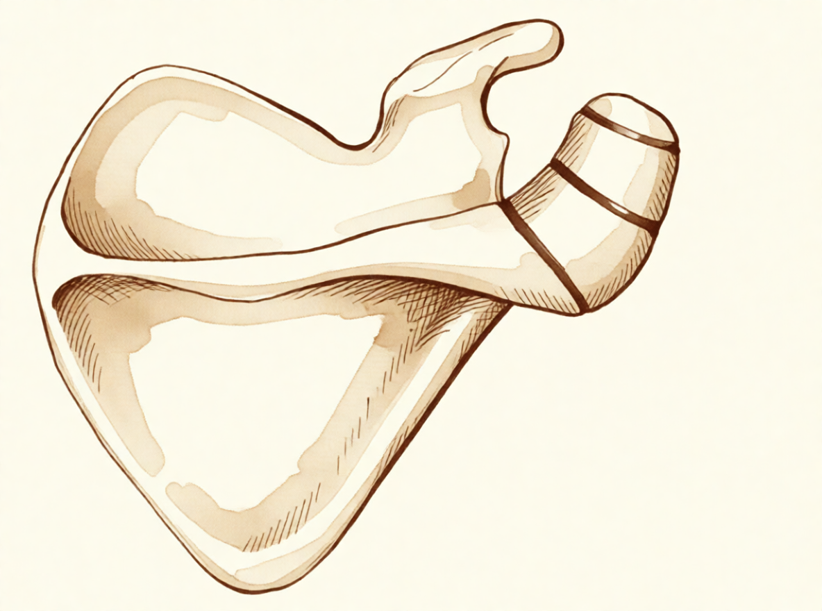
The bony roof of your shoulder does not form as one single piece of bone. It starts as three separate centres that slowly join together, beginning around age 15 and usually finished by age 25. In about 8% of people, one of these joins never closes. The gap fills with tough, gristle-like tissue instead of bone, leaving a small piece of the roof attached by a flexible link rather than solid bone.
Think of it like a plank fixed to a wall with one loose screw. It holds, but it moves a little when you lean on it. The muscle that lifts your arm out to the side attaches to this roof, so every time you raise your arm, that muscle pulls on the loose piece and it flexes slightly. That movement presses down on the tendon running beneath the roof, and the rubbing irritates it. That is why overhead activity hurts and why the pain sits at the front and side of your shoulder.
The flexible link itself can also be the problem. It can become tender in its own right, much like a worn joint, and pressing on it brings the pain on. Most people with this bone pattern never feel anything, and it does not appear to cause shoulder pain by itself or make a tendon tear more likely. But when the loose piece starts moving more than usual, either from the way it was formed or after a knock to the area, it can press on the tendon each time you lift. That is what produces the painful arc and night pain described above.
Because the loose piece sits right where the tendon lives, the two problems often appear together. If your shoulder has already been treated for a tendon problem and the pain has not settled, this unfused piece is worth checking for, since operating on the tendon alone will not quieten a moving piece of bone above it.
What we can do about it
Dr Kieran Hirpara, an upper-limb surgeon at Mater Private Hospital Rockhampton, starts with the least invasive options that suit your condition. Patients are generally referred to our clinic by their GP; if a physiotherapist has suggested you see us, you will still need a referral from your GP in order to be eligible for the Medicare rebate. At your first visit we take a history, examine your shoulder and arrange imaging where it is needed.
The first step is usually changing how you use your shoulder and giving physiotherapy a proper go. Physio aims to settle the irritation under the bony roof and build up the muscles around your shoulder so they cope better with overhead work. Give it a fair trial before thinking about anything further.
Pain medication can help you keep moving while that happens. Simple anti-inflammatories, taken as your GP advises, settle the soreness so you can do your exercises comfortably. If pain is not settling, are getting worse over weeks, wake you at night, or stop you working or using your arm, see your GP or ask for a specialist review.
When non-operative care has not given enough improvement, we consider surgery. Before that we confirm the diagnosis with plain X-rays, which are almost always enough to show the unfused piece. A view taken from below the armpit shows the shape of the joint and the front of the roof clearly, and it matters because an injury can damage the junction between the two bone pieces. An ultrasound is a simple, accurate way to check the tendon running under the roof, and an MRI scan gives a fuller picture of that tendon and the surrounding soft tissue.
The operation we most often offer is a keyhole (arthroscopic) assessment of the joint with a bursectomy, which means clearing inflamed tissue from the space under the roof, then a small open incision for bone grafting and fixation, which means holding the loose piece still so it can join to the rest of the bone. Holding it still lets the two pieces grow together. If the loose piece is small, we may remove it instead of fixing it. Fixing a larger piece needs care, because the muscle that lifts your arm pulls on it constantly and the piece is thin. Sometimes the fragment does not join after fixation, which is called non-union. We will talk through which option suits your shoulder and decide together.
What to expect
Most people with this bone pattern never have any trouble at all, and it needs no treatment. When it does cause pain, the outlook depends on whether the loose piece is stable and whether it is actually the source of your pain.
For a painful one, care usually starts without surgery. Changing how you use your shoulder, physiotherapy and simple pain relief settle things for many people. If that has not worked after a fair trial, surgery can lead to a real drop in pain and better shoulder function. When the loose piece is held still and joins to the rest of the bone, people do better on shoulder function measures than when the two pieces stay separate. Surgery is not always the answer, though. In some people with this condition and a tendon tear as well, results after surgery have been mixed, with 53% achieving satisfactory results overall.
If it is left alone and it keeps troubling you, the pain usually does not go away by itself. It can also be easy to miss: this condition is worth suspecting when shoulder pain has not settled after treatment, including after surgery for a tendon problem, because operating on the tendon alone will not quieten a moving piece of bone above it.
Recovery after surgery is gradual. You will work with your physiotherapist, building movement first and then strength, and overhead lifting will come back slowly over weeks to months. The aim is a shoulder that copes with daily life without the sharp pain on lifting you came in with.
If you notice a finger, hand or arm that becomes hot, red, swollen and painful, especially with a fever, go to the emergency department the same day. No GP referral is needed.
When to see someone
Most people with this bone pattern never need care, but see your GP or ask for a specialist review if your shoulder pain is not settling, is getting worse over weeks, wakes you at night, or stops you working or using your arm. Ask for a review sooner if the pain sits at the front and side of the shoulder, is worse with overhead activity, or keeps waking you when you lie on that side. It is also worth a review if the sore spot at the top of the shoulder is tender to press, if your arm feels weak lifting it out to the side or turning it outward, or if the shoulder hurts partway up as you raise it. Younger adults with these symptoms and no injury should be checked for this condition, especially if physiotherapy, rest from sport or an injection has not helped. If a finger, hand or arm becomes hot, red, swollen and painful, especially with a fever, go to the emergency department the same day; no GP referral is needed.
In more depth
Advanced reading: the deeper science (optional)
This section goes further than you need for your own treatment decisions. Os acromiale is worth the extra reading because it is a normal anatomical variant in a meaningful minority of people, usually causes nothing, and is frequently blamed for shoulder pain it is not responsible for.
What it is, and how common
The acromion, the bony roof over the shoulder, forms from several separate centres of bone that normally fuse in the late teens or early twenties. In some people one fails to fuse, leaving a permanent fibrous junction. That is an os acromiale, and it is present from adolescence rather than acquired.
Prevalence varies by population. A multicentre study of 6,842 patients found os acromiale is associated with rotator cuff injuries, that prevalence in the Japanese population studied was lower than in non-Asian populations, and that the fragment size tended to be smaller [1].
That association with cuff injury is worth reading carefully. It establishes that the two occur together more often than chance, not that the os causes the tear. The plausible mechanism runs both ways: an unfused fragment moves slightly under deltoid pull, which may irritate the tendon beneath — or the same shoulder mechanics that predispose to cuff disease make an unstable fragment symptomatic.
Whether it is the source of pain is the whole question
Because it is visible, permanent and abnormal-looking on imaging, an os acromiale readily becomes the reported explanation for shoulder pain. Most are silent, and most people who have one never know.
What distinguishes a symptomatic one is localised tenderness directly over the fragment, pain reproduced by pressing on it, and, where a diagnostic injection is used, relief specifically from injecting that junction rather than the subacromial space beneath. In the absence of those, the finding is best treated as incidental.
The subtypes are conventionally classified by which fusion site is involved [2], which matters surgically because the size of the mobile fragment determines whether it can be excised or must be fixed.
Why it complicates other shoulder surgery
The practical importance of an os acromiale is often less about its own symptoms than about what it does to operations planned for something else.
The deltoid attaches to the acromion, so an unfused fragment carries that pull across a mobile junction. Removing bone from the undersurface, as in acromioplasty, thins a fragment that is already unstable, and can convert a symptomless variant into a painful one or produce a nonunion. This is the strongest reason for it to be recognised on pre-operative imaging: not to treat it, but to avoid destabilising it.
Where it does require treatment, the choice is between excising a small fragment and fixing a larger one, and fixation across this junction is known to be difficult, the fragment is thin, the deltoid pulls on it continuously, and nonunion rates are appreciable. That difficulty is itself a reason to be confident the os is genuinely the pain source before operating on it.
References for the advanced reading
- Kozono N, Nishii A, Ishitani E, Mizuki Y, Kimura T, Yamamoto S, et al. Prevalence and factors associated with os acromiale: a multicenter study. JSES Int. 2025;9(5):1541-5.
- Boehm TD, Matzer M, Brazda D, Gohlke FE. Os acromiale associated with tear of the rotator cuff treated operatively. Review of 33 patients. J Bone Joint Surg Br. 2003;85(4):545-9.
Evidence & references
This is the clinical evidence summary written for health professionals. It is technical, and it lists the research this page was built from. You do not need to read it to understand your treatment or to make a decision about it.
Overview
- A symptomatic, unstable os acromiale is relatively rare despite the radiographic appearance of an os acromiale being not uncommon in patients presenting with shoulder pain [1].
- An os acromiale should always be suspected in cases of failed rotator cuff surgery or ongoing shoulder pain after failed treatment [2].
- The prevalence of os acromiale was 2.13% in Thai patients with shoulder problems who required MRI evaluation [3].
- Os acromiale is associated with rotator cuff injuries [7].
- The greater the distance of the acromioclavicular joint from the anterior edge of the acromion, the higher the likelihood of an os acromiale [15].
- The authors recommend that the acromion be carefully examined roentgenographically prior to operation in any patient with a rotator cuff tear [10].
- Surgical treatment is usually not indicated for os acromiale in the professional tennis player [9].
- Operative management of a symptomatic os acromiale that has failed initial nonoperative treatment leads to decreased symptoms and improvement in clinical outcomes [4].
- Surgical options for symptomatic os acromiale include arthroscopic sub-total excision, arthroscopic subacromial decompression of stable fragments, and open reduction and internal fixation of unstable fragments [13].
- Patients with a united os acromiale had a significantly better functional outcome as measured by the Constant score (P = .0169) [12].
- The advantages of arthroscopic internal fixation include improved visualization of the undersurface of the acromion, allowing excellent reduction, the possibility of treating concomitant shoulder pathologies, and to avoid increased morbidity of the open approach [14].
- Postoperative local tenderness at the os acromiale can be expected in 1 out of 4 patients but resolves spontaneously over time in the majority of patients [5].
- The presence of os acromiale does not appear to have a negative impact on the clinical outcomes after surgery and rTSA remains a safe and effective treatment option [6].
- The outcome of RTSA does not seem to be negatively affected by the presence of an os acromiale [11].
- Ipsilateral os acromiale may be a relative contraindication to the clavicle hook plate [16].
Anatomy & Pathophysiology
Bony Anatomy
- The acromion has three ossification centers: the metacromion (base), the mesoacromion (middle), and the preacromion (tip) [38].
- Failure of fusion of the acromial ossification centers results in os acromiale [38].
- Os acromiale is defined as incomplete fusion of secondary ossification centers, most commonly between the mesoacromion and meta-acromion [49].
- The unfused segment in os acromiale is most commonly the meso-acromion, a configuration often termed a meso-type os acromiale [22].
- The acromion, coracoacromial ligament, and coracoid process form the coracoacromial arch, a rigid bony-ligamentous structure that imparts stability to the shoulder girdle [35].
- The rotator cuff, subacromial bursa, and subdeltoid bursa pass underneath the coracoacromial arch [35].
- The scapula is attached to the axial skeleton by the acromioclavicular and sternoclavicular joints [37].
- The acromion is a flattened bony process that curves forwards from the scapular spine [37].
Prevalence and Associations
- In Thai patients with shoulder problems who required MRI evaluation, the prevalence of os acromiale was 2.13% [3].
- The radiographic appearance of an os acromiale is not uncommon in patients presenting with shoulder pain [1].
- A symptomatic, unstable os acromiale is relatively rare [1].
Pathophysiology
- Most diagnoses of os acromiale are made incidentally on axillary view radiographs of the shoulder [22].
- Primary shoulder pain is usually unrelated to the unfused os acromiale [22].
- When an os acromiale drives symptoms, the two principal causes are motion at the non-union site or an impingement-type syndrome resulting from flexion of the os fragment during deltoid contraction and arm elevation [22].
- Pain from an os acromiale due to these mechanisms has been reported in a variety of sports, particularly among throwing athletes and swimmers [22].
- Previously stable non-unions can become unstable following an episode of blunt trauma to the region [22].
- The synchondrosis of an os acromiale can be injured following trauma [24].
- Extrinsic impingement occurs when the space available for the rotator cuff is diminished, with examples including acromial fracture or pathologic os acromiale [62].
Epidemiology and Prevalence
- Liberson reviewed 1800 shoulder girdles and identified an incidence of os acromiale of 1.4% [21].
- The lesion was bilateral in 62% of patients in Liberson's review of 1800 shoulder girdles [21].
Associated Pathology and Risk Factors
- The greater the distance of the AC joint from the anterior edge of the acromion, the higher the likelihood of an os acromiale [15].
Diagnostic Considerations
- Awareness of the os acromiale in the young athlete, appropriate clinical examination, and image studies are crucial to confirm diagnosis [19].
- Appropriate radiographic investigation including axillary views is emphasized for the diagnosis of os acromiale injuries [24].
Clinical Presentation
- An os acromiale should always be suspected in cases of failed rotator cuff surgery [2].
- An os acromiale should always be suspected in cases of ongoing shoulder pain after failed treatment [2].
- Postoperative local tenderness at the os acromiale can be expected in 1 out of 4 patients [5].
- Postoperative local tenderness at the os acromiale resolves spontaneously over time in the majority of patients [5].
- The lesion was bilateral in 62% of patients in Liberson's review [21].
- The unfused segment is most commonly the meso-acromion [22].
- Any primary shoulder pain is usually unrelated to the unfused os [22].
- If the os acromiale is driving symptoms, one principle cause is motion at the non-union site [22].
- If the os acromiale is driving symptoms, another principle cause is an impingement-type syndrome resulting from flexion of the os fragment during deltoid contraction and arm elevation [22].
- Pain from an os acromiale by the mechanism of impingement has been reported in a variety of sports [22].
- Pain from an os acromiale by the mechanism of impingement has been reported in particular among throwing athletes and swimmers [22].
- Meta–os acromiale is the rarest subtype of os acromiale [23].
Investigations
Prevalence and Demographics
- Liberson reviewed 1800 shoulder girdles and identified an incidence of os acromiale of 1.4%, with the lesion being bilateral in 62% of patients [21].
- A multicenter study aimed to determine the prevalence of and factors associated with os acromiale in the Japanese population [8].
- The radiographic appearance of an os acromiale is not uncommon in patients presenting with shoulder pain, but a symptomatic, unstable os acromiale is relatively rare [1].
Radiographic Evaluation
- The acromion should be carefully examined roentgenographically prior to operation in any patient with a rotator cuff tear [10].
- Appropriate radiographic investigation for os acromiale includes axillary views [24].
- Awareness of the os acromiale in the young athlete requires appropriate clinical examination and image studies to confirm diagnosis [19].
- The standard shoulder series should include orthogonal views of the shoulder, including a true AP view in the scapular plane, an AP view, an axillary view, and a scapular Y view [60].
- The scapular Y view provides visualization of the coracoacromial arch and can reveal coracoacromial spurs, which have been closely associated with the presence of rotator cuff pathology [60].
- The scapular Y view can also show acromial shape [60].
- At least two X-ray views should be obtained for shoulder imaging: an anteroposterior in the plane of the glenoid and an axillary projection with the arm in abduction to show the relationship of the humeral head to the glenoid [52].
- The purpose of imaging of the shoulder is to help establish the diagnosis, determine the severity of the pathoanatomy, assist in surgical planning, and enable the surgeon to illustrate the condition of the shoulder to the patient [30].
- Standardized plain films are almost always sufficient to garner the information needed for shoulder evaluation [30].
- The first key view for shoulder imaging is the anteroposterior (AP) in the plane of the scapula taken so that the x-ray beam passes through the glenohumeral joint [30].
- The second key view for shoulder imaging is the axillary view taken with the arm in the functional position of elevation in the plane of the scapula and oriented so that both the spinoglenoid notch and the scapular neck are visible [30].
- The axillary view is referred to as the "truth view" because it demonstrates the glenohumeral relationships in the functional position of elevation [30].
- CT scans have the disadvantage of being taken with the arm in the adducted position, unlike the axillary truth view [30].
- The temptation to "overimage" should be resisted, obtaining only the scans or reconstructions that are necessary for the care of the patient [30].
Magnetic Resonance Imaging
- In Thai patients with shoulder problems, os acromiale was identified via Magnetic Resonance Imaging evaluation [3].
- MRI is useful to identify labral tears and rotator cuff tears, although the accuracy for these is enhanced by combining the scan with arthrography [52].
- MRI is the modality of choice for evaluating the rotator cuff, biceps, and subacromial/subdeltoid bursa [59].
- T2-weighted MRI provides better visualization of full thickness rotator cuff tears [59].
- MR arthrography is considered the benchmark for evaluation for labral tears and rarely is indicated for evaluation of rotator cuff pathology [59].
- When MRI or MR arthrography is contraindicated, CT arthrography is indicated [59].
Ultrasonography
- Ultrasonography is a simple and accurate test for identifying rotator cuff tears and calcific tendinitis [52].
- Ultrasonography is a low-cost alternative to MRI and arthrography for evaluating both skeletal and soft-tissue structures of the shoulder [59].
- Ultrasonography can provide immediate, real-time visualization of the rotator cuff, biceps tendon, and calcific deposits [59].
- Ultrasonography can be used to measure the subacromial space and detect atrophy of rotator cuff muscles [59].
- Ultrasonography is highly operator dependent and is not as useful for evaluating labral tears or rotator cuff tears that are very small or larger than 3 cm [59].
- The most commonly performed joint examination using ultrasonography is the shoulder examination, and accuracy depends on the skill of the scanner operator and an awareness of pitfalls that are encountered [50].
Clinical Associations and Diagnostic Implications
- The synchondrosis of an os acromiale can be injured following trauma, though rarely [24].
Treatment
Non-Operative
- A symptomatic os acromiale in a competitive female fastball pitcher was treated successfully with open reduction and internal fixation [27].
Operative
- Symptomatic unstable os acromiale can be effectively treated with autogenous bone-grafting and internal fixation using a rigid tension-band construct and cannulated screws, which results in a high rate of union [65].
- This new arthroscopic technique of fixation of os acromiale with absorbable screws provides promising clinical, cosmetic, and radiologic results with high patient satisfaction [28].
- After 3 to 6 years of follow-up, all 6 shoulders were rated as excellent according to criteria similar to that of the American Shoulder and Elbow Surgeons [18].
- Modified arthroscopic subacromial decompression can provide results similar to standard arthroscopic subacromial decompression for routine impingement [25].
- Meta–os acromiale is the rarest subtype of os acromiale, and special consideration must be given to the type of tension-band construct used to achieve adequate compression and fixation [23].
Complications
Post-operative and Surgical Complications
- Postoperative local tenderness at the os acromiale can be expected in 1 out of 4 patients following reverse total shoulder arthroplasty [5].
Diagnostic and Clinical Implications
Pathophysiology and Mechanisms of Symptom
- Pain from an os acromiale by the mechanism of impingement has been reported in a variety of sports, in particular among throwing athletes and swimmers [22].
Recovery
- After 3 to 6 years of follow-up, all 6 shoulders treated with osteosynthesis of an unstable mesoacromion were rated as excellent according to criteria similar to that of the American Shoulder and Elbow Surgeons [18].
- At a two to five year follow-up, all patients treated for fusion of the unstable mesoacromion had fused [69].
Key Evidence
- [L4] While the radiographic appearance of an os acromiale is not uncommon in patients presenting with shoulder pain, a symptomatic, unstable os acromiale is relatively rare. [1] (10.1097/00132589-200412000-00006)
- [L4] An os acromiale should always be suspected in cases of failed rotator cuff surgery or ongoing shoulder pain after failed treatment. [2] (10.1097/00132589-200609000-00006)
- [L3] In Thai patients with shoulder problems who required MRI evaluation, the prevalence of os acromiale was 2.13%. [3] (10.1177/23259671221078806)
- [L4] Operative management of a symptomatic os acromiale that has failed initial nonoperative treatment leads to decreased symptoms and improvement in clinical outcomes. [4] (10.1016/j.jse.2019.05.047)
- [L3] Postoperative local tenderness at the os acromiale can be expected in 1 out of 4 patients but resolves spontaneously over time in the majority of patients. [5] (10.1177/2325967120965131)
- [L4] The presence of os acromiale does not appear to have a negative impact on the clinical outcomes after surgery and rTSA remains a safe and effective treatment option. [6] (10.1016/j.xrrt.2025.01.002)
- [L3] The study supports previous findings that os acromiale is associated with rotator cuff injuries. [7] (10.1016/j.jseint.2025.05.015)
- [L3] This multicenter study aimed to determine the prevalence of and factors associated with os acromiale in the Japanese population. [8] (10.1016/j.jse.2025.01.008)
- [L4] Surgical treatment is usually not indicated for os acromiale in the professional tennis player. [9] (10.1177/2325967118773723)
- [L4] The authors recommend that the acromion be carefully examined roentgenographically prior to operation in any patient with a rotator cuff tear. [10] (10.2106/00004623-198466080-00029)
- [L4] The outcome of RTSA does not seem to be negatively affected by the presence of an os acromiale. [11] (10.1016/j.jse.2017.02.012)
- [L4] Patients with a united os acromiale had a significantly better functional outcome as measured by the Constant score (P = .0169). [12] (10.1016/s1058-2746(98)90008-8)
- [L5] Surgical options for symptomatic os acromiale include arthroscopic sub-total excision, arthroscopic subacromial decompression of stable fragments, and open reduction and internal fixation of unstable fragments. [13] (10.5435/jaaos-d-17-00011)
- [L5] The advantages of this technique are improved visualization of the undersurface of the acromion, allowing excellent reduction, the possibility of treating concomitant shoulder pathologies, and to avoid increased morbidity of the open approach. [14] (10.1097/bte.0000000000000191)
- [L4] Our data suggest that the greater the distance of the AC joint from the anterior edge of the acromion, the higher the likelihood of an os acromiale. [15] (10.1067/mse.2003.128136)
- [L4] Ipsilateral os acromiale may be a relative contraindication to the clavicle hook plate. [16] (10.1186/s12891-021-04841-1)
- [L4] After 3 to 6 years of follow-up, all 6 shoulders were rated as excellent according to criteria similar to that of the American Shoulder and Elbow Surgeons. [18] (10.1016/s1058-2746(99)90004-6)
- [L4] Awareness of the os acromiale in the young athlete, appropriate clinical examination, and image studies are crucial to confirm diagnosis. [19] (10.1016/j.jseint.2020.02.008)
- [L4] [22] (10.1302/2058-5241.4.180100)
- [L4] Meta–os acromiale is the rarest subtype of os acromiale, and special consideration must be given to the type of tension-band construct used to achieve adequate compression and fixation. [23] (10.1177/03635465211028238)
- [L4] This case highlights that the synchondrosis of an os acromiale can be injured following trauma, though rarely, and emphasizes the need for appropriate radiographic investigation including axillary views and a flexible surgical approach. [24] (10.1016/j.jse.2008.02.012)
- [L5] Increased awareness of the diagnosis and improved techniques have shown that modified arthroscopic subacromial decompression can provide results similar to standard arthroscopic subacromial decompression for routine impingement. [25] (10.1097/00132589-200206000-00003)
- [L4] A symptomatic os acromiale in a competitive female fastball pitcher was treated successfully with open reduction and internal fixation. [27] (10.1177/0363546506288305)
- [L4] This new arthroscopic technique of fixation of os acromiale with absorbable screws provides promising clinical, cosmetic, and radiologic results with high patient satisfaction. [28] (10.1016/j.jse.2011.12.011)
- [L4] Symptomatic unstable os acromiale can be effectively treated with autogenous bone-grafting and internal fixation using a rigid tension-band construct and cannulated screws, which results in a high rate of union. [65] (10.2106/00004623-199809000-00011)
- [L4] At a two to five year followup all had fused. [69] (10.1016/s1058-2746(95)80192-8)
References
[1] Treatment of the Symptomatic Os Acromiale. Techniques in Shoulder and Elbow Surgery. 2004. DOI: 10.1097/00132589-200412000-00006
[2] Management of Os Acromiale. Techniques in Shoulder and Elbow Surgery. 2006. DOI: 10.1097/00132589-200609000-00006
[3] Prevalence of Os Acromiale in Thai Patients With Shoulder Problems: A Magnetic Resonance Imaging Study. Orthopaedic Journal of Sports Medicine. 2022. DOI: 10.1177/23259671221078806
[4] Os acromiale: systematic review of surgical outcomes. Journal of Shoulder and Elbow Surgery. 2020. DOI: 10.1016/j.jse.2019.05.047
[5] Os Acromiale in Reverse Total Shoulder Arthroplasty: A Cohort Study. Orthopaedic Journal of Sports Medicine. 2020. DOI: 10.1177/2325967120965131
[6] Clinical implications of reverse total shoulder arthroplasty with an os acromiale: a systematic review. JSES Reviews, Reports, and Techniques. 2025. DOI: 10.1016/j.xrrt.2025.01.002
[7] Prevalence and factors associated with os acromiale: a multicenter study. JSES International. 2025. DOI: 10.1016/j.jseint.2025.05.015
[8] The prevalence and associated factors of os acromiale: a multicenter study. Journal of Shoulder and Elbow Surgery. 2025. DOI: 10.1016/j.jse.2025.01.008
[9] Os Acromiale in Professional Tennis Players. Orthopaedic Journal of Sports Medicine. 2018. DOI: 10.1177/2325967118773723
[10] Rotator cuff tears associated with os acromiale.. The Journal of Bone & Joint Surgery. 1984. DOI: 10.2106/00004623-198466080-00029
[11] Reverse shoulder arthroplasty in patients with os acromiale. Journal of Shoulder and Elbow Surgery. 2017. DOI: 10.1016/j.jse.2017.02.012
[12] Transacromial approach to obtain fusion of unstable os acromiale. Journal of Shoulder and Elbow Surgery. 1998. DOI: 10.1016/s1058-2746(98)90008-8
[13] Symptomatic, Unstable Os Acromiale. Journal of the American Academy of Orthopaedic Surgeons. 2018. DOI: 10.5435/jaaos-d-17-00011
[14] Arthroscopic Internal Fixation of Symptomatic Os Acromiale. Techniques in Shoulder & Elbow Surgery. 2020. DOI: 10.1097/bte.0000000000000191
[15] Relationship between os acromiale and acromioclavicular joint anatomic position. Journal of Shoulder and Elbow Surgery. 2003. DOI: 10.1067/mse.2003.128136
[16] Os acromiale may be a contraindication of the clavicle hook plate: case reports and literature review. BMC Musculoskeletal Disorders. 2021. DOI: 10.1186/s12891-021-04841-1
[18] Successful osteosynthesis of an unstable mesoacromion in 6 shoulders: A new technique. Journal of Shoulder and Elbow Surgery. 1999. DOI: 10.1016/s1058-2746(99)90004-6
[19] The unstable os acromiale: a cause of pain in the young athlete. JSES International. 2020. DOI: 10.1016/j.jseint.2020.02.008
[21] Types of os acromiale according to Liberson. 2006.
[22] Os acromiale: a review of its incidence, pathophysiology, and clinical management. EFORT Open Reviews. 2019. DOI: 10.1302/2058-5241.4.180100
[23] Rare Symptomatic Meta–Os Acromiale in an Athlete. The American Journal of Sports Medicine. 2021. DOI: 10.1177/03635465211028238
[24] Fracture of an os acromiale with associated rupture of the coracoclavicular ligaments. Journal of Shoulder and Elbow Surgery. 2008. DOI: 10.1016/j.jse.2008.02.012
[25] Arthroscopic Management of the Mesoacromion. Techniques in Shoulder and Elbow Surgery. 2002. DOI: 10.1097/00132589-200206000-00003
[27] Surgical Stabilization of Os Acromiale in a Fast-Pitch Softball Pitcher. The American Journal of Sports Medicine. 2006. DOI: 10.1177/0363546506288305
[28] Arthroscopically assisted internal fixation of the symptomatic unstable os acromiale with absorbable screws. Journal of Shoulder and Elbow Surgery. 2012. DOI: 10.1016/j.jse.2011.12.011
[30] Rockwood And Matsen S The Shoulder. Arthroscopic Management of Prearthritic and Arthritic Conditions of the Shoulder and the Postarthroplasty Shoulder > Radiographic Evaluation.
[35] Rockwood And Matsen S The Shoulder. Shoulder and Elbow Specialty Clinic Workers’ Survey > ANATOMY.
[37] Rockwood And Green S Fractures In Adults. 29: Principles of Nonunion and Bone Defect Treatment > Applied Anatomy Related to Scapular Fractures.
[38] Aaos Comprehensive Orthopaedic Review 3. Anatomy of the Shoulder, Arm, and Elbow > I. Shoulder.
[49] Miller S Review Of Orthopaedics. Genetics of musculoskeletal conditions and abnormalities are summarized in Table 1.27 > UPPER EXTREMITY > SHOULDER.
[50] Orthopaedic Knowledge Update Sports Medicine 6. Diagnostic Ultrasonography and Ultrasonography-Guided Procedures > Annotated References.
[52] Apley And Solomon S Concise System Of Orthopaedics And Trauma. INVESTIGATION.
[59] Orthopaedic Knowledge Update 13 Ebook Without Multimedia. Shoulder Anatomy and Biomechanics, Clinical Evaluation, Imaging > Clinical Evaluation > Imaging.
[60] Aaos Comprehensive Orthopaedic Review 3. Imaging of the Shoulder and Elbow > I. Shoulder.
[62] Campbell S Operative Orthopaedics 4 Volume Set. ANTERIOR CRUCIATE LIGAMENT RECONSTRUCTION WITH BONE-PATELLAR TENDON-BONE GRAFT > Developmental Stages of Impingement Syndrome.
[65] The Treatment of Symptomatic Os Acromiale. The Journal of Bone & Joint Surgery*. 1998. DOI: 10.2106/00004623-199809000-00011
[69] Successful fusion of the unstable mesoacromion: Technique and report of five cases. Journal of Shoulder and Elbow Surgery. 1995. DOI: 10.1016/s1058-2746(95)80192-8