Clinicians › Hand
Flaps and Grafts
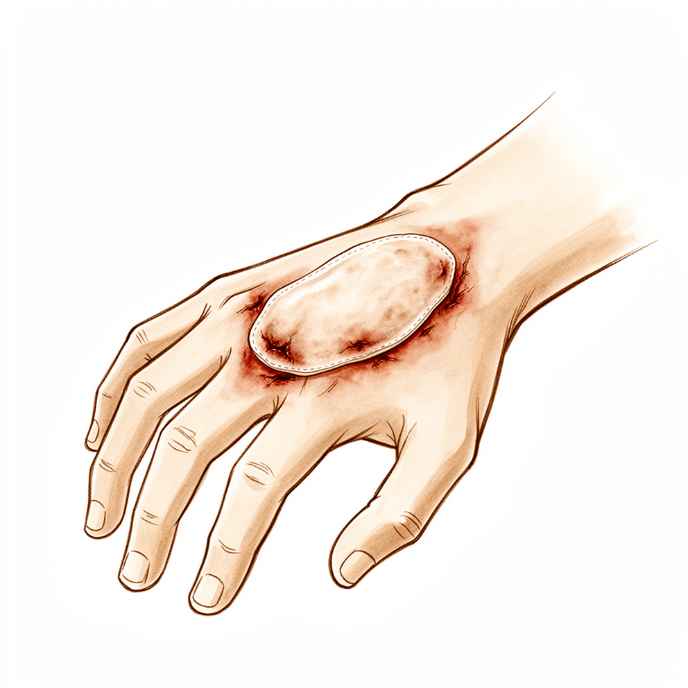
Hand reconstruction: flap and graft options for soft tissue defects, bone loss, and complex trauma—local to free flaps.
Overview
Soft tissue reconstruction of the hand and upper extremity relies on a diverse array of flaps and grafts to restore coverage, function, and aesthetics. Microsurgical techniques, including free flaps and neurovascular island flaps, offer critical advantages over non-microsurgical approaches by enabling early mobilization and providing appropriate coverage with acceptable donor morbidity [23]. Evidence supports the safety and reliability of these methods; for instance, a series of 116 Neurovascular Tranquilli-Leali Flaps reported no flap deaths [1], while long-term outcomes for homodigital unipedicle neurovascular island flaps remain satisfactory [2]. Similarly, 79 free flaps utilizing the ulnar artery distal cutaneous descending branch achieved 100% flap survival with acceptable aesthetic and functional results [4]. The Reverse Flow Shunt Restricted Arterialized Venous Free Flap has also demonstrated high survival rates and satisfactory functional outcomes in a clinical series of nine cases [11].
Selection of the optimal reconstructive option depends on defect characteristics, patient body habitus, and anatomic location [28]. A framework exists for assessing defects to select the best-suited flap using representative case examples [5]. For less extensive defects, alternative regional flaps that do not sacrifice a major limb vessel provide useful options with reduced donor morbidity and improved aesthetics [6]. Venous flaps serve as a versatile and well-tolerated option, particularly when conventional flaps are limited or unavailable, offering advantages such as ease of design, thinness, and lack of donor site morbidity [13, 140]. However, the clinical application of venous flaps remains under further investigation due to inconsistent survival in some cases [140]. The synovial flap of the superficial finger flexors should remain limited to cases where commonly used flaps are contraindicated or unusable, as larger series are needed to validate its indications [14].
Successful outcomes require efficient surgery with carefully performed vascular anastomoses outside the zone of injury [9] and adherence to basic principles and indications of replantation surgery [51]. The need for refinement and secondary surgery must be considered during initial flap selection [53]. Specific techniques, such as the reversed vascularized pedicled forearm flap for complete degloved digits [34] and the Dorso-Ulnar Osteocutaneous Reverse Flow Flap for distal thumb defects [52], provide adequate soft tissue and good functional outcomes. Primary closure of palmar island flaps for dorsolateral finger defects can be achieved without donor site morbidity [45]. Refinements in standard techniques for pedicled groin flaps help reduce patient morbidity and extend the flap's usefulness [133]. For patients who choose revision amputation or whose replants do not survive, various reconstructive options, such as toe-to-hand transfer, are available [141]. Hand transplantation and prosthetic reconstruction should not be viewed as competing options [16].
Anatomy & Pathophysiology
Osseous Architecture
The skeleton of the hand and wrist comprises 27 bones, 19 of which are long bones [40]. The hand skeleton is organized into five rays, each forming a polyarticulated chain of metacarpals and phalanges [40]. The thumb ray (first ray) is the shortest, consisting of a metacarpal and two phalanges [40]. In the sagittal plane, the thumb metacarpal makes an angle of about 45 degrees with the second metacarpal [40]. The transverse axis of the palm forms an acute angle of approximately 75 degrees with the longitudinal axis [40]. Epiphyseal plates are located at the proximal ends of the phalanges and the first metacarpal, and at the distal ends of the other metacarpals [40].
Among the metacarpals, the index metacarpal is the most firmly fixed [81]. The fifth metacarpal has a range of flexion–extension of approximately 20 degrees [81]. The ring metacarpal has about 10 degrees of mobility in flexion and extension [81].
Musculotendinous Anatomy
Control of digital posture requires a complex balance of extrinsic and intrinsic muscle forces [39]. Extrinsic extensors run through six different fibroosseous retinacular compartments at the wrist level [39]. The compartment contents are as follows: * First compartment: Contains the abductor pollicis longus and the extensor pollicis brevis [39]. * Second compartment: Contains the extensor carpi radialis longus and the extensor carpi radialis brevis [39]. * Third compartment: Contains the extensor pollicis longus, which turns abruptly radialward about Lister tubercle [39]. * Fourth compartment: Contains the extensor indicis proprius lying deep to the four tendons of the extensor digitorum communis [39]. * Fifth compartment: Contains the extensor digiti quinti [39]. * Sixth compartment: Contains the extensor carpi ulnaris tendon [39].
The extensor digitorum communis tendons of the middle, ring, and little fingers are tethered together by juncturae tendinum over the dorsum of the hand proximal to the metacarpophalangeal joint [39]. The extrinsic finger flexors are the flexor digitorum profundus and the flexor digitorum superficialis [39]. The flexor digitorum profundus inserts on the proximal volar aspect of the distal phalanx [39]. The flexor digitorum superficialis inserts via radial and ulnar slips into the proximal metaphysis of the middle phalanx [85].
There are seven interosseous muscles, four dorsal and three volar [78]. The dorsal interossei are abductors and the volar interossei are adductors [78]. The middle finger has two dorsal interossei and no volar interossei because the central axis of the hand lies within it [78]. Innervation of the flexor digitorum profundus is split: the index and middle fingers are innervated by the anterior interosseous branch of the median nerve, while the ring and little fingers are innervated by the ulnar nerve [85]. The entire flexor digitorum superficialis muscle receives innervation from the median nerve [85]. The A2 and A4 pulleys are the most essential in maintaining the mechanical advantage of the flexor tendons [85]. The tenosynovium lining the fibroosseous tunnel supplies nutrition and lubrication to the poorly vascularized flexor tendons [85].
Vascular Anatomy
The radial artery originates from the brachial artery and travels in a relatively superficial course just beneath the anterior margin of the muscle belly of the brachioradialis [99]. It gives branches to a plexus of vessels in the overlying deep fascia, which supplies the skin of the anterior and radiodorsal surfaces of the forearm [99]. The radial artery is accompanied by two or more venae comitantes with multiple anastomoses that permit reversal of flow without valvular obstruction [99]. The volar metacarpal vessels play an important part in the vascularity of the hand [136, 137].
The "princeps pollicis" artery is the terminal branch of the radial artery that crosses the first intermetacarpal space and runs along the ulnar side of the first metacarpal bone [82]. In anatomical studies, only 15% of dissections of the palmar arteries of the thumb fall into the classical "typical" category [82]. The dorsal arteries of the thumb originate from palmar arteries (princeps, commissural, or anastomoses of the superficial arcade) at the level of the first metacarpal [82].
Cutaneous Anatomy and Mechanics
The dorsal skin possesses a normal pilosebaceous system, unlike the palm [87]. It has loose connections with deeper planes, allowing free gliding and full flexion at the digital joints [87]. Flexion of the fingers produces a significant lengthening of the dorsal skin, with an average increase of 3 cm in the middle finger from extension to full flexion [87]. The dorsal and palmar areas of skin are independent due to a system of adhesions that anchors their common boundary to the underlying plane [87].
The superficial palmar fascia lies in a coronal plane beneath the palmar subcutaneous tissue and covers a triangular area of the central palm [88]. The palmaris longus tendon, when present, terminates in continuity with the fibers of the proximal corner of the superficial palmar fascia [88]. Fibers dorsal to the digital neurovascular bundle are collectively called Cleland ligament, and those palmar to the bundle are called Grayson ligament [88].
Skin dimensions for coverage are as follows: * Thumb distal to MCP: Requires about 9 cm wide and 8 cm long [80]. * Thumb and first metacarpal loss: 13 cm wide and 12 cm long [80]. * Hand palmar and dorsal surfaces: 12 cm by 10 cm [80]. * Each finger (palmar and dorsal): 7 cm by 10 cm [80]. * Both sides of hand and digits: Requires a skin of 20 cm by 20 cm for grafting or flaps [80].
Functional Biomechanics
The wrist has three axes of movement, permitting the hand to be positioned in any spatial configuration [40]. The hand presents a longitudinal and transverse concavity, giving it the shape of a cup with a palmar concavity when the thumb is placed next to the index finger [40]. The metacarpophalangeal joints are the keystones of the longitudinal arches of the hand [81]. The volar plates prevent hyperextension at the metacarpophalangeal joints [81]. The stability of the metacarpophalangeal joints is essential to the support of both the longitudinal and transverse metacarpal arches [81]. Sensation is the most important factor in thumb or fingertip repair, constituting 40% of the goal, while length and appearance account for 50% [30].
Classification
Allen’s: This four-type classification describes fingertip amputation levels. Type I involves the distal pulp only, Type II extends through the nailbed, Type III occurs at the proximal nail fold, and Type IV is located at the base of the distal phalanx [181].
Lister’s: This system categorizes distal digital amputation into zones. The distinction between Zone II and Zone III is based on the level of amputation relative to the DIP crease [182].
Urbaniak et al.: This classification grades ring avulsion injuries into three classes. Class I consists of circumferential soft tissue injury with intact circulation and innervation, Class II involves incomplete degloving with circulatory interruption, and Class III comprises digital amputation or complete soft tissue degloving [190].
Kay et al.: This modification of the Urbaniak classification subdivides Class II into arterial [a] or venous [v] insufficiency and adds a category for bone or joint injury. It designates digital amputation and total degloving as Class IV [190].
Banff Working Classification: This standardized grading scheme is used for acute rejection in composite tissue allografts [128].
Tubiana’s: This system categorizes Dupuytren’s disease based on total passive extension deficit in four stages. Stage I ranges from 0–45°, Stage II from 46–90°, Stage III from 91–135°, and Stage IV from 136–180° [201].
Merle's: This classification grades the level of thumb amputation into Type 1a, 1b, and 1c [206].
Woo et al.: This classification is proposed for the clinical description and design of venous flaps, specifically for type III flaps where Chen's classification lacks utility [97].
Other Considerations: The term 'venous flap' is considered misleading in the context of venous flap terminology and classification [97].
Clinical Presentation
The clinical assessment of hand and upper limb reconstruction focuses on tissue viability, sensory restoration, and functional recovery. In a series of 116 Neurovascular Tranquilli-Leali Flaps, there were no flap deaths [1]. Alternative regional flaps that do not sacrifice a major limb vessel are useful options for patients with less extensive defects, offering less donor morbidity and improved aesthetics [6]. Local hand flaps offer excellent coverage of soft tissue defects by replacing like with like, frequently resulting in optimum functional and aesthetic outcomes while sparing patients from more complicated repair methods [46]. Local vascular island flaps are an excellent alternative for coverage in situations where a large tissue deficiency exists after an adequate release [27].
Sensory function and appearance are critical endpoints. The thenar flap provides good sensory function and appearance when fundamental technical principles, including flap design, timing of division, and early mobilization, are respected [7]. Restoration of sensibility in free sensory flaps from the toe is closely related to the patient's age and vascular insufficiency of the flap [18]. All flaps develop a protective sensation after 12 to 18 months [20]. Reconstruction with a local neurovascular island flap has been performed in twenty-one patients to restore sensibility and function with preservation of length [49]. Homodigital artery flap reconstruction provides satisfactory texture and cosmetic appearance as well as favourable sensibility [95].
Flap survival rates vary by technique and patient population. In a series of 9 cases using the reverse flow shunt restricted arterialized venous free flap, the modified technique resulted in high flap survival rates with satisfactory functional outcomes [11]. All bone grafts consolidated and all flaps survived in the treatment of painful nonunion of the distal phalanx using bone graft and dorsal reverse adipofascial flap [15]. In a series of 34 retrograde posterior interosseous flaps, there was no total loss of a flap, but 7 (21%) developed partial ischemic necrosis localized at the periphery of the perfusion perimeter [19]. The postoperative course for the glabrous palmar flap was uneventful, and all flaps survived without major complications [24]. All flaps survived in the revision of residual deformities after primary surgery for Wassel-Flatt IV-D thumb duplication using a microsurgical free lateral great toe flap, with patients followed up for 8–12 months showing satisfactory appearance [26].
Specific flap designs demonstrate distinct advantages and complication profiles. The thenar mini-free flap provides excellent color and texture match in the reconstructed finger with minimal donor site morbidity [29]. The free digital fillet skin flap has no donor-site morbidity as opposed to distant flaps or skin grafting [32]. Primary closure of the palmar island flap can be possible without any donor site morbidity [45]. A series of 30 flaps in 28 patients for painful digital neuroma produced 86.6% good or excellent results [31]. Complications specific to the Razemon lateral digital rotation flap are infrequent and minor when they occur [103]. The presence of mild venous ischaemia during the early postoperative period was common with the thin cross finger skin flap but resolved in all cases without loss of any of the flaps [96].
Vascular anatomy and hemodynamics influence surgical planning. Dorsal metacarpal arteries are found in almost 100% of cases in the first and second metacarpal spaces, whereas they are only found in 70% of fourth metacarpal spaces [48]. End-to-side flaps presented with stable flow hemodynamics over a 14-day period [25]. The use of both true flow-through flap and flow-through conduit methods is very useful, especially in cases with few vascular resources [47]. Five described free vascularized flaps reconstruct complex soft tissue losses of the hand without harvesting major arteries, improve cosmetic appearance, augment function, and can be quite reliable with appropriate planning and microsurgical expertise [21].
Donor site morbidity and patient factors affect outcomes. Donor site complications occurred at similar rates among patients undergoing osteocutaneous and fasciocutaneous radial forearm free flap harvest [50]. Patients over 50 years of age showed significantly more complications than those under 50 in the treatment of hand defects with pedicled groin flaps [94]. One-third of pedicled groin flaps developed minor necrosis or delayed wound healing, in most cases on the proximal site of the flap [94]. Patients have tolerated the hand-to-groin position well for staged groin flaps, with no incidence of separation or significant postoperative swelling or stiffness [100]. The incidence of necrosis of the flap margin in a study of staged groin flaps in children was 17% [100].
Long-term monitoring and specific indications guide management. It is essential to maintain postoperative clinical monitoring for signs of graft degeneration in all patients with long-term vein graft insertion as part of replantation surgery [92]. An optimal thumb reconstruction should meet three main objectives: restore thumb function, recreate its aesthetics, and minimize donor-site morbidity [101]. The maximal age for complex thumb reconstruction is around 60 to 65 years old [101]. In a series of 11 patients treated with an oblique triangular flap for fingertip amputation, the average age was 44 years (range, 18–60 years) [98].
In a review of 90 operated rays for Dupuytren's disease, only one skin graft failed, and ten grafts required dressings for longer than 3 weeks [102]. The durability and functional characteristics of the vascularized fibula transfer make it an especially useful option in paediatric patients [104]. In a series of 13 patients with posttraumatic bone defects of the humerus reconstructed with vascularized fibular grafts, the time to achieve bony union averaged 6 months (range, 3-13 months) [105].
Investigations
Imaging and Vascular Assessment
Ultrasonography: Ultrasonographic examination is recommended before flap dissection to increase the safety of the procedure [48].
CT: Preoperative computed tomography angiography (CTA) and computed tomography densitometry (CDS) are accurate methods for assessing flap location and course [212].
MRI: Magnetic resonance imaging (MRI) verifies prolonged viability of flap tissue, supported by gadolinium perfusion studies [36]. Specific applications include demonstrating revascularization of the trapezium 12 months after surgery [218] and confirming revascularization in Kienböck's disease following vascularized pedicled bone grafts [179]. In the context of autologous osteochondral mosaicplasty, MRI indicates that the donor site is resurfaced with fibrous tissue [180]. For Dupuytren's disease, MRI is probably most useful for identifying additional pathology such as flexor tendon bowstringing [91]. It may also provide a quantitative noninvasive measure of cellularity in affected areas, serving as an index of biologic activity [91].
Doppler: An 8-MHz Doppler tone assessment may identify superficially displaced neurovascular bundles when Dupuytren cords lie beneath soft fleshy prominences, though false-negatives are possible [91]. Doppler imaging represents a promising improvement for identifying palmar structures, but higher resolution imaging technology is needed [91].
Monitoring and Hemodynamics
Fluorometry: Quantitative fluorometry is recommended as an adjunctive or primary technique for monitoring microvascular patency after replantation or free tissue transfer with an exposed skin surface [220].
Post-operative Radiographic Evaluation
Plain radiography: Radiographs demonstrate union of the iliac bone block and the stump without resorption [200]. There is no angular deformity of grafts nor radiographic evidence of resorption or aseptic necrosis of grafted bone at final evaluation [199]. All bones heal radiologically in less than three months following vascularised corticoperiosteal grafts [216]. Follow-up radiographs 41 months later demonstrate complete incorporation and hypertrophy of the vascularised fibular graft [225]. Clinical and radiological measurements show the efficacy of an axial flap in increasing the volume and girth of the reconstructed thumb [211].
Bone scan: Three-phase bone scans confirm union of the iliac bone block and the stump without any resorption [200].
Treatment
Non-Operative
Conservative management is limited in the context of complex soft tissue defects, where healing by secondary intention is not advised in the presence of exposed nonvascularized structures such as bone, hardware, and tendons [156]. Secondary healing of small wounds over muscle or fascia may be appropriate, especially if the medical status does not permit more complex reconstruction methods [156]. In acute burned upper extremities, splinting should continue until re-epithelialization is complete to prevent edema, contracture, scarring, and functional impediment [167]. Early and aggressive hand physical therapy and rehabilitation are critical to the success of most surgical interventions for acute burned upper extremities [167].
Operative
Indications: Surgical reconstruction is indicated for complex extremity wounds where external tissue expansion offers a technically simple and low-morbidity option for durable soft tissue coverage [227]. Combined reconstruction strategies should be considered based on defect characteristics, with acceptable complication rates and good functional recovery [129]. Bilateral hand transplantation is justified in cases of bilateral amputations with strict indications, while transplantation for the loss of one hand is not indicated [112]. The concept of "spare parts" procedures should not exclude harvesting healthy tissue if needed to achieve maximal hand reconstruction, and finger ray amputation should be postponed when possible to allow tissue use for reconstruction [123].
Surgical Approach / Technique: The use of “spare part” flaps from a non-replantable limb to cover amputation stumps in the upper extremity preserves limb length, provides durable coverage and sensation and will avoid additional donor site morbidity [207]. In composite defects of the upper extremity, the combined rib-latissimus dorsi flap is a useful option for the practicing non-microvascular orthopaedic surgeon [230]. As with all flaps, knowledge and execution of the details largely determine the outcome [10]. Despite complications such as flap necrosis or loss, microsurgeons continue to push the limits of reconstructive and replantation feasibility [3]. While microsurgery has made replantation a routine procedure with reliable viability rates, achieving a satisfactory functional result is difficult and depends on a variety of factors [118].
Specific Flap Techniques and Outcomes: The Neurovascular Tranquilli-Leali Flap has had no flap deaths in a series of 116 flaps [1]. At long-term follow-up, the patient-reported outcome measures and objective outcomes of the homodigital unipedicle neurovascular island flap are satisfactory, making it a safe and reliable flap [2]. The thenar flap provides good sensory function and appearance when fundamental technical principles (flap design, timing of division, and early mobilization) are respected [7]. In a clinical series of 9 cases, the reverse flow shunt restricted arterialized venous free flap resulted in high flap survival rates with satisfactory functional outcomes [11]. The overall flap survival rate for the anterolateral thigh free flap was 93%, with the donor site closed primarily in 8 patients (53%) and with a skin graft in 7 patients (47%) [22]. As opposed to distant flaps or skin grafting, the free digital fillet skin flap has no donor-site morbidity [32]. The distally based radial forearm fascia-fat flap preserved the radial artery and resulted in minimal morbidity with an acceptable donor site defect [37]. The dorso-ulnar osteocutaneous reverse flow flap is useful for the reconstruction of distal osteocutaneous defects of the thumb where more complex procedures are not feasible or considered as excessive [52]. The success rate of local flaps in the management of deep finger burns was 84% [107]. Flap survival was 100% and functional improvement with near normal appearance was obtained in reconstructed thumbs using microsurgical techniques for partial thumb defects [126]. Single-stage reconstruction of the subtotally amputated thumb using a synchronous neurovascular flap and Z-plasty has proved safe, reliable, and mobile [127]. For specific indications in finger-pulp amputation, homodigital pedicle island flaps give satisfying aesthetic and functional results, allowing considerable advancement [119]. The free ‘mini’ groin flap was used in ten cases of post-traumatic skin and soft tissue loss over the digits with no flap losses, and six patients were happy with the cosmetic result without requiring further debulking [143]. The Foucher flap allows coverage of large defects using a noncomplex surgical technique with little morbidity [194]. The volar cross-finger flap for distal thumb amputations resulted in all flaps and skin grafts surviving, with no instances of flap loss, infection, or donor site complication [176]. The combined use of a pedicled Scarpa’s fascia flap and a groin flap was used for simultaneous coverage of dorsal and palmar finger defects in a crush injury case [184]. The distally based pedicled thenar flap was used for severe fingertip injuries in children, with the pedicle divided at a mean of 22 days (range, 18–25) [187]. Microvascular toe transfer in children has demonstrated reliable survival of the transplant and low morbidity of the second toe donor site [174]. The reconstruction procedure combining hand-to-hand finger transfer with hallux transfer provided a satisfactory functional result in a patient with severe frostbite injury [222].
Bone Grafting and Composite Reconstruction: The report supports the use of a nonvascularized iliac crest bone graft when soft tissues are preserved or as a second stage after flap reconstruction and bone temporizing measures [12]. Nonvascularized autogenous bone grafting is capable of restoring good hand function in severe injuries with substantial phalangeal bone loss [55]. Immediate corticocancellous bone autografting can be done safely as long as radical debridement and adequate soft tissue cover of bone is also performed [75]. Vascularized bone grafts in hand surgery are useful because they rely on normal principles of fracture-healing rather than slow substitution of a nonvascularized graft [202].
Technical Principles and Complications: In less-experienced hands, omission of primary closure of the donor site in V-Y-plasty may prevent iatrogenic flap ischemia [65]. All flaps survived completely without partial or total loss of the flap in the use of adipofascial turn-over flaps for distal forearm and hand defects [70]. The overall success rate suggests that the continued incorporation of 4- to 5-mm-diameter processed nerve allografts into repair algorithms is acceptable and reasonable [121]. Coverage with a fasciocutaneous transposition flap proved to be safe and simple [122]. The patients' lack of interest in having the thumb salvaged or reconstructed was almost unanimous in rodeo roping thumb injuries [215].
Postoperative Care and Rehabilitation: Contracture of the skin, fascia, and/or muscles is the most common postburn hand deformity and can severely impede recovery [167]. Contracture release procedures, including band excision and local flaps such as the Z-plasty technique and V-Y advancement, aid physiological mobility and minimize patient discomfort [167]. Flexion contractures should be released before extension contractures to prevent the burying of fingers into the palm and to improve hygiene [167]. After contracture release, split-thickness skin grafts alone provide reliable coverage and significant range of motion improvement, especially dorsally, whereas chronic volar contractures are more likely to require flap closure to cover critical structures [167]. In the first webspace, local flap management has been shown to require fewer revision procedures than skin grafting [167]. Chronic boutonniere deformity can often retain basic functionality and is passively correctable with splinting, and invasive procedures often yield unpredictable results [167]. The importance of postoperative hand burn care cannot be overemphasized [167]. Treatment of acute radiation injury of the hands consisted of excision of nonviable tissue and resurfacing with skin grafts and local flaps [43]. Burns commonly involve the hands and severe functional problems will follow if the basic principles of treatment are ignored [139]. Skin grafting and flap coverage are frequently necessary for severe hand burns, and physiotherapy and splintage play a major role [139]. Long-term functional problems can be avoided with expert primary care, but secondary reconstruction is inevitable when the burns are deep and extensive [139]. The expertise of a hand surgical team familiar with the treatment of burns is required throughout the management of severe hand burns [139]. Active ROM exercises of the distal interphalangeal joint of the index finger were initiated before hospital discharge following volar cross-finger flap surgery [176]. The flap was divided 3 weeks after the initial procedure for volar cross-finger flaps, and active ROM exercises of the thumb and index finger were allowed immediately after flap division [176]. All patients had regained their hand function during the follow-up period and returned to their original jobs in on average 53 days (range: 45–73 days) following volar cross-finger flap surgery [176]. There was no permanent contracture of the joints of the index finger following immobilization in flexion before flap division [176]. Physiotherapy, extension splints and sensory reeducation were never required for children treated with thenar flaps for severe fingertip injuries [187]. Immediate postoperative mobilization is the most effective way of inhibiting dense scar and adhesion formation around the tendon in flexor tendon reconstruction [188].
Complications
Flap Survival and Necrosis
Flap survival rates vary by technique and patient factors. In a series of 79 free flaps using the ulnar artery distal cutaneous descending branch, survival was 100% [4]. Similarly, a clinical series of 9 cases using a reverse flow shunt restricted arterialized venous free flap observed high survival rates [11]. For fingertip injuries, a series of 32 thenar flaps provided good sensory function and appearance when fundamental technical principles were respected [7]. In 34 retrograde posterior interosseous flaps, no total flap loss occurred, though 7 (21%) developed partial ischemic necrosis following a tendency towards venous congestion [19]. This necrosis was localized at the periphery of the perfusion perimeter of the relevant septocutaneous perforator and located distally in the hand [19]. Other series report high success rates: 86.6% of 30 skin flaps for painful digital neuroma yielded good or excellent results [31], and all 9 arterialized venous toenail flaps survived completely, although one suffered 25% necrosis with nail deformity [197]. In 24 patients using free lateral great toe flaps for thumb pulp defects, 20 flaps survived (83%), with smoking identified as a potential risk factor for necrosis (P < .05) [186]. All 32 modified pivot flaps for fingertip defects survived without major complications, though two developed venous congestion; one resolved spontaneously within 3 days, and the other developed superficial partial necrosis treated conservatively with healing in 10 days [151]. The anterolateral thigh free flap demonstrated a 93% overall survival rate in 8 patients for upper-extremity reconstruction [22], while all glabrous palmar flaps survived without major complications [24].
Technical Errors and Intraoperative Complications
Technical mistakes and anatomic variations significantly impact outcomes, particularly in retrograde posterior interosseous flaps. In a series of 34 such flaps, five types of technical errors were noted [19]. These included inadequate debridement of the wound edge before application, leading to minor adjacent skin necrosis [19]; inadequate debridement of wound depth, resulting in scar contracture beneath the flap [19]; and inadequate flap width, causing benign wound or flap dehiscence [19]. Inadvertent injury to the posterior interosseus artery required microvascular repair, followed by uneventful healing [19]. Additionally, inadequate hemostasis along the pedicle necessitated revision and drainage [19]. Anatomic variations prohibiting retrograde use included the absence of septocutaneous perforators in the middle and proximal third of the forearm [19]. Another variation involved the most proximal relevant perforator originating far distally, requiring a fascial extension of the pedicle [19]. Flap geometry depends on the length of the flap island, length of the vascular pedicle, total length, and extent of the septocutaneous territory [19]. The dimensions and structure encompassed septocutaneous configuration, septo-fascio-subcutanous modality, and cases with partial necrosis [19].
Other Considerations
Long-term follow-up data for lateral arm flaps provides insight into donor and recipient site morbidity. A review of 123 lateral arm flaps performed between 1982 and 1989 in 118 patients (five patients underwent two flaps) recorded early complications including the need for reoperation, infections, flap necrosis, or loss of skin grafts [8]. The average age at surgery was 34.5 years, ranging from five to 73 years, with 89% (110/123) of patients male and 11% (13/123) female [8]. Ninety-three percent (115/123) of flaps were used in the upper limb and 6.6% (8/123) in the lower extremity [8]. Late complications assessed via telephone interviews one to seven years post-surgery included donor site appearance, elbow pain, forearm numbness, dysaesthesia in the donor site, and bulkiness of the flap at the recipient site [8]. Transfer of hair with the flap was recorded as a recipient site issue, and additional reconstructive procedures to improve the recipient site were documented [8]. Statistical comparisons utilized Fischer's exact test [8]. Records also documented the mechanism of injury, areas injured, indication for the flap, method of closing the donor site defect, flap dimensions, pedicle length, requirement for vein grafts, and any variations in normal vascular anatomy noted in operative reports [8].
Recovery
Light activity (weeks): The provided evidence does not specify a typical week range for resuming desk work, driving, or light activities of daily living.
Full activity (months): The provided evidence does not specify a month range for returning to manual work, sport, or achieving full range of motion and strength.
Complete recovery / outcome plateau (months): The provided evidence does not specify a month range for the stabilization of pain, strength, and final functional outcomes.
Rehabilitation protocol: The provided evidence does not detail specific physiotherapy phasing, immobilisation durations, weight-bearing or range-of-motion progression schedules, or timing for sling or brace removal.
Functional milestones: The provided evidence does not report validated patient-reported outcome measure trajectories or specific benchmark scores for functional recovery.
Other Considerations: Long-term follow-up of emergency free tissue transfer for severe upper extremity injuries revealed successful functional and aesthetic results, decreased morbidity and invalidity, and reduced rates of free flap failure, post-operative infection, secondary operative procedures, hospital stay, and medical expense [61]. Follow-up assessments show that the majority of zone I replantations led to satisfactory function [162]. Of 25 growing epiphyses transferred by replantation or free tissue transfer, the average length attained was 89% of normal in elective transfer, 93% of normal in replanted digits, and 92% overall over an interval varying from 27 to 81 months [219]. Reconstructive surgery was required in 15% of patients during the 10-year follow-up period after hand burns [232]. Web maintenance following tissue expansion for complete syndactyly of the first web appears superior to previous results with grafting, and secondary contraction of the flaps has not occurred over a follow-up period of up to 2 years [66]. Long-term follow-up of free vascularized fibular grafts for congenital pseudarthrosis of both forearm bones noted good morphologic integration and adaptation in the forearm [54]. A long-term review showed that the groin flap provides excellent skin cover for the hand [56, 57]. Intermediate-term outcomes for arterial grafts in upper extremity vascular reconstruction are promising, with patency rates up to 100% reported, though no long-term outcomes studies exist [58]. Prolonged viability of the retrograde radial fascial forearm flap tissue has been verified by magnetic resonance imaging scanning and gadolinium perfusion studies [36]. In a series treating painful nonunion of the distal phalanx with bone graft and dorsal reverse adipofascial flap, all bone grafts consolidated and all flaps survived [15]. A series of 79 free flaps using the ulnar artery distal cutaneous descending branch provided satisfactory reconstruction with 100% flap survival and acceptable aesthetic and functional results [4]. The thenar flap provides good sensory function and appearance when fundamental technical principles regarding flap design, timing of division, and early mobilization are respected [7]. In homodigital antegrade-flow neurovascular pedicle flaps, knowledge and execution of the details largely determine the outcome [10]. A modified reverse flow shunt restricted arterialized venous free flap technique resulted in high flap survival rates with satisfactory functional outcomes in a clinical series of 9 cases [11]. Nonvascularized iliac crest bone grafting is supported for use when soft tissues are preserved or as a second stage after flap reconstruction and bone temporizing measures [12]. The overall flap survival rate for the anterolateral thigh free flap in upper-extremity reconstruction was 93% [22]. For the anterolateral thigh free flap, the donor site was closed primarily in 8 patients (53%) and with a skin graft in 7 patients (47%) [22]. The postoperative course for the glabrous palmar flap was uneventful, with all flaps surviving without major complications [24]. In a series of microsurgical free lateral great toe flaps for thumb duplication revision, all flaps survived and patients were followed up for 8–12 months with satisfactory appearance [26]. A series of 30 skin flaps in 28 patients for painful digital neuroma produced 86.6% good or excellent results [31]. All flaps in a series of adipofascial turn-over flaps for complicated soft tissue defects of the distal forearm and hand survived completely without partial or total loss [70]. All flaps in a series of two reverse pedicle flaps for repairing soft tissue defects of the finger survived, with satisfactory sensory function [131].
Key Evidence
- [L4] There have been no flap deaths in the series of 116 flaps. [1] (10.1016/s0266-7681(95)80055-7)
- [L4] At a long-term follow-up, the patient-reported outcome measures and objective outcomes of this flap are satisfactory and it is a safe and reliable flap. [2] (10.1177/17531934231172081)
- [L5] Despite complications such as flap necrosis or loss, microsurgeons continue to push the limits of reconstructive and replantation feasibility. [3] (10.1016/j.hcl.2010.01.007)
- [L4] The series of 79 free flaps provided satisfactory reconstruction of hand wounds with 100% flap survival and acceptable aesthetic and functional results. [4] (10.1016/j.injury.2009.04.009)
- [L5] This review provides a framework for assessing defects and selecting the best-suited reconstructive option using representative flaps and case examples. [5] (10.1016/j.jhsa.2016.04.020)
- [L5] Alternative regional flaps that do not sacrifice a major limb vessel can be useful options for patients with less extensive defects, offering less donor morbidity and improved aesthetics. [6] (10.1016/j.jhsa.2006.02.024)
- [L4] It provides good sensory function and appearance when fundamental technical principles (flap design, timing of division, and early mobilization) are respected. [7] (10.1016/j.hansur.2021.04.003)
- [L4] [8] (10.1016/0266-7681(92)90086-h)
- [L5] Efficient surgery with carefully performed vascular anastomoses outside the zone of injury is paramount to the success of both replantation and free flap surgery. [9] (10.1016/j.hcl.2015.01.008)
- [L4] As with all flaps, knowledge and execution of the details largely determine the outcome. [10] (10.1016/j.jhsa.2006.06.015)
- [L4] In this clinical series of 9 cases, the modified technique resulted in high flap survival rates with satisfactory functional outcomes. [11] (10.1016/j.jhsa.2018.02.023)
- [L4] The report supports the use of a nonvascularized iliac crest bone graft when soft tissues are preserved or as a second stage after flap reconstruction and bone temporizing measures. [12] (10.1177/1753193420970162)
- [L2] Venous flaps offer a versatile and well-tolerated reconstructive option. [13] (10.1177/1753193417712879)
- [L5] However, its use should remain limited to cases where commonly used flaps are contraindicated or unusable, and larger series are needed to validate its indications. [14] (10.1016/j.main.2013.09.001)
- [L4] All bone grafts consolidated and all flaps survived. [15] (10.1097/bth.0000000000000090)
- [L5] Hand transplantation and prosthetic reconstruction should not be viewed as competing options. [16] (10.1016/j.jht.2013.10.007)
- [L4] Our investigations show that restoration of sensibility in the flap is closely related to the patient's age and vascular insufficiency of the flap. [18] (10.1016/0363-5023(89)90058-0)
- [L4] [19] (10.1016/s0363-5023(10)80112-1)
- [L5] It is well known from the literature that all flaps develop a protective sensation after 12 to 18 months, and the authors did not have any major complaints from their patients regarding this. [20] (10.1097/00130911-200012000-00008)
- [L5] The 5 described free vascularized flaps reconstruct complex soft tissue losses of the hand without harvesting major arteries, improve cosmetic appearance, augment function, and can be quite reliable with appropriate planning and microsurgical expertise. [21] (10.1016/j.hcl.2017.04.011)
- [L4] The overall flap survival rate was 93% and the donor site was closed primarily in 8 patients (53%) and with a skin graft in 7 patients (47%). [22] (10.1016/j.jhsa.2005.03.016)
- [L4] Free flap reconstructions offer critical advantages over non-microsurgical techniques, including early mobilization and appropriate coverage with acceptable donor morbidity. [23] (10.1016/j.injury.2013.01.021)
- [L4] The postoperative course was uneventful, and all the flaps survived without major complications. [24] (10.1097/bth.0b013e3181ac9183)
- [L3] End-to-side flaps presented with stable flow hemodynamics over a 14-day period. [25] (10.1016/s0363-5023(05)80008-5)
- [L4] All flaps survived and patients were followed up for 8–12 months with satisfactory appearance of the reconstructed thumbs. [26] (10.1177/17531934231222400)
- [L4] These local vascular island flaps are an excellent alternative for coverage in situations where a large tissue deficiency exists after an adequate release. [27] (10.1016/s0363-5023(96)80365-0)
- [L5] Optimal functional and aesthetic outcomes depend on optimizing flap selection based on patient body habitus, anatomic location, and defect characteristics, while utilizing adjuncts like suprafascial harvest and meticulous inset to minimize donor site morbidity. [28] (10.1016/j.hcl.2014.01.005)
- [L5] The flap provides excellent color and texture match in the reconstructed finger with minimal donor site morbidity. [29] (10.1016/0363-5023(91)90181-a)
- [L5] Sensation is the most important factor in thumb or fingertip repair, constituting 40% of the goal, while length and appearance account for 50%. [30] (10.1177/17531934211051303)
- [L4] The series of 30 flaps in 28 patients produced 86.6% good or excellent results. [31] (10.1016/0266-7681(91)90121-4)
- [L4] As opposed to distant flaps or skin grafting, there is no donor-site morbidity. [32] (10.1177/17531934221117214)
- [L4] This flap provides adequate soft tissue and good functional outcome compared with other salvage procedures. [34] (10.1016/j.jhsa.2012.01.032)
- [L4] Prolonged viability of the flap tissue has been verified by magnetic resonance imaging scanning and gadolinium perfusion studies. [36] (10.1016/s0363-5023(05)80137-6)
- [L5] The flap preserved the radial artery and resulted in minimal morbidity with an acceptable donor site defect. [37] (10.1053/jhsu.2001.22932)
- [L5] Treatment consisted of excision of nonviable tissue and resurfacing with skin grafts and local flaps. [43] (10.1016/s0363-5023(80)80108-0)
- [L4] Primary closure of the flap can be possible without any donor site morbidity. [45] (10.1097/bth.0b013e31828caa4f)
- [L5] Local hand flaps offer excellent coverage of soft tissue defects by replacing like with like, frequently resulting in optimum functional and aesthetic outcomes while sparing patients from more complicated repair methods. [46] (10.1016/j.hcl.2013.12.004)
- [L4] The use of both true flow-through flap and flow-through conduit methods is very useful, especially in cases with few vascular resources. [47] (10.1016/j.jhsg.2024.02.017)
- [L5] [48] (10.1016/s0266-7681(05)80168-8)
- [L4] Reconstruction with use of a local neurovascular island flap has been done in twenty-one patients to restore sensibility and function with preservation of length. [49] (10.1016/0363-5023(90)90160-s)
- [L3] Donor site complications occurred at similar rates among patients undergoing osteocutaneous and fasciocutaneous flap harvest. [50] (10.1016/j.jhsa.2016.07.027)
- [L5] Experience dictates that this can be achieved only if the basic principles and indications of replantation surgery are adhered to. [51] (10.1054/jhsb.2001.0595)
- [L4] Even though the indications are rare, this flap is useful for the reconstruction of distal osteocutaneous defects of the thumb where more complex procedures are not feasible or considered as excessive. [52] (10.1054/jhsb.2001.0584)
- [L5] The need for refinement and secondary surgery should be taken into consideration during the initial flap selection process. [53] (10.1016/j.hcl.2014.01.004)
- [L4] The long-term follow-up periods permit us to note a good morphologic integration and adaptation of the vascularized fibular graft in the forearm. [54] (10.1053/jhsu.1999.0604)
- [Case_report] The technique is capable of restoring good hand function in severe injuries with substantial phalangeal bone loss. [55] (10.1016/j.jhsa.2008.04.025)
- [L4] A long term review has shown that the groin flap provides excellent skin cover for the hand. [56] (10.1016/0266-7681(86)90256-1)
- [L4] A long term review has shown that the groin flap provides excellent skin cover for the hand. [57] (10.1016/0266-7681_86_90256-1)
- [L4] Intermediate-term outcomes for arterial grafts in upper extremity vascular reconstruction are promising, with patency rates up to 100% reported, though no long-term outcomes studies exist. [58] (10.1016/j.jhsa.2012.12.009)
- [L4] These flaps may also be used when more conservative procedures are indicated. [59] (10.1097/bth.0b013e3181334309)
- [L4] Long-term follow-up revealed successful functional and aesthetic results, decreased morbidity and invalidity, and reduced rates of free flap failure, post-operative infection, secondary operative procedures, hospital stay and medical expense. [61] (10.1016/s0266-7681(05)80017-8)
- [L4] [63] (10.1016/0266-7681(86)90005-7)
- [L4] In less-experienced hands, omission of primary closure of donor site may prevent iatrogenic flap ischemia. [65] (10.1097/bth.0b013e31824e1fa2)
- [L4] Web maintenance following tissue expansion appears superior to previous results with grafting, and secondary contraction of the flaps has not occurred over the follow-up period of up to 2 years. [66] (10.1016/0363-5023(94)90098-1)
- [L4] The study analyses 64 consecutive cases of advanced or exchanged homodigital neurovascular island flaps operated on by one surgeon between 1981-1986. [67] (10.1016/0266-7681_89_90127-7)
- [L4] All flaps survived completely without partial or total loss of the flap. [70] (10.1097/00130911-200106000-00002)
- [L4] A six year experience of single finger transplantation is reported. [73] (10.1016/s0363-5023(03)80442-2)
- [L4] Immediate reconstruction can be done safely as long as radical debridement and adequate soft tissue cover of bone is also performed. [75] (10.1016/j.jhsb.2004.10.020)
- [L4] The 2 cases suggest that as part of replantation surgery of a limb, it is essential to maintain postoperative clinical monitoring for signs of graft degeneration in all patients with long-term vein graft insertion. [92] (10.1016/s0363-5023(98)80024-5)
- [L3] [94] (10.1016/j.jhsa.2012.07.014)
- [L3] [95] (10.1177/1753193413515134)
- [L4] The presence of mild venous ischaemia during the early postoperative period was common with this variant but resolved in all cases without loss of any of the flaps. [96] (10.1016/j.jhsb.2007.02.010)
- [Letter] The authors comment on the terminology and classification of venous flaps, arguing that the term 'venous flap' is misleading and that Chen's classification lacks utility for type III flaps, proposing instead the Woo et al. classification for better clinical description and design. [97] (10.1177/1753193417730524)
- [L3] [98] (10.1016/j.jhsa.2008.02.022)
- [L4] [100] (10.1016/s0363-5023(81)80171-2)
- [L4] [101] (10.1177/1753193417723310)
- [L4] [102] (10.1016/s0266-7681(97)80061-7)
- [L4] Complications specific to the flap are infrequent and minor when they occur. [103] (10.1016/j.main.2013.08.005)
- [L4] The durability and functional characteristics of this flap make it an especially useful option in paediatric patients. [104] (10.1016/s0363-5023(03)80351-9)
- [L4] [105] (10.1016/j.jse.2007.12.011)
- [L4] The success rate of the flaps was 84%. [107] (10.1016/j.hansur.2019.10.070)
- [L5] Bilateral hand transplantation is justified in cases of bilateral amputations with strict indications, while transplantation for the loss of one hand is not indicated. [112] (10.1054/jhsb.2001.0674)
- [L5] While microsurgery has made replantation a routine procedure with reliable viability rates, achieving a satisfactory functional result is difficult and depends on a variety of factors. [118] (10.5435/00124635-199803000-00004)
- [L4] For specific indications in finger-pulp amputation, the homodigital pedicle island flaps give satisfying aesthetic and functional results, allowing considerable advancement. [119] (10.1016/j.main.2012.10.161)
- [L4] The overall success rate suggests that the continued incorporation of 4- to 5-mm-diameter allografts into repair algorithms is acceptable and reasonable. [121] (10.1177/1558944716646782)
- [L4] Coverage with a fasciocutaneous transposition flap proved to be safe and simple. [122] (10.2106/00004623-198971070-00005)
- [L4] The concept should not exclude harvesting healthy tissue if needed to achieve maximal hand reconstruction, and finger ray amputation should be postponed when possible to allow tissue use for reconstruction. [123] (10.1016/j.hansur.2020.10.008)
- [L4] Flap survival was 100% and functional improvement with near normal appearance was obtained in the reconstructed thumbs. [126] (10.1054/jhsb.1998.0176)
- [L4] The flaps have proved safe, reliable, and mobile. [127] (10.1016/s0363-5023(81)80014-7)
- [L5] The Banff Working Classification provides a standardized grading scheme for acute rejection in composite tissue allografts. [128] (10.1016/j.hcl.2011.08.006)
- [L4] Combined reconstruction strategies should be considered based on defect characteristics, with acceptable complication rates and good functional recovery. [129] (10.1016/j.injury.2026.113151)
- [L2] All flaps in both groups survived, with satisfactory sensory function. [131] (10.1177/1558944716660555dn)
- [L5] This refinement in the standard technique helps to reduce the patient morbidity and extend the usefulness of the flap. [133] (10.1097/bth.0b013e31827ddf47)
- [L4] The volar metacarpal vessels play an important part in the vascularity of the hand. [136] (10.1016/0266-7681(88)90140-4)
- [L4] The volar metacarpal vessels play an important part in the vascularity of the hand. [137] (10.1016/0266-7681_88_90140-4)
- [L5] [139] (10.1016/0266-7681(91)90118-8)
- [L4] Venous flaps are an optimal candidate for hand and digit reconstruction when conventional flaps are limited or unavailable, offering advantages such as ease of design, thinness, and lack of donor site morbidity, though their clinical application remains under further investigation due to inconsistent survival in some cases. [140] (10.1007/s00402-010-1107-2)
- [L5] For patients who choose revision amputation or whose replants do not survive, there are a variety of reconstructive options available, such as toe-to-hand transfer. [141] (10.1097/00130911-200212000-00003)
- [L4] [143] (10.1177/1753193408101464)
- [L4] [151] (10.1177/1753193420956320)
- [L5] [156] (10.5435/00124635-201102000-00003)
- [L4] Follow-up assessments show that the majority of zone I replantations led to satisfactory function. [162] (10.1016/j.jhsa.2008.05.005)
- [L5] [167] (10.1016/j.jhsa.2025.02.020)
- [L4] [174] (10.1016/s0266-7681(96)80177-x)
- [L4] [176] (10.1177/1753193414554752)
- [Paper] The use of these grafts have allowed for rapid reliable healing of established scaphoid nonunions and MRI evidence of revascularization of Kienböck's disease. [179] (10.1097/00130911-199806000-00003)
- [L4] However, magnetic resonance imaging indicates that the donor site is resurfaced with fibrous tissue. [180] (10.1177/0363546507306465)
- [L4] [181] (10.1016/j.jhsb.2005.04.014)
- [L4] [182] (10.1177/1753193408098904)
- [L5] [184] (10.1016/s0266-7681(97)80360-9)
- [L4] [186] (10.1016/j.jhsa.2020.10.004)
- [L4] [187] (10.1016/j.jhsb.2003.10.006)
- [L4] [188] (10.1177/1753193413520277)
- [L4] [190] (10.1016/s0266-7681(98)80123-x)
- [L4] The Foucher flap was used most often because it allows coverage of large defects using a noncomplex surgical technique with little morbidity. [194] (10.1097/bth.0b013e3181940c42)
- [L4] [197] (10.1054/jhsb.1999.0254)
- [L4] There was no angular deformity of the grafts nor radiographic evidence of resorption or aseptic necrosis of the grafted bone at the time of the final evaluation. [199] (10.1097/bth.0b013e31816d92ad)
- [L4] Radiographs and three-phase bone scans showed union of the iliac bone block and the stump without any resorption in all three patients. [200] (10.1016/s0266-7681(02)00277-2)
- [Textbook] [201] (10.1007/978-3-642-22697-7_29)
- [L5] Vascularized bone grafts in hand surgery are useful because they rely on normal principles of fracture-healing rather than slow substitution of a nonvascularized graft. [202] (10.1016/s0363-5023(05)80476-9)
- [L4] [206] (10.1016/0266-7681(94)90113-9)
- [L4] The use of “spare part” flaps from a non-replantable limb to cover amputation stumps in the upper extremity preserves limb length, provides durable coverage and sensation and will avoid additional donor site morbidity. [207] (10.1016/s0266-7681(97)80359-2)
- [L4] Clinical and radiological measurements showed the efficacy of this reconstruction in increasing the volume and girth of the reconstructed thumb. [211] (10.1016/j.jhsa.2015.02.032)
- [L4] Preoperative CTA and CDS are accurate methods for assessing the flap location and course. [212] (10.1186/s13018-020-01733-3)
- [L5] The patients' lack of interest in having the thumb salvaged or reconstructed was almost unanimous. [215] (10.1016/s0363-5023(84)80086-6)
- [L4] All of the grafts survived without complications and all of the bones healed radiologically in less than three months. [216] (10.1016/j.jhsb.2006.10.015)
- [L5] Revascularization of the trapezium was achieved as shown by MRI 12 months after surgery. [218] (10.1016/j.jhsa.2004.06.009)
- [L4] Of 25 growing epiphyses in our series that were transferred, 17 by replantation and eight by free tissue transfer supported by microvascular anastomosis, over an interval varying from 27 to 81 months, average length attained was 89% of normal in elective transfer, 93% of normal in replanted digits, and 92% overall. [219] (10.1016/s0363-5023(87)80288-5)
- [L4] The authors recommend employing quantitative fluorometry as an adjunctive or primary technique for the monitoring of microvascular patency after replantation or free tissue transfer with an exposed skin surface. [220] (10.1016/s0363-5023(87)80281-2)
- [L4] The reconstruction procedure provided a satisfactory functional result in a very active patient who refused any cosmetic prosthesis, demonstrating the usefulness of combining salvage microsurgical procedures with non-microsurgical methods. [222] (10.1054/jhsb.1999.0225)
- [L5] Follow-up radiographs 41 months later demonstrated complete incorporation and hypertrophy of the graft. [225] (10.1053/jhsu.1999.0243)
- [L4] External tissue expansion has emerged as an attractive alternative to conventional skin grafting and flap-based reconstruction for complex extremity wounds, offering a technically simple and low-morbidity option for durable soft tissue coverage. [227] (10.1016/j.jhsa.2021.07.039)
- [L4] In composite defects of the upper extremity, it is a useful option for the practicing non-microvascular orthopaedic surgeon. [230] (10.1016/j.jse.2010.08.010)
- [L4] Reconstructive surgery was required in 15% of patients during the 10-year follow-up period after hand burns. [232] (10.1016/j.jhsa.2017.02.006)
See Also
- Dupuytren's Disease
- Fingertip Injuries
- Reconstructive Surgery
References
[1] The Neurovascular Tranquilli-Leali Flap. Journal of Hand Surgery. 1995. DOI: 10.1016/s0266-7681(95)80055-7
[2] Long-term patient-reported outcome measures of fingertip coverage with a homodigital unipedicle neurovascular island flap. Journal of Hand Surgery (European Volume). 2023. DOI: 10.1177/17531934231172081
[3] Microsurgical Complications in the Upper Extremity. Hand Clinics. 2010. DOI: 10.1016/j.hcl.2010.01.007
[4] Ulnar artery distal cutaneous descending branch as free flap in hand reconstruction. Injury. 2009. DOI: 10.1016/j.injury.2009.04.009
[5] Soft Tissue Coverage of the Hand and Upper Extremity: The Reconstructive Elevator. The Journal of Hand Surgery. 2016. DOI: 10.1016/j.jhsa.2016.04.020
[6] Reconstruction of Hand Soft-Tissue Defects: Alternatives to the Radial Forearm Fasciocutaneous Flap. The Journal of Hand Surgery. 2006. DOI: 10.1016/j.jhsa.2006.02.024
[7] Fingertip injuries managed by a thenar flap: Follow-up and long-term outcomes of 32 cases. Hand Surgery and Rehabilitation. 2021. DOI: 10.1016/j.hansur.2021.04.003
[8] Complications and Morbidity of the Donor and Recipient Sites in 123 Lateral Arm Flaps. Journal of Hand Surgery. 1992. DOI: 10.1016/0266-7681(92)90086-h
[9] Management of Complications with Flap Procedures and Replantation. Hand Clinics. 2015. DOI: 10.1016/j.hcl.2015.01.008
[10] Homodigital Antegrade-Flow Neurovascular Pedicle Flaps for Sensate Reconstruction of Fingertip Amputation Injuries. The Journal of Hand Surgery. 2006. DOI: 10.1016/j.jhsa.2006.06.015
[11] Reverse Flow Shunt Restricted Arterialized Venous Free Flap. The Journal of Hand Surgery. 2018. DOI: 10.1016/j.jhsa.2018.02.023
[12] Iliac crest bone grafting for metacarpal bone defects using bridging bone block. Journal of Hand Surgery (European Volume). 2020. DOI: 10.1177/1753193420970162
[13] Venous flaps for coverage of traumatic soft tissue defects of the hand: a systematic review. Journal of Hand Surgery (European Volume). 2017. DOI: 10.1177/1753193417712879
[14] Couverture d’une perte de substance palmaire par un lambeau synovial des fléchisseurs superficiels des doigts. Chirurgie de la Main. 2013. DOI: 10.1016/j.main.2013.09.001
[15] Treatment of Painful Nonunion of the Distal Phalanx in the Finger With Bone Graft and Dorsal Reverse Adipofascial Flap Based on an Exteriorized Pedicle. Techniques in Hand & Upper Extremity Surgery. 2015. DOI: 10.1097/bth.0000000000000090
[16] Upper extremity limb loss: Functional restoration from prosthesis and targeted reinnervation to transplantation. Journal of Hand Therapy. 2014. DOI: 10.1016/j.jht.2013.10.007
[18] Restoration of sensibility in fingers repaired with free sensory flaps from the toe. The Journal of Hand Surgery. 1989. DOI: 10.1016/0363-5023(89)90058-0
[19] Retrograde posterior interosseous flap. The Journal of Hand Surgery. 1991. DOI: 10.1016/s0363-5023(10)80112-1
[20] Free Flap Coverage of Palmar Hand Wounds. Techniques in Hand & Upper Extremity Surgery. 2000. DOI: 10.1097/00130911-200012000-00008
[21] Technical Points of 5 Free Vascularized Flaps for the Hand Repairs. Hand Clinics. 2017. DOI: 10.1016/j.hcl.2017.04.011
[22] Use of the Anterolateral Thigh Free Flap for Upper-Extremity Reconstruction. The Journal of Hand Surgery. 2005. DOI: 10.1016/j.jhsa.2005.03.016
[23] Microsurgical reconstruction of soft-tissue defects in digits. Injury. 2013. DOI: 10.1016/j.injury.2013.01.021
[24] The Glabrous Palmar Flap. Techniques in Hand & Upper Extremity Surgery. 2009. DOI: 10.1097/bth.0b013e3181ac9183
[25] Hemodynamic variations between end-to-side and end-organ flap systems. The Journal of Hand Surgery. 1995. DOI: 10.1016/s0363-5023(05)80008-5
[26] Revision of residual deformities after primary surgery for Wassel-Flatt IV-D thumb duplication using a microsurgical free lateral great toe flap. Journal of Hand Surgery (European Volume). 2024. DOI: 10.1177/17531934231222400
[27] Use of forearm flaps for the severely contracted first web space in children with congenital malformations. The Journal of Hand Surgery. 1996. DOI: 10.1016/s0363-5023(96)80365-0
[28] Optimizing Functional and Aesthetic Outcomes of Upper Limb Soft Tissue Reconstruction. Hand Clinics. 2014. DOI: 10.1016/j.hcl.2014.01.005
[29] Revascularization of a finger with a thenar mini-free flap. The Journal of Hand Surgery. 1991. DOI: 10.1016/0363-5023(91)90181-a
[30] Soft and tissue repair of the hand and digital reconstruction. Journal of Hand Surgery (European Volume). 2021. DOI: 10.1177/17531934211051303
[31] Indications and Results of Skin Flaps in Painful Digital Neuroma. Journal of Hand Surgery. 1991. DOI: 10.1016/0266-7681(91)90121-4
[32] Free digital fillet skin flap in ring avulsion injuries. Journal of Hand Surgery (European Volume). 2022. DOI: 10.1177/17531934221117214
[34] Salvage of Complete Degloved Digits With Reversed Vascularized Pedicled Forearm Flap: A New Technique. The Journal of Hand Surgery. 2012. DOI: 10.1016/j.jhsa.2012.01.032
[36] The retrograde radial fascial forearm flap: Surgical rationale, technique, and clinical application. The Journal of Hand Surgery. 1995. DOI: 10.1016/s0363-5023(05)80137-6
[37] The distally based radial forearm fascia-fat flap for treatment of recurrent de Quervain's tendonitis. The Journal of Hand Surgery. 2001. DOI: 10.1053/jhsu.2001.22932
[39] A Lange Medical Book Current Diagnosis Treatment In Orthopedics Fifth Edition. 9Hand Surgery > Image DISORDERS OF THE MUSCULATURE OF THE HAND.
[40] Exam Of The Hand Wrist 2Ed. 1.1 SKELETON OF THE HAND > The osseous skeleton.
[43] Acute radiation injury of the hands: Report of a case with a twenty-one year follow-up. The Journal of Hand Surgery. 1980. DOI: 10.1016/s0363-5023(80)80108-0
[45] Palmar Island Flaps Used to Cover Dorsolateral Finger Defects. Techniques in Hand & Upper Extremity Surgery. 2013. DOI: 10.1097/bth.0b013e31828caa4f
[46] Local Flaps of the Hand. Hand Clinics. 2014. DOI: 10.1016/j.hcl.2013.12.004
[47] Flow-Through Procedure in Sequela After Complex Injuries of the Hand With Fingers’ Amputation. Journal of Hand Surgery Global Online. 2025. DOI: 10.1016/j.jhsg.2024.02.017
[48] Two New Applications for the First Dorsal Metacarpal Artery Pedicle in the Treatment of Severe Hand Injuries. Journal of Hand Surgery. 1995. DOI: 10.1016/s0266-7681(05)80168-8
[49] Local neurovascular island flap. The Journal of Hand Surgery. 1990. DOI: 10.1016/0363-5023(90)90160-s
[50] Donor Site Outcomes with Osteocutaneous versus Fasciocutaneous Radial Forearm Free Flap Harvest. The Journal of Hand Surgery. 2016. DOI: 10.1016/j.jhsa.2016.07.027
[51] Indications and Selection for Digital Amputation and Replantation. Journal of Hand Surgery. 2001. DOI: 10.1054/jhsb.2001.0595
[52] Dorso-Ulnar Osteocutaneous Reverse Flow Flap of the Thumb. Journal of Hand Surgery. 2001. DOI: 10.1054/jhsb.2001.0584
[53] Refinements and Secondary Surgery After Flap Reconstruction of the Traumatized Hand. Hand Clinics. 2014. DOI: 10.1016/j.hcl.2014.01.004
[54] Congenital pseudarthrosis of both forearm bones: Long-term results of two cases managed by free vascularized fibular graft. The Journal of Hand Surgery. 1999. DOI: 10.1053/jhsu.1999.0604
[55] Nonvascularized Autogenous Bone Graft for Extensive Phalangeal Bone Loss: Case Report. The Journal of Hand Surgery. 2008. DOI: 10.1016/j.jhsa.2008.04.025
[56] The present role of the groin flap in hand trauma in the light of a long-term review. The Journal of Hand Surgery: Journal of the British Society for Surgery of the Hand. 1986. DOI: 10.1016/0266-7681(86)90256-1
[57] The Present Role of the Groin Flap in Hand Trauma in the Light of a Long-Term Review. Journal of Hand Surgery. 1986. DOI: 10.1016/0266-7681_86_90256-1
[58] Arterial Conduits for Distal Upper Extremity Bypass. The Journal of Hand Surgery. 2013. DOI: 10.1016/j.jhsa.2012.12.009
[59] Soft Tissue Coverage of the Elbow in a Developing Country. Techniques in Hand & Upper Extremity Surgery. 2007. DOI: 10.1097/bth.0b013e3181334309
[61] Emergency Free Tissue Transfer for Severe Upper Extremity Injuries. Journal of Hand Surgery. 1995. DOI: 10.1016/s0266-7681(05)80017-8
[63] Finger reconstruction using toe transplantation. The Journal of Hand Surgery: Journal of the British Society for Surgery of the Hand. 1986. DOI: 10.1016/0266-7681(86)90005-7
[65] Safe and Easy Method to Preserve Fingertip Contour in VY-Plasty. Techniques in Hand & Upper Extremity Surgery. 2012. DOI: 10.1097/bth.0b013e31824e1fa2
[66] Tissue expansion for the treatment of complete syndactyly of the first web. The Journal of Hand Surgery. 1994. DOI: 10.1016/0363-5023(94)90098-1
[67] Homodigital Neurovascular Island Flaps for Digital Pulp Loss. Journal of Hand Surgery. 1989. DOI: 10.1016/0266-7681_89_90127-7
[70] Adipofascial Turn-Over Flap for the Reconstruction of Complicated Soft Tissue Defects of Distal Forearm and Hand. Techniques in Hand & Upper Extremity Surgery. 2001. DOI: 10.1097/00130911-200106000-00002
[73] Single finger ectopic transplantation, report of fifteen cases. The Journal of Hand Surgery. 2003. DOI: 10.1016/s0363-5023(03)80442-2
[75] Immediate Corticocancellous Bone Autografting in Segmental Bone Defects of the Hand. Journal of Hand Surgery. 2006. DOI: 10.1016/j.jhsb.2004.10.020
[78] Green S Operative Hand Surgery. Interosseous and Hypothenar Muscles.
[80] Exam Of The Hand Wrist 2Ed. Planning skin cover of the hand and forearm.
[81] Exam Of The Hand Wrist 2Ed. The arches of the hand > The metacarpal arch.
[82] Exam Of The Hand Wrist 2Ed. Techniques of investigation of the arterial supply by J P Melki > Vascularization of the thumb > Palmar aspect.
[85] A Lange Medical Book Current Diagnosis Treatment In Orthopedics Fifth Edition. 9Hand Surgery > FLEXOR TENDON INJURY.
[87] Exam Of The Hand Wrist 2Ed. The dorsal skin.
[88] Green S Operative Hand Surgery. PERTINENT ANATOMY.
[91] Dupuytren S Disease And Related Hyperproliferative Disorders. 54. The Future of Dupuytren’s Research and Treatment > 54.4 Mechanical Measurements and Procedures > 54.4.4 Imaging.
[92] Late occlusion of microvascular vein grafts in replantation. The Journal of Hand Surgery. 1998. DOI: 10.1016/s0363-5023(98)80024-5
[94] The Effectiveness of Pedicled Groin Flaps in the Treatment of Hand Defects: Results of 49 Patients. The Journal of Hand Surgery. 2012. DOI: 10.1016/j.jhsa.2012.07.014
[95] Homodigital artery flap reconstruction for fingertip amputation: a comparative study of the oblique triangular neurovascular advancement flap and the reverse digital artery island flap. Journal of Hand Surgery (European Volume). 2013. DOI: 10.1177/1753193413515134
[96] The Thin Cross Finger Skin Flap. Journal of Hand Surgery (European Volume). 2007. DOI: 10.1016/j.jhsb.2007.02.010
[97] Letter about a Published Paper. Journal of Hand Surgery (European Volume). 2018. DOI: 10.1177/1753193417730524
[98] Relationship Between Sensory Recovery and Advancement Distance of Oblique Triangular Flap for Fingertip Reconstruction. The Journal of Hand Surgery. 2008. DOI: 10.1016/j.jhsa.2008.02.022
[99] Green S Operative Hand Surgery. Radial Artery Forearm Flap.
[100] Staged groin flap in reconstruction of the pediatric hand. The Journal of Hand Surgery. 1981. DOI: 10.1016/s0363-5023(81)80171-2
[101] Microsurgical thumb repair and reconstruction. Journal of Hand Surgery (European Volume). 2017. DOI: 10.1177/1753193417723310
[102] Skin Replacement in Dupuytren’s Disease. Journal of Hand Surgery. 1997. DOI: 10.1016/s0266-7681(97)80061-7
[103] Le lambeau de rotation latéro-digital de Razemon dans les formes graves de la maladie de Dupuytren du cinquième rayon. Chirurgie de la Main. 2013. DOI: 10.1016/j.main.2013.08.005
[104] Upper extremity salvage with vascularized fibula transfer following malignant bone tumour resection. The Journal of Hand Surgery. 2003. DOI: 10.1016/s0363-5023(03)80351-9
[105] Reconstruction of posttraumatic bone defects of the humerus with vascularized fibular graft. Journal of Shoulder and Elbow Surgery. 2008. DOI: 10.1016/j.jse.2007.12.011
[107] Intérêt des lambeaux locaux dans la prise en charge des brûlures profondes des doigts : à propos de 62 cas. Hand Surgery and Rehabilitation. 2019. DOI: 10.1016/j.hansur.2019.10.070
[112] Bilateral Hand Transplantation – Indication and Rationale. Journal of Hand Surgery. 2001. DOI: 10.1054/jhsb.2001.0674
[118] Amputations of the Fingers and Hand: Indications for Replantation. Journal of the American Academy of Orthopaedic Surgeons. 1998. DOI: 10.5435/00124635-199803000-00004
[119] Évaluation fonctionnelle après lambeau homodactyle en îlot antérograde pour perte de substance pulpaire des doigts longs. À propos de 15 cas. Chirurgie de la Main. 2012. DOI: 10.1016/j.main.2012.10.161
[121] A Preliminary Assessment of the Utility of Large-Caliber Processed Nerve Allografts for the Repair of Upper Extremity Nerve Injuries. HAND. 2016. DOI: 10.1177/1558944716646782
[122] A fasciocutaneous transposition flap for coverage of defects of the lower extremity.. The Journal of Bone & Joint Surgery. 1989. DOI: 10.2106/00004623-198971070-00005
[123] Classification and clinical evaluation of "spare parts" procedures in mutilating hand injuries. Hand Surgery and Rehabilitation. 2021. DOI: 10.1016/j.hansur.2020.10.008
[126] Microsurgical Reconstruction of Partial Thumb Defects. Journal of Hand Surgery. 1999. DOI: 10.1054/jhsb.1998.0176
[127] Single-stage reconstruction of the subtotally amputated thumb: A synchronous neurovascular flap and Z-plasty. The Journal of Hand Surgery. 1981. DOI: 10.1016/s0363-5023(81)80014-7
[128] Acute and Chronic Rejection in Upper Extremity Transplantation: What Have We Learned?. Hand Clinics. 2011. DOI: 10.1016/j.hcl.2011.08.006
[129] Posterior interosseous artery flap for severe hand injuries: Outcomes of reconstruction combined with local and regional flaps. Injury. 2026. DOI: 10.1016/j.injury.2026.113151
[131] Two Reverse Pedicle Flaps for Repairing Soft Tissue Defects of the Finger. HAND. 2016. DOI: 10.1177/1558944716660555dn
[133] Tips to Orient Pedicled Groin Flap for Hand Defects. Techniques in Hand & Upper Extremity Surgery. 2013. DOI: 10.1097/bth.0b013e31827ddf47
[136] Transmetacarpal amputations and replantation: The importance of the normal vascular anatomy. The Journal of Hand Surgery: Journal of the British Society for Surgery of the Hand. 1988. DOI: 10.1016/0266-7681(88)90140-4
[137] Transmetacarpal Amputations and Replantation: The Importance of the Normal Vascular Anatomy. Journal of Hand Surgery. 1988. DOI: 10.1016/0266-7681_88_90140-4
[139] Severe Burns of the Hand: A Practical Guide to Their Management. Journal of Hand Surgery. 1991. DOI: 10.1016/0266-7681(91)90118-8
[140] Clinical applications of venous flaps in the reconstruction of hands and fingers. Archives of Orthopaedic and Trauma Surgery. 2010. DOI: 10.1007/s00402-010-1107-2
[141] Digital Replantation Including Postoperative Care. Techniques in Hand and Upper Extremity Surgery. 2002. DOI: 10.1097/00130911-200212000-00003
[143] Free ‘Mini’ Groin Flap for Digital Resurfacing. Journal of Hand Surgery (European Volume). 2009. DOI: 10.1177/1753193408101464
[151] Fingertip defect reconstruction with a modified pivot flap. Journal of Hand Surgery (European Volume). 2020. DOI: 10.1177/1753193420956320
[156] Reconstruction of Soft-tissue Injury Associated With Lower Extremity Fracture. American Academy of Orthopaedic Surgeon. 2011. DOI: 10.5435/00124635-201102000-00003
[162] Reconstruction of Circulation in the Fingertip Without Vein Repair in Zone I Replantation. The Journal of Hand Surgery. 2008. DOI: 10.1016/j.jhsa.2008.05.005
[167] The Acute Burned Upper Extremity: Evolution of Practices and Management. The Journal of Hand Surgery. 2025. DOI: 10.1016/j.jhsa.2025.02.020
[174] Toe To Hand Transfer in Children. Journal of Hand Surgery. 1996. DOI: 10.1016/s0266-7681(96)80177-x
[176] Reconstruction of the transverse and dorsal-oblique amputations of the distal thumb with volar cross-finger flap using the index finger. Journal of Hand Surgery (European Volume). 2014. DOI: 10.1177/1753193414554752
[179] Vascularized Pedicled Bone Grafts for Disorders of the Carpus. Techniques in Hand & Upper Extremity Surgery. 1998. DOI: 10.1097/00130911-199806000-00003
[180] Donor Site Evaluation after Autologous Osteochondral Mosaicplasty for Cartilaginous Lesions of the Elbow Joint. The American Journal of Sports Medicine. 2007. DOI: 10.1177/0363546507306465
[181] Restoration of Function and Sensitivity Utilizing a Homodigital Neurovascular Island Flap after Amputation Injuries of the Fingertip. Journal of Hand Surgery. 2005. DOI: 10.1016/j.jhsb.2005.04.014
[182] The Gigogne Flap: An Original Technique for an Optimal Pulp Reconstruction. Journal of Hand Surgery (European Volume). 2009. DOI: 10.1177/1753193408098904
[184] The Combined Use of a Pedicled Scarpa’s Fascia Flap and a Groin Flap for Simultaneous Coverage of Dorsal and Palmar Finger Defects. Journal of Hand Surgery. 1997. DOI: 10.1016/s0266-7681(97)80360-9
[186] Reconstruction of Thumb Pulp Defects Using Free Lateral Great Toe Flaps. The Journal of Hand Surgery. 2021. DOI: 10.1016/j.jhsa.2020.10.004
[187] Thenar Flap for Severe Finger Tip Injuries in Children. Journal of Hand Surgery. 2004. DOI: 10.1016/j.jhsb.2003.10.006
[188] Single-stage reconstruction of flexor tendons with vascularized tendon transfers. Journal of Hand Surgery (European Volume). 2014. DOI: 10.1177/1753193413520277
[190] Ring Avulsion Injuries Managed with Homodigital and Heterodigital Venous Island Conduit (VIC) Flaps. Journal of Hand Surgery. 1998. DOI: 10.1016/s0266-7681(98)80123-x
[194] Neurosensible Reconstruction of the Thumb in an Emergency Situation. Techniques in Hand & Upper Extremity Surgery. 2009. DOI: 10.1097/bth.0b013e3181940c42
[197] Aterialized Venous Toenail Flaps for Treating Nail Loss in the Fingers. Journal of Hand Surgery. 1999. DOI: 10.1054/jhsb.1999.0254
[199] Finger Joint Reconstruction with Costal Osteochondral Graft. Techniques in Hand & Upper Extremity Surgery. 2008. DOI: 10.1097/bth.0b013e31816d92ad
[200] Composite Osteocutaneous Groin Flap Combined with Neurovascular Island Flap for Thumb Reconstruction. Journal of Hand Surgery. 2003. DOI: 10.1016/s0266-7681(02)00277-2
[201] 29. The “Jacobsen Flap” for the Treatment of Stage III–IV Dupuytren’s Disease at Little Finger: Our Review of 123 Cases. Dupuytren’s Disease and Related Hyperproliferative Disorders. 2012. DOI: 10.1007/978-3-642-22697-7_29
[202] Vascularized bone graft from the second metacarpal to the thumb: A case report. The Journal of Hand Surgery. 1998. DOI: 10.1016/s0363-5023(05)80476-9
[206] A Comparative Study of the Heterodigital Neurovascular Island Flap in Thumb Reconstruction, with and without Nerve Reconnection. Journal of Hand Surgery. 1994. DOI: 10.1016/0266-7681(94)90113-9
[207] “Spare Part” Forearm Free Flaps Harvested from the Amputated Limb for Coverage of Amputation Stumps. Journal of Hand Surgery. 1997. DOI: 10.1016/s0266-7681(97)80359-2
[211] Use of an Axial Flap to Increase the Girth of Wassel IV Thumb Reconstructions. The Journal of Hand Surgery. 2015. DOI: 10.1016/j.jhsa.2015.02.032
[212] Free superficial circumflex iliac artery perforator flap with a single-pedicle bilobed design for pediatric multi-digit defect reconstruction. Journal of Orthopaedic Surgery and Research. 2020. DOI: 10.1186/s13018-020-01733-3
[215] Rodeo roping thumb injuries. The Journal of Hand Surgery. 1984. DOI: 10.1016/s0363-5023(84)80086-6
[216] Vascularised Corticoperiosteal Grafts from the Medial Femoral Condyle for Difficult Non-Unions of the Upper Limb. Journal of Hand Surgery (European Volume). 2007. DOI: 10.1016/j.jhsb.2006.10.015
[218] Osteonecrosis of the trapezium treated with a vascularized distal radius bone graft. The Journal of Hand Surgery. 2004. DOI: 10.1016/j.jhsa.2004.06.009
[219] Longitudinal epiphyseal growth after replantation and transplantation in children. The Journal of Hand Surgery. 1987. DOI: 10.1016/s0363-5023(87)80288-5
[220] Quantitative fluorometry as a monitor for microvascular surgery. The Journal of Hand Surgery. 1987. DOI: 10.1016/s0363-5023(87)80281-2
[222] Pinch Reconstruction by Hand o Hand Finger Transfer Associated with Hallux Transfer after a Severe Frostbite Injury. Journal of Hand Surgery. 1999. DOI: 10.1054/jhsb.1999.0225
[225] Septic arthritis and osteomyelitis of the wrist: Reconstruction with a vascularized fibular graft. The Journal of Hand Surgery. 1999. DOI: 10.1053/jhsu.1999.0243
[227] External Tissue Expansion in Complex Extremity Reconstruction. The Journal of Hand Surgery. 2021. DOI: 10.1016/j.jhsa.2021.07.039
[230] The use of a combined rib-latissimus dorsi flap for elbow arthrodesis and soft-tissue coverage. Journal of Shoulder and Elbow Surgery. 2011. DOI: 10.1016/j.jse.2010.08.010
[232] Indications and Predictors for Reconstructive Surgery After Hand Burns. The Journal of Hand Surgery. 2017. DOI: 10.1016/j.jhsa.2017.02.006