Clinicians › Shoulder
Assessment & Imaging
Clinical evaluation and imaging protocols for shoulder pathology, focusing on glenoid morphology and soft tissue integrity to guide surgical planning.
Overview¶
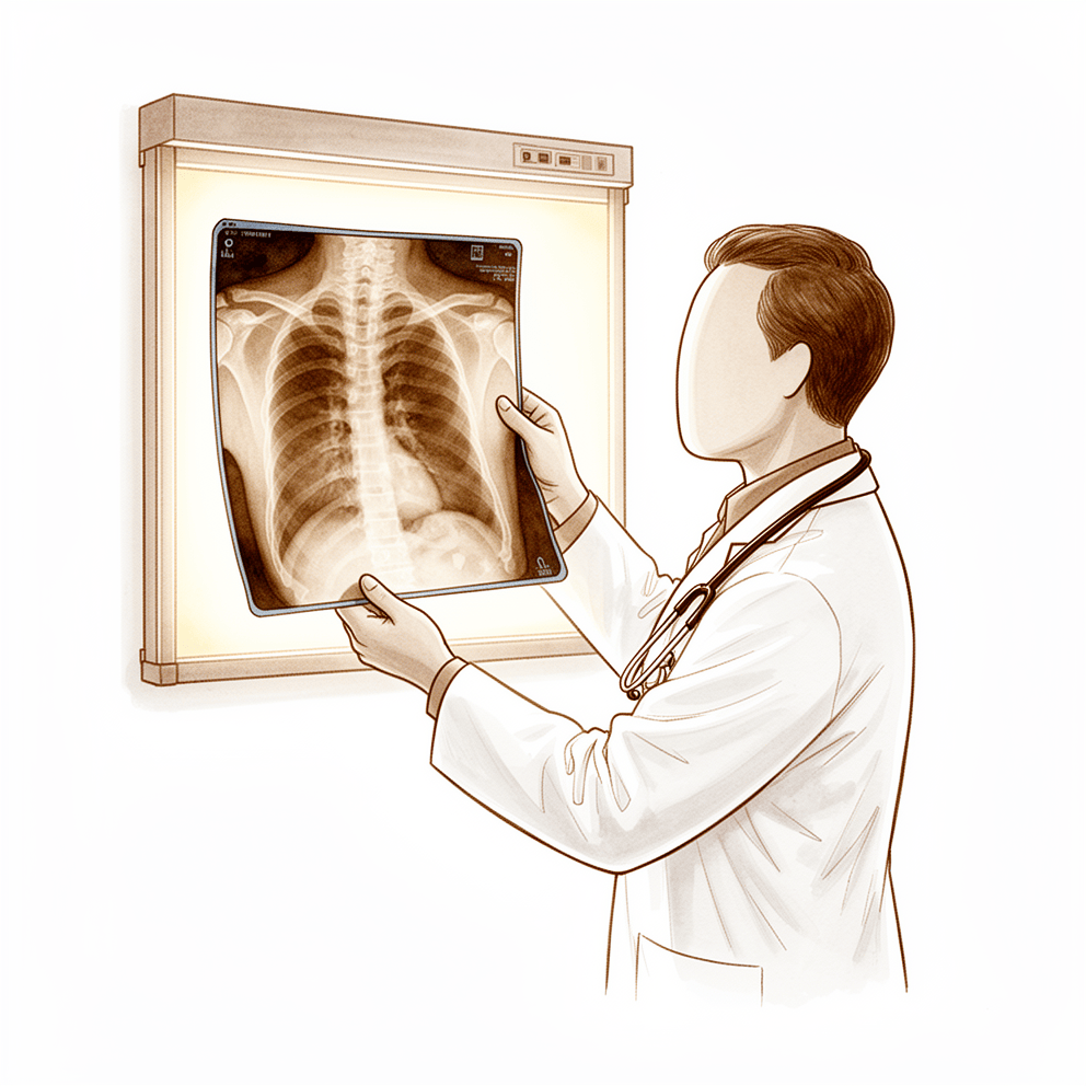
Preoperative imaging is central to the management of shoulder and spinal pathologies, yet the majority of pre-evaluation MRI scans in patients with chronic shoulder pain have no impact on clinical outcomes [1]. While advanced techniques such as CT and three-dimensional reconstructions augment plain radiographs to guide treatment decisions [32], clinicians must understand the specific indications, advantages, and disadvantages of radiographs, CT, MRI, and other modalities to safely manage spinal disorders [10]. For proximal humeral fractures, CT diagnostics allow a significantly better assessment of relevant structures than conventional diagnostics [4], and multiple radiographic views are needed to evaluate greater tuberosity displacement appropriately [53]. Optimum patient care for these fractures might require the development of new imaging modalities rather than new classification systems [24].
In glenohumeral pathology, 3-D CT is the most reliable imaging modality for quantifying glenoid bone loss, with regular CT serving as the second most reliable and reproducible option [36]. For glenohumeral osteoarthritis, the combined use of CT and MRI is recommended for preoperative planning because CT underestimates rotator cuff pathology [23]. MRI provides CT-equivalent measurements of glenoid retroversion, concavity, and BSSR after anterior shoulder dislocation, supporting its use as a viable modality for assessing key bony stability parameters and potentially reducing the need for supplemental CT [30]. However, MRI is neither accurate nor specific enough to assist in the selection of operative candidates or to choose the appropriate surgical intervention for shoulder pathology [127]. The glenoid track concept is encouraged as a routine part of preoperative evaluation for arthroscopic anterior stabilization [57], and the SHART test is suggested for addition to conventional preoperative imaging [31].
For sternoclavicular joint injuries, computed tomography is the imaging modality of choice and the gold standard for evaluating posterior dislocations [2, 16]. Postoperatively, ongoing assessment using X-rays, CT scans, or other diagnostic tests is recommended for patients experiencing pain or limited range of motion following primary anatomic and reverse total shoulder arthroplasty [3]. Routine immediate postoperative radiographs rarely identify complications (0.2%) [8], and routine PACU radiographs without specific indication may result in poor-quality images [22]. In asymptomatic pediatric patients following scoliosis surgery, routine radiographs provide low utility in guiding treatment [19]. Further clinical studies are required to establish the use of three-dimensional fluoroscopy to determine intra-articular screw penetration in proximal humeral fractures [142].
Anatomy & Pathophysiology¶
Bony Anatomy¶
The proximal humerus comprises four main parts: the humeral head, greater tuberosity, lesser tuberosity, and humeral shaft [75]. The articular head is spherical with a diameter of 37 to 57 mm [75], and its radius of curvature is approximately 25 mm, slightly larger in men than in women [87]. The articular surface is essentially spherical, with an arc of approximately 160 degrees covered by articular cartilage [87]. The most superior portion of the articular surface averages 8 mm above the greater tuberosity [75], and the superior margin is normally superior to the top of the greater tuberosity by 8 to 10 mm [87]. The head height is approximately 5.6 cm above the superior border of the pectoralis major tendon [90].
Humeral version averages 29.8 degrees, with a range of 10 to 55 degrees [75]. Proximal humeral retroversion is highly variable, ranging from 0 to 55 degrees depending on the method used for measurement [87]. The humeral head is retroverted an average of 30 degrees relative to the transepicondylar axis of the humerus [90], and averages 19 degrees of retroversion [78]. Mean humeral retroversion is around 26 degrees in healthy adults [82]. The humeral head is inclined approximately 130 degrees with respect to the humeral shaft [75], and averages 41 degrees of inclination [78]. The neck-shaft angle measures an average of 135 degrees [76]. The average neck-shaft angle is 45 degrees, with a range of 30 to 50 degrees [87]. Arthritic shoulders have a flatter neck-shaft angle close to 50 degrees [87].
The anatomic neck of the proximal humerus is located at the junction of the articular surface and the tuberosities [75]. It is located directly below the humeral head and serves as an attachment for the shoulder capsule [90]. The surgical neck represents an indistinct region below the tuberosities but above the humeral shaft [75]. The surgical neck is more distal than the anatomic neck and is more often involved in fractures [90]. The greater tuberosity serves as the attachment site for the supraspinatus, infraspinatus, and teres minor tendons [75]. The lesser tuberosity serves as the attachment site for the subscapularis tendon [75].
The glenoid is a convex structure of shallow depth shaped like an inverted pear [75]. The glenoid cavity is a shallow socket, approximately one third the size of the humeral head [76]. The subchondral bone of the glenoid is relatively flat, with the articular concavity augmented by cartilage and a circumferential labrum [78]. The glenoid diameter ranges from 18 to 30 mm superior anteroposteriorly and 21 to 35 mm inferior anteroposteriorly [87]. The superoinferior height of the glenoid ranges from 30 to 48 mm [87]. The glenoid inclination averages 4.2 degrees, with a range of -7 to 20 degrees [87]. The glenoid version averages 1.5 degrees of retroversion, with a range of 10.5 degrees anteversion to 9.5 degrees retroversion [87]. The normal position of the glenoid surface in relation to the axis of the scapular body ranges from 2 degrees of anteversion to 7 degrees of retroversion [87]. The glenoid is retroverted approximately 5 degrees relative to the scapular body [90]. The glenoid averages 5 degrees of retroversion in relation to the axis of the scapular body [78].
The glenoid articular surface radius of curvature is 2 to 3 mm larger than that of the humeral head [87]. The glenoid radius of curvature ranges from 22 to 28 mm [87]. The humeral head radius of curvature ranges from 23 to 28 mm [87]. The glenoid surface area ranges from 4 to 6 mm [87]. The humeral head surface area ranges from 11 to 19 mm [87]. The glenoid cartilage thickness is 2.16 mm [87]. The humeral head cartilage thickness is 1.44 mm [87].
The distance from the lateral base of the coracoid process to the lateral margin of the greater tuberosity is called the lateral humeral offset [87]. A significant decrease in lateral humeral offset reduces the lever arms for the deltoid and supraspinatus muscles, weakening abduction [87]. A significant increase in lateral humeral offset causes excessive tension on soft tissues, resulting in loss of motion and likely accelerating polyethylene wear [87]. Humeral articular malposition of more than 4 mm led to increased subacromial contact in a biomechanical cadaver study [87]. An offset of 8 mm in any direction significantly decreased passive range of motion in a biomechanical cadaver study [87]. The medial humeral offset ranges from 4 to 14 mm [87]. The posterior humeral offset ranges from -2 to 10 mm [87].
The scapula is triangular when viewed anteroposteriorly, with its base situated superiorly and its apex inferiorly [77]. The scapula spans the second through seventh ribs and serves as an attachment for 17 muscles [90]. The scapula is anteverted on the chest wall approximately 30 degrees relative to the body [90]. The glenoid is connected with the flat body of the scapula by the scapular neck [77]. The coracoid process curves forwards from the superior surface of the scapular neck [77]. The scapular spine ends in a flattened bony process, the acromion, which curves forwards [77]. The highest concentration of bony mass in the scapula is located in the glenoid, the scapular neck, and the lateral border of the scapular body [77]. Two bony pillars extend between the glenoid and the scapular body to transmit compressive forces from the glenoid fossa [77]. The lateral pillar connects the inferior border of the glenoid with the inferior angle [77]. The spinal pillar arises from the central part of the glenoid and continues medially to become part of the base of the scapular spine [77]. The weakest bone in the scapula is located primarily in the central part of the biomechanical body, specifically in the infraspinous fossa [77]. The weakest area of the circumference of the biomechanical body of the scapula is the spinomedial angle [77].
The acromion, coracoacromial ligament, and coracoid process form the coracoacromial arch, a rigid bony-ligamentous structure that imparts stability to the shoulder girdle [75]. The acromion is a massive structure overlying the humeral head in humans, reflecting the increasing role of the deltoid muscle in shoulder function [85]. The coracoid process has undergone an increase in size over time [85]. With the shoulder in 90 degrees of abduction, the coracoid extension over the glenohumeral joint can mechanically limit anterior translation of the humerus relative to the glenoid [85].
The clavicle is the first bone to ossify at the fifth week of gestation [78]. The clavicle is the only long bone to ossify by intramembranous ossification [78]. The medial epiphysis of the clavicle is the last ossification center to fuse, occurring at age 20 to 25 years [78]. The primary blood supply to the clavicle is periosteal, with no nutrient artery present [78]. The scapula has only one true diarthrodial articulation, the acromioclavicular joint [78]. Normal shoulder motion is approximately two-thirds glenohumeral and one-third scapulothoracic [78].
Ossification of the scapular body begins at the eighth week of gestation [78]. The acromion has three ossification centers: the metacromion, mesoacromion, and preacromion [78]. Failure of fusion of the acromial ossification centers results in os acromiale [78]. Os acromiale is incomplete fusion of secondary ossification centers, most commonly between the mesoacromion and meta-acromion [90].
The proximal humerus has three centers of ossification: the humeral head, greater tuberosity, and lesser tuberosity [78]. The formation of the humerus begins with the appearance of the cartilage anlage, which is present by the fifth week of gestation [82]. The primary ossification center for the humerus appears at about the sixth week of gestation [82]. By the time of birth, the entire humeral diaphysis is completely ossified [82]. The proximal humerus is primarily cartilaginous at birth [82]. Ossification centers for the proximal humerus can be detected with ultrasonography as early as the 38th week of gestation [82]. The ossification center for the humeral head is usually present at birth [82]. The greater tuberosity ossification center appears by 1 to 3 years of age [82]. The lesser tuberosity ossification center appears by 5 years of age [82]. Proximal humeral ossification centers fuse by 5 to 7 years of age to form the humeral head [82]. The proximal humeral physis closes by 14 to 17 years of age in girls and by 16 to 18 years in boys [82].
Humeral retroversion averages 65 degrees in infants and young children [82]. Humeral retroversion gradually decreases, approaching adult values by 11 years of age [82]. Eighty percent of subsequent humeral growth comes from the proximal humeral physis [82]. The proximal humeral physis accounts for approximately 40% of the growth of the entire upper extremity [82]. Less than 75% of growth from the proximal humerus occurs before 2 years of age [82]. More than 85% of growth from the proximal humerus occurs by 8 years of age [82]. The posteromedial metaphysis, a portion of the physis, and the epiphysis are intracapsular [82]. A large part of the proximal humeral physis is extracapsular, making it susceptible to traumatic injury [82]. The proximal humeral physis is irregularly shaped, with its apex located on the posteromedial portion of the proximal humerus [82]. The periosteum is thicker and stronger in the posteromedial portion of the proximal humerus than in the anterolateral portion [82].
The scapula has shifted caudally from the cervical position in lower animals, freeing the shoulder from the head and neck [85]. The scapular index is extremely high in pronograde animals with a long, narrow scapula [85]. The scapula is broader in humans and other primates, with the most pronounced differences observed in the infraspinatus fossa [85]. Broadening of the infraspinatus fossa has resulted in a change in the vector of muscle pull from the axillary border of the scapula to the glenoid fossa [85].
Soft Tissue Anatomy¶
The rotator cuff is a sheet of conjoined tendons closely applied over the shoulder capsule and inserting mainly into the greater tuberosity of the humerus [83]. The subscapularis is inserted into the lesser tuberosity [83]. The rotator cuff is made up of subscapularis in front, supraspinatus above, and infraspinatus and teres minor behind [83]. The rotator cuff stabilizes the head of the humerus by pulling it firmly into the glenoid whenever the deltoid lifts the arm forwards or sideways [83].
The coracoacromial arch is formed by the acromion process posterosuperiorly, the coracoid process anteriorly, and the coracoacromial ligament joining them [83]. The subacromial bursa separates the tendons from the coracoacromial arch and allows them to glide [83].
The subscapular bursa lies between the subscapularis tendon and the neck of the scapula [79]. The subscapular bursa communicates with the joint cavity between the superior and middle glenohumeral ligaments [79]. The subscapular bursa protects the tendon of the subscapularis at the point where it passes under the base of the coracoid process and over the neck of the scapula [79]. The subscapular bursa is linked to the coracoid process by a suspensory ligament [79]. In 28% of specimens dissected by Colas and colleagues, the subscapular bursae merged with the subcoracoid bursae, forming a unique wide bursa [79]. The subscapular bursa often houses loose bodies in the shoulder [79]. The subscapular bursa is a region in which synovitis of the shoulder may be most intense [79].
A soft tissue sheath consistently covers the long head of the biceps tendon to the level of the proximal margin of the pectoralis major tendon [79]. The fibro-osseous bicipital tunnel consists of three distinct anatomic zones [79]. Zone 1 of the bicipital tunnel represents the traditional bony bicipital groove beginning at the articular margin and ending at the distal margin of the subscapularis tendon [79]. Zone 2 of the bicipital tunnel extends from the distal margin of the subscapularis tendon to the proximal margin of the pectoralis major tendon and is not viewable from arthroscopy above or from subpectoral exposure below [79]. Zone 3 of the bicipital tunnel is distal to the proximal margin of the pectoralis major tendon and represents the subpectoral region [79]. The transverse humeral ligament is an important stabilizer of the biceps tendon [90].
The rotator interval is defined medially by the base of the coracoid, superiorly by the supraspinatus tendon, and inferiorly by the subscapularis tendon [78]. The rotator interval contains the coracohumeral ligament, the superior glenohumeral ligament, and the intra-articular portion of the long head of the biceps tendon [78]. Laxity of the rotator interval results in inferior laxity, known as the sulcus sign [78]. Contracture of the rotator interval is seen with adhesive capsulitis [78].
The coracohumeral ligament restricts external rotation in adduction [78]. The coracohumeral ligament is a static restraint to inferior and posterior translation in adduction and external rotation [78]. The superior glenohumeral ligament is a primary static restraint against anterior translation with the arm at the side [78]. With the coracohumeral ligament, the superior glenohumeral ligament forms a pulley that provides restraint against medial subluxation of the long head of the biceps tendon [78]. The middle glenohumeral ligament is a primary static restraint against anterior translation with the arm in external rotation and 45 degrees of abduction [78]. The anterior band of the inferior glenohumeral ligament is a primary static restraint against anterior-inferior dislocation of the glenohumeral joint in 90 degrees of abduction and external rotation [78]. The posterior band of the inferior glenohumeral ligament is a primary static restraint against posterior-inferior translation in internal rotation and adduction [78].
The superior transverse scapular ligament arises from the medial base of the coracoid overlying the suprascapular notch [78]. The suprascapular artery runs superior to the superior transverse scapular ligament [78].
Classification¶
General Imaging & Diagnostic Utility¶
Imaging findings must be interpreted with caution, as the majority of pre-evaluation MRI scans for chronic shoulder pain had no impact on the outcome [1]. High-field-strength MRI systems typically provide superior image quality compared to low-field-strength open systems [11]. Imaging abnormalities of the acromioclavicular joint and subacromial space are common in asymptomatic shoulders [21]. For proximal humeral fractures, optimum patient care might require the development of new imaging modalities rather than new classification systems [24]. Consensus was reached for five elements in the x-ray report, twenty in the MRA report, nine in the CT report, and two elements regarding MRA views and settings for anterior shoulder instability [51]. The 'long axis' measurement for acute scaphoid fractures has clear potential benefits over traditional classification systems [55]. Artificial intelligence-driven classification of proximal humerus fractures is insufficient for clinical implementation despite rigorous training based on CT imaging [134].
Rotator Cuff & Muscle Fatty Infiltration¶
Goutallier: This classification grades supraspinatus fatty infiltration on a five-point scale from grade 0 (no fat) to grade 4 (more than 50% fat) [227]. Clinical scores using a single image slice do not represent 3-dimensional muscle measurements for fatty infiltration and atrophy [49]. The correlation between the semi-quantitative MRI-based Goutallier Classification system and MR spectroscopic fat measurement is weak [56]. The intraobserver and interobserver reliability of the Goutallier classification using MRI was evaluated in a Level II study [39]. The horizontal CT image plane produced the most reliable results for evaluating supraspinatus fatty degeneration using the Goutallier classification [220]. The 3-grade grading system of the Goutallier classification demonstrated significantly better agreement than the 5-tiered system for both intraobserver and interobserver reliability [220].
Glenoid Morphology & Version¶
Walch: Intraobserver agreement using the Walch classification based on axillary radiographs was substantial and compared favorably with agreement based on CT scans [155]. The modified Walch classification represents an improvement over the original classification, but automated computer-based analysis of CT scans may be needed to further improve its value [43]. MRI is comparable to CT for the determination of glenoid version [212]. However, MRI does not accurately distinguish between Walch B2 and C classifications of glenoid morphology [212]. A new classification of glenoid bone loss for revision arthroplasty has good intra- and inter-observer reliability and good correlation between plain radiographs and CT images [123]. The established classifications by Euler and Ideberg for glenoid fractures are not capable of providing similar reliability [161].
Acromial Morphology¶
Bigliani: Intraobserver reproducability and interobserver reliability using MRI and RSOV for the Bigliani classification system are low [149]. Classification of acromial morphology on MRI is not equal to the classification on RSOV [149]. The acromial morphology classification system is an unreliable method to assess the acromion [131]. The acromial index shows no association with the presence of rotator cuff disease [131]. A system that incorporates more objective classification criteria and acknowledges the continuous nature of acromial morphologic types may improve interobserver reliability [121]. A new multi-planar classification system for acromion morphology is simple and correlates well with impingement syndrome [141].
Instability & Labral Lesions¶
Kim: The sensitivity, specificity, and reliability of magnetic resonance arthrogram for detecting Kim's lesion are substantial [60]. The interobserver reliability of Kim's classification is fair [60]. SLAP: Classification of superior labrum anterior to posterior (SLAP) lesions using MRa resulted in significant disagreement between and within raters [101]. Bankart: The percentage of grade 2 MR findings for Bankart lesions was statistically different between the normal group (6.7%) and the arthroscopic group (69.7%) [151]. FEDS: There are 16 categories within the FEDS classification for glenohumeral instability that are clinically significant [146]. Hill–Sachs: With the exception of the Hill–Sachs interval, CT and MRI show no significant differences in the diagnostic value of the HSL measurement regardless of the measurement technique [157]. All measurement techniques for HSL, with the exception of the Franceschi and Calandra classifications, provided good to very good intra- and inter-rater reliabilities with both CT and MRI [157]. A systematic review found high variability in imaging modalities and outcomes for Hill-Sachs lesions, making formal statistical analysis or meta-analysis impossible [45].
Fractures & Trauma¶
Boileau: CT improves the diagnostic accuracy but not the interobserver reliability of the Boileau classification of proximal humerus fracture sequelae [58]. The Boileau classification had poor interobserver reliability even when using a three-dimensional evaluation of the deformity [58]. LSSS: The proposed classification system for lateral scapula suspension system (LSSS) failure demonstrated satisfactory agreement and accuracy among experienced surgeons [148]. Complete disruptions (S2) in the LSSS classification are relatively common and associated with increased risk of neurovascular injury [148]. AO/OTA: Overall interobserver agreement within levels of training when evaluating the AO/OTA classification for proximal humeral fractures was fair to substantial (0.281 to 0.713) [222]. The highest levels of agreement among attending surgeons and senior residents for AO/OTA classification was with 2-D CT imaging [222]. Junior residents had the highest agreement when 3-D CT imaging was used for AO/OTA classification [222]. Elbow Osteoarthritis: The CT-based staging system for primary elbow osteoarthritis showed almost perfect interobserver and intraobserver agreement [95]. The CT-based staging system for primary elbow osteoarthritis showed high correlation with visual analogue scale scores compared to plain radiograph-based classifications [95].
Other Anatomical Classifications¶
Elbow Tendons & Ligaments: A novel classification system was developed to characterize MRI findings of tendons, ligaments, cartilage, nerves, muscles, and bones in asymptomatic elbows [46]. In the classification of asymptomatic elbow tendons, Class 1 is defined as signal intensity similar to cortical bone on PD and T2FS images [46]. Class 2 is defined as regions of signal similar to muscle on T2FS images [46]. Class 3 is defined as signal greater than muscle on T2FS images with sharply defined margins [46]. Class 4 is defined as signal greater than muscle on T2FS images with ill-defined margins [46]. In the classification of asymptomatic elbow ligaments, Class 1 is defined as signal intensity similar to cortical bone on PD and T2FS images [46]. Class 4 is defined as ligament interruption [46]. Larsen: Larsen’s classification system was used to classify glenohumeral joint destruction in rheumatoid arthritis into 6 grades (grades 0-5) [217]. The severity of greater tuberosity destruction in rheumatoid arthritis was classified into 3 grades based on depth: mild (<5 mm), moderate (5-10 mm), and severe (≥10 mm) [217]. MCL: A magnetic resonance imaging-based classification system for tear location of superficial medial collateral ligament tears was validated in a cohort of 239 patients [52]. Dejour: The Dejour classification of trochlear dysplasia shows inconsistent repeatability due to the variability of imaging modalities [216].
Clinical Presentation¶
History and Physical Examination¶
The clinical evaluation of the painful shoulder by an experienced and knowledgeable health care professional remains the most sensitive and clinically useful tool for the assessment of most patients with rotator cuff disease, glenohumeral instability, glenohumeral arthritis, and adhesive capsulitis [14]. A complete thorough history and physical examination and review of plain radiographs by an experienced examiner are sufficient to establish the diagnosis and treatment plan for most common shoulder disorders [15]. These comprehensive interactions are the initial steps set on delivering the correct diagnosis and subsequent treatment recommendations to patients [17].
The history should include the symptom location with or without associated radiation, severity, onset, duration, quality and character, aggravating or alleviating factors, and previous related treatments [17]. In the acutely injured patient, specific attention should be given to the mechanism of injury and associated injuries [17]. Clinicians must recognize that cervical spine symptoms can often mimic shoulder problems, brachial plexopathies, and peripheral compressive neuropathies [17]. Similarly, lumbar spine symptoms are often very similar to those originating from the hip, sacroiliac joint, or even vascular insufficiencies [17]. Patients with myelopathy may present with vague symptoms that could be mistaken for other systemic or neurologic disorders [17].
Physical examination findings can be definitive for specific pathologies. A precise diagnosis based on the physical examination alone is possible for supraspinatus insufficiency [109]. For acromioclavicular arthritis, surgical decision-making should rely on focused history and clinical examination rather than MRI scans [29].
Imaging Modalities and Indications¶
Imaging plays an important role in the evaluation of musculoskeletal symptoms, from evaluation of traumatic injury to tumor characterization to treatment follow-up [5]. An understanding of the available imaging modalities and indications for each is critical to ensure that the correct study is performed and the clinical question is answered [5]. Multiple image-guided interventions are available to aid in diagnosis and therapy [5].
Modality-Specific Indications: * Post-Arthroplasty Monitoring: For patients experiencing pain or limited range of motion following total shoulder arthroplasty, ongoing assessment using additional X-rays, computed tomography scans, or other diagnostic tests is recommended for effective monitoring [3]. * Pediatric Spine: Magnetic resonance is an excellent modality for imaging pathologic processes in the pediatric spine, allowing high-resolution views of osseous and soft-tissue structures [48]. * Glenohumeral Instability: The West Point view is a good screening tool for anteroinferior bony Bankart lesions, while CT should be added when the view is equivocal or difficult to obtain [116]. * Biceps Pathology: Ultrasound is more reliable than clinical tests to both confirm and rule out pathologies of the long head of the biceps [91].
Diagnostic Accuracy and Clinical Impact¶
MR Imaging is responsible for major changes in diagnosis or treatment in approximately 10% of cases and minor changes in approximately 20% of cases [12, 13]. In contrast, plain radiographs rarely alter the diagnosis or affect management in the setting of atraumatic shoulder pain, particularly in patients younger than 50 years [25]. The sensitivity and specificity of MRI evaluation for the inferior glenohumeral ligament were 94% and 82%, respectively [27].
When comparing diagnostic tools, needle diagnostic arthroscopy had better ability to rule in a diagnosis but slightly worse ability to rule out a diagnosis compared with MRI [93]. Clinicians should always correlate the findings of MRI with the patients' medical history and clinical presentation in clinical decision making [9]. The clinical utility of the lateral scapular view may need to be reassessed in the setting of nontraumatic shoulder conditions [108].
Asymptomatic Findings and Interpretation¶
Clinicians should be aware of the common anatomic findings on MRI when considering diagnostic and treatment planning for unilateral shoulder pain [20]. The prevalence of osteoarthritis of the sternoclavicular joint on computed tomography should be taken into consideration when using a CT scan to assess a patient with symptomatic SCJ pathology [28]. MRI scans are not a reliable tool for predicting symptomatic acromioclavicular arthritis [29].
Findings of asymptomatic thoracic spine MRI represent roentgenographic abnormalities only, and any clinical decisions concerning the treatment of pain in the thoracic spine usually require additional studies [122]. Clinical scores using a single image slice do not represent 3-dimensional muscle measurements [49]. The lesion of the superior glenoid labrum is predominantly seen in males with nonspecific clinical and radiographic findings [129].
Investigations¶
Imaging is integral to evaluating musculoskeletal symptoms, ranging from traumatic injury to tumor characterization and treatment follow-up [5]. Understanding available modalities and their specific indications ensures the correct study is performed to answer the clinical question [5]. The primary goals of shoulder imaging are to establish a diagnosis, determine the severity of pathoanatomy, assist in surgical planning, and illustrate the condition to the patient [41]. Because the shoulder is a three-dimensional structure that cannot be represented by a single planar view, critical relationships such as humeral head centering change with arm position [114]. Pathology may involve numerous bones and soft tissues, and overlying structures or metallic implants can complicate visualization [114]. Surgeons must adopt a judicious approach to imaging, obtaining only the scans necessary for patient care and resisting the temptation to "overimage" unless a specific research protocol dictates otherwise [41, 114]. Imaging techniques facilitate a better understanding of conditions affecting the shoulder [26].
Plain radiography: Conventional radiographs are appropriate for patients presenting with shoulder pain accompanied by a history of trauma, dislocation, night pain, or chronic shoulder pain [130]. Initially, patients are usually asked to have AP and lateral plain radiographs related to their chief complaint [126]. Plain radiographs are often the only required studies for assessing acute shoulder trauma, including fractures or dislocations [126]. They can also reveal arthritis, calcific tendinitis, and osteolysis of the distal clavicle [126]. At least two views should be obtained: an anteroposterior (AP) view in the plane of the glenoid and an axillary projection with the arm in abduction to show the relationship of the humeral head to the glenoid [102]. The standard shoulder series includes orthogonal views: a true AP view in the scapular plane, an AP view, an axillary view, and a scapular Y view [130].
The true AP view in the scapular plane visualizes the anterior greater tuberosity in profile and can reveal proximal humeral migration when the arm is held in neutral rotation with slight abduction [130]. The AP view with the arm in internal rotation visualizes the posterior aspect of the greater tuberosity and the lesser tuberosity in profile [130]. The axillary view is necessary for evaluating glenohumeral joint instability and determining humeral head position within the glenoid fossa [130]. It may detect occult, locked posterior shoulder dislocation in patients exhibiting a lack of passive external rotation [130]. Additionally, the axillary view helps evaluate glenoid morphology in glenohumeral osteoarthritis and provides good visualization of the coracoid process, acromion, and distal clavicle [130]. The scapular Y view visualizes the coracoacromial arch and can reveal coracoacromial spurs associated with rotator cuff pathology [130]. It serves as a reliable alternative for evaluating glenohumeral subluxation and dislocation [130]. The scapular Y view can also show scapular body abnormalities such as osteochondroma or fracture, as well as acromial shape [130].
Normal anatomical landmarks on radiographs include an acromiohumeral distance of 7 to 14 mm [130]. The width of the glenohumeral joint space should be symmetric superiorly and inferiorly [130]. The normal coracoclavicular distance is 1.1 to 1.3 cm [130]. Neer classified acromial morphology as type I (flat), type II (curved), and type III (hooked) [130]. Type III acromial morphology correlates with the presence of rotator cuff disease, although no direct causal relationship has been demonstrated [130]. The Neer classification has shown relatively poor interobserver reliability [130].
For shoulder arthroplasty planning, standardized plain films are almost always sufficient to garner the necessary information [41]. Properly taken plain films provide information that cannot be obtained from CT scans [41]. Proper radiographic technique is as important as proper surgical technique to achieve the desired outcome [41]. The first key view is the AP view in the plane of the scapula, taken so that the x-ray beam passes through the glenohumeral joint [41]. This view shows the superoinferior position of the humeral head relative to the glenoid, presence of osteophytes, narrowing of the joint space, degree of medial displacement of the humerus, quality of bone, presence of loose bodies, and humeral head collapse or deformity [41]. The second key view is the axillary view, taken with the arm in the functional position of elevation in the plane of the scapula [41]. This view is oriented so that both the spinoglenoid notch and the scapular neck are visible [41]. It shows a different perspective of humeral anatomy, amount of glenoid bone, shape of the glenoid, its version in relation to the plane of the scapula, and the relationship of the humeral head to the glenoid fossa [41]. The axillary view is referred to as the "truth view" because it demonstrates glenohumeral relationships in the functional position of elevation [41]. CT scans have the disadvantage of being taken with the arm in the adducted position, unlike the axillary truth view [41]. Many "axillary views" sent for consultation are taken without standardization, making it impossible to determine important features of the glenohumeral joint [41].
When taken properly, standardized anteroposterior and axillary views indicate the thickness of the cartilage space between the humerus and the glenoid, relative positions of the humeral head and glenoid, presence of osteophytes, degree of osteopenia, and extent of bony deformity and erosion [41]. Joint space narrowing is most evident on the axillary truth view as opposed to images made with the arm at the side [41]. The axillary truth view can show posterior subluxation or "functional decentering" that is not evident in images taken with the arm at the side [41]. The degree of posterior subluxation can be measured as the position of the center of the humeral head in relation to the plane of the scapula, the position of the center of the humeral head in relation to the glenoid face, or the point of contact of the humeral articular surface on the glenoid articular surface [41]. The point of contact reflects the degree of centering of the net humeral joint reaction force on the glenoid [41]. Malcentering of the joint reaction force leads to posterior instability, posterior glenoid wear, and "rocking horse" loosening of prosthetic glenoid components [41].
For proximal humerus fractures, the recommended series is the Neer trauma series, consisting of an AP view, a lateral view in the scapular plane, and a Velpeau modified axillary view [113]. The lateral radiograph in the scapular plane is the tangential Y-view of the scapula [113]. The combination of these three views allows evaluation of the shoulder joint in three separate perpendicular planes [113]. The axillary view is important for evaluating the glenoid articular surface and the relationship of the humeral head anteriorly and posteriorly [113]. The critical shoulder angle (CSA) on plain radiographs has good diagnostic performance for rotator cuff tears [33]. Radiographs provide better reproducibility and accuracy for critical shoulder angle measurement compared to MRI [112]. The inter-rater agreement between x-ray images and consensus MRI for Walch classification is fair-to-moderate, which is lower than previously reported reliability using CT scans [133].
Special Radiographic Views: The Serendipity view is indicated for the sternoclavicular joint and is taken with the patient supine using a 40° cephalic tilt view centered on the sternum [130]. The West Point view is indicated for anterior glenoid bone loss and is taken with the patient prone, involved shoulder raised above table level, centered on axilla with beam directed 25° downward and 25° medial [130]. The Zanca view is indicated for the AC joint and is taken as an AP with 10° cephalic tilt centered over the AC joint with the patient supine, using one-half the voltage of a routine shoulder AP view [130]. The Stryker notch view is indicated to evaluate Hill-Sachs lesion after dislocation and is taken with the patient supine, affected arm placed on top of head with fingers toward back of head, beam centered over coracoid process with 10° cephalic tilt [130]. The Apical oblique view is indicated to evaluate for glenoid rim fracture in instability and is taken with the patient seated, cassette placed posterior and parallel to the spine of the scapula, beam directed 45° to the plane of the thorax and 45° caudally [130]. The SHART test (shoulder hyperabduction radiological test) is suggested to be added to conventional preoperative imaging techniques [31].
CT: CT is the imaging modality of choice for evaluation of sternoclavicular joint injuries [2]. Timely recognition and treatment of these injuries are essential to prevent devastating consequences [2]. CT diagnostics allowed a significantly better assessment of relevant structures than conventional diagnostics in proximal humeral fractures [4]. CT imaging is frequently used to evaluate fractures of the shoulder, to assess for bony lesions in recurrent instability cases, or for preoperative templating for shoulder arthritis [126]. CT with three-dimensional reconstructions is the advanced imaging study of choice for determining the extent of glenoid bone loss in the setting of shoulder instability [130]. Computed tomography is a useful imaging modality for evaluating osseous Hill-Sachs lesions [160]. CT is helpful for planning fracture surgery and shoulder joint replacement [102]. On occasion, CT scanning may be necessary for detailing bony anatomy in proximal humerus fractures [113]. Significant differences in bone loss measurement between imaging modality, measurement method, and observers may lead to differences in treatment in up to 34% of cases [18]. CT underestimates rotator cuff pathology in patients with glenohumeral osteoarthritis [23]. The authors recommend the combined use of CT and MRI for preoperative planning in patients with glenohumeral osteoarthritis [23]. Measurements of glenoid defects can be done with 3D CT scans as well as MRI [138]. Differences between 3D CT and 3D MRI for assessing Latarjet procedures were small and likely not clinically significant [139]. This should be taken into consideration when using a CT scan to assess a patient with symptomatic sternoclavicular joint pathology [28].
MRI: MRI is useful to identify osteonecrosis of the humeral head, or a bone tumour [102]. MRI can identify labral tears and rotator cuff tears, although the accuracy for these is enhanced by combining the scan with arthrography [102]. MRI is the modality of choice for evaluating the rotator cuff, biceps, and subacromial/subdeltoid bursa [126]. T1-weighted MRI can reveal Hill-Sachs lesions and is often used with magnetic resonance arthrograms to provide a more detailed picture of the joint surfaces [126]. T2-weighted MRI provides better visualization of full thickness rotator cuff tears [126]. The quality of MRI images and their usefulness are markedly affected by the type of equipment employed, with high-field-strength systems typically providing superior image quality compared to low-field-strength open systems [11]. Based on data regarding glenoid morphology, version, and humeral head subluxation, a treating clinician may choose to utilize either an MRI or a CT to assess these parameters [6]. The diagnostic accuracy of MRI decreases when chronic pectoralis major tears are evaluated [169]. When using MRI for humeral head positioning, it is better to use HGA, which is a more reliable measurement value based on the comparison with CT measurements [174]. Although the reliability and diagnostic accuracy of advanced metal artifact reduction MRI for periprosthetic shoulder infection were high, these conclusions are based on a specific advanced MARS-MRI protocol interpreted by experienced musculoskeletal radiologists [159].
Traditional magnetic resonance imaging (MRI) is a diagnostic tool to complement both physical examination and standard radiographs in the management of patients with anterior shoulder instability [117]. MRI is utilized for evaluation of soft tissues, which can be performed with high contrast and spatial resolution [117]. MR accuracy in identifying labral and rotator cuff tears in the literature ranges from 70% to 100% [117]. The acquired multi-planar imaging allows for the detailed evaluation of the glenoid, labrum, joint capsule, and rotator cuff in different planes [117]. In a meta-analysis of the diagnostic test accuracy of MRA compared to MRI for the detection of glenoid labral injuries, MRA had greater diagnostic test accuracy than MRI (MRA sensitivity 88% and specificity 93% vs. MRI sensitivity 76% and specificity 87%) [117]. With standard MRI or MRA, the shoulder is routinely positioned in neutral or partial external rotation, but other alternative positions can be used to increase the sensitivity for detecting labroligamentous injuries [117]. Abduction and external rotation (ABER) of the arm is an alternative position utilized to increase the sensitivity and specificity for detecting anteroinferior labroligamentous injury [117]. Limited ROM or pain may prohibit patients from performing the ABER provocative maneuver [117]. Schreinemachers et al. found that full routine MRI or MRA examination had similar accuracy as the ABER sequence in evaluating the anteroinferior labral–ligamentous complex [117]. Tian et al. found that the sensitivity of MRA with the ABER position for detecting anteroinferior labral lesions was significantly higher than that of the MRA in neutral position and more effective in
Other Considerations: Routine immediate postoperative radiographs rarely identified postoperative complications in 0.2% of cases [8]. Routine PACU radiographs, in the absence of a specific indication, may result in poor-quality images [22]. For patients experiencing pain or limited range of motion after shoulder arthroplasty, ongoing assessment using additional X-rays, computed tomography scans, or other diagnostic tests is recommended for effective monitoring [3]. Advanced imaging techniques, including CT and three-dimensional reconstructions, augment the information obtained from plain radiographs to guide treatment decisions for acetabular fractures [32].
Treatment¶
General Principles¶
Image quality and clinical utility are markedly influenced by equipment type, with high-field-strength systems typically providing superior image quality compared to low-field-strength open systems [11]. Orthopaedic surgeons should develop a systematic approach to interpreting the entire image and create a discrete plan with radiologists on managing incidental findings [195].
Radiography¶
Routine immediate postoperative radiographs rarely identify postoperative complications, with a rate of 0.2% [8]. The lack of clinically meaningful change from routine radiography does not justify the per-patient expense, as almost all postoperative radiographs were read as normal and did not alter postoperative management [65]. Similarly, the majority of pre-evaluation MRI scans had no impact on the outcome [1]. For patients experiencing pain or limited range of motion, ongoing assessment using additional X-rays, computed tomography scans, or other diagnostic tests is recommended for effective monitoring [3].
Diagnostic Performance: Cortical Shoulder Angle (CSA) on plain radiographs has good diagnostic performance for rotator cuff tears (RCTs) [33]. In contrast, on nonstandard AP films, the diagnostic efficacy of artificial intelligence (AI) was better than that of the CSA [156]. The authors suggest adding the SHART test to conventional preoperative imaging techniques [31].
Measurement Validity: AHDs measured on radiograph and MRI should not be used interchangeably in early Hamada grades to assess outcomes of superior capsule reconstruction and similar procedures [54]. LTO healing is best assessed on radiographs; if nondisplaced or displaced nonunions are suspected, computed tomography can be a helpful additional examination [206].
Computed Tomography (CT)¶
CT diagnostics allowed a significantly better assessment of relevant structures than conventional diagnostics [4]. Timely recognition and treatment are essential to prevent devastating consequences, and computed tomography is the imaging modality of choice for evaluation [2]. CT is the gold standard, making this the most reliable method until further research establishes other modalities [16]. The authors recommend the combined use of CT and MRI for preoperative planning [23].
Technical Considerations: The 3D CT imaging is effective and reproducible in determining lesion size [135]. Risk-reduction strategies such as the widespread adoption of low-dose CT protocols should be considered [170]. Our findings highlight the importance of harmonizing imaging acquisition for obtaining consistent QIFs to study tumor imaging phenotype [152].
Magnetic Resonance Imaging (MRI)¶
Based on available data, a treating clinician may choose to utilize either an MRI or a CT to assess these parameters [6]. The sensitivity and specificity of the MRI evaluation were 94% and 82%, respectively [27]. The results indicate that MRI is the preferred strategy based on cost-effectiveness criteria, although the decision between MRI and ultrasound for an imaging center is likely to be dependent on additional factors, such as available resources and workflow [47].
Clinical Applications: A non-contrast shoulder MRI obtained in the community setting after non-dislocating shoulder trauma has a moderate sensitivity for most intraarticular pathologies when interpreted by musculoskeletal radiologists [194]. An NO-MRI scan and an NO-MRI arthrogram provide a good assessment of the structural soft-tissue injuries associated with acute and chronic SCJ injuries and provide a reliable guide to further clinical management [205]. This method of assessment is encouraged as a routine part of the preoperative evaluation of all patients under consideration for arthroscopic anterior stabilization [57].
Outcome Correlations: Clinical results showed improvements in scores and decreased pain, especially with new repair [147]. Clinical evaluation showed significant improvements in external rotation and abduction [162]. The patient became asymptomatic with conservative management despite persistent avulsion of the teres major tendon on MRI, experiencing no subsequent functional impairment during pitching [193].
Ultrasound¶
The efficacy of determination of rotator cuff tears will also increase after the introduction of shoulder ultrasonography [145]. The use of US guidance significantly improves the success rate in ACJ injection, and we recommend it for therapeutic ACJ injections in routine clinical practice [150]. Ultrasound is less expensive than the other investigations, and non-invasive [199].
Other Imaging & Assessment¶
Improvement of these kinematic changes may be a key to successful conservative treatment for symptomatic RCTs [140]. Equalization of grade 0 and grade 1 laxity improves both intra- and interobserver reproducibility, but caution is recommended when determining diagnosis and treatment based on this examination [158]. Glenoid morphology can be normalized during the intermediate to long-term postoperative period, even in shoulders with a smaller fragment [62].
Management Implications: It is reasonable to recommend conservative nonoperative management as the initial treatment even to those individuals with collapse of the humeral head [35]. It is equally important to recognize those patients in whom successful outcomes can be achieved with non-operative management [201]. Treatment remains challenging, with nonsurgical options successful in younger patients but premature degenerative changes frequently occurring, while anatomic arthroplasty can lead to subluxation and component failure [204]. Undertaking an augmented capsular plication on patients with symptomatic atraumatic SCJ instability confirmed by MRI imaging that have failed appropriate nonoperative treatment provides a satisfactory result with regard to clinical outcomes and joint stability [198]. To help reduce costs, future studies should attempt to identify the factors that predict which patients might not respond to nonoperative management and might benefit from early surgical intervention [192].
Complications¶
The provided evidence base does not contain data regarding specific postoperative complication categories such as infection, aseptic loosening, instability, periprosthetic fracture, thromboembolism, patellar or extensor-mechanism failure, stiffness, nerve palsy, wound complications, or polyethylene wear. Consequently, no incidence rates, risk factors, or management headlines for these specific entities can be reported from the supplied text.
Other Considerations:
Imaging Limitations and Diagnostic Pitfalls: Overutilization of shoulder MRI as a diagnostic screening tool in patients with chronic shoulder pain resulted in the majority of pre-evaluation scans having no impact on the outcome [1]. Routine immediate postoperative radiographs rarely identified postoperative complications, with a rate of 0.2% [8]. In a cohort of patients with adolescent idiopathic scoliosis, a large number of missing or inadequate radiographs led to repeat radiation exposure [191]. Even with a longitudinal protocol, most radiographs are of insufficient quality for critical shoulder angle measurement [64]. General sonography remains inferior to an ultrasound performed by a musculoskeletal sonographer in the detection of rotator cuff tears [207]. Patient history and specific physical examinations are more reliable than MR studies in detecting glenoid labral lesions [210]. Magnetic resonance imaging scans are not a reliable tool for predicting symptomatic acromioclavicular arthritis [29]. Clinicians should be aware of common anatomic findings on MRI when considering diagnostic and treatment planning for individuals with unilateral shoulder pain [20]. The prevalence of osteoarthritis of the sternoclavicular joint on computed tomography should be taken into consideration when using a CT scan to assess a patient with symptomatic sternoclavicular joint pathology [28].
Impact on Clinical Decision Making: Short term results show that pre-arthroplasty CT scanning of the shoulder does not influence revision rates [74].
Postoperative Monitoring and Follow-up: CT scanning is used frequently for follow-up imaging evaluation following complex spine surgery, with prevalence increasing more than two-fold from 6 months to 5 years post-surgery [61]. Sequential 3-dimensional computed tomography analysis of implant position following total shoulder arthroplasty raises concern for glenoids at risk of loosening, but further follow-up is needed to determine the long-term clinical impact of these findings [63]. Muscle atrophy and fatty infiltration are irreversible phenomena that continue even after successful repair; however, they do not have a significant influence on the functional outcome at short-term follow-up after cuff repair [67]. The imaging results showed no significant difference in cuff integrity in both groups in patients with any tear size at 6-month and minimum 2-year follow-up [69]. The clinical outcomes of reverse total shoulder arthroplasty at a minimum follow-up of 1 year were similar in the high- and lower-risk groups for iatrogenic suprascapular neuropathy by screw violation [71]. At 3 years' follow-up, pain and clinical scores improved significantly and no case of glenoid loosening occurred in revision arthroplasty with a hip-inspired computer-assisted design/computer-assisted manufacturing implant for glenoid-deficient shoulders [72]. At the most recent follow-up 2 years and 4 months after surgery for calcifying tendinitis of the infraspinatus tendon simulating a bone tumor, the patient had no pain or difficulty in daily activities, and radiographs did not show any evidence of recurrence [68].
Classification Reliability: Although the modified Walch classification represents an improvement over the original classification, automated computer-based analysis of CT scans may be needed to further improve the value of this classification [43]. Even using a three-dimensional evaluation of the deformity, the Boileau classification had a poor interobserver reliability [58].
Recovery¶
Postoperative Imaging Utility: Routine immediate postoperative radiographs rarely identify unknown complications after shoulder arthroplasty, with a rate of 0.2% [8]. For patients experiencing pain or limited range of motion following primary anatomic and reverse total shoulder arthroplasty, ongoing assessment using additional X-rays, computed tomography scans, or other diagnostic tests is recommended for effective monitoring [3].
Imaging Findings and Clinical Correlation: Muscle atrophy and fatty infiltration are irreversible phenomena that continue even after successful repair, but they do not have a significant influence on the functional outcome at short-term follow-up after cuff repair [67]. The immediate postoperative time point should be considered as the baseline to monitor the true changes of fatty infiltration after arthroscopic rotator cuff repair [235]. Early after surgery (1-3 months), worse clinical outcomes correlated with higher signal intensity on the proximal cuff tendon in patients undergoing rotator cuff repair with the suture-bridge technique [223]. Reduced perfusion of the deltoid muscle, assessed via dynamic contrast-enhanced ultrasound and elastography, was associated with limited range of motion and below-average outcome after reverse shoulder arthroplasty [226]. Imaging results showed no significant difference in cuff integrity between arthroscopic single-row and double-row rotator cuff repair groups at 6-month and minimum 2-year follow-up [69]. Sequential 3-dimensional computed tomography analysis of implant position following total shoulder arthroplasty raises concern for glenoids at risk of loosening, though further follow-up is needed to determine the long-term clinical impact [63]. The clinical outcomes of reverse total shoulder arthroplasty at a minimum follow-up of 1 year were similar in high- and lower-risk groups regarding baseplate screw penetration and risk of iatrogenic suprascapular neuropathy [71].
Rehabilitation and Long-term Prognosis: Follow-up care and rehabilitation are absolutely critical to optimizing the final functional result for fractures of the glenoid neck [182]. The natural history of first-time shoulder dislocations is bound up with arthropathy [66]. The age of the patient at the time of the initial dislocation is the most consistent and significant factor influencing prognosis, with recurrence rates of 83% in patients under twenty years and 12% in patients over fifty years [236]. Radiographic stage of III or greater for osteonecrosis of the proximal humerus was strongly associated with poor outcome, while a trend existed toward poor outcome with articular incongruity of two mm or greater [234]. Patients who underwent coracoclavicular and acromioclavicular ligament reconstruction for chronic acromioclavicular joint dislocations achieved significant improvement in shoulder function without complications related clinical symptom after a mean follow-up interval of 31.7 months [209].
Key Evidence¶
- [L4] The majority of the pre-evaluation MRI scans had no impact on the outcome. [1] (10.1016/j.jse.2004.08.002)
- [L4] For patients experiencing pain or limited range of motion, ongoing assessment using additional X-rays, computed tomography scans, or other diagnostic tests is recommended for effective monitoring. [3] (10.1016/j.jse.2024.11.010)
- [L2] CT diagnostics allowed a significantly better assessment of relevant structures than conventional diagnostics. [4] (10.1186/1471-2474-10-33)
- [L4] Based on these data, a treating clinician may choose to utilize either an MRI or a CT to assess these parameters. [6] (10.1177/1758573218768507)
- [L4] The radiology reports of routine immediate postoperative radiographs rarely identified postoperative complications (0.2%). [8] (10.1016/j.jse.2022.10.027)
- [L1] Therefore, clinicians should always correlate the findings of MRI with the patients' medical history and clinical presentation in clinical decision making. [9] (10.1186/s12891-016-1236-z)
- [L5] The quality of images and their usefulness are markedly affected by the type of equipment employed, with high-field-strength systems typically providing superior image quality compared to low-field-strength open systems. [11] (10.2106/00004623-199706000-00019)
- [L4] MR Imaging is responsible for major changes in diagnosis or treatment in approximately 10% of cases and minor changes in approximately 20% of cases. [12] (10.1016/s1058-2746(96)80292-8)
- [L4] MR Imaging is responsible for major changes in diagnosis or treatment in approximately 10% of cases and minor changes in approximately 20% of cases. [13] (10.1016/s1058-2746(95)80209-6)
- [L5] [14] (10.1016/s0894-1130(12)80075-5)
- [L3] A complete thorough history and physical examination and review of plain radiographs by an experienced examiner are sufficient to establish the diagnosis and treatment plan for most common shoulder disorders. [15] (10.1016/s1058-2746(98)90045-3)
- [L4] CT is the gold standard, making this the most reliable method until further research establishes other modalities. [16] (10.1016/j.jse.2011.07.015)
- [L3] The significant differences in bone loss measurement between imaging modality, measurement method, and observers may lead to differences in treatment in up to 34% of cases. [18] (10.1016/j.arthro.2019.06.025)
- [L4] Routine radiographs provide low utility in guiding the course of treatment for asymptomatic pediatric patients following surgery for scoliosis. [19] (10.2106/jbjs.l.01357)
- [L3] Clinicians should be aware of the common anatomic findings on MRI when considering diagnostic and treatment planning. [20] (10.1016/j.jse.2019.04.001)
- [L2] [21] (10.1186/s13018-024-05378-4)
- [L3] Routine PACU radiographs, in the absence of a specific indication, may result in poor-quality images. [22] (10.1007/s11999-012-2551-9)
- [L3] The authors recommend the combined use of CT and MRI for preoperative planning. [23] (10.1016/j.jse.2018.02.034)
- [L4] Optimum patient care might require the development of new imaging modalities rather than new classification systems. [24] (10.2106/00004623-199609000-00012)
- [L2] Plain radiographs rarely alter the diagnosis or affect management in the setting of atraumatic shoulder pain, particularly in patients younger than 50 years. [25] (10.5435/jaaos-d-16-00884)
- [Paper] Imaging techniques allow better understanding of conditions that affect the shoulder. [26] (10.1016/s0894-1130(12)80076-7)
- [L4] The sensitivity and specificity of the MRI evaluation were 94% and 82%, respectively. [27] (10.1016/j.jse.2005.02.015)
- [L3] This should be taken into consideration when using a CT scan to assess a patient with symptomatic SCJ pathology. [28] (10.1016/j.jse.2016.04.029)
- [L3] Surgical decision-making should rely on focused history and clinical examination rather than MRI scans. [29] (10.1177/1758573217724080)
- [L3] These findings support MRI as a viable modality for assessing key bony stability parameters, potentially reducing the need for supplemental CT in many clinical scenarios. [30] (10.1016/j.jse.2026.03.004)
- [L2] We suggest adding the SHART test to conventional preoperative imaging techniques. [31] (10.1007/s00167-011-1438-8)
- [L5] Advanced imaging techniques, including CT and three-dimensional reconstructions, augment the information obtained from plain radiographs to guide treatment decisions. [32] (10.5435/jaaos-d-15-00666)
- [L1] CSA on plain radiographs has good diagnostic performance for RCTs. [33] (10.1007/s00167-018-5247-1)
- [L5] The integrity of the Kaplan fibers should be routinely reviewed on MRI scans. [34] (10.1177/0363546520919986)
- [L4] It is reasonable to recommend conservative nonoperative management as the initial treatment even to those individuals with collapse of the humeral head. [35] (10.1016/s1058-2746(99)90089-7)
- [L5] Regular CT was the second most reliable and reproducible modality. [36] (10.1007/s11999-012-2607-x)
- [L2] [39] (10.1177/0363546512452714)
- [L4] Although the modified Walch classification represents an improvement over the original classification, automated computer-based analysis of CT scans may be needed to further improve the value of this classification. [43] (10.1016/j.jse.2018.09.021)
- [L1] [45] (10.1016/j.arthro.2020.08.005)
- [L4] [46] (10.1016/j.jse.2019.05.006)
- [L2] The results indicate that MRI is the preferred strategy based on cost-effectiveness criteria, although the decision between MRI and ultrasound for an imaging center is likely to be dependent on additional factors, such as available resources and workflow. [47] (10.1016/j.jse.2017.07.012)
- [L5] Magnetic resonance is an excellent modality for imaging pathologic processes in the pediatric spine, allowing high-resolution views of osseous and soft-tissue structures. [48] (10.5435/00124635-200307000-00004)
- [L2] Clinical scores using a single image slice do not represent 3-dimensional muscle measurements. [49] (10.1016/j.arthro.2015.06.035)
- [L5] Consensus was reached for five elements in the x-ray report, twenty in the MRA report, nine in the CT report, and two elements regarding MRA views and settings. [51] (10.1016/j.jseint.2024.03.012)
- [L3] [52] (10.1177/03635465251330005)
- [L5] Multiple radiographic views are needed to evaluate displacement of the greater tuberosity appropriately. [53] (10.2106/jbjs.c.01576)
- [L3] AHDs measured on radiograph and MRI should not be used interchangeably in early Hamada grades to assess outcomes of superior capsule reconstruction and similar procedures. [54] (10.1016/j.jse.2019.10.020)
- [L4] The 'long axis' measurement has clear potential benefits over traditional classification systems which should be explored in future clinical research. [55] (10.1186/s12891-018-2236-y)
- [L3] The correlation between the semi quantitative MRI based Goutallier Classification system and MR spectroscopic fat measurement is weak. [56] (10.1186/s12891-016-1216-3)
- [L3] This method of assessment is encouraged as a routine part of the preoperative evaluation of all patients under consideration for arthroscopic anterior stabilization. [57] (10.2106/jbjs.15.01099)
- [L2] However, even using a three-dimensional evaluation of the deformity, the Boileau classification had a poor interobserver reliability. [58] (10.1177/17585732221150785)
- [L4] The sensitivity, specificity, and reliability of magnetic resonance arthrogram for detecting these lesions are substantial, while the interobserver reliability of Kim's classification is fair. [60] (10.1016/j.arthro.2014.02.038)
- [L3] CT scanning is used frequently for follow-up imaging evaluation following complex spine surgery, with prevalence increasing more than two-fold from 6 months to 5 years post-surgery. [61] (10.1186/s12891-017-1420-9)
- [L4] Glenoid morphology can be normalized during the intermediate to long-term postoperative period, even in shoulders with a smaller fragment. [62] (10.2106/jbjs.n.01033)
- [L2] These findings raise concern for glenoids at risk of loosening, but further follow-up is needed to determine the long-term clinical impact of these findings. [63] (10.1016/j.jse.2017.12.012)
- [L2] Even with a longitudinal protocol, most radiographs are of insufficient quality for CSA measurement. [64] (10.1007/s11999-017-5249-1)
- [L4] The lack of clinically meaningful change from routine radiography does not justify the per-patient expense, as almost all postoperative radiographs were read as normal and did not alter postoperative management. [65] (10.1016/j.jse.2016.11.035)
- [Abstract] The natural history of the first time shoulder dislocations is bound up with arthropathy. [66] (10.1016/j.jse.2007.02.100)
- [L4] Muscle atrophy and fatty infiltration are irreversible phenomena that continue even after successful repair; however, they do not have a significant influence on the functional outcome at short-term follow-up after cuff repair. [67] (10.1016/j.jse.2020.03.040)
- [L5] At the most recent follow-up 2 years and 4 months after surgery the patient had no pain or difficulty in daily activities, and radiographs did not show any evidence of recurrence. [68] (10.1016/s1058-2746(09)80096-7)
- [L2] However, the imaging results showed no significant difference in cuff integrity in both groups in patients with any tear size at 6-month and minimum 2-year follow-up. [69] (10.1016/j.arthro.2011.07.003)
- [L3] However, the clinical outcomes of RTSA at a minimum follow-up of 1 year were similar in the high- and lower-risk groups. [71] (10.1016/j.jse.2021.10.024)
- [L4] At 3 years' follow-up, pain and clinical scores improved significantly and no case of glenoid loosening occurred. [72] (10.1016/j.jse.2013.05.004)
- [L3] Short term results shows that pre-arthroplasty CT scanning of the shoulder does not influence revision rates. [74] (10.1016/j.jse.2023.02.087)
- [L1] [91] (10.1007/s00167-022-07154-5)
- [L2] It had better ability to rule in a diagnosis but slightly worse ability to rule out a diagnosis compared with MRI. [93] (10.1016/j.arthro.2021.03.006)
- [L4] The CT-based staging system showed almost perfect interobserver and intraobserver agreement and high correlation with visual analogue scale scores compared to plain radiograph-based classifications. [95] (10.1016/j.jse.2021.03.083)
- [L4] Classification using MRa resulted in significant disagreement between and within raters. [101] (10.1016/j.jseint.2024.06.009)
- [L3] The clinical utility of the lateral scapular view may need to be reassessed in this setting. [108] (10.1016/j.xrrt.2022.01.001)
- [Paper] A precise diagnosis based on the physical examination alone is possible. [109] (10.1016/s1058-2746(96)80218-7)
- [L3] Radiographs provide better reproducibility and accuracy for CSA measurement compared to MRI. [112] (10.1007/s00167-015-3587-7)
- [L5] The West Point view is a good screening tool, while CT should be added when the view is equivocal or difficult to obtain. [116] (10.1177/03635465030310010301)
- [L4] It is concluded that a system that incorporates more objective classification criteria and acknowledges the continuous nature of acromial morphologic types may improve interobserver reliability and validate the system's use in making clinical and surgical judgments. [121] (10.1016/s1058-2746(05)80037-0)
- [L4] We emphasize that these findings represent roentgenographic abnormalities only, and any clinical decisions concerning the treatment of pain in the thoracic spine usually require additional studies. [122] (10.2106/00004623-199511000-00001)
- [L4] It has good intra- and inter-observer reliability, as well as good correlation between the results of classification by plain radiographs and CT images. [123] (10.1302/0301-620x.98b3.36664)
- [L4] This study suggests that MRI is neither accurate nor specific enough to assist in the selection of operative candidates or to assist in choosing the appropriate surgical intervention. [127] (10.1016/s1058-2746(99)90053-8)
- [L4] The lesion is predominantly seen in males with nonspecific clinical and radiographic findings. [129] (10.1016/s1058-2746(05)80015-1)
- [L3] The acromial morphology classification system is an unreliable method to assess the acromion, and the acromial index shows no association with the presence of rotator cuff disease. [131] (10.1016/j.jse.2011.09.028)
- [L3] The inter-rater agreement between x-ray images and consensus MRI is fair-to-moderate, which is lower than previously reported reliability using CT scans. [133] (10.1016/j.jse.2017.03.014)
- [L3] Despite rigorous training methodology based on CT imaging with multi-rater consensus to serve as the reference standard, artificial intelligence-driven classification is insufficient for clinical implementation. [134] (10.1302/0301-620x.106b11.bjj-2024-0264.r1)
- [L5] The 3D CT imaging is effective and reproducible in determining lesion size. [135] (10.1016/j.jse.2017.09.007)
- [L5] Measurements can be done with 3D CT scans as well as MRI. [138] (10.1016/j.jse.2007.02.115)
- [L4] Differences between 3D CT and 3D MRI were small and likely not clinically significant. [139] (10.1177/03635465231157430)
- [L4] Improvement of these kinematic changes may be a key to successful conservative treatment for symptomatic RCTs. [140] (10.1016/j.jse.2015.06.003)
- [L4] This new classification system is simple and correlates well with impingement syndrome. [141] (10.1016/s1058-2746(96)80471-x)
- [L5] Further clinical studies are required to establish this modality. [142] (10.1016/j.jse.2013.12.007)
- [L4] The efficacy of determination of rotator cuff tears will also increase after the introduction of shoulder ultrasonography. [145] (10.1186/s13018-017-0565-4)
- [L4] There are 16 categories within the FEDS classification that are clinically significant. [146] (10.1016/j.jse.2018.08.014)
- [L4] Clinical results showed improvements in scores and decreased pain, especially with new repair. [147] (10.1016/j.jse.2021.03.119)
- [L4] The proposed classification system for LSSS failure demonstrated satisfactory agreement and accuracy among experienced surgeons, with complete disruptions (S2) being relatively common and associated with increased risk of neurovascular injury. [148] (10.1016/j.injury.2013.03.001)
- [L4] Intraobserver reproducability and even more interobserver reliability using MRI and RSOV is low, classification on MRI is not equal to the classification on RSOV, clinical use of both techniques doubtful. [149] (10.1016/s1058-2746(96)80176-5)
- [L5] The use of US guidance significantly improves the success rate in ACJ injection, and we recommend it for therapeutic ACJ injections in routine clinical practice. [150] (10.1016/j.jse.2011.11.036)
- [L4] The percentage of grade 2 MR finding was statistically different between group N (6.7%) and group A (69.7%). [151] (10.1016/s1058-2746(95)80266-5)
- [L4] Our findings highlight the importance of harmonizing imaging acquisition for obtaining consistent QIFs to study tumor imaging phenotype. [152] (10.1371/journal.pone.0166550)
- [L3] In this study, intraobserver agreement using the Walch classification based on axillary radiographs was substantial and compared favorably with agreement based on CT scans. [155] (10.1016/j.jse.2017.02.015)
- [L2] In contrast, on nonstandard AP films, the diagnostic efficacy of the AI was better than that of the CSA. [156] (10.1016/j.arthro.2019.03.050)
- [L3] With regard to the intra- and inter-rater reliabilities, all measurement techniques analysed, with the exception of the Franceschi and Calandra classifications, provided good to very good reliabilities with both CT and MRI. [157] (10.1007/s00167-021-06695-5)
- [L4] Equalization of grade 0 and grade 1 laxity improves both intra- and interobserver reproducibility, but caution is recommended when determining diagnosis and treatment based on this examination. [158] (10.1177/03635465990270040901)
- [L3] Although the reliability and diagnostic accuracy were high, these conclusions are based on our specific advanced MARS-MRI protocol interpreted by experienced musculoskeletal radiologists. [159] (10.2106/jbjs.21.00912)
- [L2] Computed tomography is a useful imaging modality for evaluating osseous Hill-Sachs lesions. [160] (10.1177/0363546514549543)
- [L4] The established classifications by Euler and Ideberg are not capable of providing a similar reliability. [161] (10.1186/s12891-018-2016-8)
- [L4] Clinical evaluation showed significant improvements in external rotation and abduction. [162] (10.1016/j.jse.2005.11.004)
- [L3] The diagnostic accuracy of MRI decreases when chronic tears are evaluated. [169] (10.1016/j.jse.2015.08.037)
- [L5] Risk-reduction strategies such as the widespread adoption of low-dose CT protocols should be considered. [170] (10.1016/j.jse.2025.07.024)
- [L2] When using MRI, it is better to use HGA, which is a more reliable measurement value based on the comparison with CT measurements. [174] (10.5397/cise.2018.21.4.186)
- [L5] Follow-up care and rehabilitation are absolutely critical to optimizing the final functional result. [182] (10.1016/s1058-2746(09)80007-4)
- [L4] A large number of missing or inadequate radiographs led to repeat radiation exposure in this cohort. [191] (10.5435/jaaos-d-17-00142)
- [L4] To help reduce costs, future studies should attempt to identify the factors that predict which patients might not respond to nonoperative management and might benefit from early surgical intervention. [192] (10.1016/j.jse.2013.08.003)
- [L4] The patient became asymptomatic with conservative management despite persistent avulsion of the teres major tendon on MRI, experiencing no subsequent functional impairment during pitching. [193] (10.1177/03635465990270012401)
- [L4] A non-contrast shoulder MRI obtained in the community setting after non-dislocating shoulder trauma has a moderate sensitivity for most intraarticular pathologies when interpreted by musculoskeletal radiologists. [194] (10.1007/s00167-014-3102-6)
- [L3] Orthopaedic surgeons should develop a systematic approach to interpreting the entire image and create a discrete plan with radiologists on managing incidental findings. [195] (10.5435/jaaos-d-19-00236)
- [L4] Undertaking an augmented capsular plication on patients with symptomatic atraumatic SCJ instability confirmed by MRI imaging that have failed appropriate nonoperative treatment provides a satisfactory result with regard to clinical outcomes and joint stability. [198] (10.1016/j.jse.2025.06.003)
- [L4] Ultrasound is less expensive than the other investigations, and non-invasive. [199] (10.1016/s1058-2746(96)80208-4)
- [L5] Treatment remains challenging, with nonsurgical options successful in younger patients but premature degenerative changes frequently occurring, while anatomic arthroplasty can lead to subluxation and component failure. [204] (10.5435/jaaos-d-15-00032)
- [L4] An NO-MRI scan and an NO-MRI arthrogram provide a good assessment of the structural soft-tissue injuries associated with acute and chronic SCJ injuries and provide a reliable guide to further clinical management. [205] (10.1016/j.jseint.2025.09.001)
- [L4] LTO healing is best assessed on radiographs; if nondisplaced or displaced nonunions are suspected, computed tomography can be a helpful additional examination. [206] (10.1016/j.jse.2013.12.010)
- [L3] General sonography has improved subsequent to earlier studies but remains inferior to an ultrasound performed by a musculoskeletal sonographer. [207] (10.1177/1758573216658800)
- [L4] Patients who underwent the index procedure achieved significant improvement in shoulder function without complications related clinical symptom after a mean follow-up interval of 31.7 months. [209] (10.1016/j.jse.2024.01.019)
- [L3] Patient history and specific physical examinations are more reliable than MR studies in detecting glenoid labral lesions. [210] (10.1016/s1058-2746(96)80209-6)
- [L3] [212] (10.1016/j.jse.2016.09.024)
- [L4] [216] (10.1007/s00167-023-07612-8)
- [L3] [217] (10.1016/j.jse.2006.11.011)
- [L3] [220] (10.1016/j.jse.2008.12.014)
- [L3] [222] (10.2106/jbjs.m.00199)
- [L4] Early after surgery (1-3 months), worse clinical outcomes correlated with higher signal intensity on the proximal cuff tendon. [223] (10.1177/0363546519899357)
- [L4] Reduced perfusion was associated with limited range of motion and below-average outcome. [226] (10.1016/j.jse.2016.04.012)
- [L4] [227] (10.1186/s12891-020-03400-4)
- [L4] Radiographic stage of III or greater was strongly associated with poor outcome while a trend existed toward poor outcome with articular incongruity of two mm or greater. [234] (10.1016/s1058-2746(95)80039-5)
- [L3] The immediate postoperative time point should be considered as the baseline to monitor the true changes of fatty infiltration after repair. [235] (10.1007/s00167-017-4604-9)
- [L4] The age of the patient at the time of the initial dislocation is the most consistent and significant factor influencing prognosis, with recurrence rates of 83% in patients under twenty years and 12% in patients over fifty years. [236] (10.2106/00004623-195638050-00001)
See Also¶
- Fractures
- Rotator Cuff
- Total shoulder arthroplasty
- Os Acromiale
- Shoulder Instability
- Proximal Humerus Fracture
- Calcific Tendinitis
- Shoulder Arthroplasty
- Shoulder Arthritis
- Latarjet Procedure
- Suprascapular neuropathy
- Rotator Cuff Repair
- Reverse Shoulder Arthroplasty
References¶
[1] Overutilization of shoulder magnetic resonance imaging as a diagnostic screening tool in patients with chronic shoulder pain. Journal of Shoulder and Elbow Surgery. 2005. DOI: 10.1016/j.jse.2004.08.002
[2] 19. Epidemiology, Clinical Evaluation, Imaging, and Classification of Sternoclavicular Joint Injuries. 2011.
[3] Utility of radiographs for asymptomatic patients following primary anatomic and reverse total shoulder arthroplasty. Journal of Shoulder and Elbow Surgery. 2025. DOI: 10.1016/j.jse.2024.11.010
[4] Indications for computed tomography (CT-) diagnostics in proximal humeral fractures: a comparative study of plain radiography and computed tomography. BMC Musculoskeletal Disorders. 2009. DOI: 10.1186/1471-2474-10-33
[5] Chapter 9 Musculoskeletal Imaging Principles. 2020.
[6] Interobserver and intraobserver comparison of imaging glenoid morphology, glenoid version and humeral head subluxation. Shoulder & Elbow. 2018. DOI: 10.1177/1758573218768507
[8] Routine immediate postoperative radiographs rarely identify unknown complications after shoulder arthroplasty. Journal of Shoulder and Elbow Surgery. 2023. DOI: 10.1016/j.jse.2022.10.027
[9] Accuracy of magnetic resonance imaging in detecting lumbo-sacral nerve root compromise: a systematic literature review. BMC Musculoskeletal Disorders. 2016. DOI: 10.1186/s12891-016-1236-z
[10] Chapter 22 Imaging of the Spine. 2019.
[11] Instructional Course Lectures, The American Academy of Orthopaedic Surgeons - Magnetic Resonance Imaging of the Shoulder†. The Journal of Bone & Joint Surgery*. 1997. DOI: 10.2106/00004623-199706000-00019
[12] MR imaging of the shoulder: Clincial impact. Journal of Shoulder and Elbow Surgery. 1996. DOI: 10.1016/s1058-2746(96)80292-8
[13] MR imaging of the shoulder: Clinical impact. Journal of Shoulder and Elbow Surgery. 1995. DOI: 10.1016/s1058-2746(95)80209-6
[14] Evaluation of the Painful Shoulder. Journal of Hand Therapy. 1994. DOI: 10.1016/s0894-1130(12)80075-5
[15] The effect of shoulder magnetic resonance imaging on clinical decision making. Journal of Shoulder and Elbow Surgery. 1998. DOI: 10.1016/s1058-2746(98)90045-3
[16] Use of an O-arm intraoperative computed tomography scanner for closed reduction of posterior sternoclavicular dislocations. Journal of Shoulder and Elbow Surgery. 2012. DOI: 10.1016/j.jse.2011.07.015
[17] Chapter 47 Spine Evaluation, Clinical Examination, and Imaging. 2020.
[18] Does Bone Loss Imaging Modality, Measurement Methodology, and Interobserver Reliability Alter Treatment in Glenohumeral Instability?. Arthroscopy: The Journal of Arthroscopic & Related Surgery. 2020. DOI: 10.1016/j.arthro.2019.06.025
[19] Utility of Postoperative Radiographs for Pediatric Scoliosis. Journal of Bone and Joint Surgery. 2014. DOI: 10.2106/jbjs.l.01357
[20] Bilateral magnetic resonance imaging findings in individuals with unilateral shoulder pain. Journal of Shoulder and Elbow Surgery. 2019. DOI: 10.1016/j.jse.2019.04.001
[21] Imaging abnormalities of the acromioclavicular joint and subacromial space are common in asymptomatic shoulders: a systematic review. Journal of Orthopaedic Surgery and Research. 2025. DOI: 10.1186/s13018-024-05378-4
[22] Immediate Postoperative Radiographs After Shoulder Arthroplasty Are Often Poor Quality and Do Not Alter Care. Clinical Orthopaedics & Related Research. 2013. DOI: 10.1007/s11999-012-2551-9
[23] Computed tomography underestimates rotator cuff pathology in patients with glenohumeral osteoarthritis. Journal of Shoulder and Elbow Surgery. 2018. DOI: 10.1016/j.jse.2018.02.034
[24] Evaluation of the Neer System of Classification of Proximal Humeral Fractures with Computerized Tomographic Scans and Plain Radiographs. The Journal of Bone & Joint Surgery*. 1996. DOI: 10.2106/00004623-199609000-00012
[25] Routine Plain Radiographs in the Setting of Atraumatic Shoulder Pain: Are They Useful?. Journal of the American Academy of Orthopaedic Surgeons. 2018. DOI: 10.5435/jaaos-d-16-00884
[26] Diagnostic Imaging of the Shoulder. Journal of Hand Therapy. 1994. DOI: 10.1016/s0894-1130(12)80076-7
[27] Magnetic resonance imaging evaluation of the inferior glenohumeral ligament: Non-arthrographic imaging in abduction and external rotation. Journal of Shoulder and Elbow Surgery. 2005. DOI: 10.1016/j.jse.2005.02.015
[28] The prevalence of osteoarthritis of the sternoclavicular joint on computed tomography. Journal of Shoulder and Elbow Surgery. 2017. DOI: 10.1016/j.jse.2016.04.029
[29] Magnetic resonance imaging scans are not a reliable tool for predicting symptomatic acromioclavicular arthritis. Shoulder & Elbow. 2017. DOI: 10.1177/1758573217724080
[30] MRI Provides CT-Equivalent Measurements of Glenoid Retroversion, Concavity, and BSSR After Anterior Shoulder Dislocation. Journal of Shoulder and Elbow Surgery. 2026. DOI: 10.1016/j.jse.2026.03.004
[31] SHART: shoulder hyperabduction radiological test. Knee Surgery, Sports Traumatology, Arthroscopy. 2011. DOI: 10.1007/s00167-011-1438-8
[32] Radiographic Evaluation of Acetabular Fractures: Review and Update on Methodology. Journal of the American Academy of Orthopaedic Surgeons. 2018. DOI: 10.5435/jaaos-d-15-00666
[33] High performance of critical shoulder angle for diagnosing rotator cuff tears on radiographs. Knee Surgery, Sports Traumatology, Arthroscopy. 2018. DOI: 10.1007/s00167-018-5247-1
[34] Visualization of Proximal and Distal Kaplan Fibers Using 3-Dimensional Magnetic Resonance Imaging and Anatomic Dissection. The American Journal of Sports Medicine. 2020. DOI: 10.1177/0363546520919986
[35] Osteonecrosis of the humeral head: Relationship of disease stage, extent, and cause to natural history. Journal of Shoulder and Elbow Surgery. 1999. DOI: 10.1016/s1058-2746(99)90089-7
[36] 3-D CT is the Most Reliable Imaging Modality When Quantifying Glenoid Bone Loss. Clinical Orthopaedics & Related Research. 2013. DOI: 10.1007/s11999-012-2607-x
[39] Interobserver and Intraobserver Reliability of the Goutallier Classification Using Magnetic Resonance Imaging. The American Journal of Sports Medicine. 2012. DOI: 10.1177/0363546512452714
[41] Rockwood And Matsen S The Shoulder. Arthroscopic Management of Prearthritic and Arthritic Conditions of the Shoulder and the Postarthroplasty Shoulder > Radiographic Evaluation.
[43] Intraobserver and interobserver reliability of the modified Walch classification using radiographs and computed tomography. Journal of Shoulder and Elbow Surgery. 2019. DOI: 10.1016/j.jse.2018.09.021
[45] Accuracy and Reliability of Imaging Modalities for the Diagnosis and Quantification of Hill‐Sachs Lesions: A Systematic Review. Arthroscopy. 2020. DOI: 10.1016/j.arthro.2020.08.005
[46] Variations of magnetic resonance imaging findings in asymptomatic elbows. Journal of Shoulder and Elbow Surgery. 2019. DOI: 10.1016/j.jse.2019.05.006
[47] Cost-effectiveness of magnetic resonance imaging versus ultrasound for the detection of symptomatic full-thickness supraspinatus tendon tears. Journal of Shoulder and Elbow Surgery. 2017. DOI: 10.1016/j.jse.2017.07.012
[48] Magnetic Resonance Imaging of the Pediatric Spine. Journal of the American Academy of Orthopaedic Surgeons. 2003. DOI: 10.5435/00124635-200307000-00004
[49] Assessments of Fatty Infiltration and Muscle Atrophy From a Single Magnetic Resonance Image Slice Are Not Predictive of 3‐Dimensional Measurements. Arthroscopy. 2015. DOI: 10.1016/j.arthro.2015.06.035
[51] Substantial variability in what is considered important in the radiological report for anterior shoulder instability: a Delphi study with Dutch musculoskeletal radiologists and orthopedic surgeons. JSES International. 2024. DOI: 10.1016/j.jseint.2024.03.012
[52] Tear Location of Superficial Medial Collateral Ligament Tears: Validation of a Magnetic Resonance Imaging–Based Classification System. The American Journal of Sports Medicine. 2025. DOI: 10.1177/03635465251330005
[53] Reliability and Reproducibility of Radiographs of Greater Tuberosity Displacement. The Journal of Bone & Joint Surgery. 2005. DOI: 10.2106/jbjs.c.01576
[54] Is there a difference in the acromiohumeral distances measured on radiographic and magnetic resonance images of the same shoulder with a massive rotator cuff tear?. Journal of Shoulder and Elbow Surgery. 2020. DOI: 10.1016/j.jse.2019.10.020
[55] A new acute scaphoid fracture assessment method: a reliability study of the ‘long axis’ measurement. BMC Musculoskeletal Disorders. 2018. DOI: 10.1186/s12891-018-2236-y
[56] Comparing the MRI-based Goutallier Classification to an experimental quantitative MR spectroscopic fat measurement of the supraspinatus muscle. BMC Musculoskeletal Disorders. 2016. DOI: 10.1186/s12891-016-1216-3
[57] Clinical Validation of the Glenoid Track Concept in Anterior Glenohumeral Instability. Journal of Bone and Joint Surgery. 2016. DOI: 10.2106/jbjs.15.01099
[58] Computed tomography improves the diagnostic accuracy but not the interobserver reliability of the Boileau classification of proximal humerus fracture sequelae. Shoulder & Elbow. 2023. DOI: 10.1177/17585732221150785
[60] Arthroscopic and Magnetic Resonance Arthrogram Features of Kim's Lesion in Posterior Shoulder Instability. Arthroscopy. 2014. DOI: 10.1016/j.arthro.2014.02.038
[61] Utilization of CT scanning associated with complex spine surgery. BMC Musculoskeletal Disorders. 2017. DOI: 10.1186/s12891-017-1420-9
[62] Clinical Outcome and Glenoid Morphology After Arthroscopic Repair of Chronic Osseous Bankart Lesions. Journal of Bone and Joint Surgery. 2015. DOI: 10.2106/jbjs.n.01033
[63] Sequential 3-dimensional computed tomography analysis of implant position following total shoulder arthroplasty. Journal of Shoulder and Elbow Surgery. 2018. DOI: 10.1016/j.jse.2017.12.012
[64] Does the Critical Shoulder Angle Correlate With Rotator Cuff Tear Progression?. Clinical Orthopaedics & Related Research. 2017. DOI: 10.1007/s11999-017-5249-1
[65] Utility of postoperative radiography in routine primary total shoulder arthroplasty. Journal of Shoulder and Elbow Surgery. 2017. DOI: 10.1016/j.jse.2016.11.035
[66] Long-Term Prognosis Of First Time Anterior Shoulder Dislocation In The Young: 229 Shoulders Prospectively Followed For 25 Years. Journal of Shoulder and Elbow Surgery. 2007. DOI: 10.1016/j.jse.2007.02.100
[67] Does change in occupancy ratio and fatty infiltration of the supraspinatus influence functional outcome after single-row rotator cuff repair? A magnetic resonance imaging–based study. Journal of Shoulder and Elbow Surgery. 2020. DOI: 10.1016/j.jse.2020.03.040
[68] Calcifying tendinitis of the infraspinatus tendon simulating a bone tumor. Journal of Shoulder and Elbow Surgery. 1994. DOI: 10.1016/s1058-2746(09)80096-7
[69] Clinical Outcome and Imaging of Arthroscopic Single‐Row and Double‐Row Rotator Cuff Repair: A Prospective Randomized Trial. Arthroscopy. 2011. DOI: 10.1016/j.arthro.2011.07.003
[71] Three-dimensional analysis of baseplate screw penetration in reverse total shoulder arthroplasty: risk of iatrogenic suprascapular neuropathy by screw violation. Journal of Shoulder and Elbow Surgery. 2022. DOI: 10.1016/j.jse.2021.10.024
[72] Revision arthroplasty with a hip-inspired computer-assisted design/computer-assisted manufacturing implant for glenoid-deficient shoulders. Journal of Shoulder and Elbow Surgery. 2014. DOI: 10.1016/j.jse.2013.05.004
[74] Computed Tomography Scanning Prior To Total Shoulder Arthroplasty: Is It Really An Inevitable Prerequisite? Population-Level Analysis From The Australian Registry. Journal of Shoulder and Elbow Surgery. 2023. DOI: 10.1016/j.jse.2023.02.087
[75] Rockwood And Matsen S The Shoulder. Shoulder and Elbow Specialty Clinic Workers’ Survey > ANATOMY.
[76] A Lange Medical Book Current Diagnosis Treatment In Orthopedics Fifth Edition. 2Musculoskeletal Trauma Surgery > SHOULDER AND ARM INJURIES.
[77] Rockwood And Green S Fractures In Adults. 29: Principles of Nonunion and Bone Defect Treatment > Applied Anatomy Related to Scapular Fractures.
[78] Aaos Comprehensive Orthopaedic Review 3. Anatomy of the Shoulder, Arm, and Elbow > I. Shoulder.
[79] Rockwood And Matsen S The Shoulder. Developmental Anatomy of the Shoulder and Anatomy of the Glenohumeral Joint > Bursae.
[82] Rockwood And Matsen S The Shoulder. Fractures, Dislocations, and Acquired Problems of the Shoulder in Children > FRACTURES OF THE PROXIMAL HUMERUS.
[83] Apley And Solomon S Concise System Of Orthopaedics And Trauma. DISORDERS OF THE ROTATOR CUFF.
[85] Rockwood And Matsen S The Shoulder. Developmental Anatomy of the Shoulder and Anatomy of the Glenohumeral Joint > Development of Individual Regions.
[87] Campbell S Operative Orthopaedics 4 Volume Set. RECONSTRUCTIVE PROCEDURES OF THE SHOULDER AND ELBOW IN ADULTS > ANATOMY AND BIOMECHANICS.
[90] Miller S Review Of Orthopaedics. Genetics of musculoskeletal conditions and abnormalities are summarized in Table 1.27 > UPPER EXTREMITY > SHOULDER.
[91] Ultrasound is more reliable than clinical tests to both confirm and rule out pathologies of the long head of the biceps: a systematic review and meta‐analysis. Knee Surgery, Sports Traumatology, Arthroscopy. 2022. DOI: 10.1007/s00167-022-07154-5
[93] Needle Diagnostic Arthroscopy and Magnetic Resonance Imaging of the Shoulder Have Comparable Accuracy With Surgical Arthroscopy: A Prospective Clinical Trial. Arthroscopy. 2021. DOI: 10.1016/j.arthro.2021.03.006
[95] Intraobserver and Interobserver Reliability of the Computed Tomography-Based Radiographic Classification of Primary Elbow Osteoarthritis: Comparison with Plain Radiograph-Based Classification and Clinical Assessment. Journal of Shoulder and Elbow Surgery. 2021. DOI: 10.1016/j.jse.2021.03.083
[101] Inter-rater and intrarater reliability of superior labrum anterior to posterior lesion classification using magnetic resonance arthrography. JSES International. 2024. DOI: 10.1016/j.jseint.2024.06.009
[102] Apley And Solomon S Concise System Of Orthopaedics And Trauma. INVESTIGATION.
[108] Utility of lateral scapular radiographs in initial evaluation of nontraumatic shoulder conditions. JSES Reviews, Reports, and Techniques. 2022. DOI: 10.1016/j.xrrt.2022.01.001
[109] Assessment of supraspinatus insufficiency. Journal of Shoulder and Elbow Surgery. 1996. DOI: 10.1016/s1058-2746(96)80218-7
[112] The critical shoulder angle is associated with rotator cuff tears and shoulder osteoarthritis and is better assessed with radiographs over MRI. Knee Surgery, Sports Traumatology, Arthroscopy. 2015. DOI: 10.1007/s00167-015-3587-7
[113] A Lange Medical Book Current Diagnosis Treatment In Orthopedics Fifth Edition. 2Musculoskeletal Trauma Surgery > FRACTURES AND DISLOCATIONS AROUND THE SHOULDER.
[114] Rockwood And Matsen S The Shoulder. Developmental Anatomy of the Shoulder and Anatomy of the Glenohumeral Joint > SENIOR EDITOR COMMENTARY.
[116] Quantitative Assessment of Classic Anteroinferior Bony Bankart Lesions by Radiography and Computed Tomography. The American Journal of Sports Medicine. 2003. DOI: 10.1177/03635465030310010301
[117] Rockwood And Green S Fractures In Adults. 29: Principles of Nonunion and Bone Defect Treatment > Magnetic Resonance Imaging and Arthrography.
[121] Reliability of radiographic assessment of acromial morphology. Journal of Shoulder and Elbow Surgery. 1995. DOI: 10.1016/s1058-2746(05)80037-0
[122] Magnetic resonance imaging of the thoracic spine. Evaluation of asymptomatic individuals.. The Journal of Bone & Joint Surgery. 1995. DOI: 10.2106/00004623-199511000-00001
[123] A new classification of glenoid bone loss to help plan the implantation of a glenoid component before revision arthroplasty of the shoulder. The Bone & Joint Journal. 2016. DOI: 10.1302/0301-620x.98b3.36664
[126] Orthopaedic Knowledge Update 13 Ebook Without Multimedia. Shoulder Anatomy and Biomechanics, Clinical Evaluation, Imaging > Clinical Evaluation > Imaging.
[127] Comparison of magnetic resonance imaging and arthroscopy in the evaluation of shoulder pathology. Journal of Shoulder and Elbow Surgery. 1999. DOI: 10.1016/s1058-2746(99)90053-8
[129] An analysis of 140 injuries to the superior glenoid labrum. Journal of Shoulder and Elbow Surgery. 1995. DOI: 10.1016/s1058-2746(05)80015-1
[130] Aaos Comprehensive Orthopaedic Review 3. Imaging of the Shoulder and Elbow > I. Shoulder.
[131] Relationship of radiographic acromial characteristics and rotator cuff disease: a prospective investigation of clinical, radiographic, and sonographic findings. Journal of Shoulder and Elbow Surgery. 2012. DOI: 10.1016/j.jse.2011.09.028
[133] Can glenoid wear be accurately assessed using x-ray imaging? Evaluating agreement of x-ray and magnetic resonance imaging (MRI) Walch classification. Journal of Shoulder and Elbow Surgery. 2017. DOI: 10.1016/j.jse.2017.03.014
[134] Detection, classification, and characterization of proximal humerus fractures on plain radiographs. The Bone & Joint Journal. 2024. DOI: 10.1302/0301-620x.106b11.bjj-2024-0264.r1
[135] Three-dimensional computed tomography measurement accuracy of varying Hill-Sachs lesion size. Journal of Shoulder and Elbow Surgery. 2018. DOI: 10.1016/j.jse.2017.09.007
[138] Quantification of a glenoid defect with three-dimensional computed tomography and magnetic resonance imaging: A cadaveric study. Journal of Shoulder and Elbow Surgery. 2007. DOI: 10.1016/j.jse.2007.02.115
[139] Distal Clavicle Autograft Versus Traditional and Congruent Arc Latarjet Procedures: A Comparison of Surface Area and Glenoid Apposition With 3-Dimensional Computed Tomography and 3-Dimensional Magnetic Resonance Imaging. The American Journal of Sports Medicine. 2023. DOI: 10.1177/03635465231157430
[140] In vivo 3-dimensional analysis of scapular and glenohumeral kinematics: comparison of symptomatic or asymptomatic shoulders with rotator cuff tears and healthy shoulders. Journal of Shoulder and Elbow Surgery. 2015. DOI: 10.1016/j.jse.2015.06.003
[141] Multi-planar analysis of acromion morphology. Journal of Shoulder and Elbow Surgery. 1996. DOI: 10.1016/s1058-2746(96)80471-x
[142] Use of three-dimensional fluoroscopy to determine intra-articular screw penetration in proximal humeral fracture model. Journal of Shoulder and Elbow Surgery. 2014. DOI: 10.1016/j.jse.2013.12.007
[145] Shoulder ultrasonography performed by orthopedic surgeons increases efficiency in diagnosis of rotator cuff tears. Journal of Orthopaedic Surgery and Research. 2017. DOI: 10.1186/s13018-017-0565-4
[146] Epidemiology of the Frequency, Etiology, Direction, and Severity (FEDS) system for classifying glenohumeral instability. Journal of Shoulder and Elbow Surgery. 2019. DOI: 10.1016/j.jse.2018.08.014
[147] Magnetic Resonance Arthrography Findings in Rugby Players Undergoing Shoulder Stabilization for Glenohumeral Instability. Journal of Shoulder and Elbow Surgery. 2021. DOI: 10.1016/j.jse.2021.03.119
[148] Focussed classification of scapula fractures: Failure of the lateral scapula suspension system. Injury. 2013. DOI: 10.1016/j.injury.2013.03.001
[149] The reproducability of the bigliani classification system an assessment of interobserver reliability and intraobserver reproducability using MRI and radiography. Journal of Shoulder and Elbow Surgery. 1996. DOI: 10.1016/s1058-2746(96)80176-5
[150] The influence of ultrasound guidance in the rate of success of acromioclavicular joint injection: an experimental study on human cadavers. Journal of Shoulder and Elbow Surgery. 2012. DOI: 10.1016/j.jse.2011.11.036
[151] Detection of bankart lesion with conventional MR imaging. Journal of Shoulder and Elbow Surgery. 1995. DOI: 10.1016/s1058-2746(95)80266-5
[152] Assessing Agreement between Radiomic Features Computed for Multiple CT Imaging Settings. PLOS ONE. 2016. DOI: 10.1371/journal.pone.0166550
[155] Radiographs and computed tomography scans show similar observer agreement when classifying glenoid morphology in glenohumeral arthritis. Journal of Shoulder and Elbow Surgery. 2017. DOI: 10.1016/j.jse.2017.02.015
[156] The Effectiveness of Using the Critical Shoulder Angle and Acromion Index for Predicting Rotator Cuff Tears: Accurate Diagnosis Based on Standard and Nonstandard Anteroposterior Radiographs. Arthroscopy. 2019. DOI: 10.1016/j.arthro.2019.03.050
[157] With the exception of the Hill–Sachs interval, CT and MRI show no significant differences in the diagnostic value of the HSL measurement regardless of the measurement technique. Knee Surgery, Sports Traumatology, Arthroscopy. 2021. DOI: 10.1007/s00167-021-06695-5
[158] Intra- and Interobserver Reproducibility of the Shoulder Laxity Examination. The American Journal of Sports Medicine. 1999. DOI: 10.1177/03635465990270040901
[159] Diagnostic Performance of Advanced Metal Artifact Reduction MRI for Periprosthetic Shoulder Infection. Journal of Bone and Joint Surgery. 2022. DOI: 10.2106/jbjs.21.00912
[160] Hill-Sachs Lesions in Shoulders With Traumatic Anterior Instability. The American Journal of Sports Medicine. 2014. DOI: 10.1177/0363546514549543
[161] Intra- and interobserver reliability of glenoid fracture classifications by Ideberg, Euler and AO. BMC Musculoskeletal Disorders. 2018. DOI: 10.1186/s12891-018-2016-8
[162] Magnetic resonance imaging and clinical findings before and after tendon transfers about the shoulder in children with residual brachial plexus birth palsy. Journal of Shoulder and Elbow Surgery. 2006. DOI: 10.1016/j.jse.2005.11.004
[169] Accuracy of magnetic resonance imaging in predicting the intraoperative tear characteristics of pectoralis major ruptures. Journal of Shoulder and Elbow Surgery. 2016. DOI: 10.1016/j.jse.2015.08.037
[170] Computed tomography for preoperative shoulder arthroplasty planning: lifetime malignancy risk. Journal of Shoulder and Elbow Surgery. 2026. DOI: 10.1016/j.jse.2025.07.024
[174] Normal Range of Humeral Head Positioning on the Glenoid on Magnetic Resonance Imaging: Validation through Comparison of Computed Tomography and Magnetic Resonance Imaging. Clinics in Shoulder and Elbow. 2018. DOI: 10.5397/cise.2018.21.4.186
[182] Fractures of the glenoid neck. Journal of Shoulder and Elbow Surgery. 1994. DOI: 10.1016/s1058-2746(09)80007-4
[191] Radiographic Resource Utilization in the Initial Referral and Evaluation of Patients With Adolescent Idiopathic Scoliosis. Journal of the American Academy of Orthopaedic Surgeons. 2018. DOI: 10.5435/jaaos-d-17-00142
[192] The costs associated with the evaluation of rotator cuff tears before surgical repair. Journal of Shoulder and Elbow Surgery. 2013. DOI: 10.1016/j.jse.2013.08.003
[193] Magnetic Resonance Imaging Appearance of Teres Major Tendon Injury in a Baseball Pitcher. The American Journal of Sports Medicine. 1999. DOI: 10.1177/03635465990270012401
[194] Moderate value of non‐contrast magnetic resonance imaging after non‐dislocating shoulder trauma. Knee Surgery, Sports Traumatology, Arthroscopy. 2014. DOI: 10.1007/s00167-014-3102-6
[195] Prevalence of Incidental Benign and Malignant Lesions on Radiographs Ordered by Orthopaedic Surgeons. Journal of the American Academy of Orthopaedic Surgeons. 2020. DOI: 10.5435/jaaos-d-19-00236
[198] Augmented anterior capsular plication for type II atraumatic anterior sternoclavicular joint instability refractory to nonoperative treatment based on structural anatomic MRI findings. Journal of Shoulder and Elbow Surgery. 2026. DOI: 10.1016/j.jse.2025.06.003
[199] Assessment of shoulder pathology by dynamic ultrasound. Journal of Shoulder and Elbow Surgery. 1996. DOI: 10.1016/s1058-2746(96)80208-4
[201] 5. Chronic Glenohumeral Dislocations and Impression Fractures: Evaluation, Imaging, Treatment, and Outcomes. 2003.
[204] Glenoid Dysplasia. Journal of the American Academy of Orthopaedic Surgeons. 2016. DOI: 10.5435/jaaos-d-15-00032
[205] Magnetic resonance imaging imaging in the diagnosis of sternoclavicular joint injuries. JSES International. 2026. DOI: 10.1016/j.jseint.2025.09.001
[206] Imaging characteristics of lesser tuberosity osteotomy after total shoulder replacement: a study of 220 patients. Journal of Shoulder and Elbow Surgery. 2014. DOI: 10.1016/j.jse.2013.12.010
[207] Not all ultrasounds are created equal: general sonography versus musculoskeletal sonography in the detection of rotator cuff tears. Shoulder & Elbow. 2016. DOI: 10.1177/1758573216658800
[209] Coracoclavicular and acromioclavicular ligament reconstruction with a double-bundle semitendinosus autograft and cortical buttons for chronic acromioclavicular joint dislocations: clinical and imaging outcomes. Journal of Shoulder and Elbow Surgery. 2024. DOI: 10.1016/j.jse.2024.01.019
[210] Diagnosis of glenoid labrum tears: comparison of physical examinations and MRI. Journal of Shoulder and Elbow Surgery. 1996. DOI: 10.1016/s1058-2746(96)80209-6
[212] Magnetic resonance imaging is comparable to computed tomography for determination of glenoid version but does not accurately distinguish between Walch B2 and C classifications. Journal of Shoulder and Elbow Surgery. 2017. DOI: 10.1016/j.jse.2016.09.024
[216] Inconsistent repeatability of the Dejour classification of trochlear dysplasia due to the variability of imaging modalities: a systematic review. Knee Surgery, Sports Traumatology, Arthroscopy. 2023. DOI: 10.1007/s00167-023-07612-8
[217] The mode of destruction in shoulders with rheumatoid arthritis based on radiographic findings. Journal of Shoulder and Elbow Surgery. 2007. DOI: 10.1016/j.jse.2006.11.011
[220] Fatty infiltration of the supraspinatus: A reliability study. Journal of Shoulder and Elbow Surgery. 2009. DOI: 10.1016/j.jse.2008.12.014
[222] The Impact of Three-Dimensional CT Imaging on Intraobserver and Interobserver Reliability of Proximal Humeral Fracture Classifications and Treatment Recommendations. Journal of Bone and Joint Surgery. 2014. DOI: 10.2106/jbjs.m.00199
[223] Tendon Healing Progression Evaluated With Magnetic Resonance Imaging Signal Intensity and Its Correlation With Clinical Outcomes Within 1 Year After Rotator Cuff Repair With the Suture-Bridge Technique. The American Journal of Sports Medicine. 2020. DOI: 10.1177/0363546519899357
[226] Dynamic contrast-enhanced ultrasound and elastography assess deltoid muscle integrity after reverse shoulder arthroplasty. Journal of Shoulder and Elbow Surgery. 2017. DOI: 10.1016/j.jse.2016.04.012
[227] Threshold-based quantification of fatty degeneration in the supraspinatus muscle on MRI as an alternative method to Goutallier classification and single-voxel MR spectroscopy. BMC Musculoskeletal Disorders. 2020. DOI: 10.1186/s12891-020-03400-4
[234] Osteonecrosis of the proximal humerus. Journal of Shoulder and Elbow Surgery. 1995. DOI: 10.1016/s1058-2746(95)80039-5
[235] Changes of fatty infiltration according to the immediate postoperative time point in magnetic resonance imaging after arthroscopic rotator cuff repair. Knee Surgery, Sports Traumatology, Arthroscopy. 2017. DOI: 10.1007/s00167-017-4604-9
[236] Prognosis in Dislocations of the Shoulder. The Journal of Bone & Joint Surgery. 1956. DOI: 10.2106/00004623-195638050-00001