Clinicians › Elbow
Pronator, Lacertus and Anterior Interosseous Nerve Syndromes
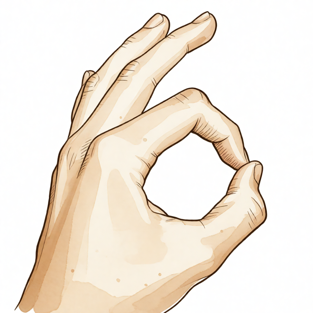
High median nerve compression: pronator syndrome (sensory, thenar/palm sparing distinguishes from CTS) and AIN palsy (pure motor, OK-sign), with observation-first management and decompression for refractory cases.
For patients: a plain-language version of this topic is available. See the patient guide.
Overview
Pronator syndrome, more accurately termed superficialis-pronator syndrome, involves median nerve compression at the pronator tunnel, a condition that is more common than lacertus syndrome [3, 4]. Lacertus syndrome is a postexertional compartment syndrome that must be distinguished from pronator syndrome through careful workup and presented as a distinct surgical target [3, 4]. The diagnosis of pronator syndrome lacks objective pathophysiology, relying instead on subjective operative findings and post-surgical relief, with a paucity of controlled trials demonstrating superiority over sham surgery [5]. Clinicians require a thorough understanding of median nerve anatomy, compression sites, and clinical findings to provide reliable diagnoses [6]. Concurrent carpal tunnel syndrome is rarely considered, and proximal compression sites are easily overlooked [10].
Conservative treatment is the first choice for median nerve compression in the form of pronator or anterior interosseous nerve syndrome [9]. Surgical decompression is indicated for pronator syndrome when symptoms persist for more than six months [1], or when activities of daily living require frequent strong pronation and electromyography confirms compression [8]. For anterior interosseous nerve syndrome, surgery is indicated after a minimum of twelve months with no signs of motor improvement [1]. A prolonged nonsurgical approach is warranted in most cases of median or anterior interosseous nerve compression in the forearm, as surgical decompression is rarely indicated [38]. Other medical causes should be considered in anterior interosseous nerve syndrome [8].
When the correct diagnosis is made, pronator syndrome can be successfully treated nonoperatively or surgically if necessary [2]. Surgical decompression can be considered for pronator syndrome if activities of daily living require frequent strong pronation and electromyography findings confirm the presence of compression [8]. The ulnar head of the pronator teres is fibrotic or fibromuscular in 71.5% of cases, and its morphology and location relative to the median nerve are important for surgical treatment [11]. The potential for fibrous compression of the median nerve at the pronator tunnel was substantiated from a series of anatomical dissections [16]. Most publications on uncommon upper extremity compression neuropathies are small retrospective series or case reports, and treatment decisions are not typically based on high levels of evidence [18].
Anatomy & Pathophysiology
Median Nerve Compression Sites
The median nerve traverses a series of musculoaponeurotic potential sites of compression from above the elbow to the mid-forearm [44]. The first potential site is the deep fascia (lacertus fibrosus) in the upper arm as it connects to the pronator teres, particularly when the pronator teres has a high origin [44]. The second site is the fibrous arch beneath the superficialis head of the pronator teres [44]. The third site is the fibrous arch beneath the deep head of the pronator teres [44]. The fourth site is the fibrous arch within the flexor digitorum superficialis [44]. The fifth site is Gantzer's muscle [44]. A fibrous arch in muscles crossing the median nerve may place the nerve at greater risk than in specimens where the nerve passes against soft muscle [44]. When the anterior interosseous nerve originates from the radial side of the median nerve, it is more susceptible to compression against a fibrous arch than if it originates on the deep or posterior aspect of the median nerve [44]. The musculoaponeurotic system formed by the multiple heads of the pronator teres and flexor superficialis can be arranged in a variable number of arches across the median nerve, ranging from no arch to two arches [44]. Clinical signs and symptoms suggestive of median nerve compression in the forearm should be considered as a continuum wherein the median nerve may be compressed at any point from proximal at the antecubital fossa to where the median nerve emerges from beneath the superficialis muscle [44].
Symptoms of pronator syndrome may result from dynamic compression of the median nerve as it passes between the heads of pronator teres or through the humeral head of the muscle [102]. Symptoms may also result from compression by the lacertus fibrosus [102], the fibrotendinous origin of flexor digitorum superficialis [102], or the supracondylar process [102]. A rare anatomical arrangement involving the humeral head of pronator teres can displace the median nerve to the anterior aspect of the medial humeral epicondyle, serving as a site of compression neuropathy [54]. The ligament of Struthers is a variant anatomy arising from the supracondylar process to attach to the medial epicondyle and is a potential site of median nerve compression [63]. The median nerve and brachial artery are located just superficial to the anteromedial joint capsule and brachialis muscle [75].
Anterior Interosseous Nerve Anatomy and Pathology
Fascicular constrictions in anterior interosseous nerve syndrome are predominantly located in the posterior/posteromedial region proximal to the elbow joint line [12]. These fascicular constrictions were identified in all MRN cases and 88% of ultrasound cases [12]. Near its site of origin, the anterior interosseous nerve is vulnerable to compression from tendinous attachments or accessory muscles [56]. Tendinous origins of the deep head of pronator teres and of flexor digitorum superficialis to the middle finger are sources of compression for the anterior interosseous nerve [56]. Accessory structures causing anterior interosseous nerve compression include the attachment of flexor digitorum superficialis to flexor pollicis longus [56]. An accessory head of the flexor pollicis longus muscle (Gantzer's muscle) is an accessory structure that can compress the anterior interosseous nerve [56]. A tendinous origin of the muscle variant palmaris profundus is an accessory structure that can compress the anterior interosseous nerve [56]. A double lacertus fibrosis is an accessory structure that can compress the anterior interosseous nerve [56]. Fractures at or around the elbow joint and direct injury to the nerve are causes of anterior interosseous nerve syndrome [56]. Compression from plaster casts is a recognized cause of anterior interosseous nerve syndrome [56]. Vascular causes have been described for anterior interosseous nerve syndrome [56]. The pronator quadratus muscle is of importance for the initiation of hand pronation [13]. Damage to the innervation of the pronator quadratus muscle risks impairment of the initiation of hand pronation [13].
Lacertus Fibrosus and Elbow Anatomy
The lacertus fibrosus is the deep fascia in the upper arm that connects to the pronator teres [44]. An intact lacertus fibrosus contributes to elbow strength [96]. The medial epicondyle forms the attachment site for the origins of the flexor pronator mass [62]. The flexor-pronator mass includes the pronator teres, flexor carpi radialis, palmaris longus, flexor carpi ulnaris, and flexor digitorum superficialis [63]. The median nerve lies in a bony groove covered and restrained by the arcuate ligament posterior to the medial epicondyle [71]. The ulnar nerve passes through the cubital tunnel at the medial column of the elbow [70]. The ulnar nerve enters the anterior forearm by traveling between the two heads of the flexor carpi ulnaris [70]. The radial nerve splits into the superficial sensory branch and the posterior interosseous nerve on the anterolateral side of the elbow [75]. The posterior interosseous nerve is just superficial to the anterior joint capsule and at the level of the radiocapitellar joint [75]. The posterior interosseous nerve courses longitudinally along the medial side of the capitellum [75]. At the level of the radial neck, the posterior interosseous nerve may come in direct contact with the joint capsule [75].
Pathophysiology and Clinical Presentation
Lacertus syndrome is a postexertional compartment syndrome [3]. Lacertus syndrome is more common than pronator syndrome [3]. Lacertus syndrome often presents with increasing pain and fullness in the flexor-pronator muscles with continued high-level activity [51]. Symptoms of lacertus syndrome resolve with rest, anywhere from 8 hours to 4 days after the cessation of activity [51]. Lacertus syndrome can include intermittent median nerve neuropathy [51]. Baseball pitchers and American football quarterbacks are the most common position players to present with lacertus syndrome [51]. The dynamic nature of lacertus syndrome, presenting after exertional activity, makes it distinct from anterior interosseous nerve and pronator syndrome [51]. The throwing elbow is a common source of nerve injuries due to the unique combination of anatomy, high forces, and sheer repetition associated with throwing sports [49].
Anterior interosseous nerve syndrome is characterized by weakness of pinch between the thumb and index finger [56]. In anterior interosseous nerve syndrome, the interphalangeal joint of the thumb and the distal interphalangeal joint of the index finger are hyperextended in the pinch position [56]. Weakness of the pronator quadratus muscle can be tested with the forearm flexed, neutralizing 75% of pronator teres action [56]. Patients with anterior interosseous nerve syndrome may complain of vague pain in the proximal forearm aggravated by exercise [56]. Pronator syndrome can be successfully treated nonoperatively or surgically when the correct diagnosis is made [2]. Surgical indications for nerve decompression in pronator syndrome include persistent symptoms for >6 months [1]. Renaut bodies are present in the median nerve at sites of potential entrapment, most commonly found proximal to the pronator arch [102]. Renaut bodies appear to displace normal endoneurial components and may be associated with low-grade axonal drop-out [102]. The presence of Renaut bodies may adversely affect the functional outcome of surgical decompression of the median nerve [102]. The function of the pronator teres and pronator quadratus cannot be isolated by elbow flexion positioning [103].
Classification
Terminology and Definitions
Superficialis-pronator syndrome: The term "pronator syndrome" should be replaced with "superficialis-pronator syndrome" to clearly distinguish it from lacertus syndrome as a separate surgical target [4]. Lacertus syndrome: This term is recommended to refer specifically to intermittent median nerve compression at the level of the lacertus fibrosus [48]. It is classified as a postexertional compartment syndrome [3]. Bennett's syndrome: The pathology originally described by Bennett should be termed "chronic exertional compartment syndrome of the pronator teres" or "Bennett's syndrome" to resolve terminology confusion [48].
Anatomical Sites of Compression
The median nerve passes through a series of musculoaponeurotic potential sites of compression in the proximal forearm, including the deep fascia (lacertus fibrosus), the fibrous arch beneath the superficialis head of the pronator teres, the fibrous arch beneath the deep head of the pronator teres, the fibrous arch within the flexor digitorum superficialis, and Gantzer's muscle [44]. When the anterior interosseous nerve takes origin from the radial side of the median nerve, it is more susceptible to compression against the fibrous arch than if it originates on the deep or posterior aspect of the median nerve [44].
Clinical Presentation and Diagnosis
Diagnostic Challenges: The diagnosis of pronator syndrome is supported by a paucity of controlled trials demonstrating that operative treatment is more effective than other treatments or sham surgery [5]. It is essential for clinicians to have a thorough understanding of median nerve anatomy, possible sites of compression, and characteristic clinical findings of pronator syndrome to provide a reliable diagnosis [6]. The diagnostic process to differentiate pronator syndrome from carpal tunnel syndrome remains a challenge due to overlapping symptoms and limited reliable information in the literature [21].
Anterior Interosseous Nerve Syndrome (AINS): AINS involves paralysis of the flexor pollicis longus, flexor digitorum profundus of the index and long fingers, and the pronator quadratus [52]. An incomplete AINS involves only the flexor pollicis longus or the flexor digitorum profundus of the index finger being either paretic or paralyzed [52]. The condition is characterized by weakness of pinch between the thumb and index finger [56].
Causes of Compression in AINS: The nerve in AINS is usually compressed by fibrous bands that most commonly originate from the deep head of the pronator teres and to the brachialis fascia [52]. Less common causes include fibrous bands from the superficial head of the pronator teres, bands from the superficialis arcade, the nerve running deep to both heads of the pronator, and compression by a double lacertus fibrosus [52]. Accessory structures that can cause compression include the attachment of flexor digitorum superficialis to flexor pollicis longus, an accessory head of the flexor pollicis longus muscle (Gantzer's muscle), a tendinous origin of the muscle variant palmaris profundus, and a double lacertus fibrosis [56]. Compression from plaster casts has been recognized as a cause of AINS [56].
Other Causes: Bilateral pronator syndrome can be caused by an anomalous tendinous origin of the ulnar belly of the pronator teres muscle [22]. An abnormal muscle in the anterior compartment of the forearm can present with compression on the median nerve [17]. Median nerve compression can occur secondary to a high insertion of pronator teres [15]. The extent of median nerve injury in Type 4 entrapment after elbow dislocation tends to be more severe than other types because of double entrapment sites [34].
Diagnostic Modalities
Fascicular constrictions in AINS were identified in all MRN cases and 88% of ultrasound cases [12]. If the activities of daily living of a patient require frequent strong pronation, and if the electromyography findings confirm the presence of compression, surgical decompression can be considered for pronator syndrome [8]. Other medical causes should be considered in AINS when electromyography findings are present [8].
Clinical Presentation
Pronator Syndrome
The clinical presentation of pronator syndrome is influenced by the morphology of the pronator teres, with the ulnar head being fibrotic or fibromuscular in 71.5% of cases [11]. This anatomical variation and its location relative to the median nerve are critical determinants for surgical treatment [11]. Median nerve compression may also result from a high insertion of the pronator teres [15]. In rare instances, a schwannoma serves as the causative agent for pronator syndrome [27].
Lacertus Syndrome
Lacertus syndrome is a postexertional compartment syndrome that is more common than pronator syndrome and can be distinguished through a careful workup [3]. Terminological clarity is essential; the term "pronator syndrome" should be replaced with "superficialis-pronator syndrome" to clearly distinguish it from lacertus syndrome as distinct surgical targets [4]. Authors recommend reserving the term "lacertus syndrome" for intermittent median nerve compression at the level of the lacertus fibrosus, while designating the pathology originally described by Bennett as chronic exertional compartment syndrome of the pronator teres or "Bennett's syndrome" [48].
Patients with median nerve compression by the lacertus fibrosus present with proximal forearm pain and weakness of the pronator teres, flexor carpi radialis, flexor pollicis longus, and pronator quadratus [23]. Electrodiagnostic studies are positive in these cases, but no sensory findings are observed distal to the forearm [23]. Median nerve symptoms can also arise secondary to lacertus fibrosus syndrome following distal biceps tendon repair [29].
Diagnostic evaluation via ultrasound presents specific challenges. US examination findings may not lead to a diagnosis of lacertus syndrome because median nerve compression in the forearm is often mild and may not produce positive US findings [47]. Consequently, negative US findings cannot rule out the diagnosis [86]. However, ultrasonography remains a valuable supplementary tool for diagnosing nerve compression pathologies by providing real-time, dynamic assessment of the median nerve and surrounding structures [86]. Physical examination findings are limited in specificity; nearly one-fifth of asymptomatic forearms exhibit tenderness upon palpation at the lacertus fibrosus [88]. Therefore, local tenderness alone lacks diagnostic specificity, and contralateral comparison is unreliable for diagnosing lacertus syndrome [88].
Anterior Interosseous Nerve Syndrome
Anterior interosseous nerve palsy is an uncommon disease of unknown etiology and pathophysiology [40]. It creates objective weakness and electrophysiological abnormalities [40]. Fascicular constrictions above the elbow typify anterior interosseous nerve syndrome [12]. These constrictions were identified in all MRN cases and 88% of ultrasound cases, predominantly in the posterior/posteromedial region proximal to the elbow joint line [12].
The clinical manifestations of proximal entrapment of the median nerve include aching pain in the anterior forearm, loss of key and tip pinch strength, including the typical inability to make an 'OK' sign, and loss of fine motor skills and a sense of clumsiness [87]. Less frequently, paraesthesias in the median nerve innervated region of the hand similar to a carpal tunnel syndrome may be present [87]. Nerve conduction studies are usually normal and thus unhelpful in proximal entrapment of the median nerve [87]. Ultrasound has been reported to identify the site of compression by virtue of the change in the diameter of the nerve [87]. However, in the absence of a mass or haematoma causing compression, it may not be easy to make a diagnosis with ultrasonography since the fibrous bands or scar tissue may be too small to visualize [87].
If the activities of daily living of a patient require frequent strong pronation, and if the electromyography findings confirm the presence of compression, surgical decompression can be considered against the background of pronator syndrome [8]. In contrast, other medical causes should be considered in AIN syndrome [8].
Investigations
Diagnostic Challenges and Limitations: The diagnostic workup for uncommon compression syndromes of the radial, ulnar, and median nerves is constrained by the limited quality of available literature. Most publications on these conditions are small retrospective series or case reports [18]. Consequently, treatment decisions are not typically based on high levels of evidence [18].
MRI: Magnetic resonance imaging provides high diagnostic yield for specific pathologies. In one institution, MRI identified the cause of medial elbow pain in 20 (95%) of 21 throwing athletes [55].
Angiography: Angiography may provide critical diagnostic clues when other modalities are inconclusive. In a case of neurilemmoma, angiography provided the only clue that the anterior interosseous nerve was involved, whereas all other findings suggested the tumor arose from the median nerve [108].
Electrodiagnostic Studies: Electrodiagnostic testing is central to confirming nerve compression. Three patients with compression of the median nerve by the lacertus fibrosus presented with positive electrodiagnostic studies but no sensory findings distal to the forearm [23]. If electromyography findings confirm the presence of compression in a patient with frequent strong pronation requirements, surgical decompression can be considered for pronator syndrome [8].
Other Considerations: Anatomical and morphological factors must be considered in the differential diagnosis. An abnormal muscle in the anterior compartment of the forearm can compress the median nerve [17]. Clinically, patients with compression of the median nerve by the lacertus fibrosus present with proximal forearm pain and weakness of the pronator teres, flexor carpi radialis, flexor pollicis longus, and pronator quadratus [23]. Median nerve symptoms secondary to lacertus fibrosus syndrome can occur following distal biceps tendon repair [29].
Treatment
Non-Operative
Conservative management is the initial standard of care for both pronator syndrome and anterior interosseous nerve syndrome (AINS). For AINS, the typical nonsurgical regimen includes rest, splinting, and observation [30]. Because most patients improve without surgical intervention, observation for a period of several months is suggested before considering decompression [30]. In two reported cases, AINS palsy following open capsular release for elbow stiffness recovered completely with conservative management [111]. For pronator syndrome, treatment is largely nonsurgical and consists of activity modification, anti-inflammatory medication, corticosteroid injections, stretching, and periods of splinting [77]. Conservative options also include physical therapy, nerve gliding, therapeutic ultrasound, nonsteroidal anti-inflammatory drugs, and corticosteroid injections [73]. Most sources recommend 3 to 6 months of conservative treatment before planning surgical interventions [73]. Several studies reported improvement in 29% to 70% of patients with pronator syndrome treated conservatively [73]. A trial of nonsurgical treatment is warranted in all patients, with surgical decompression reserved for those who do not respond or continue to progress despite conservative treatment [116].
Operative
Indications: Surgical decompression is indicated for pronator syndrome when conservative therapy fails [77] or when symptoms persist for >6 months [1]. For AINS, surgery is recommended for patients who have a space-occupying mass in the area or who fail a several-month course of nonsurgical treatment [30]. Additionally, surgical indications include a minimum of 12 months with no signs of motor improvement [1].
Surgical Approach / Technique: The surgical approach for pronator syndrome or AINS is performed through a lazy-S–type incision that begins with an exploration of the median nerve proximal to the antecubital flexion crease [30]. After the median nerve is released proximally, it is dissected free from the overlying lacertus fibrosus, the humeral head of the pronator teres, and the proximal fascial edge of the FDS arch [30]. Gantzer's accessory FPL muscle, if present, should be released during surgical decompression [30]. In patients with AINS, the AIN should be thoroughly decompressed from any overlying tissue and completely visualized as it travels into the distal forearm [30]. Endoscopic proximal median nerve decompression enables the complete decompression of the median nerve in the distal aspect of the arm and proximal aspect of the forearm through a small incision [77]. The endoscopically assisted, minimally invasive approach adequately and safely decompressed all anatomical points of compression and improved DASH scores [25]. The author is a proponent of the endoscopic technique with minimal damage and foresees its wide indication in the future for pronator syndrome [24].
Outcomes: Complete exposure open median nerve release provides complete symptomatic relief in the majority of patients, with some patients experiencing recurrent symptoms of pain and paresthesia [73]. Surgical decompression provides satisfactory outcomes for patients with persistent forearm pain and median nerve symptoms [115]. When the correct diagnosis is made, pronator syndrome can be successfully treated surgically if necessary [2].
Complications
Nerve palsy: Anterior interosseous nerve palsy is a recognized complication following shoulder arthroscopy, closed fractures of the proximal ulna, and Galeazzi fractures [20]. Damage to the innervation of the pronator quadratus muscle, which is critical for initiating hand pronation, represents a specific risk associated with anterior interosseous nerve anatomy [13]. In the context of elbow dislocation, median nerve injury in Type 4 entrapment tends to be more severe than in other types due to the presence of double entrapment sites [34].
Other Considerations: Compression of the median nerve by the lacertus fibrosus can present with weakness of the pronator teres, flexor carpi radialis, flexor pollicis longus, and pronator quadratus [23]. Anomalous anatomical structures contribute to these syndromes; specifically, an anomalous tendinous origin of the ulnar belly of the pronator teres muscle can cause bilateral pronator syndrome [22], while a bifid reversed palmaris longus can cause median nerve compression [120]. Additionally, a high insertion of the pronator teres is a cause of median nerve compression [15]. Regarding anterior interosseous nerve compression, fibrous bands originating from the deep head of the pronator teres and extending to the brachialis fascia are the most common cause [52]. Less common causes include fibrous bands from the superficial head of the pronator teres, bands from the superficialis arcade, the nerve running deep to both heads of the pronator, and compression by a double lacertus fibrosus [52].
Recovery
Other Considerations: Pronator teres symptoms disappeared in 93% of cases following the mini-invasive technique [7]. Full recovery of the ulnar nerve was achieved six months after the surgery [26].
Key Evidence
- [Paper] Surgical indications for nerve decompression include persistent symptoms for >6 months in patients with pronator syndrome or for a minimum of 12 months with no signs of motor improvement in those with anterior interosseous nerve syndrome. [1] (10.5435/jaaos-21-05-268)
- [L5] When the correct diagnosis is made, pronator syndrome can be successfully treated nonoperatively or surgically, if necessary. [2] (10.1016/s0278-5919(05)70267-2)
- [L5] Lacertus syndrome is a postexertional compartment syndrome that is more common than pronator syndrome and can be distinguished with a careful workup. [3] (10.1016/j.csm.2020.03.004)
- [L5] Lacertus syndrome and superficialis-pronator syndrome should be clearly presented as distinct targets for surgical release, and the term pronator syndrome should be replaced with superficialis-pronator syndrome. [4] (10.1177/17531934211024092)
- [L5] The diagnosis of pronator syndrome lacks objective pathophysiology and is supported only by subjective operative findings and relief after surgery, with a paucity of controlled trials demonstrating that operative treatment is more effective than other treatments or sham surgery. [5] (10.1016/j.jhsa.2011.02.014)
- [L5] It is essential for clinicians to have a thorough understanding of median nerve anatomy, possible sites of compression, and characteristic clinical findings of pronator syndrome to provide a reliable diagnosis and treat their patients. [6] (10.1016/j.jhsa.2020.07.006)
- [L4] Pronator teres symptoms disappeared in 93% of cases following the mini-invasive technique. [7] (10.1016/j.jhsa.2012.05.033)
- [L5] If the activities of daily living of a patient require frequent strong pronation, and if the electromyography findings confirm the presence of compression, surgical decompression can be considered against the background of pronator syndrome, whereas other medical causes should be considered in AIN syndrome. [8] (10.1177/17531934221080018)
- [L5] Conservative treatment stays the first choice for median nerve compression either in the form of pronator or anterior interosseous nerve syndrome. [9] (10.1016/j.main.2004.10.024)
- [L4] Concurrent carpal tunnel syndrome and pronator syndrome are rarely considered and proximal compression sites are easily overlooked. [10] (10.1016/j.otsr.2016.10.009)
- [L5] The ulnar head of the pronator teres is fibrotic or fibromuscular in 71.5% of cases, and its morphology and location relative to the median nerve are important for surgical treatment of pronator syndrome. [11] (10.1016/j.otsr.2016.08.016)
- [L4] These constrictions were identified in all MRN cases and 88% of ultrasound cases, predominantly in the posterior/posteromedial region proximal to the elbow joint line. [12] (10.1002/mus.26768)
- [L5] This risks damage of the innervation of this muscle of importance for initiation of hand pronation. [13] (10.1016/j.jhsb.2005.06.017)
- [L4] [15] (10.1111/j.1758-5740.2010.00051.x)
- [L4] The potential for fibrous compression of the median nerve at the pronator tunnel was substantiated from a series of anatomical dissections. [16] (10.1016/s0363-5023(79)80104-5)
- [L5] The paper describes a 19-year-old patient with an abnormal muscle in the anterior compartment of the forearm that compressed the median nerve. [17] (10.1177/1753193413485011)
- [L4] This article reviews uncommon compression syndromes of the radial, ulnar, and median nerves, noting that most publications are small retrospective series or case reports and treatment decisions are not typically based on high levels of evidence. [18] (10.1016/j.hcl.2013.04.014)
- [L4] [20] (10.1007/s00402-011-1322-5)
- [L4] The diagnostic process to differentiate pronator syndrome from carpal tunnel syndrome remains a challenge due to overlapping symptoms and limited reliable information in the literature; this review provides a comprehensive clinical comparison to aid in establishing appropriate diagnosis and treatment. [21] (10.3390/diagnostics12102433)
- [L5] We report the case of a patient with bilateral pronator syndrome caused by an anomalous tendinous origin of the ulnar belly of the pronator teres muscle. [22] (10.1016/0363-5023(93)90373-b)
- [L4] Three patients with compression of the median nerve by the lacertus fibrosus presented with proximal forearm pain, weakness of the pronator teres, flexor carpi radialis, flexor pollicis longus, and pronator quadratus, and positive electrodiagnostic studies, but no sensory findings distal to the forearm. [23] (10.1016/s0363-5023(86)80015-6)
- [L4] The author is a proponent of the endoscopic technique with minimal damage and foresees its wide indication in the future for pronator syndrome. [24] (10.1177/17531934221076298)
- [L4] The endoscopically assisted, minimally invasive approach to treat pronator syndrome adequately and safely decompressed all anatomical points of compression and improved DASH scores. [25] (10.1016/j.jhsa.2012.02.023)
- [L5] Full recovery of the ulnar nerve was achieved six months after the surgery. [26] (10.1142/s2424835516720036)
- [L5] [27] (10.1007/s12593-013-0115-1)
- [L4] With clinical evaluation as the reference standard, electrodiagnostic, ultrasound, and MRI are not helpful in making a diagnosis of pronator syndrome concurrent with carpal tunnel syndrome. [28] (10.1016/j.jhsa.2020.06.006)
- [Case_report] This case presents a unique instance of median nerve symptoms secondary to lacertus fibrosus syndrome after distal biceps tendon repair. [29] (10.1016/j.xrrt.2024.08.009)
- [L5] [30] (10.1016/j.jhsa.2009.10.017)
- [L4] An isolated anterior interosseous nerve injury may not by itself be an indication for urgent surgery. [32] (10.2106/jbjs.n.00136)
- [L4] The transverse incision and surgical technique allows for satisfactory decompression of all the structures involved in pronator teres syndrome. [33] (10.1016/0266-7681(94)90047-7)
- [L5] The extent of median nerve injury in Type 4 tends to be more severe than the other three types because of the double entrapment sites. [34] (10.1016/0266-7681(94)90127-9)
- [Case_report] Resection of a segment of the persistent median artery is the recommended treatment for pronator syndrome associated with this anomaly. [35] (10.2106/00004623-198769020-00026)
- [L5] Surgical decompression of the median nerve or the AIN in the forearm is rarely indicated; a prolonged nonsurgical approach is warranted in most cases. [38] (10.5435/jaaos-d-16-00010)
- [L5] [40] (10.1016/j.jhsa.2010.08.018)
- [L5] [44] (10.1016/0266-7681(87)90189-6)
- [L5] US examination can be challenging and its findings may not lead to diagnosis of LS, as median nerve compression in the forearm is often mild and may not produce positive US findings. [47] (10.1177/17531934251370272)
- [L5] The authors recommend that the term 'lacertus syndrome' refer to intermittent median nerve compression at the level of the lacertus fibrosus, while the pathology originally described by Bennett should be termed chronic exertional compartment syndrome of the pronator teres or 'Bennett's syndrome' to resolve current terminology confusion. [48] (10.1177/17531934231170347)
- [L5] The throwing elbow is a common source of nerve injuries due to the unique combination of anatomy, high forces, and sheer repetition associated with throwing sports. [49] (10.1016/j.csm.2004.04.012)
- [L5] [51] (10.1016/j.radcr.2021.02.022)
- [L4] [52] (10.1016/s0363-5023(85)80240-9)
- [L5] This rare anatomical arrangement resulted in displacement of the median nerve to the anterior aspect of the medial humeral epicondyle and, as far as we are aware, has not previously been described as a site of compression neuropathy. [54] (10.1016/0266-7681(94)90241-0)
- [L3] MRI from our institution was able to identify the cause of medial elbow pain in 20 (95%) of 21 throwing athletes. [55] (10.1016/s1058-2746(95)80208-8)
- [L5] [56] (10.1016/s0266-7681(05)80231-1)
- [L5] [73] (10.1016/j.xrrt.2024.10.001)
- [L4] [77] (10.1016/j.jhsg.2021.03.006)
- [L4] [86] (10.1177/17531934251346595)
- [L4] [87] (10.1177/1753193417726214)
- [L5] [88] (10.1177/17531934251382852)
- [L3] An intact lacertus fibrosus contributes to elbow strength and should be preserved in distal biceps tendon repair. [96] (10.1007/s00167-019-05673-2)
- [L4] [102] (10.1054/jhsb.1998.0177)
- [L4] The findings suggest that the function of the PT and PQ cannot be isolated by elbow flexion positioning. [103] (10.1016/j.jhsa.2025.09.013)
- [L5] Angiography provided the only clue that the anterior interosseous nerve was involved, whereas all other findings suggested the neurilemmoma arose from the median nerve. [108] (10.1016/0266-7681(91)90141-a)
- [L4] The palsy in these patients recovered completely with conservative management. [111] (10.1016/j.jhsa.2008.10.019)
- [L4] Surgical decompression provides satisfactory outcomes for patients with persistent forearm pain and median nerve symptoms. [115] (10.1177/1558944719874137)
- [L5] A trial of nonsurgical treatment is warranted in all patients, with surgical decompression reserved for those who do not respond or continue to progress despite conservative treatment. [116] (10.1097/00130911-200212000-00010)
- [L5] The authors state they are not aware of any previous description of this particular anomaly associated with compression of the median nerve. [120] (10.1016/s0363-5023(87)80007-2)
See Also
References
[1] Pronator Syndrome and Anterior Interosseous Nerve Syndrome. Journal of the American Academy of Orthopaedic Surgeons. 2013. DOI: 10.5435/jaaos-21-05-268
[2] PRONATOR SYNDROME. Clinics in Sports Medicine. 2001. DOI: 10.1016/s0278-5919(05)70267-2
[3] The Lacertus Syndrome of the Elbow in Throwing Athletes. Clinics in Sports Medicine. 2020. DOI: 10.1016/j.csm.2020.03.004
[4] Median nerve compression: lacertus syndrome versus superficialis-pronator syndrome. Journal of Hand Surgery (European Volume). 2021. DOI: 10.1177/17531934211024092
[5] Pronator Syndrome. The Journal of Hand Surgery. 2011. DOI: 10.1016/j.jhsa.2011.02.014
[6] Proximal Median Nerve Compression: Pronator Syndrome. The Journal of Hand Surgery. 2020. DOI: 10.1016/j.jhsa.2020.07.006
[7] New Mini-invasive Decompression for Pronator Teres Syndrome. The Journal of Hand Surgery. 2012. DOI: 10.1016/j.jhsa.2012.05.033
[8] Two clinical observations: pronator syndrome in violinists and anterior interosseous nerve syndrome with pure motor loss. Journal of Hand Surgery (European Volume). 2022. DOI: 10.1177/17531934221080018
[9] Compression du nerf médian au coude. Chirurgie de la Main. 2004. DOI: 10.1016/j.main.2004.10.024
[10] Concurrent carpal tunnel syndrome and pronator syndrome: A retrospective study of 21 cases. Orthopaedics & Traumatology: Surgery & Research. 2017. DOI: 10.1016/j.otsr.2016.10.009
[11] Morphology and morphometry of the ulnar head of the pronator teres muscle in relation to median nerve compression at the proximal forearm. Orthopaedics & Traumatology: Surgery & Research. 2016. DOI: 10.1016/j.otsr.2016.08.016
[12] Fascicular constrictions above elbow typify anterior interosseous nerve syndrome. Muscle & Nerve. 2019. DOI: 10.1002/mus.26768
[13] An Anatomical Study of the Anterior Interosseous Nerve and its Innervation of the Pronator Quadratus Muscle. Journal of Hand Surgery. 2005. DOI: 10.1016/j.jhsb.2005.06.017
[15] Median Nerve Compression Secondary to a High Insertion of Pronator Teres. Shoulder & Elbow. 2010. DOI: 10.1111/j.1758-5740.2010.00051.x
[16] Median nerve entrapment syndrome in the proximal forearm. The Journal of Hand Surgery. 1979. DOI: 10.1016/s0363-5023(79)80104-5
[17] Abnormal muscle of the distal anterior forearm presenting with compression on the median nerve. Journal of Hand Surgery (European Volume). 2013. DOI: 10.1177/1753193413485011
[18] Uncommon Upper Extremity Compression Neuropathies. Hand Clinics. 2013. DOI: 10.1016/j.hcl.2013.04.014
[20] Anterior interosseous nerve syndrome: retrospective analysis of 14 patients. Archives of Orthopaedic and Trauma Surgery. 2011. DOI: 10.1007/s00402-011-1322-5
[21] How to Differentiate Pronator Syndrome from Carpal Tunnel Syndrome: A Comprehensive Clinical Comparison. Diagnostics. 2022. DOI: 10.3390/diagnostics12102433
[22] Bilateral pronator syndrome associated with anomalous heads of the pronator teres muscle: A case report. The Journal of Hand Surgery. 1993. DOI: 10.1016/0363-5023(93)90373-b
[23] Median nerve compression neuropathy by the lacertus fibrosus: Report of three cases. The Journal of Hand Surgery. 1986. DOI: 10.1016/s0363-5023(86)80015-6
[24] Re: Tang JB. Median nerve compression: lacertus syndrome versus superficialis-pronator syndrome. J Hand Surg Eur. 2021, 46: 1017–22. Journal of Hand Surgery (European Volume). 2022. DOI: 10.1177/17531934221076298
[25] Endoscopically Assisted Decompression for Pronator Syndrome. The Journal of Hand Surgery. 2012. DOI: 10.1016/j.jhsa.2012.02.023
[26] Acute Compartment Syndrome in the Forearm with Trans-Ulnar Single Incision. The Journal of Hand Surgery (Asian-Pacific Volume). 2016. DOI: 10.1142/s2424835516720036
[27] Pronator Syndrome Due to Schwannoma. Journal of Hand and Microsurgery. 2015. DOI: 10.1007/s12593-013-0115-1
[28] Clinical, Radiological, and Electrodiagnostic Diagnosis of Pronator Syndrome Concurrent With Carpal Tunnel Syndrome. The Journal of Hand Surgery. 2020. DOI: 10.1016/j.jhsa.2020.06.006
[29] Acute median nerve compression secondary to lacertus fibrosis syndrome following distal biceps tendon reconstruction: a case report. JSES Reviews, Reports, and Techniques. 2025. DOI: 10.1016/j.xrrt.2024.08.009
[30] Unusual Compression Neuropathies of the Forearm, Part II: Median Nerve. The Journal of Hand Surgery. 2009. DOI: 10.1016/j.jhsa.2009.10.017
[32] Supracondylar Humeral Fractures with Isolated Anterior Interosseous Nerve Injuries: Is Urgent Treatment Necessary?. The Journal of Bone and Joint Surgery-American Volume. 2014. DOI: 10.2106/jbjs.n.00136
[33] A Transverse Skin Incision Approach for Decompression of Pronator Teres Syndrome. Journal of Hand Surgery. 1994. DOI: 10.1016/0266-7681(94)90047-7
[34] Type 4 Median Nerve Entrapment after Elbow Dislocation. Journal of Hand Surgery. 1994. DOI: 10.1016/0266-7681(94)90127-9
[35] Pronator syndrome associated with a persistent median artery. A case report.. The Journal of Bone & Joint Surgery. 1987. DOI: 10.2106/00004623-198769020-00026
[38] Ulnar Tunnel Syndrome, Radial Tunnel Syndrome, Anterior Interosseous Nerve Syndrome, and Pronator Syndrome. Journal of the American Academy of Orthopaedic Surgeons. 2017. DOI: 10.5435/jaaos-d-16-00010
[40] Anterior Interosseous Nerve Syndrome. The Journal of Hand Surgery. 2010. DOI: 10.1016/j.jhsa.2010.08.018
[44] Musculoaponeurotic variations along the course of the median nerve in the proximal forearm. The Journal of Hand Surgery: Journal of the British Society for Surgery of the Hand. 1987. DOI: 10.1016/0266-7681(87)90189-6
[47] Re: Fang J, Zhang LQ, Tang JB. Incidence of local tenderness at the lacertus fibrosus in healthy people. Journal of Hand Surgery (European Volume). 2025. DOI: 10.1177/17531934251370272
[48] Lacertus syndrome: one term – two different pathologies. Journal of Hand Surgery (European Volume). 2023. DOI: 10.1177/17531934231170347
[49] Nerve injuries in the throwing elbow. Clinics in Sports Medicine. 2004. DOI: 10.1016/j.csm.2004.04.012
[51] Lacertus syndrome: Use of pre- and post-exercise MRI to aid in diagnosis and treatment. Radiology Case Reports. 2021. DOI: 10.1016/j.radcr.2021.02.022
[52] The incomplete anterior interosseous nerve syndrome. The Journal of Hand Surgery. 1985. DOI: 10.1016/s0363-5023(85)80240-9
[54] Median Nerve Compression within the Humeral Head of Pronator Teres. Journal of Hand Surgery. 1994. DOI: 10.1016/0266-7681(94)90241-0
[55] Diagnostic utility of MRI in elite throwing athletes with medial elbow pain. Journal of Shoulder and Elbow Surgery. 1995. DOI: 10.1016/s1058-2746(95)80208-8
[56] An Anomaly of the Median Artery Associated with the Anterior Interosseous Nerve Syndrome. Journal of Hand Surgery. 1992. DOI: 10.1016/s0266-7681(05)80231-1
[62] Orthopaedic Knowledge Update 13 Ebook Without Multimedia. Anatomy, Biomechanics, Physical Examination, and Imaging of the Elbow > Anatomy > Bony Anatomy.
[63] Miller S Review Of Orthopaedics. SECTION 16 PATELLAR TRACKING IN TOTAL KNEE ARTHROPLASTY > TABLE 2.3 Shoulder Spaces.
[70] A Lange Medical Book Current Diagnosis Treatment In Orthopedics Fifth Edition. 2Musculoskeletal Trauma Surgery > INJURIES AROUND THE ELBOW.
[71] Orthopaedic Knowledge Update Trauma. Distal Humerus Fracture > Anatomy.
[73] Median nerve entrapment neuropathy: a review on the pronator syndrome. JSES Reviews, Reports, and Techniques. 2025. DOI: 10.1016/j.xrrt.2024.10.001
[75] Green S Operative Hand Surgery. PERTINENT ANATOMY AND PORTALS.
[77] Endoscopic Proximal Median Nerve Decompression: An Alternative Treatment for Pronator Syndrome. Journal of Hand Surgery Global Online. 2021. DOI: 10.1016/j.jhsg.2021.03.006
[86] Incidence of local tenderness at the lacertus fibrosus in healthy people. Journal of Hand Surgery (European Volume). 2025. DOI: 10.1177/17531934251346595
[87] The scratch collapse test in the diagnosis of compression of the median nerve in the proximal forearm. Journal of Hand Surgery (European Volume). 2017. DOI: 10.1177/1753193417726214
[88] Re: Fang J, Zhang LQ, Tang JB. Incidence of local tenderness at the lacertus fibrosus in healthy people. Journal of Hand Surgery (European Volume). 2025. DOI: 10.1177/17531934251382852
[96] An intact lacertus fibrosus improves strength after reinsertion of the distal biceps tendon. Knee Surgery, Sports Traumatology, Arthroscopy. 2019. DOI: 10.1007/s00167-019-05673-2
[102] Microscopic Anatomy of the Posterior Interosseous and Median Nerves at Sites of Potential Entrapment in the Forearm. Journal of Hand Surgery. 1999. DOI: 10.1054/jhsb.1998.0177
[103] Rethinking Muscle Testing: Dynamic EMG Studies of Elbow Positioning as a Differentiator of Pronator Teres and Quadratus Functions. The Journal of Hand Surgery. 2026. DOI: 10.1016/j.jhsa.2025.09.013
[108] A Very Large Neurilemmoma of the Anterior Interosseous Nerve. Journal of Hand Surgery. 1991. DOI: 10.1016/0266-7681(91)90141-a
[111] Anterior Interosseous Nerve Palsy After Open Capsular Release for Elbow Stiffness: Report of 2 Cases. The Journal of Hand Surgery. 2009. DOI: 10.1016/j.jhsa.2008.10.019
[115] Median Nerve Compression in the Forearm: A Clinical Diagnosis. HAND. 2019. DOI: 10.1177/1558944719874137
[116] Radial Tunnel Syndrome. Techniques in Hand and Upper Extremity Surgery. 2002. DOI: 10.1097/00130911-200212000-00010
[120] Median nerve compression secondary to bifid reversed palmaris longus. The Journal of Hand Surgery. 1987. DOI: 10.1016/s0363-5023(87)80007-2