Patients › Elbow
Elbow Ligament Reconstruction (Stabilisation)
Why this operation has been suggested
Dr Kieran Hirpara, an upper-limb surgeon at Mater Private Hospital Rockhampton, starts with the least invasive options that suit your condition. Elbow ligament reconstruction is an operation that replaces or tightens a worn or torn ligament with a new piece of tissue, so the elbow holds together properly again. We usually suggest it when your elbow still feels unstable after other treatment, or when it needs to be held awkwardly, bent past 50 to 60 degrees, just to stay in place. Some injuries, such as a dislocation with broken bones, may need surgery straight away to hold the joint steady while it heals.
Patients are generally referred to our clinic by their GP; if a physiotherapist has suggested you see us, you will still need a referral from your GP in order to be eligible for the Medicare rebate. We assess your history, examine your elbow, and arrange scans where needed. If the ligament on the outside of the elbow is the problem, reconstruction restores normal elbow function and, in about 85% of cases, restores stability. The aim is a stable, functional, pain-free elbow that moves well enough for daily life.
Before the operation
We plan your operation around scans. These usually include X-rays, and sometimes an MRI (a scan that shows soft tissues such as ligaments) or an ultrasound. Before the day itself, there are a few practical things to sort out. Stop eating and drinking seven hours before your operation. We ask for seven hours rather than six so you can be brought forward if the theatre list runs early. Your surgeon will tell you which medications to stop and when. Bring a list of everything you take. Arrange someone to drive you home, and wear loose, comfortable clothing. If you have other medical conditions, you may need blood tests or a review with the anaesthetist (the doctor who gives your anaesthetic).
On the day
You arrive at the hospital's surgical admissions unit, where you are checked in and prepared for theatre. You will meet the anaesthetist, who will go through your medical history and answer any questions. This operation is done under general anaesthetic. A regional nerve block is sometimes added for post-operative pain relief; the anaesthetist will discuss this with you on the day. You are then taken into the operating theatre, where the operation is performed.
When the operation is finished, you wake up in the recovery area. Nurses will monitor you while the anaesthetic wears off. Once you are stable, you will either move to a ward or go home, depending on the procedure and how your recovery is going. How long you stay is covered in 'After the operation'.
What the operation involves
The exact steps depend on which ligament is torn and whether any bones were injured. Your surgeon repairs or rebuilds the damaged structures one at a time, working from the deepest layer to the surface. Broken pieces of bone are held in place with small metal implants, and if the bone at the top of the radius (one of the two forearm bones) is badly broken, it may be replaced with an implant rather than repaired. Torn ligaments are stitched back onto the bone, or rebuilt with a new piece of tendon tissue if they are too worn to hold stitches.
The ligament on the outside of the elbow, which does most of the work keeping the joint steady, gets particular attention. Sometimes the surgeon can tighten and re-stitch it through small keyhole cuts using a camera inside the joint. Other times a slightly bigger cut is needed, and a new ligament is built from a tendon graft, anchored to the bone in a way that spares the surrounding muscles and leaves the joint capsule (the lining of the joint) untouched. If the ligament on the inside of the elbow is the problem, the graft is shaped like a fan to match your own ligament, and care is taken not to disturb the bony bump on the inner elbow where muscles attach.
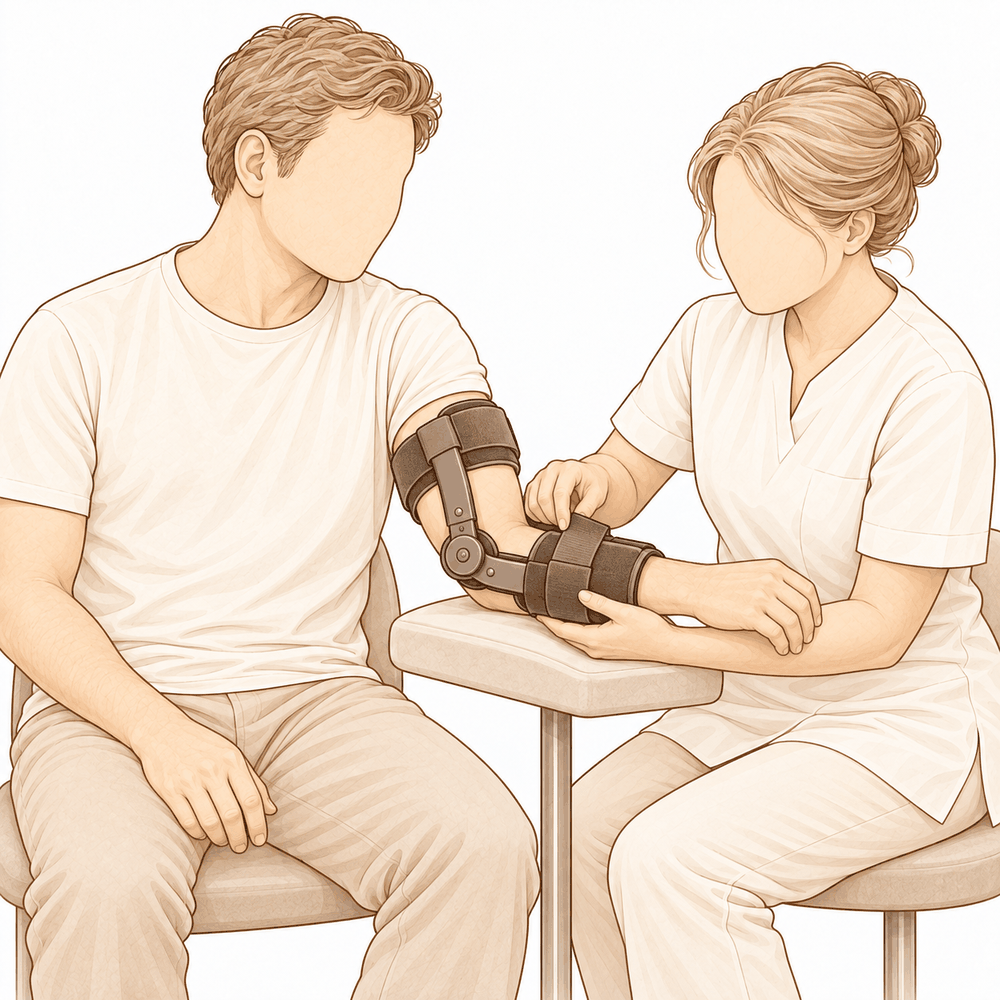
If your elbow is still loose once everything is repaired, a temporary hinge may be placed across the joint to hold it steady while the tissues heal. The cuts are closed with stitches and covered with a dressing. Early movement is part of the plan: the repair is designed to be strong enough for your elbow to start moving soon after surgery, because keeping it still for more than three weeks tends to leave it stiff.
After the operation
You will wake up in the recovery ward, where nurses keep an eye on you as the anaesthetic wears off. Pain relief is tailored to you, and your nurses will check in regularly to keep you comfortable. Your arm will be in a sling or a protective dressing, and you can get up and move around with help soon after. Please arrange for someone to stay with you for the first 24 hours. Your team will tell you whether you go home the same day or stay one night in hospital. We leave the dressing on for about 10 days; please do not take it off before then unless we tell you to. We change or remove it when we see you.
Recovery
For the first few days your elbow will be sore and swollen, and the skin may bruise down towards your hand. This is normal and settles steadily. Keeping your hand raised on pillows, even while you sleep, helps the swelling go down. Take the pain relief as directed rather than waiting for the pain to build.
You will go home with your arm in a sling for comfort. You can get up, walk around, and use your hand for light tasks such as eating and typing as soon as it feels comfortable. The dressing stays on for about 10 days; we change or remove it when we see you. Your first hand therapy appointment is with Ruby Doolan at Extend Rehabilitation. She is a hand therapist who will guide your exercises and make any splint you need. Early, gentle movement is the goal: the repair is designed to be strong enough for your elbow to start bending soon after surgery, because keeping it still too long tends to leave it stiff.
As the swelling settles, the exercises grow. You will work on straightening and bending the elbow, then turning your palm up and down. Grip strength comes back later. Once you can grip and lift without pain, everyday tasks feel normal again, and heavier work, sport and gym training follow as your strength returns. Your timeline may differ; your surgeon and your therapist will guide you.
What can go wrong
Most patients do well, but problems can occasionally happen. Your surgeon and the team monitor you closely to spot any issue early.
The nerve that runs along the inside of your elbow can be irritated by the operation. This can cause tingling, pins and needles, or numbness in your little and ring fingers. Some of this is expected while the nerve block wears off in the first 24 hours. If numbness or weakness lasts beyond that, call the clinic. If you cannot move your hand or fingers once the block has worn off, call the clinic.
The new ligament can loosen or fail, which makes the elbow feel unstable again, as if it might give way. If your elbow starts to feel loose or slips out of place, contact the clinic.
The elbow can become stiff after surgery. You might notice it not straightening or bending as far as it should, or feeling tight and hard to move. Raise this at your next review, as exercises or further treatment may help.
Infection is uncommon but serious. Watch for fever, redness that spreads out from the wound, or fluid or pus leaking from it. Call the clinic the same day if you notice any of these. Pain that keeps getting worse despite your pain medicines also needs a same-day call to the clinic.
Where a tendon graft is taken from, that spot can be sore or slow to settle, and the skin there can occasionally get infected. If the graft site becomes red, painful or leaks fluid, call the clinic the same day.
Some elbows develop arthritis in the joint over time after a bad injury. This can feel like a deep ache, or a grinding or clicking sensation when you move. Mention it at your next review so it can be assessed.
The complications table on this page lists typical rates if you want the specifics.
When to call us
Call the clinic if you notice fever, spreading redness around the wound, or fluid or pus leaking from it. Call the same day if pain keeps getting worse despite your pain medicines. Go to the emergency department if you have calf swelling or pain, or shortness of breath, as these can be signs of a blood clot. Numbness or weakness in the first 24 hours after a nerve block is expected. If it lasts beyond that, call the clinic. If you cannot reach us out of hours or on a weekend, go to your nearest emergency department.
Evidence & references
This is the clinical evidence summary written for health professionals. It is technical, and it lists the research this page was built from. You do not need to read it to understand your treatment or to make a decision about it.
Overview
- Surgical ligament reconstruction or repair is the most favorable treatment option for restoration of normal elbow function in posterolateral rotatory instability [4].
- Repair and reconstruction of the lateral ulnar collateral ligament offers practical and often successful solutions for patients with posterolateral rotatory instability of the elbow [10].
- Improving arthroscopic techniques provide a satisfactory alternative in stabilizing the elbow [7].
- The gravity varus test has biomechanical plausibility and confirms that pronation stabilizes the lateral elbow [1].
- An all-arthroscopic reconstruction technique for the lateral ulnar collateral ligament is reproducible and avoids residual instability [2].
- Open posterolateral ligament plication and lateral ulnar collateral ligament repair using an all-suture construct allows for complete posterolateral stabilization of the elbow with a single implant and bone preservation [3].
- Suture-augmented lateral ulnar collateral ligament and radial collateral ligament reconstruction provides a reproducible, anatomically based construct that restores posterolateral elbow stability and addresses the complex spectrum of lateral-sided injuries observed in posterolateral rotatory instability [5].
- Further follow-up and wider experience of the Surgilig/LockDown technique is required to assess its long-term functional outcome in comparison with more established surgical techniques for stabilization of posterolateral elbow instability [6].
- Arthroscopic lateral ulnar collateral ligament plication/reconstruction with augmented lateral collateral ligament imbrication allows effective management of elbow instability while promoting quicker patient recovery and long-term functional restoration [8].
- Acute repair of the lateral ulnar collateral ligament with a cortical button resulted in satisfactory clinical outcomes and successful restoration of elbow stability [9].
- The use of suture button fixation for repair of the lateral ulnar collateral ligament in terrible triad injuries has not been previously described [11].
- Corrective open-wedge osteotomy with structural bone allograft restored radial head alignment, eliminated dynamic subluxation, and achieved stable elbow function without the need for ligament reconstruction in complex posterolateral rotatory instability due to malunited radius fracture [12].
- Reconstruction of the lateral ulnar collateral ligament with a tendon graft offers an alternative that restores stability through a dynamic “sling effect” rather than rigid constraint [13].
Anatomy & Pathophysiology
Bony Anatomy
- The elbow is a trocho-ginglymoid joint consisting of medial and lateral articulations that provide bony stability [22].
- The ulnohumeral joint is formed by the articulation of the trochlea with the ulna within the greater sigmoid notch [22].
- The ulnohumeral joint provides highly congruent anatomy through almost 180° of articular contact, except for a bare area of the greater sigmoid notch devoid of cartilage [22].
- The radiocapitellar joint is formed by the articulation of the capitellum and the radial head [22].
- The radial head is a concave elliptical structure covered with articular cartilage along the radiocapitellar joint and approximately 270° of the articular margin [22].
- The radius is held in close approximation to the ulna at the proximal radioulnar joint by the annular ligament [22].
- The distal humeral articulation is angled 30° from the longitudinal axis of the humerus [22].
- The axis of rotation is angulated 5° to 7° in the coronal plane relative to the epicondylar axis, with the medial side more distal than the lateral side [22].
- The articular surface of the distal humerus is angled 30 degrees anterior to the humeral shaft axis [28].
- In full extension, 60% of axial load is transmitted through the radiocapitellar joint [28].
- The medial column of the distal humerus diverges from the humeral shaft at a 45-degree angle in the coronal plane [30].
- The lateral column of the distal humerus diverges from the humeral shaft at a 20-degree angle in the coronal plane [30].
- The trochlea is covered by articular cartilage anteriorly, inferiorly, and posteriorly, creating an arc of almost 270 degrees [33].
- The olecranon fossa and coronoid fossa are separated by a thin bony septum which is occasionally absent [33].
- The ulna medially bends approximately 8° at 8 cm from the tip of the olecranon [22].
- The articulation to the tip of the coronoid is approximately 30° from the long axis of the ulna in the sagittal plane [22].
Ligamentous Anatomy
- The lateral collateral ligament complex consists of the radial collateral ligament, the lateral ulnar collateral ligament (LUCL), and the annular ligament [33].
- The radial collateral ligament originates from an isometric point on the lateral epicondyle and fans out to attach to the annular ligament [33].
- The LUCL arises from an isometric point on the lateral epicondyle and attaches to the crista supinatoris of the proximal ulna [33].
- The annular ligament attaches to the anterior and posterior margins of the lesser sigmoid notch [33].
- The LUCL origin center is located 10.7 mm from the lateral epicondyle [21].
- The LUCL insertion is located 3.3 mm from the apex of the supinator crest [21].
- The LUCL acts as a posterior buttress for the radial head and the ulna to prevent subluxation [17].
- The LUCL is considered the principal constraint of the elbow joint against posterolateral rotatory instability (PLRI) [17].
- The lateral collateral ligament contributes 14% of varus stability of the elbow with the joint in full extension [37].
- The lateral collateral ligament contributes 9% of varus stability of the elbow with the joint in 90 degrees of flexion [37].
- The medial ulnar collateral ligament (MUCL) is the primary restraint to valgus stress within functional elbow range of motion [28].
- The MUCL originates on the posterior medial epicondyle and inserts on the sublime tubercle of the medial coronoid process [28].
- The anterior bundle of the MUCL is the primary restraint to valgus stress within functional elbow range of motion [28].
- The posterior bundle of the MUCL is the primary restraint to valgus stress with the elbow in maximal flexion [28].
- The common extensor tendon is a secondary stabilizer of the lateral elbow [16].
- The joint capsule is a secondary stabilizer of the elbow [19].
- The radiocapitellar articulation is a secondary stabilizer of the elbow [19].
Biomechanics & Pathophysiology
- Elbow stability is determined by primary and secondary stabilizers, with injury to these structures causing instability [19].
- The three primary stabilizers of the elbow are the ulnohumeral articulation, the MUCL, and the LUCL complex [19].
- Secondary stabilizers of the elbow include the radiocapitellar articulation, common flexor tendon, common extensor tendon, and joint capsule [19].
- Tensile forces are present at the medial elbow while compressive forces are present at the lateral elbow [23].
- The capsule allows maximum distension at approximately 70 to 80 degrees of flexion [23].
- The anterior capsule attaches at a point approximately 6 mm distal to the tip of the coronoid [23].
- Pronation stabilizes the lateral elbow [1].
- The LUCL is taut in both flexion and extension [37].
- Disruption of the LUCL results in posterolateral rotatory instability [37].
- Failure of primary stabilizers results in increased stress on secondary stabilizers, leading to capsular contractures, chondromalacia, osteophytes, and loose bodies [37].
- Ulnar nerve symptoms may develop from traction, scarring, or osteophyte impingement following primary stabilizer failure [37].
- The typical injury pattern for traumatic elbow dislocation involves a valgus internal rotation force of the forearm on a slightly flexed extremity [37].
- In traumatic elbow dislocation, structures are disrupted on the lateral side first, progressing medially as more force is applied [37].
- Posterolateral structures are most commonly affected when recurrence or persistence of instability results from traumatic dislocation [37].
- A coronoid fracture in association with disruption of the posterior band of the ulnar collateral ligament can result in symptomatic posteromedial instability [37].
- The normal elbow has a range of motion from 0° to 140° from extension to flexion [19].
- The normal elbow has a range of motion of 75° and 85° in pronation and supination respectively [19].
- A functional arc for elbow flexion and extension is 100° [19].
- A functional arc for forearm rotation is 100° [19].
- The normal valgus carrying angle of the elbow is 5 to 10 degrees for men and 10 to 15 degrees for women [28].
- The overall valgus angle of the elbow in extension, including the ulna, is 10 to 17 degrees [33].
- The distal humerus articular surface is internally rotated 3 to 8 degrees axially [33].
- As the elbow flexes, it internally rotates resulting in slight varus alignment [33].
Classification
- The lateral ulnar collateral ligament (LUCL) acts as a posterior buttress for the radial head and the ulna to prevent subluxation [17].
- Pronation stabilizes the lateral elbow in a cadaveric model of posterolateral rotatory instability [1].
Clinical Presentation
- The gravity varus test has biomechanical plausibility for evaluating posterolateral rotatory instability of the elbow [1].
- Complex posterolateral rotatory instability can be caused by a malunited radius fracture [12].
- Four-dimensional computed tomography scan can be used to diagnose complex posterolateral rotatory instability due to malunited radius fracture [12].
- Dynamic subluxation is a feature of complex posterolateral rotatory instability associated with malunited radius fracture [12].
Investigations
Physical Examination
- Pronation stabilizes the lateral elbow during the gravity varus test [1].
- Elbow stability is determined by primary stabilizers, which include the ulnohumeral articulation, the medial ulnar collateral ligament (MUCL), and the lateral ulnar collateral ligament (LUCL) complex [19].
- Secondary stabilizers of the elbow include the radiocapitellar articulation, the common flexor tendon, the common extensor tendon, and the joint capsule [19].
- The physical examination for elbow pathology is directed by the history and the location of patient pain in the anterior, posterior, medial, or lateral aspect of the elbow [19].
- The LUCL is considered the principal constraint of the elbow joint against posterolateral rotatory instability [17].
- The lateral ulnar collateral ligament injury is the cause of recurrent instability following elbow dislocation [41].
Imaging
- Plain radiographs remain the hallmark and best screening test for elbow evaluation [19].
- Standard radiographic views for elbow evaluation include AP, lateral, and oblique views [36].
- CT is helpful when assessing for malunion architecture and the location and pattern of osteophytes or loose bodies [36].
- Three-dimensional CT is used to check for heterotopic ossification [36].
- CT is not necessary when elbow stiffness is entirely soft-tissue related [36].
- CT is beneficial if any joint incongruity or abnormal bony anatomy is present [36].
- MRI can be used to evaluate ligaments and tendons, but it is rarely indicated for elbow stiffness [36].
- Magnetic resonance evaluation of the elbow includes imaging of ligament complexes [20].
- MRI findings in acute elbow dislocation provide insight into the mechanism of injury [20].
- MR evaluation of instability patterns emphasizes the soft-tissue lesions that result from dislocation [20].
- Four-dimensional computed tomography scan can be used for diagnosis in complex posterolateral rotatory instability due to malunited radius fracture [12].
Treatment
Operative Techniques and Constructs
- Surgical ligament reconstruction or repair is the most favorable treatment option for restoration of normal elbow function [4].
- Repair and reconstruction of the lateral ulnar collateral ligament (LUCL) offers practical and often successful solutions for patients with posterolateral rotatory instability (PLRI) of the elbow [10].
- An all-arthroscopic reconstruction technique for the LUCL is reproducible and achieves reconstruction while avoiding residual instability [2].
- Open posterolateral ligament plication and LUCL repair using an all-suture construct allows for complete posterolateral stabilization of the elbow with a single implant and bone preservation [3].
- A suture-augmented LUCL and radial collateral ligament reconstruction method provides a reproducible, anatomically based construct that restores posterolateral elbow stability and addresses the complex spectrum of lateral-sided injuries observed in PLRI [5].
- Acute repair of the LUCL with a cortical button resulted in satisfactory clinical outcomes and successful restoration of elbow stability [9].
- Arthroscopic LUCL plication/reconstruction with augmented lateral collateral ligament imbrication is a minimally invasive method that allows effective management of elbow instability while promoting quicker patient recovery and long-term functional restoration [8].
- Arthroscopic-assisted LUCL reconstruction provides less insult and dissection to the soft tissue at the lateral side of the elbow while being an excellent tool to diagnose any concomitant intraarticular pathologies [15].
- A knotless, onlay technique for LUCL reconstruction performs reconstruction with a tendon graft without violation of the extensor origin and soft tissue envelop [16].
- The use of knotless anchors and an onlay technique shortens operative time, reduces the required surgical exposure, and removes the risk of tunnel osteolysis or fracture and resultant graft failure while maintaining a broad bone surface for graft incorporation [16].
- A minimally-invasive dissection prevents iatrogenic injury to the common extensor origin, an important secondary stabilizer of the lateral elbow, and the articular capsule [16].
- Remaining extracapsular with a minimally-invasive technique avoids plication of the capsular structures or risk of formation of intra-articular adhesions, theoretically reducing the risk of any postoperative loss of range of motion [16].
Outcomes and Considerations
- The docking technique originally described by Jones et al. in 2012 is the most common method in use in contemporary practice for LUCL reconstruction [16].
- The docking technique has shown recurrent instability rates as high as 25% [16].
- A minimally-invasive approach allows for earlier rehabilitation and return of range of motion, reduced postoperative pain, and reduced operative time as a repair of the common extensor origin is not required following LUCL reconstruction [16].
- Fluoroscopic guidance can help to reduce injuries to unintended structures that could be foreseen due to a limited exposure [16].
- Postoperative stiffness is a known complication of LUCL reconstruction and occurs not uncommonly [16].
Non-Ligamentous Interventions
- Corrective open-wedge osteotomy with structural bone allograft restored radial head alignment, eliminated dynamic subluxation, and achieved stable elbow function without the need for ligament reconstruction in cases of complex PLRI due to malunited radius fracture [12].
Instability and Recurrence
- The docking technique for LUCL reconstruction has shown recurrent instability rates as high as 25% [16].
- The knotless, onlay technique for LUCL reconstruction removes the risk of tunnel osteolysis or fracture and resultant graft failure [16].
- The arthroscopic technique for LUCL reconstruction achieves reconstruction and avoids residual instability [2].
- The all-suture construct for posterolateral ligament plication and LUCL repair allows for complete posterolateral stabilization of the elbow [3].
- The suture-augmented LUCL and radial collateral ligament reconstruction method restores posterolateral elbow stability [5].
- Acute repair of the LUCL with a cortical button resulted in successful restoration of elbow stability [9].
- Repair and reconstruction of the LUCL offers practical and often successful solutions for patients with PLRI of the elbow [10].
- Reconstruction of the lateral ulnar collateral ligament with a tendon graft restores stability through a dynamic “sling effect” rather than rigid constraint [13].
Stiffness and Range of Motion
- The minimally-invasive, extracapsular technique for LUCL reconstruction theoretically reduces the risk of postoperative loss of range of motion by avoiding plication of capsular structures or risk of formation of intra-articular adhesions [16].
- The minimally-invasive method for managing elbow instability promotes quicker patient recovery and long-term functional restoration [8].
Iatrogenic Injury and Soft Tissue
- The minimally-invasive dissection for LUCL reconstruction prevents iatrogenic injury to the common extensor origin, an important secondary stabilizer of the lateral elbow [16].
- The minimally-invasive dissection for LUCL reconstruction prevents iatrogenic injury to the articular capsule [16].
- The arthroscopic-assisted LUCL reconstruction provides less insult and dissection to the soft tissue at the lateral side of the elbow [15].
Long-Term Outcomes
Recovery
- The minimally invasive arthroscopic method allows effective management of elbow instability while promoting quicker patient recovery and long-term functional restoration [8].
- The all-arthroscopic reconstruction technique is reproducible and achieves the reconstruction of the lateral ulnar collateral ligament as well as avoids residual instability [2].
- The open posterolateral ligament plication and lateral ulnar collateral ligament repair technique allows for complete posterolateral stabilization of the elbow with a single implant and bone preservation [3].
- The suture-augmented lateral ulnar collateral ligament and radial collateral ligament reconstruction method provides a reproducible, anatomically based construct that restores posterolateral elbow stability and addresses the complex spectrum of lateral-sided injuries observed in posterolateral rotatory instability [5].
- Reconstruction of the lateral ulnar collateral ligament with a tendon graft offers an alternative, restoring stability through a dynamic “sling effect” rather than rigid constraint [13].
- Clinical practice supports formal reconstruction of the lateral ulnar collateral ligament for chronic late presenting injury with posterolateral rotatory instability in the patient with open physes, without concern for causing significant growth arrest [14].
Key Evidence
- [L5] These findings support the biomechanical plausibility of the test and confirm that pronation stabilizes the lateral elbow. [1] (10.1016/j.jseint.2026.101792)
- [L5] The presented arthroscopic technique is reproducible and achieves the reconstruction of the LUCL of the elbow as well as avoids residual instability. [2] (10.1016/j.eats.2024.103096)
- [L5] The technique allows for complete posterolateral stabilization of the elbow with a single implant and bone preservation. [3] (10.1016/j.eats.2024.103172)
- [L4] Surgical ligament reconstruction or repair is the most favorable treatment option for restoration of normal elbow function. [4] (10.1016/s1058-2746(03)00091-0)
- [L5] The described method provides a reproducible, anatomically based construct that restores posterolateral elbow stability and addresses the complex spectrum of lateral-sided injuries observed in PLRI. [5] (10.1016/j.eats.2025.103797)
- [L4] Further follow-up and wider experience of the technique is required to assess its long-term functional outcome in comparison with more established surgical techniques for stabilization of posterolateral elbow instability. [6] (10.1097/bte.0000000000000025)
- [L5] Improving arthroscopic techniques provide a satisfactory alternative in stabilizing the elbow. [7] (10.1016/s0278-5919(05)70246-5)
- [L5] This minimally invasive method allows effective management of elbow instability while promoting quicker patient recovery and long-term functional restoration. [8] (10.1016/j.eats.2025.103529)
- [L4] The study found that acute repair of the LUCL with a cortical button resulted in satisfactory clinical outcomes and successful restoration of elbow stability. [9] (10.1097/bte.0000000000000178)
- [L5] Repair and reconstruction of the LUCL now offers practical and often successful solutions for patients with PLRI of the elbow. [10] (10.1016/j.csm.2004.06.010)
- [L4] The use of suture button fixation for repair of lateral ulnar collateral ligament has not been previously described. [11] (10.1016/j.eats.2023.10.004)
- [L5] Corrective open-wedge osteotomy with structural bone allograft restored radial head alignment, eliminated dynamic subluxation, and achieved stable elbow function without the need for ligament reconstruction. [12] (10.1016/j.xrrt.2026.100840)
- [L5] Reconstruction of the lateral ulnar collateral ligament with a tendon graft offers an alternative, restoring stability through a dynamic “sling effect” rather than rigid constraint. [13] (10.1002/atn2.70037)
- [L5] Clinical practice supports formal reconstruction of the LUCL for chronic late presenting injury with PLRI in the patient with open physes, without concern for causing significant growth arrest. [14] (10.1097/bth.0b013e3181dd88f3)
- [L5] It provides less insult and dissection to the soft tissue at the lateral side of the elbow while being an excellent tool to diagnose any concomitant intraarticular pathologies. [15] (10.1016/j.eats.2024.103101)
- [L5] [16] (10.1002/atn2.70135)
- [L5] On the basis of these morphological features, the LUCL might act a posterior buttress for the radial head and the ulna to prevent its subluxation, and therefore it is considered to be the principal constraint of the elbow joint against PLRI. [17] (10.1177/1558944716660555mm)
References
[1] Biomechanical Evaluation of the Gravity Varus Test in a Cadaveric Model of Posterolateral Rotatory Instability of the Elbow. JSES International. 2026. DOI: 10.1016/j.jseint.2026.101792
[2] Posterolateral Elbow Dislocation: An All‐Arthroscopic Reconstruction of the Lateral Ulnar Collateral Ligament. Arthroscopy Techniques. 2024. DOI: 10.1016/j.eats.2024.103096
[3] Open Posterolateral Ligament Plication and Lateral Ulnar Collateral Ligament Repair in Posterolateral Rotatory Instability of the Elbow Using an All‐Suture Construct. Arthroscopy Techniques. 2024. DOI: 10.1016/j.eats.2024.103172
[4] Surgical reconstruction for posterolateral rotatory instability of the elbow. Journal of Shoulder and Elbow Surgery. 2003. DOI: 10.1016/s1058-2746(03)00091-0
[5] Suture‐Augmented Lateral Ulnar Collateral Ligament and Radial Collateral Ligament Reconstruction for Subacute and Chronic Posterolateral Rotatory Instability. Arthroscopy Techniques. 2025. DOI: 10.1016/j.eats.2025.103797
[6] Surgical Repair of Posterolateral Rotatory Instability of the Elbow Using Surgilig/LockDown. Techniques in Shoulder & Elbow Surgery. 2014. DOI: 10.1097/bte.0000000000000025
[7] Posterolateral Rotatory Instability of the Elbow. Clinics in Sports Medicine. 2001. DOI: 10.1016/s0278-5919(05)70246-5
[8] Arthroscopic Lateral Ulnar Collateral Ligament Plication/Reconstruction With Augmented Lateral Collateral Ligament Imbrication. Arthroscopy Techniques. 2025. DOI: 10.1016/j.eats.2025.103529
[9] Clinical Outcomes of Lateral Ulnar Collateral Ligament Repair of the Elbow Using a Cortical Button. Techniques in Shoulder & Elbow Surgery. 2019. DOI: 10.1097/bte.0000000000000178
[10] PLRI: posterolateral rotatory instability of the elbow. Clinics in Sports Medicine. 2004. DOI: 10.1016/j.csm.2004.06.010
[11] Suture Button Repair for Lateral Ulnar Collateral Ligament in Terrible Triad Injuries: Surgical Technique. Arthroscopy Techniques. 2024. DOI: 10.1016/j.eats.2023.10.004
[12] Complex posterolateral rotatory instability due to malunited radius fracture: diagnosis with four-dimensional computed tomography scan and corrective osteotomy. JSES Reviews, Reports, and Techniques. 2026. DOI: 10.1016/j.xrrt.2026.100840
[13] Arthroscopic Lateral Collateral Ligament Reconstruction With Tendon Graft in High‐Grade Atraumatic Posterolateral Rotatory Instability in Elbows. Arthroscopy Techniques. 2026. DOI: 10.1002/atn2.70037
[14] Surgical Treatment of Posterolateral Rotatory Instability of the Elbow in Children. Techniques in Hand & Upper Extremity Surgery. 2010. DOI: 10.1097/bth.0b013e3181dd88f3
[15] Arthroscopic‐Assisted Lateral Ulnar Collateral Ligament Reconstruction for Posterolateral Rotatory Instability of the Elbow: A Technical Note. Arthroscopy Techniques. 2024. DOI: 10.1016/j.eats.2024.103101
[16] Lateral Ulnar Collateral Ligament Reconstruction Through a Mini‐Invasive Approach. Arthroscopy Techniques. 2026. DOI: 10.1002/atn2.70135
[17] Morphology of the Lateral Ulnar Collateral Ligament of the Elbow. HAND. 2016. DOI: 10.1177/1558944716660555mm
[19] Orthopaedic Knowledge Update 13 Ebook Without Multimedia. Anatomy, Biomechanics, Physical Examination, and Imaging of the Elbow > Summary and Conclusions.
[20] Orthopaedic Knowledge Update Sports Medicine 6. Magnetic Resonance Imaging of the Elbow > Annotated References.
[21] Orthopaedic Knowledge Update 13 Ebook Without Multimedia. Anatomy, Biomechanics, Physical Examination, and Imaging of the Elbow > Annotated References.
[22] Orthopaedic Knowledge Update 13 Ebook Without Multimedia. Anatomy, Biomechanics, Physical Examination, and Imaging of the Elbow > Anatomy > Bony Anatomy.
[23] Miller S Review Of Orthopaedics. SECTION 16 PATELLAR TRACKING IN TOTAL KNEE ARTHROPLASTY > TABLE 2.3 Shoulder Spaces.
[28] Miller S Review Of Orthopaedics. ELBOW.
[30] A Lange Medical Book Current Diagnosis Treatment In Orthopedics Fifth Edition. 2Musculoskeletal Trauma Surgery > INJURIES AROUND THE ELBOW.
[33] Rockwood And Green S Fractures In Adults. 29: Principles of Nonunion and Bone Defect Treatment > Pathoanatomy and Applied Anatomy Related to Distal Humerus Fractures.
[36] Aaos Comprehensive Orthopaedic Review 3. Elbow Stiffness* > IV. Evaluation.
[37] Campbell S Operative Orthopaedics 4 Volume Set. POSTERIOR SURGICAL APPROACH FOR QUADRILATERAL SPACE SYNDROME > MCLAUGHLIN PROCEDURE > ARTHROSCOPIC SURGERY.
[41] A Lange Medical Book Current Diagnosis Treatment In Orthopedics Fifth Edition. 2Musculoskeletal Trauma Surgery > ELBOW DISLOCATION.