Clinicians › Hand
Fingertip Injuries
Crush, laceration, nail-bed and amputation injuries of the fingertip and their management.
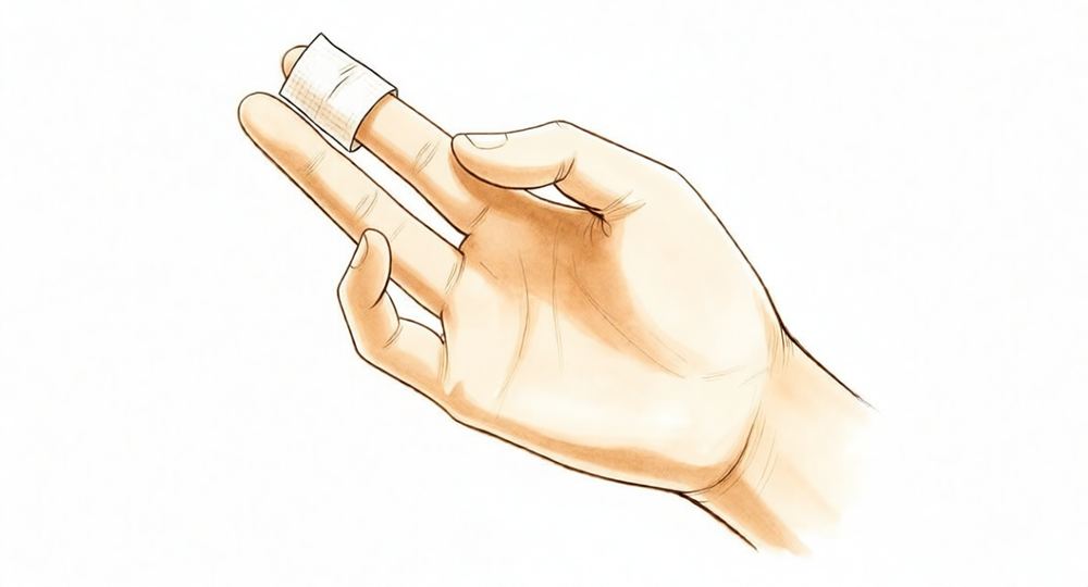
For patients: a plain-language version of this topic is available. See the patient guide.
Overview
Fingertip injuries are common in children, predominantly occurring at home in doors or windows, and represent a significant but mostly preventable burden [7]. These injuries require the same meticulous care as all other hand surgery, aiming for coverage with high-quality skin and optimal sensibility [9]. A thorough understanding of the involved structures generally leads to esthetic and functional results [8]. While a simple and efficient treatment method is recommended for all injury types [1], the best treatment for composite defects remains undetermined due to a lack of prospective randomized trials and disparate retrospective data [60]. Reconstructive options depend on the location of damage [13], and specific techniques such as the purse-string suture are recommended to preserve the nail [50]. The open method has a definite place in certain cases [4], while silver sulphadiazine dressings offer advantages by eliminating the need for hospital admission and operating theatre time [17].
Fingertip replantation is recommended to achieve the best appearance and functional outcome [29], and is particularly advised in paediatric patients whenever possible, despite technical demands [41]. However, strong evidence from randomized controlled trials supporting very distal finger replantation is lacking, largely because well-designed prospective studies are difficult to conduct [33]. Secondary procedures are often necessary following hand and digit replants [31]. The Fingertip Injury Outcome Score (FIOS) is the most complete fingertip-specific instrument and should be adopted as the primary standard for outcome reporting and long-term follow-up [3].
Specific reconstructive techniques have defined indications. The extended step-advancement flap is a viable alternative to replantation [59], and the parallelogram flap is preferred for reconstruction with bone exposure [14]. The reversed digital artery island flap is better indicated for large dorsal defects of the middle and third phalanx rather than fingertip injuries [42]. Conservation of amputated fingertips provides new possibilities for late reconstruction [6]. In a series from 1976 to 1986, 74 patients with 96 injured fingers treated with a deepithelialized pedicle flap placed subcutaneously resulted in a well-contoured fingertip in all patients [5]. The low incidence of infection (2.5%) and lack of meaningful difference between groups call into question prophylactic antibiotic prescribing after distal fingertip injuries [12].
Anatomy & Pathophysiology
General Hand Architecture & Function
The hand serves as both an information-gathering organ and an organ of execution, functioning efficiently only when the proximal joints of the limb are stable yet mobile [76]. Located at the extremity of the upper limb, which acts as its vector, the hand confers uniqueness to the upper extremity [76]. The dorsal surface is convex and aesthetically important, while the concave palmar surface is the functional aspect [76]. The distal half of the hand divides into five digits that flex toward the palm; these digits converge during closing (flexion and adduction) and diverge during opening (extension and abduction) [76]. The thumb occupies a more proximal and lateral position, permitting movement inward and outward from the palm, while the four fingers represent the distal extension of the carpometacarpal part of the hand [76]. Finger movement hinges are located at the thenar crease and the transverse distal palmar crease rather than at the bases of the digits [76]. When fully extended and touching, digit tips describe a regular curve with peripheral digits being shortest; when extended and separated, the tips lie on a circle centered on the head of the third metacarpal [76].
The hand comprises 19 bones, 17 articulations, and 19 intrinsic muscles, along with a similar number of tendons activated by forearm muscles [76]. Functional prehension requires a stable wrist and at least two sensate digits capable of opposing with power [57]. Painless and full hand function depends on the seamless integration of joints, muscles, and nerves to complete basic tasks [24].
Osseous Skeleton
The skeleton of the hand and wrist consists of 27 bones, 19 of which are long bones, divided into five rays forming polyarticulated chains of metacarpals and phalanges [39]. Each metacarpal base articulates with the distal carpal row, while the carpus articulates with the forearm via its proximal row [39]. The radioulnocarpal articulation possesses two axes of movement, supplemented by a third axis of pronation and supination from the forearm, allowing the hand to assume any spatial configuration for grasping [39]. The radial ray (first ray) is the shortest, comprising a metacarpal and two phalanges, and continues the external carpal column formed by the scaphoid and trapezium [39]. The trapezium is angled anteriorly so that the first metacarpal forms a 45-degree angle with the second metacarpal in the sagittal plane [39]. The remaining four rays are of unequal length, each formed by a metacarpal and three phalanges [39].
Metacarpal length varies, with the thumb metacarpal being the shortest and the index finger metacarpal the longest, while others decrease in length from the third to the fifth digits [39]. The proximal and middle phalanges of the middle and ring fingers are longer than those of the index finger, making the long and ring fingers generally longer than the index finger [39]. Relative skeletal segment lengths vary with fist opening and closing; in flexion, digital extremes converge toward the thumb pulp for pinch or the thenar eminence base for power grip [39]. Ulnar digits deviate obliquely toward the palm, with deviations toward the median axis occurring primarily at the metacarpophalangeal and proximal interphalangeal articulations [39]. The two ulnar metacarpals, especially the fifth, exhibit slightly more flexion–rotation mobility to compensate for their lack of length [39].
The hand skeleton presents longitudinal and transverse concavities, forming a cup shape when the thumb is placed next to the index finger [39]. When the thumb spreads to grasp an object, this cup becomes a gutter with a major oblique axis following the thumb crease [39]. The transverse axis of the palm corresponds to the metacarpophalangeal articulations and is oblique, more distal at the index metacarpophalangeal joint and more proximal at the fifth, forming an acute angle of approximately 75 degrees with the longitudinal axis [39]. Epiphyseal plates are located at the proximal ends of the phalanges and the first metacarpal, and at the distal ends of the other metacarpals [39].
The index metacarpal is the most firmly fixed [81]. The ring metacarpal acts as a transitional element to the fifth metacarpal, possessing about 10 degrees of mobility in flexion and extension [81]. The fifth metacarpal is semi-independent, articulating with the hamate and restrained radially by its articulation with the base of the fourth metacarpal, with a flexion–extension range of approximately 20 degrees [81]. The second to fifth metacarpals are bound by fibrous structures, the most distal being the deep transverse intermetacarpal ligament, also known as the interglenoid ligament [81]. This ligament ties together the anterior “glenoid ligaments” of the metacarpophalangeal articulations, known as volar plates [81]. The longitudinal arches consist of a fixed carpometacarpal portion and a mobile digital portion, with the metacarpophalangeal articulations serving as keystones [81]. Thick anterior glenoid capsules, or volar plates, prevent hyperextension at the metacarpophalangeal joints and are interconnected by the transverse interglenoid ligament [81]. Stability of the metacarpophalangeal joints is essential for supporting both the longitudinal and transverse metacarpal arches [81]. The thumb ray exhibits considerable mobility and independence, while the index ray has a degree of independence at the phalangeal level due to its flexor and extensor muscle arrangement [81].
Musculature & Extensor Mechanism
Digital posture control requires a complex balance of extrinsic and intrinsic muscle forces [38]. Extrinsic muscles originate outside the hand and insert on the hand or carpus, functioning as flexors or extensors, while intrinsic muscles have both origin and insertion within the hand and contribute to both digital flexion and extension [38]. Extrinsic extensors run through six fibroosseous retinacular compartments at the wrist [38].
Compartment 1 (Most Radial): Contains the abductor pollicis longus and extensor pollicis brevis [38]. The abductor pollicis longus has multiple slips inserting at the base of the thumb metacarpal to radially abduct the thumb [38]. The extensor pollicis brevis inserts on the dorsum of the proximal aspect of the thumb’s proximal phalanx, actively extending the thumb metacarpophalangeal joint [38].
Compartment 2: Contains the extensor carpi radialis longus and extensor carpi radialis brevis [38]. The extensor carpi radialis longus inserts on the index metacarpal, dorsiflexing and radially deviating the wrist [38]. The extensor carpi radialis brevis inserts into the base of the middle metacarpal, providing balanced wrist dorsiflexion [38].
Compartment 3: Contains the extensor pollicis longus, which runs longitudinally down the forearm and turns abruptly radialward about Lister tubercle [38]. It inserts on the distal phalanx, providing forceful extension of the thumb interphalangeal joint [38]. Its oblique course provides a substantial adduction component to its pull [38].
Compartment 4: Contains the extensor indicis proprius, lying deep to the four tendons of the extensor digitorum communis [38]. The extensor indicis proprius inserts on the index finger ulnar to the extensor digitorum communis [38]. It may be recognized at the wrist level as possessing the most distal muscle belly of any digital extensor tendon [38].
Compartment 5: Contains the extensor digiti quinti, which inserts on the little finger ulnar to the extensor digitorum communis insertion [38].
Compartment 6: Contains the extensor carpi ulnaris, which inserts at the base of the little finger metacarpal, providing wrist extension and ulnar deviation [38].
The extensor indicis proprius, extensor digitorum communis, and extensor digiti quinti each play a role in digital extension at the metacarpophalangeal, proximal interphalangeal, and distal interphalangeal joints [38]. The extensor digitorum communis inserts on the index, middle, ring, and, in some cases, little fingers [38]. The principal bony insertion of extrinsic digital extensors is on the dorsal proximal aspect of the middle phalanx [38]. Metacarpophalangeal joint extension is provided by extrinsic extensor force transmitted through the sagittal bands [38]. Distal interphalangeal joint extension is achieved through conjoined lateral bands composed of tendinous slips from extrinsic and intrinsic tendons [38]. The extensor digitorum communis tendons of the middle, ring, and little fingers are tethered together by juncturae tendinum over the dorsum of the hand proximal to the metacarpophalangeal joint [38].
Digital extensor tendons are stabilized over the mid-line of the metacarpophalangeal joint by attachment to sagittal band fibers, which insert onto the volar proximal phalanx and the lateral borders of the volar plate [38]. These fibers form a sling that transmits proximal extrinsic extensor tension to the proximal phalanx, permitting metacarpophalangeal joint extension without a direct tendinous insertion onto the proximal phalanx [38]. The sagittal bands keep the extrinsic extensor as far as possible from the center of rotation of the metacarpophalangeal joint, maximizing mechanical efficiency [38]. Rupture or attenuation of sagittal band fibers allows the extrinsic extensor tendon to sublux to the ulnar side of the metacarpal head, causing ulnar deviation of the finger [38].
There are seven interosseous muscles: four dorsal and three volar [78]. The anatomic axis of the hand coincides with the axis of the third metacarpal [78]. The dorsal interossei are abductors, lying to the radial side of the index and middle fingers and the ulnar side of the middle and ring fingers [78]. The little finger is abducted by the abductor digiti quinti [78]. The volar interossei are adductors, lying to the ulnar side of the index finger and the radial side of the ring and little fingers [78]. The middle finger has two dorsal interossei (abductors) and no volar interossei (adductors) because the central axis of the hand lies within it [78].
Each dorsal interosseous muscle, with the exception of the third, has two muscle heads [78]. The superficial head arises dorsally from the shaft of contiguous metacarpals and inserts deeply by a medial tendon onto the lateral tubercle of the base of the proximal phalanx [78]. It abducts and weakly flexes the proximal phalanx, with no direct effect on the middle or distal phalanges [78]. The deep head forms a lateral tendon, or lateral band, at the level of the metacarpophalangeal joint [78]. It flexes and weakly abducts the proximal phalanx while extending the middle and distal phalanges [78]. At the middle of the proximal phalanx, transverse fibers arch dorsally from each lateral band to join over the dorsum of the finger, flexing the proximal phalanx [78]. Oblique fibers (spiral fibers) from the lateral bands sweep over the distal third of the proximal phalanx to insert onto the lateral tubercles at the base of the middle phalanx, extending the middle phalanx (proximal interphalangeal joint) [78]. The lateral bands are joined by the lateral slips of the extensor tendon to form the conjoined lateral band [78]. The two conjoined lateral bands to each finger unite at the distal third of the middle phalanx to form the terminal tendon, which inserts at the base of the distal phalanx to extend it [78].
The flexor digiti quinti brevis is structurally and functionally similar to the deep head of the dorsal interossei, forming the ulnar lateral band of the little finger [78]. The three volar interossei arise from adjacent surfaces of contiguous metacarpal shafts [78]. Each volar interosseous muscle has only one head and none insert onto the proximal phalanx [78]. They form the ulnar lateral band of the index finger and the radial lateral band of the ring and little fingers [78]. The volar interossei send oblique or spiral fibers that insert onto the base of the middle phalanx at its lateral tubercle [78]. All lateral bands are joined by the lateral slips of the extensor tendon to form a conjoined lateral band and finally a terminal tendon extending to the distal phalanx [78]. The abductor digiti quinti and flexor digiti quinti brevis are similar in structure and function to the superficial and deep heads of the dorsal interossei, respectively, and arise from the fifth metacarpal [78]. The abductor digiti quinti inserts onto the ulnar lateral tubercle at the base of the proximal phalanx of the little finger [78]. The flexor digiti quinti forms the ulnar lateral band [78]. The opponens digiti quinti lies deepest, arising from the pisohamate ligament and the hook of the hamate, and inserting onto the ulnar side of the diaphysis of the fifth metacarpal, where it flexes and supinates the fifth metacarpal [78].
Flexor Tendon Anatomy
The extrinsic finger flexors are the flexor digitorum profundus and the flexor digitorum superficialis [38]. The flexor digitorum profundus inserts on the proximal volar aspect of the distal phalanx [38]. It flexes the distal interphalangeal joint as well as the proximal interphalangeal and metacarpophalangeal joints [38]. The flexor digitorum superficialis acts as a flexor of the proximal interphalangeal and metacarpophalangeal joints [38].
Classification
PNB: The PNB classification separates fingertip injuries into their effect on the pulp, nail, and bone [108]. It provides a three-digit number that accurately describes the injury for documentation, treatment instructions, and referral indications [108]. A study assessed the inter-observer reliability and repeatability of the PNB classification using a classical assessment design where observations were classified by several observers at several times [25]. In this reliability study, patients under 18 were excluded [25].
Allen: The Allen classification is a simple method of classification that has been consistently used to grade fingertip amputations [179]. However, it cannot identify suitable patients with volar oblique amputations who are best suited for neurovascular island flaps [179].
Arterial System-Based: A three-dimensional concept classification based on the damaged arterial system is useful to predict the site of the ruptured artery to be anastomosed [100]. This arterial system-based classification can make distal fingertip replantation easier [100].
SATT: The SATT classification system is based first on identifying the severity of the initial injury and determining whether the involved parts are viable [160]. It takes into account anatomical localisation (isolated vs. extended) and topography (volar vs. dorsal) to direct management [160]. The system also considers the type of injury (sharp vs. crush-avulsed) to ensure the hand will be functional to the full extent possible [160].
Ring Avulsion: Ring avulsion injuries are classified into Class I, Class II, and Class III based on circulation status and tissue viability [176]. Class I injuries have adequate circulation where standard bone and soft tissue treatment is sufficient [176]. Class II injuries have inadequate circulation where vessel repair preserves viability [176]. Class III injuries involve complete degloving or amputation where judgment is required regarding revascularization [176]. Class IIA injuries are defined as those in which only digital arteries are damaged but all other structures are intact and functional [176].
Lister: The Lister classification of distal digital amputation divides injuries into zones, with Zone II and Zone III being palmarly orientated in the context of Gigogne flap reconstruction [157].
Ishikawa: The Ishikawa classification is used to determine the level of amputation of the fingertip [72].
Other Considerations: The mechanism of injury for fingertip amputations can be categorized as crush, avulsion, or sharp [72].
Clinical Presentation
History and Epidemiology
Hand injuries are common presentations in the emergency department [102]. Fingertip injuries in athletes may arise from a myriad of causes on the field and may occur in virtually any sport [26]. Two of the most common fingertip injuries in athletes are mallet and jersey fingers [26]. Specific mechanisms include firework-related trauma, which involves burns to the fingers, hand, and wrist (26.7%), injuries to the eye (14.9%), and open injuries to the hand and wrist (6.5%) [23]. Snowblower-related injuries lead to multiple types of trauma, mainly to the longer fingers [55]. Weakened rings allow avoiding complete finger avulsion but are the cause of severe and complex lesions that require specialized management [51]. Patients presenting with abuse are significantly more likely to have fingertip injuries during childhood compared with those without recorded abuse [18].
Clinical Evaluation
Assessment of patients with fingertip injuries should include a focused history including age, sex, handedness, mechanism of injury, occupation, smoking status, medical comoridities, tetanus vaccination, and previous operations on the affected hand [47]. The examiner should elicit subjective symptoms including numbness, weakness, or pain during the evaluation of fingertip injuries [47]. A careful physical examination is essential to direct care and future testing if indicated for hand and wrist injuries [24]. Patients often have difficulty accurately describing their symptoms and may incorrectly attribute pathology to a perceived deficit [24]. The Pulp Nail Bone (PNB) classification for fingertip injuries has been assessed for inter-observer reliability and repeatability [25].
Symptoms and Outcomes
Patients continued to experience symptoms long-term after treatment of open fingertip injury, particularly 'cold intolerance' or 'Trauma-Induced Cold Associated Symptoms' (TICAS) [22]. Function was not significantly affected in some cases despite long-term symptoms after open fingertip injury [22]. A correlation exists between experienced loss of function and an estimated diagnosis of major depression in patients with a fingertip injury [19]. Sensation recovery is of primary importance for fingertip injuries [16]. Sensation is the most important factor in thumb or fingertip repair, constituting 40% of the goal, while length and appearance account for 50% [52]. Ectopic nail formation should be included within the differential diagnosis when evaluating distal finger masses, especially in cases of previous distal digital trauma [110]. The Fingertip Injuries Outcome Score (FIOS) is the most complete fingertip-specific outcome instrument and should be adopted as the primary standard for outcome reporting and long-term follow-up [3]. A FIOS score of 12 or less indicates excellent, 13–18 indicates good, 19–24 indicates fair, and greater than 24 indicates poor [21].
Investigations
MRI: Magnetic resonance imaging aids in the diagnosis of a glomus tumor when it presents in an unusual location within the fingertip [162].
Other Considerations: Fingertip injuries in pediatric patients and those without distal phalanx fractures achieve better cosmesis scores [71]. The rare presentation of subungual exostosis in a finger often stimulates an extensive differential diagnosis due to lack of recognition, creating potential for misdiagnosis and overaggressive treatment [53].
FIOS is the most complete fingertip-specific outcome instrument and should be adopted as the primary standard for outcome reporting and long-term follow-up across fingertip injuries [3]. Fingertip injuries in children are common and result in significant burden, yet are mostly preventable, with most injuries occurring at home in a door or window [7]. Patients presenting with abuse are significantly more likely to have fingertip injuries during childhood compared with those without recorded abuse, suggesting these injuries may be ones of abuse or neglect [18].
Treatment
General Principles and Outcomes
The precise management of adult fingertip injuries depends on the degree of injury, with a variety of operative and non-operative techniques available for successful treatment [15]. These injuries must be treated with the same care as other hand surgery, prioritizing coverage of the fingertip with high-quality skin and optimal sensibility [9]. Treatment approaches vary significantly between Asian and European surgeons; Europeans favor conservative management and spontaneous regeneration for fingertip defects, while Asians more frequently utilize flaps and replantation [141]. Long-term symptoms, particularly 'cold intolerance' or 'Trauma-Induced Cold Associated Symptoms' (TICAS), may persist after treatment of open fingertip injuries, although function is not significantly affected in some cases [22]. Outcomes are evaluated using the Fingertip Injuries Outcome Score (FIOS), where a score of 12 or less indicates excellent, 13–18 indicates good, 19–24 indicates fair, and greater than 24 indicates poor [21].
Non-Operative
Conservative treatment is recommended as a safe and simple option after fingertip loss, even when bone is exposed in the wound [20]. Conservative nonsurgical treatment of fingertip amputations, with or without bone exposure, can be treated successfully without surgical intervention [143]. Fingertip amputation injuries have the potential for regeneration through healing by secondary intention under semiocclusive dressing conditions [134]. The semiocclusive dressing is a suitable alternative for treating fingertip injuries [152]. A semipermeable dressing causes less discomfort during removal for dressing changes, particularly in early healing stages, and appears to enhance healing times [155]. Management with silver sulphadiazine dressings offers advantages including the removal of the need for hospital admission and operating theatre time [17]. Conservative treatment of thumb tip injuries with semi-occlusive dressing yields excellent results regarding the restoration of contour and sensibility in pulp injuries [48].
Operative
Indications: Fingertip replantation is recommended to obtain the best appearance and gain better functional outcomes [29]. Although technically demanding, paediatric fingertip replantation is recommended whenever possible due to achievable good outcomes [41]. Age alone should not be an absolute contraindication to finger replantation [92]. Results confirm positive long-term functional outcomes of thumb replantation interventions [28]. Functional results of successfully replanted fingers are rated from fair to excellent, with better outcomes in younger patients [62]. There is a lack of strong evidence, such as randomized controlled trials, to support clinical experience with very distal finger replantation, though conducting well-designed prospective studies for this procedure is considered highly difficult [33]. Despite microsurgical advances and high surgical expertise, the finger survival rate after ring avulsion injuries remains mostly influenced by the extent of intrinsic damage [35].
Surgical Approach / Technique: The parallelogram flap is a better choice for reconstruction of fingertip injury with bone exposure compared to the homodigital island flap [14]. The free dorsal middle phalangeal finger flap provides excellent sensory and aesthetic recovery for severe fingertip injuries [36]. The deepithelialized pedicle flap placed subcutaneously for fingertip reconstruction results in a well-contoured fingertip in all patients [5]. The split-thickness nail bed flap graft effectively achieves aesthetic and functional repair of a distal partial digit defect [125]. The volar cross-finger flap using the index finger for transverse and dorsal-oblique amputations of the distal thumb results in all flaps and skin grafts surviving, with no instances of flap loss, infection, or donor site complication [138]. The Moberg flap, first dorsal metacarpal artery island flap, and reverse flow homodigital flaps play roles in the management of thumb tip injuries [48]. The traditional armamentarium of flaps for thumb tip injuries has been expanded and replaced by a wide variety of flaps with more versatility and less donor side morbidity [48]. It is recommended that the purse-string suture technique be applied to all fingertip injuries to preserve the nail [50]. When considering reconstructive options for hand injuries involving the fingertip, the location of damage should be considered [13]. The authors do not agree with the system of freeing the caught hand by rotating the handle in the reverse direction due to the potential risk of damaging vital structures [2].
Complications
Infection: The incidence of infection following distal fingertip amputation and crush injury is 2.5% [12]. There is no meaningful difference in outcomes between groups regarding prophylactic antibiotic prescribing after distal fingertip injuries [12]. An animal bite of an index finger that was primarily sutured became infected [166].
Cold Intolerance and Vasospasm: Patients continue to experience symptoms long-term after treatment of open fingertip injury, particularly 'cold intolerance' or 'Trauma-Induced Cold Associated Symptoms' (TICAS) [22]. The incidence of cold-induced vasospasm after replanted digital amputations has been reported to be as high as 100% [159]. Cold-induced vasospasm in replanted digits does not improve with time [159]. Patients with severe cold-induced vasospasm problems did not experience improvement over time [159].
Neuropathic Pain and Sensory Deficits: One in three patients reporting long-term neuropathic pain following primary terminalization for acute fingertip injuries [64]. Sixteen patients with twenty fingertip injuries treated with volar V-Y plasty reported an average estimate of sensitivity of 73% of normal [10].
Necrosis and Graft Failure: In a series of 7 fingers treated with nonmicrosurgical replantation using a subcutaneous pocket, three fingers developed total necrosis [37]. In a series of 7 fingers treated with nonmicrosurgical replantation using a subcutaneous pocket, one finger survived completely but became atrophic after 4 months [37]. In a series of fingertip replantations using a single volar arteriovenous anastomosis, partial necrosis occurred in 1 case [70]. Phalangization for thumb reconstruction generally yielded poor results with repeated ulceration and no increase in span [150].
Malignancy: Subungual melanoma has been reported to follow a single injury to the digit after an interval of between nine months and seven years [170]. The average interval between a single digit injury and the development of subungual melanoma is 2.9 years [170].
Deformity: Posttraumatic ectopic nail can be distinguished from one of congenital origin by the history of injury [149].
Recovery
Outcome Assessment and Functional Results: The Fingertip Injuries Outcome Score (FIOS) is the most complete fingertip-specific outcome instrument and should be adopted as the primary standard for outcome reporting and long-term follow-up across fingertip injuries [3]. Primary terminalization for acute fingertip injuries is associated with excellent long-term patient-reported outcomes, high levels of satisfaction, and a low rate of complications [64].
Replantation and Reconstruction Outcomes: Long-term functional outcomes of thumb replantation interventions are positive [28], with results confirming satisfactory general upper limb function, handgrip and pinch strength, and social and work reintegration [145]. Fingertip replantation is recommended to obtain the best appearance and to gain better functional outcome compared to amputation closure [29]. In a series of 7 fingers treated with nonmicrosurgical replantation using a subcutaneous pocket, only one survived completely but became atrophic after 4 months, while three fingers developed total necrosis [37]; in all cases of this technique, a protracted period of hand therapy was necessary before full recovery was attained [169]. The results of delaying replantation of digits overnight give results comparable with those of immediate replantation in selected cases [174]. Amputations of the distal phalanx and the thumb, being male, and ischemia time of greater than 12 hours seem to have a somewhat worse prognosis for digit replantation [184]. In a series of 3 digits treated with cross-finger dermal pocketing to augment venous outflow for distal fingertip replantation, all digits survived and patients regained baseline active motion 6 weeks after the procedure [183]. Early series of microvascular reconstruction of the traumatized thumb demonstrated survival in 13 of 15 transfers, with average two-point discrimination of 15 mm and interphalangeal joint range of motion 70% less than normal [196]. The aesthetic and functional outcomes of reconstructed thumbs and fingers using the vascularized half–big toenail flap significantly improved, and donor site functional morbidity was minimum [135]. The reconstructed finger exhibited satisfactory sensation and functional scores during follow-up using a staged hand-foot flap reciprocity protocol [127]. Despite microsurgical advances and high levels of surgical expertise, the finger survival rate after ring avulsion injuries is mostly influenced by the extent of intrinsic damage [35].
Rehabilitation and Follow-up Protocols: Prophylactic antibiotics were continued for 1 week following secondary thumb reconstruction via ectopic banking of bony phalanges [75]. Follow-up for this procedure began 1 week after discharge [75]. Kirschner wires were left in place until evidence of fusion was noted on radiographic examination, which was usually seen after approximately 6 weeks in secondary thumb reconstruction via ectopic banking [75]. Examinations of the reconstructed thumb focused on infection, range of motion of the IP and metacarpophalangeal joints, bone union of the graft, and signs of osseous resorption [75]. The best results with a shock absorbing finger cap were obtained when the cap was fitted within weeks rather than months of the finger-tip becoming painful [74]. At 6 months, very significant progress was observed with complete and total recovery of active flexion amplitudes of the proximal and distal interphalangeal joints of 4 long fingers following secondary tendon surgery [177].
Key Evidence
- [L4] We recommend this method for the management of all types of fingertip injuries. [1] (10.1016/s0266-7681(05)80019-1)
- [L4] The authors do not agree with the system of freeing the caught hand by rotating the handle in the reverse direction due to the potential risk of damaging vital structures. [2] (10.1016/0266-7681(85)90045-2)
- [L3] FIOS is the most complete fingertip-specific outcome instrument and should be adopted as the primary standard for outcome reporting and long-term follow-up across fingertip injuries. [3] (10.2106/jbjs.rvw.25.00128)
- [L3] This prospective study shows that the open method has a definite place in the treatment of certain fingertip injuries. [4] (10.1016/s0363-5023(82)80042-7)
- [L4] From 1976 through 1986, 74 patients with 96 injured fingers were treated with this technique, and in all patients the result was a well-contoured fingertip. [5] (10.1016/0363-5023(92)90375-y)
- [L4] The conservation of these finger-tips provides the hand surgeon with new possibilities for late reconstruction of an injured digit. [6] (10.1016/s0020-1383(73)80022-1)
- [L4] Fingertip injuries in children are common and result in significant burden, yet are mostly preventable, with most injuries occurring at home in a door or window. [7] (10.1177/1558944716670139)
- [L5] The appropriate understanding of the involved structures of a fingertip injury as well as the meticulous management of these injuries can generally lead to a very esthetic and functional fingertip. [8] (10.1016/s0749-0712(02)00075-6)
- [L5] Injuries to the fingertip must be treated with the same care as is used for all other hand surgery, providing coverage to the tip of the finger with good quality of skin and with the best sensibility possible. [9] (10.1016/s0749-0712(21)01040-4)
- [L4] Sixteen patients with twenty fingertip injuries reported an average estimate of sensitivity of 73% of normal. [10] (10.1016/0266-7681(85)90011-7)
- [L3] The low incidence of infection (2.5%) and lack of a meaningful difference between groups call into question prophylactic antibiotic prescribing after these distal fingertip injuries. [12] (10.1016/j.jhsg.2023.07.010)
- [L5] Reconstructive options for hand injuries, when considering fingertip injuries, the location of damage should be considered. [13] (10.1016/j.hcl.2020.09.002)
- [L2] This method is a better choice for reconstruction of fingertip injury. [14] (10.1186/s13018-022-03214-1)
- [Paper] However, the precise management of a fingertip injury in adults depends on the degree of injury itself, and a number of operative and non-operative techniques may be successfully employed. [15] (10.1016/j.injury.2017.10.042)
- [L5] Sensation recovery is of primary importance for fingertip injuries. [16] (10.1177/1753193419876496)
- [L4] The management of fingertip injuries by this method has a number of advantages, including the removal of the need for hospital admission and operating theatre time. [17] (10.1016/s0020-1383(99)00296-x)
- [L3] Patients presenting with abuse are significantly more likely to have fingertip injuries during childhood compared with those without recorded abuse, which suggests that these injuries may be ones of abuse or neglect. [18] (10.1016/j.jhsg.2019.09.001)
- [L3] We have found a correlation between experienced loss of function and an estimated diagnosis of major depression in patients with a fingertip injury. [19] (10.1177/15589447211060456)
- [L4] Conservative treatment is recommended as a safe and simple treatment after loss of a fingertip, even when bone is exposed in the wound. [20] (10.1016/0020-1383(87)90138-0)
- [L4] [21] (10.1016/j.jhsg.2024.02.018)
- [L4] Patients continued to experience symptoms long-term after treatment of open fingertip injury, particularly 'cold intolerance' or 'Trauma-Induced Cold Associated Symptoms' (TICAS), although function was not significantly affected in some cases. [22] (10.1177/175899830701200302)
- [L4] [23] (10.1016/j.jhsa.2014.08.041)
- [L4] [25] (10.1016/j.jhsb.2006.11.015)
- [L5] [26] (10.1016/j.hcl.2020.09.012)
- [L4] Results confirm and strengthen evidence of positive long-term functional outcomes of thumb replantation interventions. [28] (10.1016/j.injury.2020.11.006)
- [L3] We recommend attempting fingertip replantation not only to obtain the best appearance but also to gain better functional outcome. [29] (10.1016/j.jhsa.2006.02.020)
- [Paper] Secondary procedures are often necessary following hand and digit replants. [31] (10.1055/s-0039-1681981)
- [L5] The authors state there is a lack of strong evidence such as randomized controlled trials to support clinical experience with very distal finger replantation, though they believe it is highly difficult to conduct well-designed prospective studies for this procedure. [33] (10.1177/1753193419873554)
- [L4] Despite microsurgical advances and high levels of surgical expertise the finger survival rate after ring avulsion injuries still seems to be mostly influenced by the extent of intrinsic damage. [35] (10.1007/s00402-020-03576-3)
- [L5] The free dorsal middle phalangeal finger flap provides excellent sensory and aesthetic recovery for severe fingertip injuries. [36] (10.1053/jhsu.2003.50064)
- [L4] Of the 7 fingers treated, only one survived completely but became atrophic after 4 months, while three fingers developed total necrosis. [37] (10.1016/j.jhsa.2004.10.013)
- [L4] Although technically demanding, paediatric fingertip replantation is recommended, whenever possible, because of the good outcomes achievable. [41] (10.1177/17531934211002476)
- [L4] The best indication of the reversed digital artery island flap is not the treatment of fingertip injuries, but rather the coverage of large defects of the dorsal aspects of the middle and third phalanx. [42] (10.1016/0363-5023(94)90032-9)
- [L5] [47] (10.5435/jaaos-d-24-00818)
- [L5] [48] (10.1016/j.jhsa.2014.09.028)
- [L4] It is recommended that this technique be applied to all fingertip injuries to preserve the nail. [50] (10.1097/bth.0b013e3181f60dc0)
- [L4] Weakened rings allow avoiding complete finger avulsion but are the cause of severe and complex lesions that require specialized management. [51] (10.1016/j.main.2012.10.007)
- [L5] Sensation is the most important factor in thumb or fingertip repair, constituting 40% of the goal, while length and appearance account for 50%. [52] (10.1177/17531934211051303)
- [L5] The rare presentation in a finger often stimulates an extensive differential diagnosis due to lack of recognition, and the potential for misdiagnosis and overaggressive treatment has prompted this case report. [53] (10.1016/0363-5023(92)90352-p)
- [L4] Snowblower-related injuries lead to multiple types of trauma, mainly to the longer fingers. [55] (10.1016/j.main.2014.06.002)
- [L5] The hand requires a stable wrist and at least two sensate digits that can oppose with some power for functional prehension. [57] (10.1016/s0749-0712(02)00130-0)
- [L4] It is a viable alternative to replantation of the fingertip. [59] (10.1016/j.jhsa.2010.10.008)
- [L5] There is insufficient evidence to determine the best treatment method for composite defects of the fingertips due to the lack of prospective randomized trials and disparate retrospective case series. [60] (10.1016/j.jhsa.2008.07.001)
- [L4] Functional results of successfully replanted fingers were rated from fair to excellent, with better outcomes in younger patients. [62] (10.1016/j.main.2012.10.006)
- [L4] Primary terminalization for acute fingertip injuries is associated with excellent long-term patient-reported outcomes, high levels of satisfaction, and a low rate of complications, despite one in three patients reporting long-term neuropathic pain. [64] (10.1177/17531934241247276)
- [L4] In 3 cases the replanted fingertip survived completely; partial necrosis occurred in 1 case. [70] (10.1053/jhsu.2001.28939)
- [L3] Fingertip injuries in pediatric patients and fingertip injuries without distal phalanx fractures achieved better cosmesis scores. [71] (10.1016/j.jhsg.2023.04.002)
- [L4] [72] (10.1016/s0266-7681(96)80020-9)
- [L4] The best results were obtained when the cap was fitted within weeks rather than months of the finger-tip becoming painful, but there were exceptions to this in that a few long-standing cases were helped. [74] (10.1016/0266-7681(91)90122-5)
- [L4] [75] (10.1016/j.jhsa.2022.06.027)
- [L3] Age alone should not be an absolute contraindication to finger replantation. [92] (10.1016/j.jhsa.2011.01.031)
- [Paper] This three-dimensional concept classification, based on the damaged arterial system, is useful to predict the site of the ruptured artery to be anastomosed and can make distal fingertip replantation easier. [100] (10.1007/s12593-012-0086-7)
- [L4] [102] (10.1016/j.annemergmed.2004.10.012)
- [L5] The PNB classification separates fingertip injuries into their effect on the pulp, nail, and bone, providing a three-digit number that accurately describes the injury for documentation, treatment instructions, and referral indications. [108] (10.1054/jhsb.1999.0305)
- [L5] Ectopic nail formation should be included within the differential diagnosis when evaluating distal finger masses, especially in cases of previous distal digital trauma. [110] (10.1016/j.jhsa.2005.12.025)
- [L4] The outcomes showed that this technique effectively achieves aesthetic and functional repair of a distal partial digit defect. [125] (10.1016/j.jhsa.2020.02.018)
- [L4] The reconstructed finger exhibited satisfactory sensation and functional scores during follow-up, while concurrently addressing the repair of the foot donor site. [127] (10.1016/j.injury.2025.112745)
- [L4] Fingertip amputation injuries have a potential for regeneration through healing by secondary intention under semiocclusive dressing conditions. [134] (10.1142/s242483552050023x)
- [L4] The aesthetic and functional outcomes of the reconstructed thumbs and fingers significantly improved, and donor site functional morbidity was minimum. [135] (10.1016/j.jhsg.2020.05.005)
- [L4] [138] (10.1177/1753193414554752)
- [L5] Treatment approaches for common hand problems vary significantly between Asian and European surgeons, with Europeans favoring conservative management and spontaneous regeneration for fingertip defects while Asians more frequently utilize flaps and replantation. [141] (10.1016/j.hcl.2017.04.010)
- [L4] This study demonstrated that conservative nonsurgical treatment of fingertip amputations with or without bone exposure can be treated successfully without surgical intervention. [143] (10.1016/j.jht.2013.08.018)
- [L4] The long-term results of thumb replantation confirm satisfactory outcomes in terms of general upper limb function, handgrip and pinch strength, and social and work reintegration. [145] (10.1016/j.injury.2012.11.009)
- [L5] The history of injury quickly distinguishes a posttraumatic ectopic nail from one of congenital origin. [149] (10.1016/j.jhsa.2012.05.008)
- [L4] The paper reviews 51 thumb reconstructions performed between 1972 and 1981, noting that phalangization generally yielded poor results with repeated ulceration and no increase in span. [150] (10.1016/0266-7681(84)90030-5)
- [L4] The semiocclusive dressing is a suitable alternative in treating fingertip injuries. [152] (10.1177/1558944716660555da)
- [L1] A semipermeable dressing has been shown to cause less discomfort to patients on removal for changes of dressing in finger-tip injuries, particularly in the early stages of healing, and healing times appear enhanced by the use of such a dressing. [155] (10.1016/0266-7681(87)90077-5)
- [L4] [157] (10.1177/1753193408098904)
- [L4] [159] (10.1016/s0266-7681(05)80059-2)
- [L4] [160] (10.1016/j.injury.2008.06.007)
- [L5] Magnetic resonance imaging has been shown to help in the diagnosis of a glomus tumor in an unusual location in a fingertip. [162] (10.1016/0363-5023(90)90072-y)
- [L4] [166] (10.1016/0020-1383(81)90015-2)
- [L4] In all cases a protracted period of hand therapy was necessary before full recovery was attained. [169] (10.1016/j.jhsa.2005.09.005)
- [L4] [170] (10.1016/0266-7681(84)90053-6)
- [L4] The results of delaying replantation of digits overnight give results comparable with those of immediate replantation in selected cases. [174] (10.1016/j.jhsa.2018.03.047)
- [L4] [176] (10.1016/s0363-5023(84)80053-2)
- [L5] At 6 months, very significant progress was observed with complete and total recovery of active flexion amplitudes of the proximal and distal interphalangeal joints of 4 long fingers. [177] (10.1016/j.hansur.2018.10.074)
- [L4] [179] (10.1142/s0218810417500071)
- [L4] In our series, 3 digits survived and patients regained baseline active motion 6 weeks after the procedure. [183] (10.1097/bth.0000000000000051)
- [L1] The amputations of the distal phalanx and the thumb, being male, and ischemia time of greater than 12 hours seem to have a somewhat worse prognosis. [184] (10.1097/01.bth.0000225005.64605.17)
- [L5] Early series demonstrated survival in 13 of 15 transfers, with average two-point discrimination of 15 mm and interphalangeal joint range of motion 70% less than normal. [196] (10.1016/s0749-0712(21)00469-8)
References
[1] A Simple and Efficient Treatment for Fingertip Injuries. Journal of Hand Surgery. 1995. DOI: 10.1016/s0266-7681(05)80019-1
[2] Severe mincer injuries. The Journal of Hand Surgery: Journal of the British Society for Surgery of the Hand. 1985. DOI: 10.1016/0266-7681(85)90045-2
[3] Outcome Scores for Fingertip Injuries. JBJS Reviews. 2025. DOI: 10.2106/jbjs.rvw.25.00128
[4] Open treatment of fingertip injuries in adults. The Journal of Hand Surgery. 1982. DOI: 10.1016/s0363-5023(82)80042-7
[5] Deepithelialized pedicle flap placed subcutaneously for fingertip reconstruction. The Journal of Hand Surgery. 1992. DOI: 10.1016/0363-5023(92)90375-y
[6] Preservation of amputated finger-tips. Injury. 1973. DOI: 10.1016/s0020-1383(73)80022-1
[7] Fingertip Injuries in Children: Epidemiology, Financial Burden, and Implications for Prevention. HAND. 2016. DOI: 10.1177/1558944716670139
[8] Acute nail bed injuries. Hand Clinics. 2002. DOI: 10.1016/s0749-0712(02)00075-6
[9] COMPLICATIONS OF FINGERTIP INJURIES. Hand Clinics. 1994. DOI: 10.1016/s0749-0712(21)01040-4
[10] Sensitivity following volar V-Y plasty for fingertip amputations. The Journal of Hand Surgery: Journal of the British Society for Surgery of the Hand. 1985. DOI: 10.1016/0266-7681(85)90011-7
[12] Antibiotic Prophylaxis in the Management of Distal Fingertip Amputation and Crush Injury. Journal of Hand Surgery Global Online. 2023. DOI: 10.1016/j.jhsg.2023.07.010
[13] Microsurgical Free Tissue Options for Fingertip Reconstruction. Hand Clinics. 2021. DOI: 10.1016/j.hcl.2020.09.002
[14] Parallelogram flap versus homodigital island flap in the treatment of fingertip defects with bone exposure: a prospective controlled study. Journal of Orthopaedic Surgery and Research. 2022. DOI: 10.1186/s13018-022-03214-1
[15] Management of partial fingertip amputation in adults: Operative and non operative treatment. Injury. 2017. DOI: 10.1016/j.injury.2017.10.042
[16] Fingertip repair methods: choices for different fingers and sides emphasizing sensation. Journal of Hand Surgery (European Volume). 2019. DOI: 10.1177/1753193419876496
[17] Late review of the use of silver sulphadiazine dressings for the treatment of fingertip injuries. Injury. 2000. DOI: 10.1016/s0020-1383(99)00296-x
[18] Pediatric Fingertip Injuries: Association With Child Abuse. Journal of Hand Surgery Global Online. 2020. DOI: 10.1016/j.jhsg.2019.09.001
[19] A Threshold QuickDASH Score for Estimating a Diagnosis of Major Depression in Patients With Fingertip Injuries in the American and Dutch Population. HAND. 2021. DOI: 10.1177/15589447211060456
[20] Conservative treatment of fingertip injuries. Injury. 1987. DOI: 10.1016/0020-1383(87)90138-0
[21] Retrospective Analysis of Functional Outcome of Distal Fingertip Replants Without Heparin. Journal of Hand Surgery Global Online. 2025. DOI: 10.1016/j.jhsg.2024.02.018
[22] Patient-Reported Outcomes and Functional Assessment After Treatment of Open Fingertip Injuries. The British Journal of Hand Therapy. 2007. DOI: 10.1177/175899830701200302
[23] Firework-Related Injuries of the Hand. The Journal of Hand Surgery. 2015. DOI: 10.1016/j.jhsa.2014.08.041
[24] Orthopaedic Knowledge Update 13 Ebook Without Multimedia. Anatomy, Evaluation, Clinical Examination, and Imaging > Evaluation and Clinical Examination: Current Concepts.
[25] Reliability of the Pulp Nail Bone (PNB) Classification for Fingertip Injuries. Journal of Hand Surgery (European Volume). 2007. DOI: 10.1016/j.jhsb.2006.11.015
[26] Fingertip Injuries in Athletes, Musicians, and Other Special Cases. Hand Clinics. 2021. DOI: 10.1016/j.hcl.2020.09.012
[28] Long-term clinical results of 33 thumb replantations. Injury. 2020. DOI: 10.1016/j.injury.2020.11.006
[29] A Retrospective Study of Functional Outcomes After Successful Replantation Versus Amputation Closure for Single Fingertip Amputations. The Journal of Hand Surgery. 2006. DOI: 10.1016/j.jhsa.2006.02.020
[31] Characteristics of Secondary Procedures following Digit and Hand Replantation. Journal of Hand and Microsurgery. 2019. DOI: 10.1055/s-0039-1681981
[33] Finger amputations and pulp defects distal to the distal interphalangeal joint. Journal of Hand Surgery (European Volume). 2019. DOI: 10.1177/1753193419873554
[35] Digital avulsion injuries: epidemiology and factors influencing finger preservation. Archives of Orthopaedic and Trauma Surgery. 2020. DOI: 10.1007/s00402-020-03576-3
[36] The pulp ring avulsion lesion as a new indication for the free dorsal middle phalangeal finger flap: A case report. The Journal of Hand Surgery. 2003. DOI: 10.1053/jhsu.2003.50064
[37] Nonmicrosurgical Replantation Using a Subcutaneous Pocket for Salvage of the Amputated Fingertip. The Journal of Hand Surgery. 2005. DOI: 10.1016/j.jhsa.2004.10.013
[38] A Lange Medical Book Current Diagnosis Treatment In Orthopedics Fifth Edition. 9Hand Surgery > Image DISORDERS OF THE MUSCULATURE OF THE HAND.
[39] Exam Of The Hand Wrist 2Ed. 1.1 SKELETON OF THE HAND > The osseous skeleton.
[41] Comparative assessment of fingertip replantation in paediatric and adult patients within a single institution. Journal of Hand Surgery (European Volume). 2021. DOI: 10.1177/17531934211002476
[42] A Reversed Digital Artery Island Flap for the Treatment of Fingertip Injuries. The Journal of Hand Surgery. 1994. DOI: 10.1016/0363-5023(94)90032-9
[47] Fingertip Injuries: A Review and Update on Management. Journal of the American Academy of Orthopaedic Surgeons. 2024. DOI: 10.5435/jaaos-d-24-00818
[48] Management of Thumb Tip Injuries. The Journal of Hand Surgery. 2015. DOI: 10.1016/j.jhsa.2014.09.028
[50] Purse-string Suture as a Complementary Technique with Conventional Flaps in Repairing Fingertip Amputation. Techniques in Hand & Upper Extremity Surgery. 2011. DOI: 10.1097/bth.0b013e3181f60dc0
[51] Doigts d’alliance partiels : description des lésions induites par des bagues fragilisées, à propos de huit cas cliniques. Chirurgie de la Main. 2012. DOI: 10.1016/j.main.2012.10.007
[52] Soft and tissue repair of the hand and digital reconstruction. Journal of Hand Surgery (European Volume). 2021. DOI: 10.1177/17531934211051303
[53] Subungual exostosis of the fingers. The Journal of Hand Surgery. 1992. DOI: 10.1016/0363-5023(92)90352-p
[55] Snowblower injuries to the hand. Chirurgie de la Main. 2014. DOI: 10.1016/j.main.2014.06.002
[57] Biomechanics and hand trauma: what you need. Hand Clinics. 2003. DOI: 10.1016/s0749-0712(02)00130-0
[59] Extended Step-Advancement Flap for Avulsed Amputated Fingertip—A New Technique to Preserve Finger Length: Case Series. The Journal of Hand Surgery. 2011. DOI: 10.1016/j.jhsa.2010.10.008
[60] Fingertip Reconstruction. The Journal of Hand Surgery. 2008. DOI: 10.1016/j.jhsa.2008.07.001
[62] Avulsions digitales complètes par bague (Ring Finger stade 4), bilan d’un centre SOS mains sur 15 ans. Chirurgie de la Main. 2012. DOI: 10.1016/j.main.2012.10.006
[64] Long-term outcomes after terminalization for acute fingertip injuries. Journal of Hand Surgery (European Volume). 2024. DOI: 10.1177/17531934241247276
[70] Fingertip replantation using a single volar arteriovenous anastomosis and drainage with a transverse tip incision. The Journal of Hand Surgery. 2001. DOI: 10.1053/jhsu.2001.28939
[71] Appearance Outcomes of Repositioned Native Nail Plate as a Free Graft and Artificial Nail Plate in the Reconstruction of the Fingertip Injuries. Journal of Hand Surgery Global Online. 2023. DOI: 10.1016/j.jhsg.2023.04.002
[72] A Neurovascular Island Flap for Volar-Oblique Fingertip Amputations. Journal of Hand Surgery. 1996. DOI: 10.1016/s0266-7681(96)80020-9
[74] Shock Absorbing Finger Cap. Journal of Hand Surgery. 1991. DOI: 10.1016/0266-7681(91)90122-5
[75] Secondary Thumb Reconstruction via Ectopic Banking of Bony Phalanges From a Nonreplantable Amputated Thumb: A Follow-Up Study. The Journal of Hand Surgery. 2024. DOI: 10.1016/j.jhsa.2022.06.027
[76] Exam Of The Hand Wrist 2Ed. INTRODUCTION.
[78] Green S Operative Hand Surgery. Interosseous and Hypothenar Muscles.
[81] Exam Of The Hand Wrist 2Ed. The arches of the hand > The metacarpal arch.
[92] Adverse Events Following Digital Replantation in the Elderly. The Journal of Hand Surgery. 2011. DOI: 10.1016/j.jhsa.2011.01.031
[100] Classification of Distal Fingertip Amputation Based on the Arterial System for Replantation. Journal of Hand and Microsurgery. 2013. DOI: 10.1007/s12593-012-0086-7
[102] Non–Work-Related Finger Amputations in the United States, 2001-2002. Annals of Emergency Medicine. 2005. DOI: 10.1016/j.annemergmed.2004.10.012
[108] A New Classification for Fingertip Injuries. Journal of Hand Surgery. 2000. DOI: 10.1054/jhsb.1999.0305
[110] Posttraumatic Ectopic Nail: Case Report. The Journal of Hand Surgery. 2006. DOI: 10.1016/j.jhsa.2005.12.025
[125] Split-Thickness Nail Bed Flap Graft in the Management of Distal Partial Defect of the Nail Bed Combined With Soft Tissue. The Journal of Hand Surgery. 2020. DOI: 10.1016/j.jhsa.2020.02.018
[127] Staged hand-foot flap reciprocity: A microsurgical protocol utilizing great toenail flap for finger defect reconstruction. Injury. 2025. DOI: 10.1016/j.injury.2025.112745
[134] Experience with the Use of Splint Caps for the Management of Fingertip Amputation Injuries. The Journal of Hand Surgery (Asian-Pacific Volume). 2020. DOI: 10.1142/s242483552050023x
[135] Aesthetic Reconstruction of Fingers and Thumbs With the Vascularized Half–Big Toenail Flap With Minimum Donor Site Morbidity. Journal of Hand Surgery Global Online. 2020. DOI: 10.1016/j.jhsg.2020.05.005
[138] Reconstruction of the transverse and dorsal-oblique amputations of the distal thumb with volar cross-finger flap using the index finger. Journal of Hand Surgery (European Volume). 2014. DOI: 10.1177/1753193414554752
[141] Common Hand Problems with Different Treatments in Countries in Asia and Europe. Hand Clinics. 2017. DOI: 10.1016/j.hcl.2017.04.010
[143] Conservative Wound Care Treatment of Fingertip Amputation Injuries With and Without Bone Exposure. Journal of Hand Therapy. 2014. DOI: 10.1016/j.jht.2013.08.018
[145] Functional and subjective results of 20 thumb replantations. Injury. 2013. DOI: 10.1016/j.injury.2012.11.009
[149] Posttraumatic Double Nail Deformity. The Journal of Hand Surgery. 2012. DOI: 10.1016/j.jhsa.2012.05.008
[150] Experience with Thumb Reconstruction. Journal of Hand Surgery. 1984. DOI: 10.1016/0266-7681(84)90030-5
[152] Therapy of Fingertip Injuries. HAND. 2016. DOI: 10.1177/1558944716660555da
[155] The use of semipermeable dressings in fingertip injuries. The Journal of Hand Surgery: Journal of the British Society for Surgery of the Hand. 1987. DOI: 10.1016/0266-7681(87)90077-5
[157] The Gigogne Flap: An Original Technique for an Optimal Pulp Reconstruction. Journal of Hand Surgery (European Volume). 2009. DOI: 10.1177/1753193408098904
[159] Cold-Induced Vasospasm after Digital Replantation does not Improve with Time. Journal of Hand Surgery. 1995. DOI: 10.1016/s0266-7681(05)80059-2
[160] Versatility of the island forearm flap in the management of extensive skin defects of the hand. Injury. 2008. DOI: 10.1016/j.injury.2008.06.007
[162] Magnetic resonance imaging of a glomus tumor of the fingertip. The Journal of Hand Surgery. 1990. DOI: 10.1016/0363-5023(90)90072-y
[166] Time off work after hand injury. Injury. 1981. DOI: 10.1016/0020-1383(81)90015-2
[169] Nonmicrosurgical Replantation Using a Subcutaneous Pocket for Salvage of the Amputated Fingertip. The Journal of Hand Surgery. 2006. DOI: 10.1016/j.jhsa.2005.09.005
[170] Subungual Melanoma Following a Single Injury. Journal of Hand Surgery. 1984. DOI: 10.1016/0266-7681(84)90053-6
[174] Immediate Versus Overnight-Delayed Digital Replantation: Comparative Retrospective Cohort Study of Survival Outcomes. The Journal of Hand Surgery. 2018. DOI: 10.1016/j.jhsa.2018.03.047
[176] Class IIA ring avulsion injuries: An absolute indication for microvascular repair. The Journal of Hand Surgery. 1984. DOI: 10.1016/s0363-5023(84)80053-2
[177] Tubulure de perfusion siliconée en lieu et place de tiges de Hunter dans la chirurgie secondaire des tendons fléchisseurs en zone 2 dans un environnement chirurgical à ressources limitées. Hand Surgery and Rehabilitation. 2018. DOI: 10.1016/j.hansur.2018.10.074
[179] Functional Outcomes of Antegrade Homodigital Neurovascular Island Flaps for Fingertip Amputation. The Journal of Hand Surgery (Asian-Pacific Volume). 2017. DOI: 10.1142/s0218810417500071
[183] Cross-Finger Dermal Pocketing to Augment Venous Outflow for Distal Fingertip Replantation. Techniques in Hand & Upper Extremity Surgery. 2014. DOI: 10.1097/bth.0000000000000051
[184] A Meta-analysis of Success Rates for Digit Replantation. Techniques in Hand & Upper Extremity Surgery. 2006. DOI: 10.1097/01.bth.0000225005.64605.17
[196] MICROVASCULAR RECONSTRUCTION OF THE TRAUMATIZED THUMB. Hand Clinics. 1999. DOI: 10.1016/s0749-0712(21)00469-8