Clinicians › Hand
PIP joint replacement
Surgeon-side topic for pip joint replacement. Backed by 354 articles from the corpus, retrieved via combined MeSH + title-text matching.
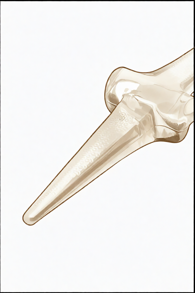
For patients: a plain-language version of this topic is available. See the patient guide.
Overview
Proximal interphalangeal (PIP) joint arthroplasty addresses symptomatic osteoarthritis, with silicone implant arthroplasty remaining the treatment of choice and demonstrating 90% implant survivorship at an average of 10 years postoperatively [4]. While precise therapeutic recommendations are limited by inconsistent postoperative management parameters across studies [1], initial reports for PIP and metacarpophalangeal surface replacement implants are encouraging, particularly as component loosening typical of earlier designs has not been a problem to date [8]. Surface replacement arthroplasty holds promise for the future, offering motion and stability for the index finger unattainable with silicone arthroplasty [13], and the CapFlex-PIP implant demonstrates favourable medium-term results in this context [23]. However, the relatively high complication rate means that the question of PIP resurfacing arthroplasty is not yet settled [17], and results are overall satisfying only when postoperative complications and reoperation incidence are noticeable and disclosed to patients preoperatively [15].
Clinical outcomes for pyrocarbon implants are variable despite substantially good survivorship, with 85% survival at 5 years of follow-up and high patient satisfaction [2, 59]. Approximately 1 in 5 PIP joint arthroplasties with a pyrocarbon implant will require revision surgery by 5 years, and 1 in 3 will undergo more than 1 operation [7]. Reoperations following primary non-constrained PIP arthroplasties are common [9], and minimizing postoperative complications after MCP and PIP joint arthroplasty is one avenue to decrease health care costs [26]. Silicone arthroplasty is an excellent option for the management of chronic PIP joint dislocations to improve pain and restore function while maintaining acceptable long-term viability [22], and excellent functional results and patient satisfaction can be gained using silastic PIP joint arthroplasty in the index finger [31]. The minimum 2 years of follow-up evaluation of the SLFJ implant PIP joint arthroplasty demonstrated good pain relief and good overall patient satisfaction while maintaining joint range of motion [30].
Mobilization of the PIP joint at zero to 5 days following joint replacement is recommended [19]. Results for PIP arthroplasty through a volar approach were as good as or better than the published results for PIP arthroplasty through a dorsal approach [11], leading to pain relief whilst maintaining the range of motion without any loss of extension [12]. Preliminary results encourage considering combining concomitant arthrodesis of the distal interphalangeal joint with surface replacement arthroplasty of the proximal interphalangeal joint to increase range of motion, despite study limitations of small sample size and short follow-up [20]. PIP denervation was associated with encouraging midterm outcomes in selected patients with symptomatic PIP joint osteoarthritis, with 81% of joints not requiring conversion to arthroplasty or arthrodesis over a 4-year follow-up [3]. Treatment of the long finger may be a relative contraindication to PIPJ arthroplasty [18], and ultimate salvage for the failed PIP joint arthroplasty may require arthrodesis or even amputation [6].
Anatomy & Pathophysiology
Bony Anatomy
The hand skeleton comprises 27 bones, 19 of which are long bones [49]. This structure is organized into five rays, each forming a polyarticulated chain of metacarpals and phalanges [49]. The base of each metacarpal articulates with the distal carpal row [49]. The radioulnocarpal articulation possesses two axes of movement, supplemented by a third axis of pronation and supination from the forearm [49]. These three wrist axes permit the hand to assume any spatial configuration necessary for grasping [49]. The radial ray is the shortest, consisting of a metacarpal and two phalanges [49]. The remaining four digital rays each contain four skeletal segments: a metacarpal and three phalanges [49].
Metacarpal length varies by digit; the thumb metacarpal is the shortest, the index finger metacarpal is the longest, and lengths decrease from the third to the fifth digits [49]. The proximal and middle phalanges of the middle and ring fingers are longer than those of the index finger [49]. Consequently, the long finger and usually the ring finger are longer than the index finger [49]. Epiphyseal plates are located at the proximal ends of the phalanges and the first metacarpal, and at the distal ends of the other metacarpals [49]. The hand contains 19 bones, 17 articulations, and 19 muscles situated entirely within the hand, along with a comparable number of tendons activated by forearm muscles [65].
Soft Tissue Anatomy
Extrinsic muscles originate outside the hand and insert on the hand or carpus, functioning as either flexors or extensors [48]. Intrinsic muscles have both origin and insertion within the hand and contribute to both digital flexion and extension [48]. The extrinsic extensors traverse six fibroosseous retinacular compartments at the wrist [48].
Compartment 1: Contains the abductor pollicis longus and extensor pollicis brevis [48]. The abductor pollicis longus inserts at the base of the thumb metacarpal via multiple slips to radially abduct the thumb [48]. The extensor pollicis brevis inserts on the dorsum of the proximal aspect of the thumb proximal phalanx to actively extend the metacarpophalangeal joint [48].
Compartment 2: Contains the extensor carpi radialis longus and extensor carpi radialis brevis [48]. The extensor carpi radialis longus inserts on the index metacarpal to dorsiflex and radially deviate the wrist [48]. The extensor carpi radialis brevis inserts into the base of the middle metacarpal to provide balanced wrist dorsiflexion [48].
Compartment 3: Contains the extensor pollicis longus [48]. This tendon runs longitudinally down the forearm, turns abruptly radialward about Lister tubercle—a dorsal prominence on the distal radius—and inserts on the distal phalanx to provide forceful extension of the thumb interphalangeal joint [48]. Its oblique course provides a substantial adduction component to its pull [48].
Compartment 4: Contains the extensor indicis proprius, which lies deep to the four tendons of the extensor digitorum communis [48]. The extensor indicis proprius inserts on the index finger ulnar to the extensor digitorum communis [48]. It may be recognized at the wrist level by possessing the most distal muscle belly of any digital extensor tendon [48].
Compartment 5: Contains the extensor digiti quinti, which inserts on the little finger ulnar to the extensor digitorum communis insertion [48]. The extensor digitorum communis inserts on the index, middle, ring, and, in some cases, little fingers [48]. The tendons of the middle, ring, and little fingers are tethered together by juncturae tendinum over the dorsum of the hand proximal to the metacarpophalangeal joint [48]. The extensor indicis proprius, extensor digitorum communis, and extensor digiti quinti each contribute to digital extension at the metacarpophalangeal, proximal interphalangeal, and distal interphalangeal joints [48].
Compartment 6: Contains the extensor carpi ulnaris, which inserts at the base of the little finger metacarpal to provide wrist extension and ulnar deviation [48].
The principal bony insertion of extrinsic digital extensors is on the dorsal proximal aspect of the middle phalanx [48]. Metacarpophalangeal joint extension is achieved by extrinsic extensor force transmitted through the sagittal bands [48]. Distal interphalangeal joint extension is achieved through conjoined lateral bands composed of tendinous slips from extrinsic and intrinsic tendons [48]. Digital extensor tendons are stabilized over the mid-line of the metacarpophalangeal joint by attachment to sagittal band fibers [48]. These fibers insert onto the volar proximal phalanx and the lateral borders of the volar plate, forming a sling that transmits proximal extrinsic extensor tension to the proximal phalanx [48]. This mechanism permits metacarpophalangeal joint extension without a direct tendinous insertion onto the proximal phalanx [48]. The sagittal bands keep the extrinsic extensor as far as possible from the center of rotation of the metacarpophalangeal joint, maximizing mechanical efficiency [48]. Rupture or attenuation of these fibers allows the extrinsic extensor tendon to sublux to the ulnar side of the metacarpal head, causing ulnar deviation of the finger [48].
The extrinsic finger flexors are the flexor digitorum profundus and flexor digitorum superficialis [48]. The flexor digitorum profundus inserts on the proximal volar aspect of the distal phalanx, flexing the distal interphalangeal, proximal interphalangeal, and metacarpophalangeal joints [48]. The flexor digitorum superficialis acts as a flexor of the proximal interphalangeal and metacarpophalangeal joints [48].
The intrinsic musculature includes seven interosseous muscles: four dorsal and three volar [67]. The anatomic axis of the hand coincides with the axis of the third metacarpal [67]. The dorsal interossei are abductors and lie to the radial side of the index and middle fingers and the ulnar side of the middle and ring fingers [67]. The little finger is abducted by the abductor digiti quinti [67]. The volar interossei are adductors and lie to the ulnar side of the index finger and the radial side of the ring and little fingers [67]. The middle finger possesses two dorsal interossei and no volar interossei because the central axis of the hand lies within it [67].
Each dorsal interosseous muscle, with the exception of the third, has two muscle heads [67]. The superficial head arises dorsally from the shaft of contiguous metacarpals and inserts deeply via a medial tendon onto the lateral tubercle of the base of the proximal phalanx [67]. It abducts and weakly flexes the proximal phalanx, with no direct effect on the middle or distal phalanges [67]. The deep head forms a lateral tendon, or lateral band, at the level of the metacarpophalangeal joint [67]. It flexes and weakly abducts the proximal phalanx while extending the middle and distal phalanges [67]. At the middle of the proximal phalanx, transverse fibers arch dorsally from each lateral band to join over the dorsum of the finger, flexing the proximal phalanx [67]. More distally, oblique spiral fibers from the lateral bands sweep over the distal third of the proximal phalanx to insert onto the lateral tubercles at the base of the middle phalanx, extending the middle phalanx at the proximal interphalangeal joint [67]. Further distally, the lateral bands are joined by the lateral slips of the extensor tendon to form the conjoined lateral band [67]. The two conjoined lateral bands unite at the distal third of the middle phalanx to form the terminal tendon, which inserts at the base of the distal phalanx to extend it [67].
The flexor digiti quinti brevis is structurally and functionally similar to the deep head of the dorsal interossei, forming the ulnar lateral band of the little finger [67]. The three volar interossei arise from adjacent surfaces of contiguous metacarpal shafts [67]. Each volar interosseous muscle has a single head and none insert onto the proximal phalanx [67]. They form the ulnar lateral band of the index finger and the radial lateral band of the ring and little fingers [67]. The volar interossei send oblique or spiral fibers that insert onto the base of the middle phalanx at its lateral tubercle [67]. All lateral bands are joined by the lateral slips of the extensor tendon to form a conjoined lateral band and finally a terminal tendon extending to the distal phalanx [67]. The abductor digiti quinti and flexor digiti quinti brevis are similar in structure and function to the superficial and deep heads of the dorsal interossei, respectively, and arise from the fifth metacarpal [67]. The abductor digiti quinti inserts onto the ulnar lateral tubercle at the base of the proximal phalanx of the little finger [67]. The flexor digiti quinti forms the ulnar lateral band [67]. The opponens digiti quinti lies deepest, arising from the pisohamate ligament and the hook of the hamate, and inserts onto the ulnar side of the diaphysis of the fifth metacarpal to flex and supinate it [67].
The flexor digitorum profundus originates from the proximal ulna and interosseous membrane [74]. In the forearm, it divides into a radial component supplying the index finger and an ulnar component supplying the middle, ring, and little fingers [74]. The flexor digitorum profundus and flexor pollicis longus form the deep compartment of the volar forearm [74]. As these tendons travel through the carpal tunnel, they occupy the floor of the tunnel [74]. The tenosynovial sheath of the flexor pollicis longus is continuous with the radial bursa, while the sheath to the little finger is continuous with the ulnar digital bursa [74]. In some patients, these bursae communicate, allowing a horseshoe abscess to spread between the thumb and little finger if infection occurs in the flexor tendon sheath of either digit [74]. The lumbricals originate from the radial side of the index, middle, ring, and little fingers in the palm [74]. The profundus tendon passes through the bifurcation of the flexor digitorum superficialis before inserting into the proximal palmar base of the distal phalanx [74]. Innervation of the flexor digitorum profundus for the index and middle fingers is via the anterior interosseous branch of the median nerve, while the ring and little fingers are innervated by the ulnar nerve [74]. The flexor digitorum profundus provides digital flexion at both the proximal and distal interphalangeal joints [74].
The flexor digitorum superficialis has two heads: a radial head originating from the proximal radius shaft and a humeral ulnar head originating from the medial humeral epicondyle and coronoid process of the ulna [74]. Each digit has a corresponding independent superficialis muscle [74]. As the tendons pass through the carpal tunnel, those of the middle and ring fingers are more superficial and central than those of the index and little fingers [74]. In the proximal finger, the flexor digitorum superficialis tendon bifurcates around the flexor digitorum profundus at the beginning of the A2 pulley [74]. The tendon slips reunite distally at the Camper chiasm, with approximately half of the fibers staying on the ipsilateral side and half crossing to the contralateral side [74]. The flexor digitorum superficialis inserts via radial and ulnar slips into the proximal metaphysis of the middle phalanx [74]. The entire muscle receives innervation from the median nerve [74]. Its primary function is digital flexion at the proximal interphalangeal joint [74].
The flexor pollicis longus originates from a radial head on the proximal radius and interosseous membrane, and an accessory head on the coronoid process of the ulna and medial epicondyle of the humerus [74]. In the palm, its tendon traverses between the abductor pollicis brevis and flexor pollicis brevis [74]. It inserts into the proximal base of the thumb distal phalanx and is innervated by the anterior interosseous branch of the median nerve [74]. The flexor pollicis longus flexes both the interphalangeal and metacarpophalangeal joints of the thumb [74].
As flexor tendons pass distal to the metacarpal neck, they enter the fibroosseous tunnel, or digital flexor sheath, which extends distally to the proximal aspect of the distal phalanx [74]. The tendinous sheath consists of annular pulleys providing mechanical stability and cruciate pulleys providing flexibility [74]. The first, third, and fifth annular pulleys (A1, A3, and A5) are located over the metacarpophalangeal, proximal interphalangeal, and distal interphalangeal joints, respectively [74]. The second and fourth pulleys (A2 and A4) are situated over the middle portion of the proximal and middle phalanges [74]. The A2 and A4 pulleys are the most essential in maintaining the mechanical advantage of the flexor tendons [74]. The tenosynovium lining the fibroosseous tunnel supplies nutrition and lubrication to the poorly vascularized flexor tendons [74]. Proximal to the sheath, tendons are well vascularized by the peritenon [74]. Within the sheath, tendon vascularity is supplied via the vincula system, comprising the vinculum longus and brevis [74].
The superficial palmar fascia lies in a coronal plane beneath the palmar subcutaneous tissue [77]. It covers a triangular area of the central palm, with the proximal corner facing directly proximal [77]. The palmaris longus tendon, when present, terminates in continuity with the fibers of this proximal corner [77]. From this common point, four central bands of fascia extend distally toward each of the fingers [77]. There is no central band for the thumb [77]. Confluent proximally, the central bands separate and diverge just distal to the transverse retinacular ligament [77].
Classification
PIP-Kellgren: A proposed functional-radiological classification system is designed to stratify surgical candidates and standardize severity assessment [96].
Bone Resorption Scoring: A radiological scoring system for bone resorption has been validated specifically in the context of PIP joint replacement using the Self-Locking Finger Joint prosthesis [136].
Clinical Presentation
PIP joint arthroplasty is indicated for joints with osteoarthritis destruction or post-traumatic conditions [45]. Pre-requisites for the procedure include intact tendons and at least some residual joint stability [45]. Corrections of a lateral deviation beyond 30° are difficult and likely to fail in PIP joint arthroplasty [45]. PIP joint arthroplasty should be proposed exceptionally if the PIP joint arthritis causes invalidating functional pain [91].
Non-constrained PIP joint arthroplasties are designed to provide durable, functional, and painless range of motion in cases of symptomatic arthritis associated with pain, stiffness, and/or deformity [5]. Silicone implant arthroplasty remains the treatment of choice for the symptomatic osteoarthritic PIP joint [4]. Silicone implant arthroplasty of the PIP joint is a common and generally successful surgery for the correction of painful stiffness due to underlying arthropathy [40]. Pain relief and patient satisfaction following silicone implant arthroplasty of the PIP joint are generally reliable [40]. Silicone arthroplasty for osteoarthritis of the PIP remains a good option for pain relief [42]. Treatment of MCP and PIP osteoarthritis with an anatomically neutral implant can provide reliable, long-term pain relief and maintenance of function [41].
Outcomes following PIP joint arthroplasties are less predictable than large joint replacements [5]. Reported reoperation rates for PIP joint arthroplasties range from 6% to 58% [5]. The majority of reoperations following non-constrained PIP joint arthroplasties are due to extensor mechanism dysfunction [5]. Common etiologies for reoperations following non-constrained PIP joint arthroplasties include component loosening, collateral ligament failure, and volar plate contracture [5]. Reoperations due to collateral ligament failure are more common in patients with inflammatory arthritis than in patients with primary or posttraumatic osteoarthritis [5]. Younger age leads to higher revision rates after PIP arthroplasty, particularly in the posttraumatic setting [32].
Surgical approaches requiring division of the extensor tendon, such as the Chamay and central slip reflecting approaches, are associated with higher rates of extensor mechanism dysfunction [5]. The extensor tendon splitting approach and the lateral approach are associated with lower rates of extensor mechanism problems [5]. Insertion of a silicone implant arthroplasty for the PIP joint can easily be performed through a lateral approach, affording early movement and few complications without leading to instability [27]. The volar approach to PIP SRA can result in excellent range of motion, function, and pain relief with minimal complications in active patients with osteoarthritis or posttraumatic arthritis [28]. Results from PIP arthroplasty through a volar approach are as good as or better than the published results for PIP arthroplasty through a dorsal approach [11].
Swan neck deformity and boutonniere can occur after surface replacement arthroplasty of the PIPJ due to loss of palmar plate integrity after the palmar approach or loss of extensor mechanism integrity after a dorsal approach [14]. When present preoperatively, swan neck deformity and boutonniere deformity are very difficult to correct when performing PIPJ implant arthroplasty [14]. PJI is uncommon after MCP or PIP arthroplasties [25].
PIP denervation is associated with encouraging midterm outcomes in selected patients with symptomatic PIP joint osteoarthritis [3]. 81% of joints treated with PIP denervation did not require conversion to arthroplasty or arthrodesis over a 4-year follow-up [3].
Clinical outcomes for PIP joint replacements with pyrocarbon implants are variable [2]. Approximately 1 in 5 PIP joint arthroplasties with a pyrocarbon implant will require revision surgery by 5 years [7]. 1 in 3 PIP joint arthroplasties with a pyrocarbon implant will undergo more than 1 operation [7]. Pyrocarbon PIPJ arthroplasty carries a risk of early complications necessitating revision surgeries [33]. The authors of one study no longer recommend the pyrocarbon implant for the treatment of osteoarthritis of the PIPJ [24].
Surface replacement arthroplasty of the PIP joint offers motion and stability for the index finger unattainable with silicone arthroplasty [13]. The results of surface replacement arthroplasty of the PIP joint are overall satisfying [15]. Postoperative complications and incidence of reoperations for surface replacement arthroplasty are noticeable and should be mentioned to patients in the preoperative setting [15]. The kinematics of the PIP joint after replacement are similar to that of the normal joint [16]. PIP joint surface replacement arthroplasty tends to achieve better anatomical stability compared to flexible silicone implants [29].
Implant survivorship for silicone implant arthroplasty is 90% at an average of 10 years postoperatively [4]. Patients returned to work after a median of 8 weeks following PIP arthroplasty [10]. It is not possible to provide precise recommendations concerning therapeutic strategies that need to be adopted following PIP joint arthroplasty based on available knowledge due to a lack of information on postoperative management and the use of different parameters in studies [1]. The authors recommend mobilization of the PIP joint at zero to 5 days following joint replacement [19]. The authors recommend simultaneous surgical intervention in case of severe painful OA of the PIP and DIP joints of the same digit [51].
Investigations
Clinical Examination: A systematic method for approaching the physical examination of the hand is essential due to the number of structures in a small space [47]. Clinical evaluation of the hand and wrist requires combining patient history with a careful physical examination to pinpoint or narrow the scope of possible pathologic processes [47].
Other Considerations: Diagnostic tests such as imaging and serum laboratory studies are useful in determining hand pathology but can be expensive, time consuming, and often nonspecific [47]. The kinematics of the PIP joint after replacement were similar to that of the normal joint [16].
Treatment
Non-Operative
The provided evidence does not detail specific conservative management protocols such as weight loss, physiotherapy, NSAIDs, or injections for PIP joint arthritis.
Operative
Indications: PIP joint arthroplasty is indicated for primary degenerative or post-traumatic arthritis in elderly, less-active patients to maintain motion and avoid arthrodesis [56]. It is a widely accepted procedure for joints with osteoarthritis destruction or post-traumatic conditions [45]. Pre-requisites for the procedure include intact tendons and at least some residual joint stability [45]. Pyrocarbon PIP arthroplasty is specifically recommended for post-traumatic painful joints [57]. While arthrodesis was historically considered the treatment of choice for the index finger to provide forceful key pinch against the thumb [38], total arthroplasty with a Tactys1 implant is no longer a formal contraindication for index fingers, including cases of clinodactyly greater than 58 degrees [101].
Surgical Approach / Technique: The volar approach yields results as good as or better than the dorsal approach [11], with one study identifying no statistical difference in mean postoperative range of motion, complication incidence, or revision surgery between the two [53]. A simplified lateral hinge approach provides adequate exposure without collateral ligament detachment, carries a low risk of complications, and allows early mobilization [88]. Approaches requiring division of the extensor tendon, such as the Chamay and central slip reflecting techniques, are associated with higher rates of extensor mechanism dysfunction [5]. In contrast, the extensor tendon splitting approach and the lateral approach demonstrate lower rates of extensor mechanism problems [5]. A C-shaped dorsal incision that conserves the extensor mechanism and collateral ligaments facilitates post-operative rehabilitation [61]. The dorsal chevron flap technique incorporates the "dorsal plate" of the central slip, which functions similarly to the patella to improve the extensor moment arm, aid joint stability, and prevent attrition rupture [60]. Comparative data show the palmar approach results in a total active motion of 87° and extensor lag of 9°, compared to 70° TAM and 13° extensor lag for the chevron approach, 69° TAM and 12° extensor lag for the longitudinal approach, and 52° TAM and 17° extensor lag for the dorsolateral approach [60]. Swan neck deformity can occur after surface replacement due to loss of palmar plate integrity following a palmar approach [14], while boutonniere deformity can result from loss of extensor mechanism integrity after a dorsal approach [14].
Implant Selection: Silicone implant arthroplasty remains the treatment of choice for symptomatic osteoarthritic PIP joints, with an implant survivorship of 90% at an average of 10 years postoperatively [4]. It is a common and generally successful surgery for painful stiffness due to underlying arthropathy, with reliable pain relief and patient satisfaction [40]. Pyrolytic carbon implants are no longer recommended for the treatment of PIP joint osteoarthritis [24]. However, pyrocarbon hemiarthroplasty appears to be a viable alternative to total joint arthroplasty for PIP joint arthritis [89]. In a series of 18 PIP joints treated with pyrolytic carbon resurfacing arthroplasty, there were no failures or revisions, with only two joints showing evidence of loosening [21]. The revision rate for the LPM prosthesis was higher than in published series for other PIP joint implants, and close surveillance of all patients with this prosthesis currently in situ is recommended [44].
Complications and Reoperations: Outcomes following PIP joint arthroplasties are less predictable, with reported reoperation rates ranging from 6% to 58% [5]. The majority of reoperations following non-constrained PIP joint arthroplasties were due to extensor mechanism dysfunction [5]. Other common etiologies for reoperations included component loosening, collateral ligament failure, and volar plate contracture [5]. Reoperations due to collateral ligament failure were more common in patients with inflammatory arthritis than in those with primary or posttraumatic osteoarthritis [5]. Passive manipulation serves as an alternative to surgical release for select PIP joint extension contractures [141].
Rehabilitation and Recovery: Rehabilitation for the index finger after PIP arthroplasty involves beginning functional exercises later and prolonging functional splinting to protect the radial collateral ligament [45]. The goal of index-finger rehabilitation is optimized function rather than maximum mobility [45]. The procedure for superficialis sling (flexor digitorum superficialis tenodesis) for swan neck reconstruction may be performed through various approaches with the PIP joint positioned in 20-30 degrees of flexion [36].
Adjunctive Procedures: Preliminary results encourage considering combining distal interphalangeal joint arthrodesis with surface replacement arthroplasty of the proximal interphalangeal joint to increase range of motion following PIP joint arthroplasty, despite study limitations of small sample size and short follow-up [20].
Complications
General Complication and Reoperation Rates: Published complication rates for silicone PIP arthroplasties range from 7% to 9% [132], while rates for PIP surface replacements reach up to 30% [132]. Reported reoperation rates for silicone implants range from 1% to 16% [132], compared to 6% to 58% for surface replacements [5, 132]. Approximately one in three PIP joint arthroplasties with a pyrocarbon implant undergoes more than one operation by 5 years [7]. Revision arthroplasty is associated with a 70% 5-year survival and a high incidence of complications [121]. The LPM prosthesis demonstrated a higher revision rate than other published PIP joint implants [44], with a 29% revision rate at a maximum follow-up of 6 years [64]. An additional 20% of patients with the LPM prosthesis experienced adverse clinical and radiological outcomes at the same follow-up period [64]. In pyrolytic carbon PIP arthroplasty, 28% of patients required a second procedure and 8% required a revision arthroplasty [147]. A total of 13% of joints treated with pyrocarbon implants required a secondary surgical procedure [150].
Extensor Mechanism and Deformity: Extensor mechanism dysfunction was the majority cause of reoperations in non-constrained PIP joint arthroplasties [5]. Surgical approaches requiring division of the extensor tendon, specifically the Chamay and central slip reflecting approaches, were associated with higher rates of extensor mechanism dysfunction [5]. Conversely, the extensor tendon splitting approach and the lateral approach had lower rates of extensor mechanism problems compared to approaches requiring tendon division [5]. Swan neck deformity can occur after surface replacement arthroplasty due to loss of palmar plate integrity after a palmar approach [14], while boutonniere deformity can occur due to loss of extensor mechanism integrity after a dorsal approach [14]. Both swan neck and boutonniere deformities are very difficult to correct during PIPJ implant arthroplasty if present preoperatively [14]. Recurrence of swan neck deformity was observed in 2 out of 9 cases in a series of Swanson implant PIP arthroplasties performed via a volar approach [37], with one case requiring surgical revision [37]. Postoperative contracture was observed in 3 out of 15 cases following surface replacement arthroplasty using a volar approach [86]. Intra-operative fracture or fragility of the central slip insertion on the dorsal rim of the middle phalanx was present in all 3 cases of postoperative contracture [86]. Elongation of the central slip insertion developed into flexion contracture with a range of motion of 20 degrees in 2 cases [86], while ectopic ossification on the dorsum of the PIP joint developed into a fixed extension contracture of 10 degrees in 1 case [86]. All 3 cases of postoperative contracture following volar approach surface replacement required further surgery [86].
Implant and Structural Failures: Component loosening, collateral ligament failure, and volar plate contracture were common etiologies for reoperations in non-constrained PIP joint arthroplasties [5]. Postoperative dislocation was the second most common etiology for revision surgery in border digit PIP arthroplasties, occurring in 6 cases [39]. Implant fracture occurred in 1 case of border digit PIP arthroplasty revision surgery [39]. Implant fracture and night-time paresthesia were complications observed in 1 case of Swanson implant PIP arthroplasty via a volar approach [37]. A fracture of a silicone implant occurred 3 years after arthroplasty in 1 patient, requiring salvage by arthrodesis [115]. One patient required amputation at the PIP level 1 year after silicone arthroplasty due to joint stiffness resulting in functional disability [115]. The only noncemented joint in a volar approach surface replacement series demonstrated progressive subsidence of both components [28]. Intraoperative fractures occurred in 7% of multi-digit PIP joint arthroplasties performed during a single operation [50], while postoperative fractures occurred in 1% of these cases [50]. Intraoperative fractures occurred in 12% of multi-digit PIP joint arthroplasties performed on two or more different dates [50], with postoperative fractures occurring in 2% of these cases [50].
Infection and Pain: Periprosthetic joint infection is uncommon after PIP arthroplasties [25]. Infections were uncommon etiologies for reoperations in non-constrained PIP joint arthroplasties [5]. Infections occurred in 3% of multi-digit PIP joint arthroplasties performed during a single operation [50] and in 4% of those performed on two or more different dates [50]. Infection was the third most common etiology for revision surgery in border digit PIP arthroplasties, occurring in 3 cases [39]. Pain was the main cause for reoperations in a series reported by Jennings et al. [5]. Persistent pain was an etiology for reoperation in 2 cases of a series of 53 primary PIP joint nonconstrained arthroplasties [5]. Pain and stiffness were the most common etiologies for border digit arthroplasty revision surgery, occurring in 10 cases [39]. Stiffness due to extensor tendon adhesions was an etiology for reoperation in 3 cases of a series of 53 primary PIP joint nonconstrained arthroplasties [5].
Salvage and Management: Secondary tenolysis was performed in 4 cases of a series of silicone PIP arthroplasties for posttraumatic arthritis [115]. Resection of the volar plate was performed in cases of postoperative contracture following volar approach surface replacement [86].
Recovery
Rehabilitation protocol: A controlled motion rehabilitation program guides occupational therapy over a twelve-week postoperative timeline. This protocol promotes watchful progression to optimal PIP joint motion while protecting the extensor tendon repair [108].
Functional milestones: In a series involving dermofasciectomy and PIP joint replacement for Dupuytren's disease, a plateau of function was reached at three months, with active flexion to 85 degrees [144].
Other Considerations: Postoperative complications and the incidence of reoperations for surface replacement arthroplasty of the PIP joint are noticeable and should be mentioned to patients in the preoperative setting [15]. The majority of reoperations in a specific series were due to extensor mechanism dysfunction [5]. Other common etiologies for reoperations included component loosening, collateral ligament failure, and volar plate contracture [5]. Surgical approaches that required division of the extensor tendon, namely the Chamay and the central slip reflecting approaches, were associated with higher rates of extensor mechanism dysfunction [5].
Key Evidence
- [L4] It is not possible to provide precise recommendations concerning therapeutic strategies that need to be adopted following PIP joint arthroplasty based on available knowledge due to a lack of information on postoperative management and the use of different parameters in studies. [1] (10.1016/j.hansur.2019.10.174)
- [L4] Despite substantially good survivorship, clinical outcomes for PIP joint replacements with pyrocarbon implants are variable. [2] (10.1302/2058-5241.2.160041)
- [L3] PIP denervation was associated with encouraging midterm outcomes in selected patients with symptomatic PIP joint osteoarthritis, with 81% of joints not requiring conversion to arthroplasty or arthrodesis over a 4-year follow-up. [3] (10.1016/j.jhsa.2026.06.006)
- [L4] With an implant survivorship of 90% at average of 10 years postoperatively, silicone implant arthroplasty remains the treatment of choice for the symptomatic osteoarthritic PIP joint. [4] (10.1016/j.jhsa.2013.11.008)
- [L4] [5] (10.1016/j.jhsa.2011.06.002)
- [L5] Ultimate salvage for the failed PIP joint arthroplasty may require arthrodesis or even amputation. [6] (10.1016/j.hcl.2017.12.011)
- [L4] Approximately 1 in 5 PIP joint arthroplasties with a pyrocarbon implant will require revision surgery by 5 years, and 1 in 3 will undergo more than 1 operation. [7] (10.1016/j.jhsa.2018.06.020)
- [L5] Initial reports of PIP and MCP joint surface replacement implants are encouraging, particularly because component loosening typical of earlier designs has not been a problem to date. [8] (10.5435/00124635-200309000-00002)
- [L4] Reoperations following primary non-constrained PIP arthroplasties are common. [9] (10.1016/s0363-5023(11)60049-x)
- [L3] Patients returned to work after a median of 8 weeks following PIP arthroplasty. [10] (10.1177/15589447221141485)
- [L4] These results were as good as or better than the published results for PIP arthroplasty through a dorsal approach. [11] (10.1097/bth.0b013e31818c0f71)
- [L4] Arthroplasty with a volar approach in the treatment of PIP arthritis leads to pain relief whilst maintaining the range of motion without any loss of extension. [12] (10.1016/j.main.2014.07.004)
- [L4] Surface replacement arthroplasty of the PIP joint holds promise for the future, offering motion and stability for the index finger unattainable with silicone arthroplasty. [13] (10.1016/j.jhsa.2008.06.008)
- [L4] [14] (10.1177/17531934241265837)
- [L4] The results of surface replacement arthroplasty of the PIP joint are overall satisfying; however, postoperative complications and incidence of reoperations are noticeable and should be mentioned to the patients in the preoperative setting. [15] (10.1007/s11552-009-9246-z)
- [L5] The kinematics of the PIP joint after replacement were similar to that of the normal joint. [16] (10.1067/jhsu.2000.jhsu25a0305)
- [L4] However, the relatively high complication rate means that the question of PIP resurfacing arthroplasty is not yet settled. [17] (10.1016/j.hansur.2017.08.002)
- [L1] Treatment of the long finger may be a relative contraindication to PIPJ arthroplasty. [18] (10.1177/1558944718791186)
- [L4] The authors recommend mobilization of the PIP joint at zero to 5 days following joint replacement. [19] (10.1016/s0894-1130(88)80004-8)
- [L4] The authors state that their preliminary results encourage considering combining the two operations to increase range of motion following PIP joint arthroplasty, despite study limitations of small sample size and short follow-up. [20] (10.1177/1753193420954371)
- [L4] There were no failures or revisions in these 18 PIPJs, and only 2 joints had evidence of loosening. [21] (10.1016/j.jhsa.2006.02.018)
- [L5] Silicone arthroplasty is an excellent option for the management of chronic PIP joint dislocations to improve pain and restore function while maintaining acceptable long-term viability. [22] (10.1097/bth.0b013e31820f8b53)
- [L4] The CapFlex-PIP implant demonstrates favourable medium-term results in surface replacing arthroplasty of the proximal interphalangeal joint. [23] (10.1177/1753193420977244)
- [L4] Based on these findings, the authors no longer recommend this implant for the treatment of osteoarthritis of the PIPJ. [24] (10.1016/s0363-5023(10)60073-1)
- [L4] PJI is uncommon after MCP or PIP arthroplasties. [25] (10.1016/j.jhsa.2024.12.008)
- [L3] Minimizing postoperative complications after MCP and PIP joint arthroplasty is one avenue to decrease health care costs. [26] (10.1016/j.jhsa.2019.11.002)
- [L4] Insertion of a silicone implant arthroplasty for the PIP joint can easily be performed through a lateral approach, affording early movement and few complications without leading to instability. [27] (10.1016/j.jhsa.2007.04.011)
- [L4] [28] (10.1016/j.jhsa.2011.03.003)
- [L3] PIP joint surface replacement arthroplasty tends to achieve better anatomical stability compared to flexible silicone implants. [29] (10.1016/j.hansur.2020.02.013)
- [L4] The minimum 2 years of follow-up evaluation of the SLFJ implant PIP joint arthroplasty demonstrated good pain relief and good overall patient satisfaction while maintaining joint range of motion. [30] (10.1177/1558944717726136)
- [L4] The series shows that excellent functional results and patient satisfaction can be gained using silastic PIP joint arthroplasty in the index finger. [31] (10.1177/1558944720921468)
- [L3] Younger age leads higher revision rates after PIP arthroplasty, particularly in the posttraumatic setting. [32] (10.5435/jaaos-d-17-00109)
- [L3] Pyrocarbon PIPJ arthroplasty has a risk of early complications necessitating revision surgeries. [33] (10.1007/s00402-020-03592-3)
- [L5] The procedure may be performed through various approaches with the PIP joint positioned in 20-30 degrees of flexion. [36] (10.1016/j.jhsa.2015.07.018)
- [L4] [37] (10.1016/j.hansur.2017.11.008)
- [L4] [38] (10.1016/j.jhsa.2014.03.033)
- [L4] [39] (10.1177/1753193420926127)
- [L4] Silicone implant arthroplasty of the PIP joint remains a common and generally successful surgery for the correction of painful stiffness due to underlying arthropathy, with pain relief and patient satisfaction being generally reliable. [40] (10.1016/j.jhsa.2007.04.013)
- [L4] Treatment of MCP and PIP osteoarthritis with an anatomically neutral implant can provide reliable, long-term pain relief and maintenance of function. [41] (10.1016/j.jhsa.2008.11.005)
- [L4] Silicone arthroplasty for osteoarthritis of the PIP remains a good option for pain relief. [42] (10.1177/1558944718769427)
- [L4] The revision rate for the LPM prosthesis was higher than in published series for other PIP joint implants, with close surveillance of all patients with this prosthesis currently in situ recommended. [44] (10.1177/1753193407087864)
- [L5] [45] (10.1302/2058-5241.4.180042)
- [L4] [50] (10.1177/1753193418765691)
- [L3] The authors recommend simultaneous surgical intervention in case of severe painful OA of the PIP and DIP joints of the same digit. [51] (10.1177/17531934231191255)
- [L3] The study identified no statistical difference in mean postoperative ROM, incidence of complications, or revision surgery between volar and dorsal approaches for PIP arthroplasty. [53] (10.1177/1558944719861718)
- [L4] Surface replacement arthroplasty of the PIPJ is indicated for primary degenerative or post-traumatic arthritis in elderly, less-active patients to maintain motion and avoid arthrodesis. [56] (10.1016/j.jhsa.2007.04.012)
- [L4] We recommend arthroplasty for this group of patients. [57] (10.1016/s0363-5023(03)80384-2)
- [L4] The survival of pyrocarbon PIP joint arthroplasty was 85% at 5 years of follow-up, with high patient satisfaction. [59] (10.1016/j.jhsa.2012.02.012)
- [L4] [60] (10.1016/s0266-7681(98)80026-0)
- [L4] [61] (10.1054/jhsb.2000.0541)
- [L4] The revision rate of 29% and a further 20% rate of adverse clinical and radiological outcomes for the LPM PIPJ prosthesis at a maximum follow-up of 6 years is unacceptable. [64] (10.1177/1753193408094920)
- [L4] [86] (10.1142/s2424835516500326)
- [L4] The simplified lateral hinge approach allows for a simple, adequate exposure for PIPJ replacement, does not require detachment of the collateral ligaments, has a low risk of complications, and allows for early mobilization after surgery. [88] (10.1097/bth.0000000000000094)
- [L4] Pyrocarbon hemiarthroplasty appears to be a viable alternative to total joint arthroplasty in the treatment of PIP joint arthritis. [89] (10.1016/j.jhsa.2014.12.016)
- [L4] It should be proposed exceptionally if the PIP joint arthritis causes invalidating functional pain. [91] (10.1177/15589447211030962)
- [L4] The proposed PIP-Kellgren functional-radiological classification system may help stratify surgical candidates and standardize severity assessment. [96] (10.1016/j.jhsg.2025.100911)
- [L4] Total arthroplasty of the PIP with a Tactys1 is no longer a formal contraindication for the index fingers and in cases of clinodactyly of more than 58. [101] (10.1016/j.hansur.2019.03.001)
- [L5] This controlled motion rehabilitation program has been useful in our practice to help guide occupational therapy over a twelve-week postoperative timeline in a manner that is simple and promotes watchful progression to the timely achievement of optimal PIP joint motion while protecting the extensor tendon repair. [108] (10.1016/j.jht.2019.04.003)
- [L4] [115] (10.1053/jhsu.1999.jhsu24a0073)
- [L3] Revision arthroplasty was associated with a 70% 5-year survival but with a high incidence of complications. [121] (10.1016/j.jhsa.2015.05.015)
- [L1] [132] (10.1177/1753193418770606)
- [L4] [136] (10.1177/17531934251414329)
- [L4] Passive manipulation is an alternative to surgical release for select PIP joint extension contractures. [141] (10.1016/j.jhsa.2022.01.023)
- [L5] A plateau of function was reached at three months with active flexion to 85 degrees. [144] (10.1016/s0266-7681(85)80060-7)
- [L4] Twenty-eight percent of patients required a second procedure and 8% required a revision arthroplasty. [147] (10.1016/j.jhsa.2006.10.017)
- [L4] A total of 13% of the joints required a secondary surgical procedure. [150] (10.1016/j.jhsa.2009.08.010)
See Also
- Dislocations
- PIP Joint Arthritis
- Extensor Tendon Repair
- Dupuytren's Disease
References
[1] Rééducation après arthroplastie prothétique de l’articulation interphalangienne proximale des doigts longs. Hand Surgery and Rehabilitation. 2019. DOI: 10.1016/j.hansur.2019.10.174
[2] Proximal interphalangeal (PIP) joint replacements with pyrolytic carbon implants in the hand. EFORT Open Reviews. 2017. DOI: 10.1302/2058-5241.2.160041
[3] Is PIP Denervation for Osteoarthritis a Durable Solution? Results from a Multicenter Outcome Study. The Journal of Hand Surgery. 2026. DOI: 10.1016/j.jhsa.2026.06.006
[4] Long-Term Results of Swanson Silicone Arthroplasty for Proximal Interphalangeal Joint Osteoarthritis. The Journal of Hand Surgery. 2014. DOI: 10.1016/j.jhsa.2013.11.008
[5] Reoperations Following Proximal Interphalangeal Joint Nonconstrained Arthroplasties. The Journal of Hand Surgery. 2011. DOI: 10.1016/j.jhsa.2011.06.002
[6] Salvaging a Failed Proximal Interphalangeal Joint Implant. Hand Clinics. 2018. DOI: 10.1016/j.hcl.2017.12.011
[7] Medium-Term Outcomes With Pyrocarbon Proximal Interphalangeal Arthroplasty: A Study of 170 Consecutive Arthroplasties. The Journal of Hand Surgery. 2018. DOI: 10.1016/j.jhsa.2018.06.020
[8] New-Generation Implant Arthroplasties of the Finger Joints. Journal of the American Academy of Orthopaedic Surgeons. 2003. DOI: 10.5435/00124635-200309000-00002
[9] Reoperations following Proximal Interphalangeal Joint Unconstrained Arthroplasty. The Journal of Hand Surgery. 2011. DOI: 10.1016/s0363-5023(11)60049-x
[10] Type of Work and Preoperative Ability to Perform Work Affect Return to Usual Work Following Proximal Interphalangeal Joint Arthroplasty for Osteoarthritis. HAND. 2022. DOI: 10.1177/15589447221141485
[11] The Volar Approach to Proximal Interphalangeal Joint Arthroplasty. Techniques in Hand & Upper Extremity Surgery. 2009. DOI: 10.1097/bth.0b013e31818c0f71
[12] Proximal interphalangeal joint arthroplasty with Neuflex® implants: Relevance of the volar approach and early rehabilitation. Chirurgie de la Main. 2014. DOI: 10.1016/j.main.2014.07.004
[13] Surface Replacement Arthroplasty of the Proximal Interphalangeal Joint Using the PIP-SRA Implant: Results, Complications, and Revisions. The Journal of Hand Surgery. 2008. DOI: 10.1016/j.jhsa.2008.06.008
[14] Proximal interphalangeal joint arthroplasty: current trends and evidence-based practice. Journal of Hand Surgery (European Volume). 2024. DOI: 10.1177/17531934241265837
[15] Proximal Interphalangeal Joint Replacement with Surface Replacement Arthroplasty (SR–PIP): Functional Results and Complications. HAND. 2009. DOI: 10.1007/s11552-009-9246-z
[16] Kinematics of the proximal interphalangeal joint of the finger after surface replacement. The Journal of Hand Surgery. 2000. DOI: 10.1067/jhsu.2000.jhsu25a0305
[17] Results of Toccata® resurfacing PIP joint arthroplasty. A series of 32 cases at a mean follow-up of 5.9 years. Hand Surgery and Rehabilitation. 2017. DOI: 10.1016/j.hansur.2017.08.002
[18] Prosthetic Arthroplasty of Proximal Interphalangeal Joints for Treatment of Osteoarthritis and Posttraumatic Arthritis: Systematic Review and Meta-Analysis Comparing the Three Ulnar Digits With the Index Finger. HAND. 2018. DOI: 10.1177/1558944718791186
[19] A New Early-Mobilization Splint for Proximal Interphalangeal Joint Replacements. Journal of Hand Therapy. 1988. DOI: 10.1016/s0894-1130(88)80004-8
[20] Concomitant arthrodesis of the distal interphalangeal joint with surface replacement arthroplasty of the proximal interphalangeal joint: a comparative study in 11 patients. Journal of Hand Surgery (European Volume). 2020. DOI: 10.1177/1753193420954371
[21] Pyrolytic Carbon Proximal Interphalangeal Joint Resurfacing Arthroplasty. The Journal of Hand Surgery. 2006. DOI: 10.1016/j.jhsa.2006.02.018
[22] Silicone Arthroplasty for Chronic Proximal Interphalangeal Joint Dislocations. Techniques in Hand & Upper Extremity Surgery. 2011. DOI: 10.1097/bth.0b013e31820f8b53
[23] Surface replacing arthroplasty of the proximal interphalangeal joint using the CapFlex-PIP implant: a prospective study with 5-year outcomes. Journal of Hand Surgery (European Volume). 2020. DOI: 10.1177/1753193420977244
[24] Pyrolytic Carbon Arthroplasty of the PIP Joint: Not all it's Cracked up to be. The Journal of Hand Surgery. 2010. DOI: 10.1016/s0363-5023(10)60073-1
[25] Incidence and Presentation of Periprosthetic Joint Infection After Primary Metacarpophalangeal and Proximal Interphalangeal Arthroplasty. The Journal of Hand Surgery. 2025. DOI: 10.1016/j.jhsa.2024.12.008
[26] National Prevalence of Complications and Cost of Small Joint Arthroplasty for Hand Osteoarthritis and Post-Traumatic Arthritis. The Journal of Hand Surgery. 2020. DOI: 10.1016/j.jhsa.2019.11.002
[27] Lateral Approach to Proximal Interphalangeal Joint Implant Arthroplasty. The Journal of Hand Surgery. 2007. DOI: 10.1016/j.jhsa.2007.04.011
[28] Surface Replacement Arthroplasty of the Proximal Interphalangeal Joint Using a Volar Approach: Case Series. The Journal of Hand Surgery. 2011. DOI: 10.1016/j.jhsa.2011.03.003
[29] Lateral stability in healthy proximal interphalangeal joints versus surface replacement and silicone arthroplasty: Results of a three-dimensional motion analysis study. Hand Surgery and Rehabilitation. 2020. DOI: 10.1016/j.hansur.2020.02.013
[30] Outcomes of Surface Replacement Proximal Interphalangeal Joint Arthroplasty Using the Self Locking Finger Joint Implant: Minimum Two Years Follow-up. HAND. 2017. DOI: 10.1177/1558944717726136
[31] The Long-term Results of Proximal Interphalangeal Joint Arthroplasty of the Osteoarthritic Index Finger. HAND. 2020. DOI: 10.1177/1558944720921468
[32] Proximal Interphalangeal Joint Arthroplasty in Young Patients. Journal of the American Academy of Orthopaedic Surgeons. 2019. DOI: 10.5435/jaaos-d-17-00109
[33] Outcome of proximal interphalangeal joint replacement with pyrocarbon implants: a long-term longitudinal follow-up study. Archives of Orthopaedic and Trauma Surgery. 2020. DOI: 10.1007/s00402-020-03592-3
[36] Superficialis Sling (Flexor Digitorum Superficialis Tenodesis) for Swan Neck Reconstruction. The Journal of Hand Surgery. 2015. DOI: 10.1016/j.jhsa.2015.07.018
[37] Comparison of proximal interphalangeal arthroplasty outcomes with Swanson implant performed by volar versus dorsal approach. Hand Surgery and Rehabilitation. 2018. DOI: 10.1016/j.hansur.2017.11.008
[38] Silicone Proximal Interphalangeal Joint Arthroplasty for Primary Osteoarthritis Using a Volar Approach. The Journal of Hand Surgery. 2014. DOI: 10.1016/j.jhsa.2014.03.033
[39] Results of proximal interphalangeal arthroplasty: border digits versus middle digits. Journal of Hand Surgery (European Volume). 2020. DOI: 10.1177/1753193420926127
[40] The Dorsal Approach to Silicone Implant Arthroplasty of the Proximal Interphalangeal Joint. The Journal of Hand Surgery. 2007. DOI: 10.1016/j.jhsa.2007.04.013
[41] Anatomically Neutral Silicone Small Joint Arthroplasty for Osteoarthritis. The Journal of Hand Surgery. 2009. DOI: 10.1016/j.jhsa.2008.11.005
[42] Proximal Interphalangeal Joint Silicone Arthroplasty for Osteoarthritis: Midterm Outcomes. HAND. 2018. DOI: 10.1177/1558944718769427
[44] TWO TO FIVE YEAR FOLLOW-UP OF THE LPM CERAMIC COATED PROXIMAL INTERPHALANGEAL JOINT ARTHROPLASTY. Journal of Hand Surgery (European Volume). 2008. DOI: 10.1177/1753193407087864
[45] The proximal interphalangeal joint: arthritis and deformity. EFORT Open Reviews. 2019. DOI: 10.1302/2058-5241.4.180042
[47] Orthopaedic Knowledge Update 13 Ebook Without Multimedia. Anatomy, Evaluation, Clinical Examination, and Imaging > Evaluation and Clinical Examination: Current Concepts.
[48] A Lange Medical Book Current Diagnosis Treatment In Orthopedics Fifth Edition. 9Hand Surgery > Image DISORDERS OF THE MUSCULATURE OF THE HAND.
[49] Exam Of The Hand Wrist 2Ed. 1.1 SKELETON OF THE HAND > The osseous skeleton.
[50] Impact of multi- versus single finger proximal interphalangeal joint arthroplasty: analysis of 249 fingers treated in 15 years. Journal of Hand Surgery (European Volume). 2018. DOI: 10.1177/1753193418765691
[51] Does distal interphalangeal joint arthrodesis affect proximal interphalangeal joint arthroplasty outcomes in the same finger?. Journal of Hand Surgery (European Volume). 2023. DOI: 10.1177/17531934231191255
[53] Comparison of Volar and Dorsal Approach for PIP Arthroplasty. HAND. 2019. DOI: 10.1177/1558944719861718
[56] Surface Replacement Arthroplasty of the Proximal Interphalangeal Joint. The Journal of Hand Surgery. 2007. DOI: 10.1016/j.jhsa.2007.04.012
[57] Pyrocarbon PIP arthroplasty for post trauma painful joints. The Journal of Hand Surgery. 2003. DOI: 10.1016/s0363-5023(03)80384-2
[59] Pyrocarbon Proximal Interphalangeal Joint Arthroplasty: Minimum Two-Year Follow-Up. The Journal of Hand Surgery. 2012. DOI: 10.1016/j.jhsa.2012.02.012
[60] Dorsal Chevron Flap for Exposure of the Proximal Interphalangeal Joint. Journal of Hand Surgery. 1998. DOI: 10.1016/s0266-7681(98)80026-0
[61] A Conservative Approach for Proximal Interphalangeal Joint Arthroplasty. Journal of Hand Surgery. 2001. DOI: 10.1054/jhsb.2000.0541
[64] A Report on the Early Failure of the LPM Proximal Interphalangeal Joint Replacement. Journal of Hand Surgery (European Volume). 2008. DOI: 10.1177/1753193408094920
[65] Exam Of The Hand Wrist 2Ed. INTRODUCTION.
[67] Green S Operative Hand Surgery. Interosseous and Hypothenar Muscles.
[74] A Lange Medical Book Current Diagnosis Treatment In Orthopedics Fifth Edition. 9Hand Surgery > FLEXOR TENDON INJURY.
[77] Green S Operative Hand Surgery. PERTINENT ANATOMY.
[86] Post-Operative Contracture of the Proximal Interphalangeal Joint after Surface Replacement Arthroplasty Using a Volar Approach. The Journal of Hand Surgery (Asian-Pacific Volume). 2016. DOI: 10.1142/s2424835516500326
[88] A Simplified Lateral Hinge Approach to the Proximal Interphalangeal Joint. Techniques in Hand & Upper Extremity Surgery. 2015. DOI: 10.1097/bth.0000000000000094
[89] Pyrolytic Carbon Hemiarthroplasty in the Management of Proximal Interphalangeal Joint Arthritis. The Journal of Hand Surgery. 2015. DOI: 10.1016/j.jhsa.2014.12.016
[91] Arthroplasty of the Proximal Interphalangeal Joint With the TACTYS Prosthesis: Clinical and Radiographic Results With a Mean Follow-up of 5 Years. HAND. 2022. DOI: 10.1177/15589447211030962
[96] Short-Term Outcomes of Digitalis Silicone Implant for Proximal Interphalangeal Joint Osteoarthritis and a Proposed Functional-Radiological Classification. Journal of Hand Surgery Global Online. 2026. DOI: 10.1016/j.jhsg.2025.100911
[101] Arthroplasty of the proximal interphalangeal joint with the Tactys® modular prosthesis: Results in case of index finger and clinodactyly. Hand Surgery and Rehabilitation. 2019. DOI: 10.1016/j.hansur.2019.03.001
[108] A new controlled motion program for rehabilitation of the proximal interphalangeal joint arthroplasty. Journal of Hand Therapy. 2021. DOI: 10.1016/j.jht.2019.04.003
[115] Proximal interphalangeal joint silicone arthroplasty for posttraumatic arthritis. The Journal of Hand Surgery. 1999. DOI: 10.1053/jhsu.1999.jhsu24a0073
[121] Revision Proximal Interphalangeal Arthroplasty: An Outcome Analysis of 75 Consecutive Cases. The Journal of Hand Surgery. 2015. DOI: 10.1016/j.jhsa.2015.05.015
[132] Complications, reoperations and revisions after proximal interphalangeal joint arthroplasty: a systematic review and meta-analysis. Journal of Hand Surgery (European Volume). 2018. DOI: 10.1177/1753193418770606
[136] Proximal interphalangeal joint replacement with the Self-Locking Finger Joint prosthesis: mid-term outcomes and validation of a radiological scoring system for bone resorption. Journal of Hand Surgery (European Volume). 2026. DOI: 10.1177/17531934251414329
[141] Passive Manipulation for Proximal Interphalangeal Joint Extension Contractures. The Journal of Hand Surgery. 2023. DOI: 10.1016/j.jhsa.2022.01.023
[144] Dermofasciectomy and proximal interphalangeal joint replacement in Dupuytren's disease. The Journal of Hand Surgery: British & European Volume. 1985. DOI: 10.1016/s0266-7681(85)80060-7
[147] Pyrolytic Carbon Proximal Interphalangeal Joint Arthroplasty: Results With Minimum Two-Year Follow-Up Evaluation. The Journal of Hand Surgery. 2007. DOI: 10.1016/j.jhsa.2006.10.017
[150] Outcomes of Proximal Interphalangeal Joint Pyrocarbon Implants. The Journal of Hand Surgery. 2010. DOI: 10.1016/j.jhsa.2009.08.010