Clinicians › Hip
Degenerative Conditions
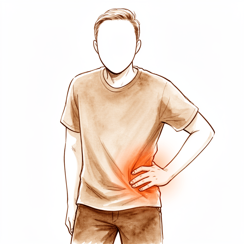
Hip osteoarthritis and osteonecrosis of the femoral head: pathophysiology, epidemiological trends, and management of joint degeneration.
Overview
Degenerative conditions of the spine and joints necessitate a nuanced understanding of patient characteristics and pathology to select effective treatment approaches [1, 2]. In the spine, various consecutive injections for conservative management are safe and reduce pain while improving quality of life [9]. For lumbar disorders, pre-surgery physiotherapy protocols aim to improve treatment outcomes [16]. Posterior decompression with instrumented fusion is safe and effective for patients aged 80 years and older with degenerative lumbar conditions [23]. Arthrodesis remains the best surgical treatment for persistently painful degenerative back, though it increases morbidity and mortality rates and carries a risk of non-union [26].
In the hip and knee, appropriate surgical indications are paramount. For periacetabular osteotomy (PAO), indications based on preoperative intra-articular cartilage degeneration are critical for long-term success [4]. Hip arthroscopy outcomes in older patients can equal those in younger patients if surgical indications are proper, specifically in the absence of degenerative articular cartilage changes [37]. However, surgeons should proceed with caution regarding hip arthroscopy for femoroacetabular impingement in patients at least 50 years of age, requiring careful evaluation of symptoms, hip disease, and expectations [19]. Patients undergoing primary hip arthroscopy must be counseled regarding the potential progression of degenerative change leading to arthroplasty and the potential for revision surgery [50].
For knee disorders, degenerative joint disease involves various processes requiring evidence-based care [2]. There is a growing body of evidence supporting the use of platelet-rich plasma (PRP) in selected indications [24]. In shoulder pathology, arthroscopic Bankart repair yields satisfactory functional outcomes at 11-20 years of follow-up despite a high incidence of postoperative degenerative changes in the long term [5].
Anatomy & Pathophysiology
Osseous Morphology and Dysplasia
Hip dysplasia represents a pathoanatomic osseous morphology associated with hip instability, which may be partially attributable to hip capsular thickness [73]. High rates of femoroacetabular impingement (FAI) morphologic characteristics are present in patients with hip instability [82]. Complex dynamic interplay exists between the hip and spinopelvic parameters; specifically, a cam deformity, acetabular undercoverage, and increased spinopelvic angles are predictive of a symptomatic hip state [79].
Capsular Integrity and Kinetics
Management of the hip capsule must allow for improved exposure without compromising stability and kinematics of the hip [46]. Most biomechanical evidence supports capsulotomy repair or reconstruction to improve hip distractive stability at the end of hip arthroscopic surgery [63]. The literature indicates a potential role for the hip joint capsule in mechanics via mechanoreceptors, though nomenclature is inconsistent and proprioceptive roles cannot be reliably confirmed as no study has reported type I-III mechanoreceptors [67]. Treatment of a hip capsular injury with platelet-rich plasma and bone marrow aspirate concentrate therapy resulted in marked improvement in kinematic and kinetic performance measures and MRI appearance of the torn hip capsule and gluteus minimus tendon [54].
Spinopelvic Biomechanics and Kinematics
Planning and measurement of the intended position of the acetabular component in the supine position may fail to predict clinically significant changes in its orientation during functional activities due to individual pelvic kinematics [61]. With advancing age, spinopelvic biomechanics demonstrate decreased spinal mobility and increased pelvic/hip mobility [55]. Observed hip flexion in the asymptomatic hips of young women is substantially less than has been historically reported [59]. Hip impingement in those with FAI syndrome may routinely occur at hip flexion angles below 90° in neutral rotation, with males engaging at higher flexion angles than females [83].
Systemic and Contralateral Effects
Patients with severe unilateral osteoarthritis of the knee are at risk from abnormal biomechanics in the contralateral knee and possibly both hips [48]. Intrinsic sarcomere changes contribute to the development of hip displacement in cerebral palsy [86]. Hip pain in the absence of osteoarthritis may be due to a complex combination of mechanical stresses, both dynamic and static [53]. Understanding of the etiology and pathology of hip instability has increased as new information has emerged [22]. Knowledge of the etiology and evolving research is essential to understand the spectrum of hip disease [22].
Functional Outcomes and Rehabilitation
Hip arthroscopic surgery yields meaningful improvements in hip function in the majority of patients, regardless of sex [87]. No biomechanical changes favoring arthroscopy were detected compared to physical therapist-led care, suggesting that personalized hip therapy elicits greater changes in hip moments during walking at 12-month follow-up [41]. After an ACL injury prevention program, athletes that exhibit the greatest reduction in knee abduction moments exhibit greater hip adduction excursion at baseline and show corresponding improvements in hip flexion and knee abduction kinematics and hip flexion moments [69].
Classification
Thorough understanding of clinical aging indices is essential for managing degenerative spine diseases, particularly in selecting effective treatment approaches for an aging society [1]. Degenerative joint disease of the knee involves various disease processes requiring understanding of pathology, diagnosis, and treatment options to provide evidenced-based care [2]. Proper diagnosis and treatment of complex hip pathologies has the potential to alter early degenerative changes in the adult hip [6]. Comprehensive diagnosis and management of the spectrum of posterior hip diseases is required to understand the etiology and evolving research on intra- and extraarticular hip complaints [8]. Understanding subchondral vascular physiology is key to better MRI classification, prevention, control, prognosis, and treatment of osteoarthritis and other bone diseases [29].
Beck: Represents the current standard for arthroscopic evaluation of intraarticular disease in femoroacetabular impingement [42]. The Beck classification demonstrates substantial interobserver reliability [42]. A proposed novel classification with only two disease categories may not adequately characterize the spectrum of intra-articular abnormalities [42].
Acetabular Rim Labrochondral: The reliability of arthroscopic classification of acetabular rim labrochondral disease is similar to previously reported arthroscopic disease classifications in the knee and shoulder [57]. The reliability of arthroscopic classification of acetabular rim labrochondral disease seems appropriate for future outcome reporting [57].
Cartilage Lesions: A universal and definitive grading system for cartilage lesions is necessary [56]. Measurement devices are needed for objective cartilage grading in questionable cases [56].
Lumbar Degenerative Spondylolisthesis: Both the CARDS and French classification systems have acceptable reliability and validity for lumbar degenerative spondylolisthesis [64].
Degenerative Lumbar Scoliosis: Degenerative lumbar scoliosis is distinct from adult idiopathic scoliosis [65]. Degenerative lumbar scoliosis is clearly associated with degenerative disk disease, facet arthropathy, and hypertrophy of the ligamenta flava [65].
Avascular Necrosis: Symptoms and the JIC classification are risk factors for collapse progression in avascular necrosis after pediatric femoral neck fracture [68].
Greater Trochanteric Pain Syndrome (GTPS): The intraoperative classification system and treatment algorithm for surgical treatment of various Greater Trochanteric Pain Syndrome (GTPS) types leads to favorable patient-reported outcomes [71].
Paravertebral Muscle Degeneration: There are three types of pathological changes in patients with paravertebral muscle degeneration [74]. The three types of pathological changes in paravertebral muscle degeneration may help to decide on targeted treatments for low back pain [74].
Other Considerations: A universal and definitive grading system for cartilage lesions is necessary [56]. Measurement devices are needed for objective cartilage grading in questionable cases [56].
Clinical Presentation
Degenerative joint disease of the knee is a common condition [2]. Degenerative cervical spondylosis is a common age-related condition [7], yet radiographic evidence of degenerative cervical spondylosis is frequent in asymptomatic adults [7]. There is no relationship between the progression of degeneration on MRI and the development of clinical symptoms in the cervical spine, with the exception of an association between foraminal stenosis and upper-limb pain [10].
A clinical diagnosis of hip osteoarthritis was found in approximately 22% of young patients undergoing hip arthroscopy within 2 years [36]. Increasing symptoms and decreased function related to degenerative hip disease may occur fifteen to twenty years after Colonna arthroplasty with concomitant femoral shortening and rotational osteotomy [14]. Rapidly destructive osteoarthritis of the hip joint represents an uncommon subset of osteoarthritis [15]. Regular clinical and radiological review is required to assess the speed of progression in rapidly destructive osteoarthritis of the hip joint [15].
Extra-articular etiologies of pain represent an important subset of hip disorders [13]. Extra-articular etiologies of hip pain can be accurately identified through physical examination and imaging [13]. Understanding the etiology and pathology of hip instability is essential to understand the spectrum of hip disease [22].
MRI serves as a noninvasive tool that overcomes the shortcomings of radiography by detecting preclinical disease and subtle early abnormalities in articular cartilage [12]. Magnetic resonance imaging has created an important role for reproducible, noninvasive, and objective evaluation and monitoring of cartilage in the setting of trauma, degenerative arthritides, and surgical treatment for cartilage injury [21]. Although many OA-related biomarkers are currently available, none can be considered a surrogate marker of clinical and imaging features for the diagnosis or prognosis of the disease at this time [18].
Careful evaluation of symptoms, hip disease, and expectations is needed before proceeding with hip arthroscopy for femoroacetabular impingement in patients at least 50 years of age [19]. Recognition of associations between clinical and radiographic characteristics and hip disease patterns is important for patient selection, surgical planning, and patient counseling [35]. A thorough history and physical examination, coupled with selective diagnostic testing, can differentiate between symptomatic osteoarthritis of the hip and degenerative lumbar spinal stenosis [3]. The recognition of both hip and lumbar spine pathologies may help reduce the likelihood of misdiagnosis [11]. Management of both hip and lumbar spine pathologies in the appropriate sequence may help reduce the likelihood of persistent symptoms [11].
Current non-surgical managements for osteoarthritis do not change the clinical course or arrest disease progression [20]. Joint replacement is indicated for end-stage osteoarthritis [20].
Investigations
Plain radiography: Radiography remains a foundational tool, though it has limitations in detecting early pathology. MRI serves as a noninvasive alternative that overcomes these shortcomings by detecting preclinical disease and subtle early abnormalities [12]. Regular clinical and radiological review is essential to prevent rapid loss of bone stock without the surgeon being aware in rapidly destructive osteoarthritis of the hip joint [15].
MRI: Clinical magnetic resonance imaging (MRI) is the method of choice for the non-invasive evaluation of articular cartilage defects [78]. It also serves as the method of choice for the follow-up of cartilage repair procedures [78]. MRI has created an undeniably important role for reproducible, noninvasive, and objective evaluation and monitoring of cartilage in the setting of trauma, degenerative arthritides, and surgical treatment for cartilage injury [21]. It is evolving as a complete answer to cartilage-imaging requirements for lesion description, treatment planning, and outcome measurement [12]. 3.0T MRI T2 mapping technology can be used to determine the degree of acetabular cartilage degeneration [75]. This technology can effectively monitor the disease course of acetabular cartilage degeneration [75]. Understanding subchondral vascular physiology will be key to better MRI classification and prevention, control, prognosis and treatment of osteoarthritis and other bone diseases [29].
CT: There is no specific evidence provided for CT in this section.
Bone scan: There is no specific evidence provided for bone scan in this section.
Tomosynthesis: There is no specific evidence provided for tomosynthesis in this section.
Aspiration: There is no specific evidence provided for aspiration in this section.
Laboratory: None of the currently available OA-related biomarkers can be considered a surrogate marker of clinical and imaging features for the diagnosis or prognosis of the disease [18].
Other Considerations: Thorough understanding of clinical aging indices is essential for managing degenerative spine diseases and selecting effective treatment approaches [1]. Degenerative joint disease of the knee requires understanding of pathology, diagnosis, and treatment options to provide evidenced-based care [2]. Selective diagnostic testing helps prioritize management and determine the order in which to address hip osteoarthritis and lumbar spinal stenosis [3]. Proper diagnosis and treatment of complex hip pathologies demonstrates promising results with the potential to alter early degenerative changes in the adult hip [6]. Understanding the etiology of and evolving research on intra- and extraarticular hip complaints requires comprehensive diagnosis and management of the spectrum of posterior hip diseases [8]. Extra-articular etiologies of pain represent an important subset of hip disorders that can be accurately identified through physical examination and imaging [13].
Degenerative cervical spondylosis is a common age-related condition with radiographic evidence frequent in asymptomatic adults [7]. There was no relationship between the progression of degeneration on MRI and the development of clinical symptoms in a 20-year prospective longitudinal study of cervical spine degeneration [10]. An association was found between foraminal stenosis and upper-limb pain in a 20-year prospective longitudinal study of cervical spine degeneration [10]. Imaging the sacroiliac joint may be unreliable for diagnosing pain sources, as degenerative changes are common in asymptomatic patients [51]. Orthopaedists should remain skeptical and interpret radiologic changes of the sacroiliac joint with caution [51].
The management of both hip and lumbar spine pathologies in the appropriate sequence may help reduce the likelihood of persistent symptoms [11]. Abnormal preoperative MRI findings do not have an influence on the outcome of UKA when modern radiographic and clinical criteria are met [77]. Postoperative improvements in clinical and MRI outcomes after autologous osteochondral transfer (AOT) at the early term follow-up were maintained through a mean follow-up of 4 years [30].
Treatment
Non-Operative Management
Intra-articular injections for degenerative spine diseases are safe, decreasing pain and improving quality of life [9]. For knee osteoarthritis, intra-articular mesenchymal stem cells offer dose-dependent symptomatic relief with established safety and ease of use [38]. Pre-surgery physiotherapy improves treatment outcomes for patients with degenerative lumbar spine disorder scheduled for surgery [16].
Nonoperative treatment remains the mainstay for articular cartilage injury of the hip, though scientific support for biologic injections in hip cartilage disease is heterogeneous [72]. For degenerative hip abductor lesions, nonoperative management is a valid long-term option for partial tears, showing low risk of progression or fatty infiltration and clinical outcomes similar to operative treatment [76]. Small, asymptomatic, medially-placed lesions of non-traumatic osteonecrosis of the femoral head may be treated with observation alone, whereas larger lesions carry a 25% to 50% risk of progression [80].
For shoulder osteoarthritis, nonoperative modalities should be utilized before surgical options, particularly in moderate-to-mild disease [85].
Operative Management
Indications: Thorough understanding of disease characteristics is essential for selecting effective treatments in the aging population [1]. Arthroscopy is not recommended for evolved osteoarthritis in hip femoroacetabular impingement patients [47]. The most common indication for microfracture as an adjunct to hip arthroscopy is a full-thickness, focal chondral defect (Outerbridge grade IV) [49]. Surgical treatments like arthroplasty are considered effective for severe cases of shoulder osteoarthritis [85].
Surgical Approach / Technique: Arthrodesis is the best surgical treatment for persistently painful degenerative back, though it increases morbidity and mortality rates and carries a risk of non-union [26]. Open posterior lumbar surgery effectively reduces pain and improves lumbar curve with considerable satisfaction [34]. Both the Topping-off technique and lumbar fusion surgery achieve satisfactory clinical outcomes for lumbar degenerative diseases [43]. The non-fusion procedure using PEEK rod systems might be a viable alternative for lumbar degenerative diseases [70]. In lumbar degenerative diseases, oblique lumbar interbody fusion (OLIF) offers advantages over transforaminal lumbar interbody fusion (TLIF) in improving lumbar function, restoring foramen and disc height, and shortening length of stay [45].
Implant Selection: Proper indication for autologous osteochondral grafting relies on identifying and simultaneously correcting malalignment and/or traumatic changes in affected joints [44].
Adjuncts / Outcomes: Despite high incidence of long-term postoperative degenerative changes, functional outcomes remain satisfactory after arthroscopic Bankart repair [5]. In degenerative rotator cuff tears, surgical repair did not improve functional outcome more than conservative treatment at one year [39]. The degree of cage subsidence following posterior single-segment lumbar interbody fusion was not associated with clinical efficacy [40]. Physical therapy management of osteochondritis dissecans can incorporate a full spectrum of conservative, nonoperative, and postoperative care [66].
Complications
Adjacent Segment Degeneration: Lumbar fusion surgery is associated with adjacent segment degeneration and disease development, primarily driven by iatrogenic ligament and muscle damage [90]. Additionally, hip arthrodesis carries a risk of degenerative disease in adjacent joints [58].
Progressive Osteoarthritis: Long-term knee instability causes proliferative and degenerative changes and persistent pain [17]. In the hip, intra-articular corticosteroid injections carry risks of rapidly progressive osteoarthritis and femoral head collapse in patients with and without pre-existing osteoarthritis [52]. Most literature reports adverse outcomes from these injections in patients with pre-existing osteoarthritis, although adverse outcomes also occur in patients without pre-existing osteoarthritis [52].
Degenerative Disease Patterns: Lumbar degeneration is more advanced and degenerative diseases such as discogenic pain occur more frequently among professional baseball players in their 30s [81]. Regarding hip morphology, degenerative change occurred earliest in patients with developmental dysplasia of the hip (DDH), whereas the natural history of patients with femoroacetabular impingement (FAI) was quite similar to structurally normal hips [32]. FAI morphology is common in young adults and predisposes to later osteoarthritis, but more long-term data are needed to define the natural history of pincer deformities and FAI in younger cohorts [88].
Other Considerations: There is limited evidence on long-term functional outcomes and failure rates of hip preservation procedures beyond the 10-year mark [25]. Greater long-term follow-up is necessary to assess the efficacy of hip arthroscopic surgery in altering the natural history and progressive degenerative changes associated with femoroacetabular impingement (FAI) [28]. Further refinement of knowledge regarding the natural history and treatment outcomes of hip joint disorders requires more longitudinal studies with adequate length of follow-up and, whenever possible, comparative designs [31].
Recovery
Light activity (weeks): Evidence does not specify a precise week range for light activity or return to desk work across the degenerative conditions reviewed. However, conservative management with consecutive spinal injections is safe and leads to decreased pain and improved quality of life [9]. For endoscopic repair of gluteal tendon tears, patient-reported functional outcomes improve at follow-up at least 1 year postoperatively [60].
Full activity (months): Specific month ranges for return to manual work or sport are not defined in the provided evidence. Long-term instability causes proliferative and degenerative changes and persistent pain in the knee [17]. Patients with untreated focal chondral defects (FCDs) of the knee joint are more likely to experience progression of cartilage damage, although studies did not demonstrate the development of radiographically evident osteoarthritis within 2 years of follow-up [93].
Complete recovery / outcome plateau (months): Postoperative improvements in clinical and MRI outcomes after autologous osteochondral transfer (AOT) at early term follow-up were maintained through a mean follow-up of 4 years [30]. Survivorship for patients with borderline dysplasia undergoing primary hip arthroscopy was 98.2% at midterm follow-up and 76.3% at long-term follow-up [27]. While short-term and intermediate-term results for hip preservation are favorable, there is limited evidence on long-term functional outcomes and failure rates beyond the 10-year mark [25]. Arthritis is a prognostic indicator of poor long-term outcomes after hip arthroscopy in athletes [62]. Patients with underlying inflammatory conditions have similar 2-year outcomes after hip arthroscopy for intra-articular pathology compared with patients with no history of inflammatory disease [94].
Rehabilitation protocol: The provided evidence does not detail specific rehabilitation protocols, including PT phasing, immobilisation duration, or weight-bearing progression.
Functional milestones: Appropriate surgical indications based on preoperative intra-articular cartilage degeneration are paramount to achieving long-term success in periacetabular osteotomy [4]. A thorough history and physical examination, coupled with selective diagnostic testing, can differentiate between symptomatic hip osteoarthritis and degenerative lumbar spinal stenosis to help prioritize management and determine the order of addressing these conditions [3]. Thorough understanding of patient characteristics is essential for selecting effective treatment approaches for degenerative spine diseases in an aging society [1].
Other Considerations: Adjacent segment disease occurs in approximately 3% of patients per year, with an expected incidence of 25% within the first 10 years following cervical spine fusion [92]. There was no relationship between the progression of degeneration on MRI and the development of clinical symptoms in the cervical spine, with the exception of an association between foraminal stenosis and upper-limb pain [10]. Collapse progression on the symptomatic side is a poor prognostic factor for the natural history of contralateral osteonecrosis of the femoral head [91].
Key Evidence
- [L3] Thorough understanding of these characteristics is essential when managing degenerative spine diseases, particularly in the selection of effective treatment approaches for the increasingly aging society in the future. [1] (10.1186/s12891-025-09185-8)
- [L5] A thorough history and physical examination, coupled with selective diagnostic testing, can differentiate between symptomatic osteoarthritis of the hip and degenerative lumbar spinal stenosis to help prioritize management and determine the order in which to address these conditions. [3] (10.5435/jaaos-20-07-434)
- [L4] Appropriate surgical indications based on preoperative intra-articular cartilage degeneration are paramount to achieving long-term success in PAO. [4] (10.1016/j.arthro.2021.01.060)
- [L4] Despite the high incidence of postoperative degenerative changes in the long term, functional outcomes remain satisfactory. [5] (10.1016/j.jse.2025.04.015)
- [L5] Degenerative cervical spondylosis is a common age-related condition with radiographic evidence frequent in asymptomatic adults. [7] (10.2106/00004623-200706000-00026)
- [L5] Understanding the etiology of and evolving research on intra- and extraarticular hip complaints requires comprehensive diagnosis and management of the spectrum of posterior hip diseases. [8] (10.5435/jaaos-d-15-00629)
- [L4] Various consecutive spinal injections for conservative treatment of degenerative spine diseases are safe and lead to a decrease in pain and improvement in quality of life. [9] (10.1186/s12891-022-05970-x)
- [L3] There was no relationship between the progression of degeneration on MRI and the development of clinical symptoms, with the exception of an association found between foraminal stenosis and upper-limb pain. [10] (10.2106/jbjs.17.01347)
- [L5] The recognition of both hip and lumbar spine pathologies may help reduce the likelihood of misdiagnosis, and the management of both entities in the appropriate sequence may help reduce the likelihood of persistent symptoms. [11] (10.5435/jaaos-d-15-00740)
- [L5] MRI is evolving as a complete answer to cartilage-imaging requirements for lesion description, treatment planning, and outcome measurement, serving as a noninvasive tool that overcomes the shortcomings of radiography by detecting preclinical disease and subtle early abnormalities. [12] (10.2106/jbjs.rvw.15.00093)
- [L4] However, increasing symptoms and decreased function related to degenerative hip disease may occur fifteen to twenty years after the procedure. [14] (10.2106/00004623-199701000-00009)
- [L4] The authors postulate that these cases represent an uncommon subset of osteoarthritis and regular review, both clinically and radiologically, are required to assess speed of progression and prevent rapid loss of bone stock without the surgeon being aware. [15] (10.1186/1749-799x-3-3)
- [L2] The study findings will help improve the treatment of patients with degenerative lumbar spine disorder scheduled for surgery. [16] (10.1186/s12891-016-1126-4)
- [L5] Although many OA-related biomarkers are currently available, none can be considered as a surrogate marker of clinical and imaging features for the diagnosis or prognosis of the disease at this time. [18] (10.1186/1471-2474-16-s1-s2)
- [L5] Careful evaluation of symptoms, hip disease, and expectations is needed before proceeding, and surgeons should proceed with caution in patients at least 50 years of age. [19] (10.1016/j.arthro.2019.07.002)
- [L5] Current non-surgical managements for osteoarthritis do not change the clinical course or arrest disease progression, while joint replacement is indicated for end-stage disease. [20] (10.1530/eor-2025-0050)
- [L5] Magnetic resonance imaging has created an undeniably important role for reproducible, noninvasive, and objective evaluation and monitoring of cartilage in the setting of trauma, degenerative arthritides, and surgical treatment for cartilage injury. [21] (10.1177/0363546505281938)
- [L5] Understanding of the etiology and pathology of hip instability has increased as new information has emerged; knowledge of the etiology and evolving research is essential to understand the spectrum of hip disease. [22] (10.5435/jaaos-20-04-190)
- [L4] With proper patient selection, posterior decompression with instrumented fusion can be safe and effective for patients 80 years of age and older with degenerative lumbar conditions. [23] (10.1186/s12891-016-1239-9)
- [L5] There is a growing body of evidence to support the use of PRP in selected indications for knee disorders. [24] (10.1302/2058-5241.2.160004)
- [L5] While short-term and intermediate-term results are favorable, there is limited evidence on long-term functional outcomes and failure rates beyond the 10-year mark. [25] (10.1007/s00167-023-07409-9)
- [L5] Arthrodesis is at present the best surgical treatment for the persistently painful degenerative back, though it increases morbidity and mortality rates and carries a risk of non-union. [26] (10.2106/00004623-196345070-00016)
- [L4] Survivorship at midterm follow-up was 98.2% and 76.3% at long-term follow-up. [27] (10.1016/j.arthro.2022.12.030)
- [L5] While favorable short-term and midterm clinical outcomes have been reported after arthroscopic treatment of prearthritic hip lesions, greater long-term follow-up is necessary to assess the efficacy of hip arthroscopic surgery in altering the natural history and progressive degenerative changes associated with FAI. [28] (10.1177/0363546513476281)
- [L4] Understanding subchondral vascular physiology will be key to better MRI classification and prevention, control, prognosis and treatment of osteoarthritis and other bone diseases. [29] (10.1530/eor-23-0002)
- [L4] Postoperative improvements in clinical and MRI outcomes after AOT at the early term follow-up were maintained through a mean follow-up of 4 years. [30] (10.1177/23259671251356267)
- [L5] Further refinement of knowledge regarding the natural history and treatment outcomes of disorders about the hip joint will require more longitudinal studies with adequate length of follow-up and, whenever possible, comparative designs. [31] (10.1177/0363546513488357)
- [L3] Degenerative change occurred earliest in patients with DDH, whereas the natural history of patients with FAI was quite similar to structurally normal hips. [32] (10.1007/s11999-016-4815-2)
- [L3] Open posterior lumbar surgery is an effective treatment for degenerative lumbar disease which provides pain reduction and lumbar curve improvement with a considerable satisfaction rate. [34] (10.1186/s12891-022-06066-2)
- [L3] The recognition of these associations between clinical and radiographic characteristics and hip disease patterns is important for patient selection, surgical planning, and patient counseling. [35] (10.1177/0363546510384787)
- [L3] A clinical diagnosis of hip osteoarthritis was found in approximately 22% of young patients undergoing hip arthroscopy within 2 years. [36] (10.1186/s12891-019-2646-5)
- [L5] Hip arthroscopy outcomes in older patients can equal outcomes in younger patients with proper surgical indications, specifically in the absence of degenerative articular cartilage changes. [37] (10.1016/j.arthro.2023.03.006)
- [L1] The symptomatic treatment effect was found to be dose dependent, and the efficacy of SVF injections, in combination with its safety and ease of use, supports its use as a treatment option for symptomatic knee osteoarthritis. [38] (10.1177/2325967120s00127)
- [L1] In patients with degenerative rotator cuff tears, surgical repair did not improve functional outcome more than conservative treatment at one year. [39] (10.2106/jbjs.15.01240)
- [L3] However, the degree of cage subsidence was not associated with clinical efficacy. [40] (10.1186/s12891-022-05930-5)
- [L1] No biomechanical changes favoring arthroscopy were detected, suggesting that personalized hip therapy elicits greater changes in hip moments during walking at 12-month follow-up. [41] (10.1177/03635465221120388)
- [L5] The Beck classification represents the current standard for arthroscopic evaluation of intraarticular disease in femoroacetabular impingement, demonstrating substantial interobserver reliability, while the proposed novel classification with only two disease categories may not adequately characterize the spectrum of intra-articular abnormalities. [42] (10.1177/0363546513480110)
- [L3] Both the Topping-off technique and lumbar fusion surgery achieved satisfactory clinical outcomes in treating lumbar degenerative diseases. [43] (10.1186/s12891-025-09316-1)
- [Paper] Proper indication relies on identifying and simultaneously correcting malalignment and/or traumatic changes in affected joints. [44] (10.1016/j.injury.2008.01.041)
- [L1] In the treatment of lumbar degenerative diseases, compared with TLIF, OLIF has more advantages in terms of improving the lumbar function, restoring the foramen height and disc height, and shortening the length of stay. [45] (10.3389/fsurg.2024.1374134)
- [L5] The management of the capsule is critical and must allow for improved exposure without compromising stability and kinematics of the hip. [46] (10.1016/j.arthro.2011.08.288)
- [L1] The authors do not recommend arthroscopy in cases of evolved osteoarthritis. [47] (10.5435/jaaos-d-17-00380)
- [L3] Patients with severe unilateral OA of the knee are at risk from abnormal biomechanics in the contralateral knee, and possibly both hips. [48] (10.1302/0301-620x.95b3.30850)
- [L4] The most common indication used in the literature for microfracture is a full-thickness, focal chondral defect (Outerbridge grade IV). [49] (10.1016/j.arthro.2015.06.041)
- [L4] Patients should be counseled regarding the potential progression of degenerative change leading to arthroplasty as well as the potential for revision surgery. [50] (10.1177/0363546514562563)
- [L5] Imaging the sacroiliac joint may be unreliable for diagnosing pain sources, as degenerative changes are common in asymptomatic patients; orthopaedists should remain skeptical and interpret radiologic changes with caution. [51] (10.1016/j.arthro.2019.06.003)
- [L5] Adverse outcomes occur in patients without pre-existing osteoarthritis, but most of the available literature reports these outcomes in patients with pre-existing osteoarthritis. [52] (10.1016/j.asmr.2025.101169)
- [L5] Hip pain in the absence of osteoarthritis may be due to a complex combination of mechanical stresses, both dynamic and static. [53] (10.1016/j.arthro.2010.07.022)
- [L4] He also demonstrated marked improvement in kinematic and kinetic performance measures and MRI appearance of the torn hip capsule and gluteus minimus tendon. [54] (10.1007/s00167-012-2232-y)
- [L3] With advancing age, spinopelvic biomechanics demonstrate decreased spinal mobility and increased pelvic/hip mobility. [55] (10.1302/0301-620x.106b8.bjj-2023-1197.r1)
- [L4] A universal and definitive grading system for lesions is necessary, and measurement devices are needed for objective cartilage grading in questionable cases. [56] (10.1007/s00402-009-0868-y)
- [L3] This level of reliability is similar to previously reported arthroscopic disease classifications in the knee and shoulder and seems appropriate for future outcome reporting. [57] (10.1177/0363546512457157)
- [L4] Observed hip flexion in the asymptomatic hips of young women is substantially less than has been historically reported. [59] (10.2106/jbjs.19.01088)
- [L1] Patient-reported functional outcomes were improved at follow-up at least 1 year postoperatively. [60] (10.1016/j.arthro.2022.06.031)
- [L4] Planning and measurement of the intended position of the acetabular component in the supine position may fail to predict clinically significant changes in its orientation during functional activities, as a consequence of individual pelvic kinematics. [61] (10.1302/0301-620x.99b2.bjj-2016-0098.r1)
- [L4] However, arthritis is a prognostic indicator of poor long-term outcomes. [62] (10.1177/0363546509337705)
- [L4] Most biomechanical evidence supports capsulotomy repair or reconstruction to improve hip distractive stability at the end of hip arthroscopic surgery. [63] (10.1177/03635465231208193)
- [L4] Both CARDS and French classification systems have acceptable reliability and validity. [64] (10.1186/s12891-019-2753-3)
- [L5] Degenerative lumbar scoliosis is distinct from adult idiopathic scoliosis and is clearly associated with degenerative disk disease, facet arthropathy, and hypertrophy of the ligamenta flava. [65] (10.5435/00124635-200305000-00004)
- [Paper] Physical therapy management of osteochondritis dissecans can incorporate a full spectrum of conservative, nonoperative, and postoperative care. [66] (10.1016/j.csm.2014.01.001)
- [L1] The literature indicates a potential role for the hip joint capsule in mechanics via mechanoreceptors, though nomenclature is inconsistent and proprioceptive roles cannot be reliably confirmed as no study has reported type I-III mechanoreceptors. [67] (10.1371/journal.pone.0229128)
- [L4] And the symptoms and the JIC classification are other two risk factors of collapse progression. [68] (10.1186/s13018-020-02037-2)
- [L1] After an ACL-IPP, athletes that exhibit the greatest reduction in knee abduction moments exhibit greater hip adduction excursion at baseline and show corresponding improvements in hip flexion and knee abduction kinematics and hip flexion moments. [69] (10.1007/s00167-018-5158-1)
- [L4] The non-fusion procedure using PEEK rod systems might be a viable alternative for treatment of lumbar degenerative diseases. [70] (10.1186/s12891-016-0913-2)
- [L3] This validation study supports that the classification system and treatment algorithm for surgical treatment of various GTPS types leads to favorable patient-reported outcomes. [71] (10.1016/j.arthro.2021.01.058)
- [Paper] Nonoperative treatment remains the mainstay of management for patients with articular cartilage injury of the hip, and there is a heterogeneity of support in the scientific literature regarding the efficacy of biologic injections for cartilage disease of the hip. [72] (10.1016/j.csm.2017.02.010)
- [L5] Hip dysplasia is a pathoanatomic osseous morphology associated with hip instability that may, in part, be due to hip capsular thickness. [73] (10.1016/j.arthro.2024.07.012)
- [L4] There are three types of pathological changes in patients with paravertebral muscle degeneration, which may help to decide on targeted treatments for low back pain. [74] (10.1186/s12891-021-04734-3)
- [L4] 3.0T MRI T2 mapping technology can be used to determine the degree of acetabular cartilage degeneration, which can effectively monitor the disease course. [75] (10.1186/s13018-024-04898-3)
- [L4] Nonoperative treatment might be a valid long-term option for degenerative hip abductor lesions, especially for partial tears, which demonstrated a low risk of clinically relevant progression or muscle fatty infiltration and similar clinical outcomes to those reported in the literature for operatively treated hip abductor tendon lesions. [76] (10.1177/03635465221135759)
- [L3] The results of this study suggest abnormal preoperative MRI findings do not have an influence on the outcome of UKA when modern radiographic and clinical criteria are met. [77] (10.1016/j.arth.2013.05.011)
- [Paper] Clinical magnetic resonance imaging (MRI) is the method of choice for the non-invasive evaluation of articular cartilage defects and the follow-up of cartilage repair procedures. [78] (10.1016/j.injury.2008.01.043)
- [L2] Complex dynamic interplay exists between the hip and spinopelvic parameters; a cam deformity, acetabular undercoverage, and increased spinopelvic angles are predictive of a hip symptomatic state. [79] (10.1177/0363546518800825)
- [L2] The review provides an up-to-date, evidence-based guide to the management, both non-operative and operative, of non-traumatic osteonecrosis of the femoral head, emphasizing that small, asymptomatic, medially-placed lesions may be treated with observation alone while larger lesions have a 25% to 50% risk of progression. [80] (10.1302/0301-620x.99b10.bjj-2017-0233.r2)
- [L3] However, among players in their 30s, lumbar degeneration was more advanced, and degenerative diseases such as discogenic pain occurred more frequently. [81] (10.1177/23259671221125513)
- [L4] High rates of FAI morphologic characteristics are present in patients with hip instability. [82] (10.1016/j.arthro.2015.07.021)
- [L4] Hip impingement in those with FAIS may routinely occur at hip flexion angles below 90° in neutral rotation, with males engaging at higher flexion angles than females. [83] (10.1002/arj.70005)
- [L5] The article provides an overview of available treatments for shoulder osteoarthritis, noting that nonoperative modalities should be utilized before surgical options, particularly for patients with moderate-to-mild disease, while surgical treatments like arthroplasty are considered effective for severe cases. [85] (10.1155/2013/370231)
- [L3] These findings suggest intrinsic sarcomere changes contribute to the development of hip displacement. [86] (10.1186/s13018-019-1239-1)
- [L2] Hip arthroscopic surgery yields meaningful improvements in hip function in the majority of patients, regardless of sex. [87] (10.1177/0363546515610535)
- [L5] FAI is defined as a pathologic mechanical process involving morphologic abnormalities and vigorous motion that damages soft-tissue structures; while FAI morphology is common in young adults and predisposes to later OA, more long-term data are needed to define the natural history of pincer deformities and FAI in younger cohorts. [88] (10.5435/00124635-201300001-00004)
- [L1] Ligament and muscle damage are the most impactful iatrogenic factors contributing to adjacent segment degeneration and disease development. [90] (10.1186/s13018-025-05561-1)
- [L3] Collapse progression on the symptomatic side is a poor prognostic factor for the natural history of contralateral osteonecrosis of the femoral head. [91] (10.1016/j.arth.2021.08.005)
- [L5] Adjacent segment disease occurs in approximately 3% of patients per year, with an expected incidence of 25% within the first 10 years following fusion. [92] (10.5435/jaaos-21-01-3)
- [L3] Patients with untreated FCDs of the knee joint are more likely to experience a progression of cartilage damage, although the studies included in this review did not demonstrate the development of radiographically evident OA within 2 years of follow-up. [93] (10.1177/2325967118801931)
- [L3] Patients with underlying inflammatory conditions have similar 2-year outcomes after hip arthroscopy for intra-articular pathology compared with patients with no history of inflammatory disease. [94] (10.1016/j.arthro.2020.01.017)
References
[1] Comparative analysis of surgically treated degenerative cervical and lumbar spine diseases using multiple clinical aging indices. BMC Musculoskeletal Disorders. 2025. DOI: 10.1186/s12891-025-09185-8
[2] Chapter 39 Degenerative Conditions of the Knee. 2020.
[3] Hip-spine Syndrome. Journal of the American Academy of Orthopaedic Surgeons. 2012. DOI: 10.5435/jaaos-20-07-434
[4] Preoperative Rather Than Postoperative Intra‐Articular Cartilage Degeneration Affects Long‐Term Survivorship of Periacetabular Osteotomy. Arthroscopy. 2021. DOI: 10.1016/j.arthro.2021.01.060
[5] Long-term outcomes of arthroscopic Bankart repair: risk of recurrence and osteoarthritis at 11-20 years of follow-up. Journal of Shoulder and Elbow Surgery. 2026. DOI: 10.1016/j.jse.2025.04.015
[6] Chapter 35 Early Degenerative Changes of the Hip. 2020.
[7] Degenerative Cervical Spondylosis. The Journal of Bone & Joint Surgery. 2007. DOI: 10.2106/00004623-200706000-00026
[8] Current Concepts Review: Evaluation and Management of Posterior Hip Pain. Journal of the American Academy of Orthopaedic Surgeons. 2018. DOI: 10.5435/jaaos-d-15-00629
[9] Complications of injections in conservative treatment of degenerative spine disease: a prospective unicentric study. BMC Musculoskeletal Disorders. 2022. DOI: 10.1186/s12891-022-05970-x
[10] A 20-Year Prospective Longitudinal Study of Degeneration of the Cervical Spine in a Volunteer Cohort Assessed Using MRI. Journal of Bone and Joint Surgery. 2018. DOI: 10.2106/jbjs.17.01347
[11] Differentiating Hip Pathology From Lumbar Spine Pathology: Key Points of Evaluation and Management. Journal of the American Academy of Orthopaedic Surgeons. 2017. DOI: 10.5435/jaaos-d-15-00740
[12] Magnetic Resonance Imaging of Articular Cartilage. JBJS Reviews. 2016. DOI: 10.2106/jbjs.rvw.15.00093
[13] Chapter 13 Extra-articular Hip Disorders. 2019.
[14] Colonna Arthroplasty with Concomitant Femoral Shortening and Rotational Osteotomy. Long-Term Results. The Journal of Bone and Joint Surgery-American Volume*. 1997. DOI: 10.2106/00004623-199701000-00009
[15] Rapidly destructive osteoarthritis of the hip joint: a case series. Journal of Orthopaedic Surgery and Research. 2008. DOI: 10.1186/1749-799x-3-3
[16] PREPARE: Pre-surgery physiotherapy for patients with degenerative lumbar spine disorder: a randomized controlled trial protocol. BMC Musculoskeletal Disorders. 2016. DOI: 10.1186/s12891-016-1126-4
[17] Instability of the Knee: A LONG-TERM EXPERIMENTAL STUDY IN DOGS.. The Journal of Bone and Joint Surgery. American Volume. 1971.
[18] Biomarkers of prognosis and efficacy of treatment in OA. BMC Musculoskeletal Disorders. 2015. DOI: 10.1186/1471-2474-16-s1-s2
[19] Hip Arthroscopy for Femoroacetabular Impingement Patients Older Than 50 Years—Proceed With Caution. Arthroscopy. 2019. DOI: 10.1016/j.arthro.2019.07.002
[20] Considerations in modern regenerative medicine for osteoarthritis. EFORT Open Reviews. 2025. DOI: 10.1530/eor-2025-0050
[21] Magnetic Resonance Imaging of Articular Cartilage. The American Journal of Sports Medicine. 2006. DOI: 10.1177/0363546505281938
[22] Hip Instability. Journal of the American Academy of Orthopaedic Surgeons. 2012. DOI: 10.5435/jaaos-20-04-190
[23] Surgical outcomes after instrumented lumbar surgery in patients of eighty years of age and older. BMC Musculoskeletal Disorders. 2016. DOI: 10.1186/s12891-016-1239-9
[24] Platelet-rich plasma (PRP) for knee disorders. EFORT Open Reviews. 2017. DOI: 10.1302/2058-5241.2.160004
[25] The growth of hip preservation as a speciality. Knee Surgery, Sports Traumatology, Arthroscopy. 2023. DOI: 10.1007/s00167-023-07409-9
[26] Surgical Treatment of Degenerative Disease of the Back. The Journal of Bone & Joint Surgery. 1963. DOI: 10.2106/00004623-196345070-00016
[27] Mid‐ and Long‐Term Outcomes Are Favorable for Patients With Borderline Dysplasia Undergoing Primary Hip Arthroscopy: A Systematic Review. Arthroscopy. 2022. DOI: 10.1016/j.arthro.2022.12.030
[28] Hip Arthroscopic Surgery. The American Journal of Sports Medicine. 2013. DOI: 10.1177/0363546513476281
[29] Walking on water: subchondral vascular physiology explains how joints work and why they become osteoarthritic. EFORT Open Reviews. 2023. DOI: 10.1530/eor-23-0002
[30] Autologous Osteochondral Transfer Demonstrates Satisfactory Clinical Outcomes and Durable Cartilage Properties: A Mean 4-Year Follow-up Using Quantitative MRI. Orthopaedic Journal of Sports Medicine. 2025. DOI: 10.1177/23259671251356267
[31] Modern-Day Hipsters. The American Journal of Sports Medicine. 2013. DOI: 10.1177/0363546513488357
[32] The John Charnley Award: Redefining the Natural History of Osteoarthritis in Patients With Hip Dysplasia and Impingement. Clinical Orthopaedics & Related Research. 2017. DOI: 10.1007/s11999-016-4815-2
[34] Risk factors for failed back surgery syndrome following open posterior lumbar surgery for degenerative lumbar disease. BMC Musculoskeletal Disorders. 2022. DOI: 10.1186/s12891-022-06066-2
[35] Clinical and Radiographic Predictors of Intra-articular Hip Disease in Arthroscopy. The American Journal of Sports Medicine. 2010. DOI: 10.1177/0363546510384787
[36] The two-year incidence of hip osteoarthritis after arthroscopic hip surgery for femoroacetabular impingement syndrome. BMC Musculoskeletal Disorders. 2019. DOI: 10.1186/s12891-019-2646-5
[37] Editorial Commentary: Hip Arthroscopy Outcomes in Older Patients Can Equal Outcomes in Younger Patients With Proper Surgical Indications. Arthroscopy. 2023. DOI: 10.1016/j.arthro.2023.03.006
[38] Clinical Efficacy Of Intra-articular Mesenchymal Stem Cells For The Treatment Of Knee Osteoarthritis: A Double Blinded, Prospective, Randomized, Controlled Clinical Trial. Orthopaedic Journal of Sports Medicine. 2020. DOI: 10.1177/2325967120s00127
[39] Surgical Repair Did Not Improve Functional Outcomes More Than Conservative Treatment for Degenerative Rotator Cuff Tears. Journal of Bone and Joint Surgery. 2016. DOI: 10.2106/jbjs.15.01240
[40] Forearm T-score as a predictor of cage subsidence in patients with degenerative lumbar spine disease following posterior single-segment lumbar interbody fusion. BMC Musculoskeletal Disorders. 2022. DOI: 10.1186/s12891-022-05930-5
[41] Comparison of Walking Biomechanics After Physical Therapist–Led Care or Hip Arthroscopy for Femoroacetabular Impingement Syndrome: A Secondary Analysis From a Randomized Controlled Trial. The American Journal of Sports Medicine. 2022. DOI: 10.1177/03635465221120388
[42] Arthroscopic Classification of Acetabular Rim Labrochondral Disease: Letter to the Editor. The American Journal of Sports Medicine. 2013. DOI: 10.1177/0363546513480110
[43] Can Wallis topping-off surgery reduce radiographic adjacent segment degeneration? a single-center study with at least 8 years of long-term follow-up. BMC Musculoskeletal Disorders. 2025. DOI: 10.1186/s12891-025-09316-1
[44] Autologous osteochondral grafting—Technique and long-term results. Injury. 2008. DOI: 10.1016/j.injury.2008.01.041
[45] Fusion rate and complications of oblique lumbar interbody fusion and transforaminal lumbar interbody fusion in the treatment of lumbar degenerative diseases: a meta-analysis. Frontiers in Surgery. 2024. DOI: 10.3389/fsurg.2024.1374134
[46] Capsular Management During Hip Arthroscopy: From Femoroacetabular Impingement to Instability. Arthroscopy. 2011. DOI: 10.1016/j.arthro.2011.08.288
[47] Outcome of Surgical Treatment of Hip Femoroacetabular Impingement Patients with Radiographic Osteoarthritis: A Meta-analysis of Prospective Studies. Journal of the American Academy of Orthopaedic Surgeons. 2019. DOI: 10.5435/jaaos-d-17-00380
[48] The effect of osteoarthritis of the knee on the biomechanics of other joints in the lower limbs. The Bone & Joint Journal. 2013. DOI: 10.1302/0301-620x.95b3.30850
[49] Indications and Outcomes for Microfracture as an Adjunct to Hip Arthroscopy for Treatment of Chondral Defects in Patients With Femoroacetabular Impingement: A Systematic Review. Arthroscopy. 2015. DOI: 10.1016/j.arthro.2015.06.041
[50] Does Primary Hip Arthroscopy Result in Improved Clinical Outcomes?. The American Journal of Sports Medicine. 2015. DOI: 10.1177/0363546514562563
[51] Editorial Commentary: Consider the Sacroiliac Joint: Imaging a Source of Pain Can Feel Compelling. Orthopaedists Would Do Well to Remain Skeptical. Arthroscopy. 2019. DOI: 10.1016/j.arthro.2019.06.003
[52] Risks of Intra‐articular Hip Corticosteroid Injections Include Rapidly Progressive Osteoarthritis and Femoral Head Collapse in Patients With and Without Pre‐existing Osteoarthritis: A Systematic Review. Arthroscopy, Sports Medicine, and Rehabilitation. 2025. DOI: 10.1016/j.asmr.2025.101169
[53] Static and Dynamic Mechanical Causes of Hip Pain. Arthroscopy. 2010. DOI: 10.1016/j.arthro.2010.07.022
[54] Treatment of a hip capsular injury in a professional soccer player with platelet‐rich plasma and bone marrow aspirate concentrate therapy. Knee Surgery, Sports Traumatology, Arthroscopy. 2012. DOI: 10.1007/s00167-012-2232-y
[55] Hip-spine parameters change with increasing age. The Bone & Joint Journal. 2024. DOI: 10.1302/0301-620x.106b8.bjj-2023-1197.r1
[56] How valid is the arthroscopic diagnosis of cartilage lesions? Results of an opinion survey among highly experienced arthroscopic surgeons. Archives of Orthopaedic and Trauma Surgery. 2009. DOI: 10.1007/s00402-009-0868-y
[57] The Reliability of Arthroscopic Classification of Acetabular Rim Labrochondral Disease. The American Journal of Sports Medicine. 2012. DOI: 10.1177/0363546512457157
[58] Chapter 43 Nonarthroplasty Surgical Treatment of the Hip. 2019.
[59] Impingement-Free Hip Flexion in Asymptomatic Young Adult Women. Journal of Bone and Joint Surgery. 2020. DOI: 10.2106/jbjs.19.01088
[60] Labral Tears and Chondral Lesions Are Common Comorbidities Identified During Endoscopic Repair of Gluteal Tendon Tears for Greater Trochanteric Pain Syndrome: A Systematic Review. Arthroscopy. 2022. DOI: 10.1016/j.arthro.2022.06.031
[61] Variation in functional pelvic tilt in patients undergoing total hip arthroplasty. The Bone & Joint Journal. 2017. DOI: 10.1302/0301-620x.99b2.bjj-2016-0098.r1
[62] Hip Arthroscopy in Athletes. The American Journal of Sports Medicine. 2009. DOI: 10.1177/0363546509337705
[63] The Contribution of Soft Tissue and Bony Stabilizers to the Hip Suction Seal: A Systematic Review of Biomechanical Studies. The American Journal of Sports Medicine. 2024. DOI: 10.1177/03635465231208193
[64] Comparison of the French and CARDS classifications for lumbar degenerative spondylolisthesis: reliability and validity. BMC Musculoskeletal Disorders. 2019. DOI: 10.1186/s12891-019-2753-3
[65] Degenerative Lumbar Scoliosis: Evaluation and Management. Journal of the American Academy of Orthopaedic Surgeons. 2003. DOI: 10.5435/00124635-200305000-00004
[66] Physical Therapy Management of Patients with Osteochondritis Dissecans. Clinics in Sports Medicine. 2014. DOI: 10.1016/j.csm.2014.01.001
[67] Innervation of the hip joint capsular complex: A systematic review of histological and immunohistochemical studies and their clinical implications for contemporary treatment strategies in total hip arthroplasty. PLOS ONE. 2020. DOI: 10.1371/journal.pone.0229128
[68] The clinical and radiographic characteristics of avascular necrosis after pediatric femoral neck fracture: a systematic review and retrospective study of 115 patients. Journal of Orthopaedic Surgery and Research. 2020. DOI: 10.1186/s13018-020-02037-2
[69] Hip biomechanics differ in responders and non-responders to an ACL injury prevention program. Knee Surgery, Sports Traumatology, Arthroscopy. 2018. DOI: 10.1007/s00167-018-5158-1
[70] Non-fusion procedure using PEEK rod systems for lumbar degenerative diseases: clinical experience with a 2-year follow-up. BMC Musculoskeletal Disorders. 2016. DOI: 10.1186/s12891-016-0913-2
[71] Intraoperative Classification System Yields Favorable Outcomes for Patients Treated Surgically for Greater Trochanteric Pain Syndrome. Arthroscopy. 2021. DOI: 10.1016/j.arthro.2021.01.058
[72] A Critical Review. Clinics in Sports Medicine. 2017. DOI: 10.1016/j.csm.2017.02.010
[73] Editorial Commentary: Decreased Hip Ligament Thickness in Patients With Dysplasia May Contribute to Hip Instability, Above and Beyond Associated Acetabular Under‐coverage. Arthroscopy. 2024. DOI: 10.1016/j.arthro.2024.07.012
[74] An assessment of morphological and pathological changes in paravertebral muscle degeneration using imaging and histological analysis: a cross-sectional study. BMC Musculoskeletal Disorders. 2021. DOI: 10.1186/s12891-021-04734-3
[75] The clinical application value of 3.0T magnetic resonance T2 mapping imaging in evaluating the degree of acetabular cartilage degeneration in joint replacement surgery running title: MRI and acetabular cartilage degeneration. Journal of Orthopaedic Surgery and Research. 2024. DOI: 10.1186/s13018-024-04898-3
[76] Natural History of Degenerative Hip Abductor Tendon Lesions. The American Journal of Sports Medicine. 2022. DOI: 10.1177/03635465221135759
[77] Abnormal Preoperative MRI Does Not Correlate with Failure of UKA. The Journal of Arthroplasty. 2013. DOI: 10.1016/j.arth.2013.05.011
[78] Magnetic resonance imaging for diagnosis and assessment of cartilage defect repairs. Injury. 2008. DOI: 10.1016/j.injury.2008.01.043
[79] Can We Discriminate Symptomatic Hip Patients From Asymptomatic Volunteers Based on Anatomic Predictors? A 3-Dimensional Magnetic Resonance Study on Cam, Pincer, and Spinopelvic Parameters. The American Journal of Sports Medicine. 2018. DOI: 10.1177/0363546518800825
[80] An evidence-based guide to the treatment of osteonecrosis of the femoral head. The Bone & Joint Journal. 2017. DOI: 10.1302/0301-620x.99b10.bjj-2017-0233.r2
[81] Low Back Pain and Lumbar Degeneration in Japanese Professional Baseball Players. Orthopaedic Journal of Sports Medicine. 2022. DOI: 10.1177/23259671221125513
[82] Does Femoroacetabular Impingement Cause Hip Instability? A Systematic Review. Arthroscopy. 2015. DOI: 10.1016/j.arthro.2015.07.021
[83] Impingement in Patients With Femoroacetabular Impingement Syndrome Routinely Occurs Below 90° of Hip Flexion. Arthroscopy. 2026. DOI: 10.1002/arj.70005
[85] Shoulder Osteoarthritis. Arthritis. 2013. DOI: 10.1155/2013/370231
[86] Relationship of muscle morphology to hip displacement in cerebral palsy: a pilot study investigating changes intrinsic to the sarcomere. Journal of Orthopaedic Surgery and Research. 2019. DOI: 10.1186/s13018-019-1239-1
[87] Sex Differences in Self-Reported Hip Function Up to 2 Years After Arthroscopic Surgery for Femoroacetabular Impingement. The American Journal of Sports Medicine. 2015. DOI: 10.1177/0363546515610535
[88] Femoroacetabular Impingement: Defining the Condition and its Role in the Pathophysiology of Osteoarthritis. Journal of the American Academy of Orthopaedic Surgeons. 2013. DOI: 10.5435/00124635-201300001-00004
[90] Impact of iatrogenic alterations on adjacent segment degeneration after lumbar fusion surgery: a systematic review. Journal of Orthopaedic Surgery and Research. 2025. DOI: 10.1186/s13018-025-05561-1
[91] Collapse Progression or Cessation Affects the Natural History of Contralateral Osteonecrosis of the Femoral Head. The Journal of Arthroplasty. 2021. DOI: 10.1016/j.arth.2021.08.005
[92] Adjacent Segment Disease Following Cervical Spine Surgery. Journal of the American Academy of Orthopaedic Surgeons. 2012. DOI: 10.5435/jaaos-21-01-3
[93] Do Focal Chondral Defects of the Knee Increase the Risk for Progression to Osteoarthritis? A Review of the Literature. Orthopaedic Journal of Sports Medicine. 2018. DOI: 10.1177/2325967118801931
[94] Clinical Outcomes of Hip Arthroscopy in Patients With Systemic Inflammatory Diseases Compared With Matched Controls at a Minimum of 2‐Year Follow‐Up. Arthroscopy. 2020. DOI: 10.1016/j.arthro.2020.01.017