Clinicians › Elbow
Osteochondritis Dissecans of the Capitellum
Capitellar OCD: Panner vs OCD, stable/unstable lesions, imaging for stability, and the ladder from rest to debridement/microfracture, fixation and osteochondral autograft.
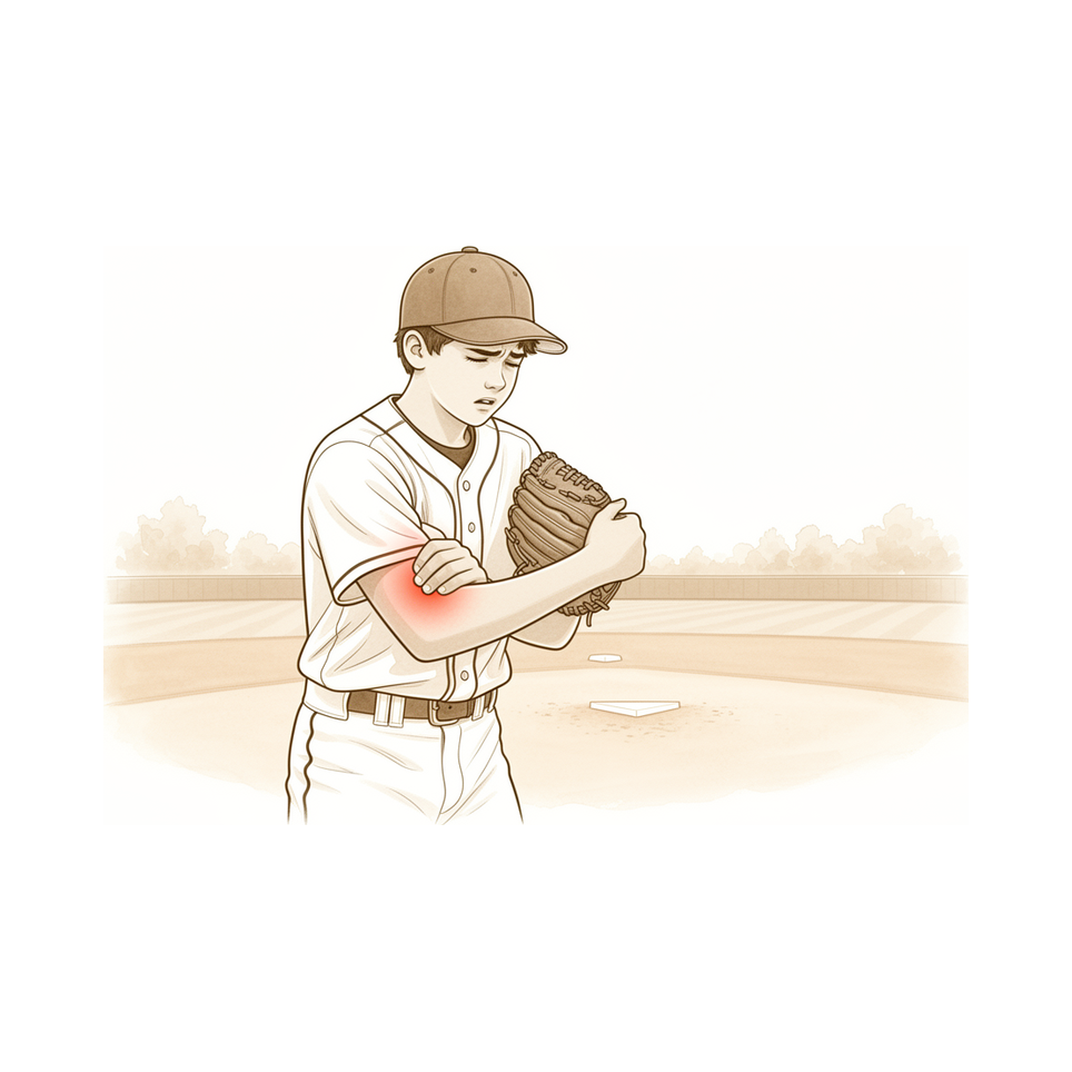
For patients: a plain-language version of this topic is available. See the patient guide.
Overview
Osteochondritis dissecans (OCD) of the capitellum is a debilitating injury that primarily affects immature athletes and is rarely encountered in adults [1]. The condition is frequently associated with repetitive throwing in the adolescent athlete, a mechanism that often leads to long-term problems [24]. Despite appropriate and aggressive treatment, the prognosis remains guarded [6]. Long-term results demonstrate continued elbow symptoms and degenerative joint disease in approximately 50% of patients [6]. Given these outcomes, it may be wise to counsel symptomatic pitchers and catchers to consider switching to another position or sport until outcomes for capitellar OCD improve [24].
Diagnosis is aided by MRI or CT due to the low accuracy of conventional radiographs [3]. In one series, OCD of the capitellum was detected during the initial interpretations of radiographic examinations in seven of 15 patients [5]. The true cause, natural history, and optimal treatment of this condition remain unknown [2]. Clinicians should exercise caution when assigning grades using magnetic resonance classifications for capitellar OCD [15].
Management depends on lesion stability and skeletal maturity. OCD of the capitellum can be classified as stable or unstable [10]. Stable lesions with an open growth plate and good motion heal with rest [10]. Conversely, unstable lesions with a closed growth plate, fragmentation, or restricted motion require surgery [10].
Anatomy & Pathophysiology
Bony Anatomy
The elbow is a trocho-ginglymoid joint comprising medial and lateral articulations that provide bony stability [55]. The ulnohumeral joint forms where the trochlea articulates with the ulna within the greater sigmoid notch, while the radiocapitellar joint forms between the capitellum and the radial head [55]. The radial head is a concave elliptical structure covered with articular cartilage along the radiocapitellar joint and approximately 270° of the articular margin [55]. The distal humeral articulation is angled 30° from the longitudinal axis [55], with the articular surface angled 30 degrees anterior to the humeral shaft axis [67]. The axis of rotation is 5° to 7° angulated in the coronal plane to the epicondylar axis, positioning the medial side more distal than the lateral side [55].
The distal humerus consists of medial and lateral columns [67], with the trochlea and capitellum comprising the articular component [70]. The medial column diverges from the humeral shaft at a 45-degree angle, whereas the lateral column diverges at a 20-degree angle [69]. In the sagittal plane, the lateral column curves anteriorly, creating a 35 to 40 degrees angle with the shaft [72]. In the coronal plane, the trochlea is more distal than the capitellum, resulting in a valgus alignment of 4 to 8 degrees [72]. The articular surface of the capitellum begins at the most distal aspect of the lateral column and encompasses an arc of approximately 180 degrees in the sagittal plane [72]. The posterior aspect of the lateral column is relatively flat and wide, permitting safe hardware placement due to the absence of cartilage [72].
The ulnohumeral joint allows flexion-extension, while the radiocapitellar joint allows forearm rotation [69]. The trochlea possesses a 300-degree arc of cartilage [69] and is covered by articular cartilage anteriorly, inferiorly, and posteriorly, creating an arc of almost 270 degrees [72]. Radiographically, the radial head should line up with the capitellum at all arm positions on all views [56]. In full extension, 60% of axial load is transmitted through the radiocapitellar joint [67].
Ligamentous Anatomy
The medial ulnar collateral ligament (MUCL) is the primary restraint to valgus stress within functional elbow range of motion [67]. It originates on the posterior medial epicondyle and inserts on the sublime tubercle of the medial coronoid process [67], with the posterior bundle serving as the primary restraint to valgus stress when the elbow is in maximal flexion [67]. The lateral collateral ligament complex consists of the radial collateral ligament, the lateral ulnar collateral ligament (LUCL), and the annular ligament [72]. The LUCL arises from an isometric point on the lateral epicondyle and attaches to the crista supinatoris of the proximal ulna [72], functioning as a posterolateral stabilizer [56]. The radial collateral ligament originates from an isometric point on the lateral epicondyle and fans out to attach to the annular ligament [72]. The LCL complex functions as an important restraint to varus and posterolateral rotatory instability [72]. The anterior capsule attaches at a point approximately 6 mm distal to the tip of the coronoid [56].
Pathophysiology & Etiology
Osteochondritis dissecans (OCD) of the capitellum is an uncommon disorder seen primarily in the adolescent overhead athlete [49]. Unlike Panner disease, which is a self-limiting condition of the immature capitellum, OCD is multifactorial [49]. The exact etiology is not fully defined [49], though causes include trauma, ischemia, constitution, and genetic predisposition [29]. OCD likely results from microtrauma in the setting of cartilage mismatch and vascular susceptibility [49], affecting immature athletes who undergo repetitive compression of the radiocapitellar joint [49]. Elbow valgus torque increases contact pressure in the radiocapitellar joint [94]. Various theories have been proposed to explain the pathogenesis, but little is known about the early changes [29]. This localized lesion involves a segment of articular cartilage with disruption of associated subchondral bone [49]. The natural history of OCD is poorly understood [49], and degenerative joint disease may develop over time [49]. Over mid- to long-term follow-up, the entire elbow joint underwent cylindrical-like morphological changes, leading to restricted motion in surgically treated OCD cases [86].
Classification
Minami Classification: The Minami classification describes the appearance of the capitellum on plain radiographs [44]. It is considered the most reliable system for classifying different stages of osteochondritis dissecans (OCD) of the humeral capitellum [112]. Type 1 lesions present with flattening of the capitellum or cystic changes in the capitellum [44]. Type 2 lesions are defined by clear subchondral detachment or fragment splitting in the capitellum [44].
Modified MRI Staging System: A modified MRI staging system classifies capitellar OCD lesions into five stages based on T2-weighted MRI findings [99]. Stage 1 is defined as a normally shaped capitellum with several spotted areas of high signal intensity that is lower than that of cartilage [99]. Stage 2 is defined as a normally shaped capitellum with several spotted areas of higher intensity than that of cartilage [99]. Stage 3 is defined as discontinuity and noncircularity of the chondral surface signal of the capitellum with no high signal interface apparent between the lesion and the floor [99]. Stage 4 is defined as a lesion separated by a high intensity line in comparison with cartilage [99]. Stage 5 is defined as a capitellar lesion displaced from the floor or defect of the capitellar lesion noted [99].
Other Considerations: Osteochondral lesions located in the posterolateral capitellum were associated with an advanced stage [22].
Clinical Presentation
Osteochondritis dissecans (OCD) of the capitellum is a critical elbow injury in adolescent overhead throwing athletes [133]. It is a painful condition that often affects young throwing athletes [36]. The condition causes pain, motion deficits, and activity limitations [38]. Panner's disease and osteochondritis dissecans likely represent a continuum of disordered endochondral ossification, with presentation and prognosis dependent primarily on age at onset [31].
Osteochondritis dissecans of the capitellum has only a slight tendency to heal, and instability can cause failure of the lesion to heal [17]. An osseous defect of the posterolateral corner of the capitellum is an uncommon finding that in each instance was associated with chronic posterolateral rotatory instability of the elbow [129]. Conversely, osteochondritis dissecans lesions of the capitellum, both small and large, did not alter the ulnohumeral kinematics and stability with intact collateral ligaments [16].
Investigations
Plain radiography: Conventional radiographs remain the hallmark and best screening test for elbow evaluation [41]. However, they have low accuracy for diagnosing osteochondritis dissecans of the capitellum [3]. In one series, the condition was detected during initial interpretations of radiographic examinations in seven of 15 patients [5]. Capitellum fractures may be easily missed on conventional radiographs if not suspected [137]. The presence of an osteochondral fragment undetected on radiography may adversely affect the result [132].
MRI: Magnetic resonance imaging (MRI) aids in the diagnosis of osteochondritis dissecans of the capitellum [3]. MRI can be used to evaluate ligaments and tendons, but it is rarely indicated for elbow stiffness evaluation [76]. Osteochondral lesions in the posterolateral capitellum were associated with an advanced stage based on MRI analysis [22].
CT: Computed tomography (CT) aids in the diagnosis of osteochondritis dissecans of the capitellum [3]. In an adolescent considered at risk for osteochondritis dissecans with lateral elbow joint pain, CT is the imaging modality of choice for diagnosing and staging the condition [130]. CT is helpful when assessing for malunion architecture and the location and pattern of osteophytes or loose bodies [76]. Radiological imaging including CT scans cannot reliably detect interposed capitellar cartilage fragments, which become apparent only during surgery [138].
Treatment
Non-Operative
Conservative management is the standard of care for stable osteochondritis dissecans (OCD) lesions of the capitellum [21]. Successful non-operative treatment is achievable when initiated in the early stages of the disease [20]. If identified early enough, most capitellar OCD lesions can be managed conservatively with favorable outcomes [52].
Operative
Indications: Surgery is indicated for patients with stable lesions who fail nonoperative treatment, or for those with unstable lesions, particularly when mechanical symptoms are present [38]. Unstable lesions specifically require surgical management [21].
Surgical Approach / Technique: Arthroscopy is useful for evaluating lesion stability and performing debridement, eliminating the need for more extensive open procedures associated with complications [13]. Arthroscopic debridement and microfracture can produce good to excellent outcomes in the majority of patients with osteochondritic lesions [12]. This approach is associated with a low incidence of progressive radiographic changes [12] and high rates of return to sport in the short-term [12]. In adolescent baseball players, arthroscopic surgery for symptomatic capitellar OCD can provide excellent rating scores with intermediate follow-up [11], although it does not assure return to baseball [11]. Microfracture is indicated for stage II or III lesions that are less than 1 cm in diameter and show an intact lateral buttress of capitellar cartilage [52]. For example, arthroscopic debridement and microfracture was performed on an 11-year-old swimmer with capitellar OCD after failing nonoperative treatment [27].
Osteochondral autograft transplantation (OAT) remains a superior surgical treatment for symptomatic chondral and osteochondral pathology of the capitulum [23]. OAT demonstrates excellent clinical and radiographic outcomes [7], is associated with minimal short-term donor site morbidity [7], and yields a high level of return to the sport [7]. For high-level athletes, OAT has excellent reported outcomes [23] and high rates of return to play [23]. Current evidence suggests that OAT may lead to better and more consistent outcomes than previously described methods for treating large OCD lesions of the capitellum [36]. Clinicians can counsel injured youth baseball players that capitellar OCD lesions indicated for autologous osteochondral transplantation can have similar or better return-to-sport outcomes than lesions indicated for fixation or debridement plus microfracture [30].
Surgical osteochondral allograft transplantation of the capitellum is a viable option for even large lesions (>10 mm) [9]. This technique has minimal morbidity [9] and good return of function [9]. A ligament-sparing technique for fresh osteochondral allograft transplantation is described to treat unstable osteochondritis dissecans of the capitellum [18].
Other surgical techniques include closed-wedge osteotomy of the lateral humeral condyle, which provides good long-term clinical and radiographic results for capitellar OCD across all lesion types [8]. Arthroscopic mosaicplasty is an option for large lesions that allow the radial head to engage within the capitellar lesion [38]. Drilling is a surgical treatment option for stable intact lesions [38], while chondroplasty with drilling is a surgical treatment option for unstable lesions [38]. Internal fixation is a surgical treatment option for unstable in situ osteochondritis dissecans lesions of the capitellum [63]. Extra-articular core tunneling and local autogenous bone grafting is a treatment for osteochondritis dissecans lesion of the capitellum with intact articular cartilage and subchondral bone deficiency [64].
Implant Selection: Treatment of large, unstable osteochondritis dissecans lesions of the capitellum in adolescent athletes allows reliable return to high level of sports [4]. This treatment is safe [4] and has excellent long-term clinical outcomes [4]. Similarly, treatment of large, unstable OCD lesions of the capitellum in adolescent athletes allows reliable return to play [48], is safe [48], and has good clinical outcomes at short-term follow-up [48]. Treatment of large, unstable OCD lesions of the capitellum in adolescent athletes allows for a high rate of return to sport [66] and has good clinical outcomes with long-term follow-up [66]. Harvesting osteochondral grafts did not exert adverse effects on donor knee function in young athletes at 2 years after undergoing osteochondral autograft transplantation for capitellar osteochondritis dissecans [19]. Costal osteochondral autograft gave satisfactory results for advanced osteochondritis dissecans of the humeral capitellum with extensive lesions affecting the lateral wall [14]. Both patients had generally good clinical outcomes with arthroscopic débridement and autologous osteochondral graft to the capitellum [26].
Surgical Technique and Portals: Use of dual direct lateral portals is safe and practical for arthroscopic treatment of capitellar osteochondritis dissecans [32].
Postoperative Rehabilitation: Postoperative recovery and rehabilitation for capitellar OCD occurs in 4 phases: healing/immobilization, motion, strengthening, and sport-specific training [61]. For arthroscopic debridement or microfracture, immobilization is not routinely used [61]. For larger procedures, operative extremities may be immobilized in a sling for up to 2 -3 weeks but rarely longer [61]. Some authors immobilize in a long-arm splint or cast [61]. The goal is generally full ROM by 6 -8 weeks postoperatively [61]. Shoulder isometrics are typically permitted within the first few weeks after surgery [61]. For larger cases, elbow isometrics may begin once elbow ROM is restored [61]. For simple debridement and microfracture, return to sport-specific training typically begins as soon as symptoms permit, usually around 1 -3 months [61].
For microfracture specifically, patients are provided with a soft, supportive sling postoperatively [52]. The dressing is changed on postoperative day 2 [52]. Passive and gentle active ROM is initiated within the first week [52]. The goal is achieving full ROM by 6 weeks postoperatively [52]. Gentle shoulder, scapular, wrist, and hand strengthening is initiated during weeks 2 to 6 [52]. Light elbow resistance training is initiated at week 6 [52]. Patients are not allowed to return to contact sports or throwing for a minimum of 6 months after microfracture surgery [52].
Prognosis and Outcomes: Long-term results demonstrate continued elbow symptoms in approximately 50% of patients with osteochondritis dissecans of the capitellum [6]. Long-term results also demonstrate degenerative joint disease in approximately 50% of patients with osteochondritis dissecans of the capitellum [6].
Complications
Long-term Outcomes and Degeneration
Long-term results for osteochondritis dissecans of the capitellum demonstrate continued elbow symptoms and degenerative joint disease in approximately 50% of patients despite appropriate and aggressive treatment [6]. In a cohort of 31 patients with a mean follow-up of 23 years, 42% complained of pain and/or reduced range of motion, and one-third had radiographic degenerative changes [106]. The natural history of osteochondritis dissecans of the capitellum is poorly understood, and degenerative joint disease may develop over time [49].
Surgical Complications and Morbidity
Donor-site morbidity after osteochondral autologous transplantation for capitellar osteochondritis dissecans was reported in a considerable group of patients [47]. Arthroscopy is useful for evaluating the stability of the lesion and performing debridement in cases of osteochondritis dissecans of the capitellum, thereby eliminating the need for a more extensive open procedure associated with complications [13]. Concerns persist regarding the healing potential of advanced lesions treated with fragment fixation and bone grafting [49].
Diagnostic and Prognostic Factors
Conventional radiographs have low accuracy for diagnosing osteochondritis dissecans of the capitellum, with detection occurring in only seven of 15 patients during initial interpretations [5]. Repetitive throwing in the adolescent athlete often leads to long-term problems, and until outcomes improve, it may be wise to counsel symptomatic pitchers and catchers to consider switching to another position or sport [24]. Arthroscopic surgery for symptomatic osteochondritis dissecans of the capitellum in adolescent baseball players can provide excellent rating scores with intermediate follow-up but does not assure return to baseball [11].
Recovery
Other Considerations: The natural history of osteochondritis dissecans of the capitellum is characterized by only a slight tendency to heal [17]. Instability of the lesion can cause failure to heal [17]. Long-term results demonstrate continued elbow symptoms and degenerative joint disease in approximately 50% of patients [6].
Surgical Outcomes: Osteochondral autograft transplantation demonstrates excellent clinical and radiographic outcomes [7]. Surgical osteochondral allograft transplantation of the capitellum is associated with minimal morbidity [9] and good return of function [9]. OAT is associated with excellent reported outcomes and rates of return to play for high-level athletes [23]. Autologous osteochondral mosaicplasty for advanced lesions of capitellar osteochondritis dissecans can provide satisfactory clinical and radiographic results [25].
Key Evidence
- [L5] Osteochondritis dissecans (OCD) of the capitellum occurs in immature athletes and is rarely found in adults. [1] (10.1016/s0278-5919(05)70270-2)
- [L5] The true cause, natural history, and optimal treatment of osteochondritis dissecans of the capitellum remain unknown. [2] (10.1177/0363546509354969)
- [L5] Osteochondritis dissecans of the capitellum is a debilitating injury in young athletes, with diagnosis aided by MRI or CT due to low accuracy of conventional radiographs. [3] (10.1177/17585732231190011)
- [L4] Treatment of large, unstable osteochondritis dissecans lesions of the capitellum in adolescent athletes allows reliable return to high level of sports, is safe and has excellent long-term clinical outcomes. [4] (10.1177/2325967114s00036)
- [L4] Osteochondritis dissecans of the capitellum was detected during the initial interpretations of the radiographic examinations of the elbow in seven of 15 patients. [5] (10.1007/s00256-005-0899-6)
- [L5] Despite appropriate and aggressive treatment, the prognosis for osteochondritis dissecans of the capitellum remains guarded, with long-term results demonstrating continued elbow symptoms and degenerative joint disease in approximately 50% of patients. [6] (10.1016/j.jse.2009.11.058)
- [L4] In the treatment of osteochondritis dissecans of the capitellum, osteochondral autograft transplantation demonstrates excellent clinical and radiographic outcomes, with minimal short-term donor site morbidity and a high level of return to the sport. [7] (10.1016/j.jhsa.2021.02.024)
- [L4] Closed-wedge osteotomy of the lateral humeral condyle provides good long-term clinical and radiographic results for osteochondritis dissecans of the capitellum across all lesion types. [8] (10.1016/j.jse.2019.05.016)
- [L5] Surgical osteochondral allograft transplantation of the capitellum is a viable option for even large lesions (>10 mm), with minimal morbidity and good return of function. [9] (10.1016/j.eats.2024.102997)
- [L2] Osteochondritis dissecans of the capitellum can be classified as stable or unstable; stable lesions with an open growth plate and good motion heal with rest, while unstable lesions with a closed growth plate, fragmentation, or restricted motion require surgery. [10] (10.2106/jbjs.f.00622)
- [L4] Arthroscopic surgery for symptomatic osteochondritis dissecans of the capitellum in adolescent baseball players can provide excellent rating scores with intermediate follow-up but does not assure return to baseball. [11] (10.1177/03635465020300040401)
- [L4] Arthroscopic debridement and microfracture can produce good to excellent outcomes in the majority of patients with osteochondritic lesions of the capitellum, with a low incidence of progressive radiographic changes and high rates of return to sport in the short-term. [12] (10.1016/j.arthro.2010.04.034)
- [L4] Arthroscopy is useful for evaluating the stability of the lesion and performing debridement in cases of osteochondritis dissecans of the capitellum, thereby eliminating the need for a more extensive open procedure associated with complications. [13] (10.2152/jmi.61.233)
- [L4] Costal osteochondral autograft gave satisfactory results for advanced osteochondritis dissecans of the humeral capitellum with extensive lesions affecting the lateral wall. [14] (10.1016/j.jse.2014.06.047)
- [L4] One should be cautious when assigning grades using magnetic resonance classifications for capitellar osteochondritis dissecans. [15] (10.1177/1758573218821151)
- [L5] Osteochondritis dissecans lesions of the capitellum, both small and large, did not alter the ulnohumeral kinematics and stability with intact collateral ligaments. [16] (10.1016/j.jhsa.2010.09.037)
- [L3] Osteochondritis dissecans of the capitellum has only a slight tendency to heal, and instability can cause failure of the lesion to heal. [17] (10.1177/03635465990270060701)
- [L4] The authors describe a ligament-sparing technique for fresh osteochondral allograft transplantation to treat unstable osteochondritis dissecans of the capitellum. [18] (10.1097/bte.0000000000000066)
- [L4] Harvesting osteochondral grafts did not exert adverse effects on donor knee function in young athletes at 2 years after undergoing osteochondral autograft transplantation for capitellar osteochondritis dissecans. [19] (10.1177/0363546510388386)
- [L3] Osteochondrosis of the humeral capitellum can be successfully treated conservatively if treatment is begun in an early stage of the disease. [20] (10.1177/0363546507312168)
- [L5] The article explores osteochondritis dissecans of the elbow regarding aetiology, clinical presentation, diagnostics, surgical techniques, complications, clinical outcomes, and future directions, noting that stable lesions are generally treated conservatively while unstable lesions require surgical management. [21] (10.1136/jisakos-2015-000008)
- [L3] Osteochondral lesions in the posterolateral capitellum were associated with an advanced stage. [22] (10.1177/2325967120946269)
- [Paper] OAT remains a superior surgical treatment for symptomatic chondral and osteochondral pathology of the capitulum, with excellent reported outcomes and rates of return to play for high-level athletes. [23] (10.1016/j.eats.2020.07.022)
- [L5] Repetitive throwing in the adolescent athlete often leads to long-term problems, and until outcomes for osteochondritis dissecans of the capitellum improve, it may be wise to counsel symptomatic pitchers and catchers to consider switching to another position or sport. [24] (10.1016/j.arthro.2020.02.029)
- [L4] Autologous osteochondral mosaicplasty for advanced lesions of capitellar osteochondritis dissecans can provide satisfactory clinical and radiographic results. [25] (10.1016/j.jse.2010.09.001)
- [L4] Both patients had generally good clinical outcomes with arthroscopic débridement and autologous osteochondral graft to the capitellum. [26] (10.1016/j.jses.2018.02.005)
- [L5] The patient, an 11-year-old swimmer with capitellar osteochondritis dissecans, underwent arthroscopic debridement and microfracture after failing nonoperative treatment. [27] (10.1016/j.xrrt.2026.100760)
- [L4] [29] (10.2106/00004623-199806000-00014)
- [L4] Clinicians can counsel injured youth baseball players that capitellar osteochondritis dissecans lesions indicated for autologous osteochondral transplantation can have similar or better return-to-sport outcomes than osteochondritis dissecans lesions indicated for fixation or debridement + microfracture. [30] (10.1177/03635465261443986)
- [L5] Panner's disease and osteochondritis dissecans likely represent a continuum of disordered endochondral ossification with presentation and prognosis dependent primarily on age at onset. [31] (10.5435/00124635-200407000-00005)
- [L5] Use of dual direct lateral portals is safe and practical for arthroscopic treatment of capitellar osteochondritis dissecans. [32] (10.1016/j.arthro.2007.01.029)
- [L4] [36] (10.1177/1558944716643293)
- [L4] [38] (10.1097/01.bte.0000235401.59599.31)
- [Paper] [44] (10.1177/1941738112444707)
- [L1] Donor-site morbidity after OATS for capitellar osteochondritis dissecans was reported in a considerable group of patients. [47] (10.1007/s00167-017-4516-8)
- [L4] Treatment of large, unstable OCD lesions of the capitellum in adolescent athletes allows reliable return to play, is safe, and has good clinical outcomes at short-term follow-up. [48] (10.1016/j.jse.2015.03.014)
- [L5] [49] (10.5435/00124635-201009000-00007)
- [Paper] [52] (10.1016/j.eats.2016.01.030)
- [L4] [61] (10.1007/s12178-019-09528-8)
- [L4] [63] (10.1016/s0363-5023(12)60060-4)
- [L4] [64] (10.1016/j.xrrt.2023.09.005)
- [L4] Treatment of large, unstable OCD lesions of the capitellum in adolescent athletes is safe, allows for a high rate of return to sport, and has good clinical outcomes with long-term follow-up. [66] (10.1016/j.jseint.2026.101680)
- [L3] Over mid- to long-term follow-up, the entire elbow joint underwent cylindrical-like morphological changes, leading to restricted motion. [86] (10.1177/23259671251339180)
- [L5] Elbow valgus torque increases contact pressure in the radiocapitellar joint. [94] (10.1177/0363546513490652)
- [L2] [99] (10.1177/0363546514532604)
- [L5] [106] (10.1007/s00167-015-3518-7)
- [L4] The Minami Classification was the most reliable for classifying different stages of OCD of the humeral capitellum. [112] (10.1016/j.jse.2015.03.029)
- [L4] An osseous defect of the posterolateral corner of the capitellum is an uncommon finding that in each instance was associated with chronic posterolateral rotatory instability of the elbow. [129] (10.2214/ajr.07.3739)
- [L3] In an adolescent considered at risk for OCD with lateral elbow joint pain, CT is the imaging modality of choice for diagnosing and staging OCD of the capitellum. [130] (10.1177/03635465221129598)
- [L5] The presence of an osteochondral fragment undetected on radiography may adversely affect the result. [132] (10.1016/0020-1383(75)90118-7)
- [L5] Osteochondritis dissecans (OCD) of the humeral capitellum is a critical elbow injury in adolescent overhead throwing athletes. [133] (10.1016/j.jos.2017.11.013)
- [L4] Capitellum fractures may be easily missed on conventional radiographs, if not suspected. [137] (10.1097/bpo.0000000000001208)
- [L4] Radiological imaging including CT scans cannot reliably detect interposed capitellar cartilage fragments, which become apparent only during surgery. [138] (10.1007/s00402-021-03895-z)
References
[1] OSTEOCHONDRITIS DISSECANS OF THE HUMERAL CAPITELLUM. Clinics in Sports Medicine. 2001. DOI: 10.1016/s0278-5919(05)70270-2
[2] Osteochondritis Dissecans of the Capitellum. The American Journal of Sports Medicine. 2010. DOI: 10.1177/0363546509354969
[3] Update in diagnosis, treatment, and prevention of osteochondritis dissecans of the capitellum. Shoulder & Elbow. 2023. DOI: 10.1177/17585732231190011
[4] Osteochondral Autograft Plug Transfer for Treatment of Osteochondritis Dissecans of the Capitellum in Adolescent Athletes. Orthopaedic Journal of Sports Medicine. 2014. DOI: 10.1177/2325967114s00036
[5] Radiography of the elbow for evaluation of patients with osteochondritis dissecans of the capitellum. Skeletal Radiology. 2005. DOI: 10.1007/s00256-005-0899-6
[6] Osteochondritis dissecans of the capitellum. Journal of Shoulder and Elbow Surgery. 2010. DOI: 10.1016/j.jse.2009.11.058
[7] Outcomes of Osteochondral Autograft Transplantation in Pediatric Patients With Osteochondritis Dissecans of the Capitellum. The Journal of Hand Surgery. 2021. DOI: 10.1016/j.jhsa.2021.02.024
[8] Long-term results of closed-wedge osteotomy of the lateral humeral condyle for osteochondritis dissecans of the capitellum. Journal of Shoulder and Elbow Surgery. 2019. DOI: 10.1016/j.jse.2019.05.016
[9] Osteochondral Allograft Transplantation to the Capitellum: Technical Considerations of a Mega Osteochondritis Dissecans Technique. Arthroscopy Techniques. 2024. DOI: 10.1016/j.eats.2024.102997
[10] Classification, Treatment, and Outcome of Osteochondritis Dissecans of the Humeral Capitellum. The Journal of Bone & Joint Surgery. 2007. DOI: 10.2106/jbjs.f.00622
[11] Arthroscopic Surgery for Isolated Capitellar Osteochondritis Dissecans in Adolescent Baseball Players: Minimum Three-Year Follow-Up. The American Journal of Sports Medicine. 2002. DOI: 10.1177/03635465020300040401
[12] Arthroscopic Debridement and Microfracture of Capitellar Osteochondritis Dissecans of the Elbow (SS‐24). Arthroscopy. 2010. DOI: 10.1016/j.arthro.2010.04.034
[13] State of the Art: Elbow Arthroscopy: Review of the Literature and Application for Osteochondritis Dissecans of the Capitellum. The Journal of Medical Investigation. 2014. DOI: 10.2152/jmi.61.233
[14] Costal osteochondral autograft for reconstruction of advanced-stage osteochondritis dissecans of the capitellum. Journal of Shoulder and Elbow Surgery. 2014. DOI: 10.1016/j.jse.2014.06.047
[15] Interobserver reliability of the classification of capitellar osteochondritis dissecans using magnetic resonance imaging. Shoulder & Elbow. 2019. DOI: 10.1177/1758573218821151
[16] Osteochondral Lesions of the Capitellum Do Not Affect Elbow Kinematics and Stability With Intact Collateral Ligaments: An In Vitro Biomechanical Study. The Journal of Hand Surgery. 2011. DOI: 10.1016/j.jhsa.2010.09.037
[17] Nonoperative Treatment of Osteochondritis Dissecans of the Humeral Capitellum. The American Journal of Sports Medicine. 1999. DOI: 10.1177/03635465990270060701
[18] Fresh Osteochondral Allograft Transplantation for the Treatment of Unstable Osteochondritis Dissecans of the Capitellum in the Elbow. Techniques in Shoulder & Elbow Surgery. 2016. DOI: 10.1097/bte.0000000000000066
[19] Functional Recovery of the Donor Knee After Autologous Osteochondral Transplantation for Capitellar Osteochondritis Dissecans. The American Journal of Sports Medicine. 2010. DOI: 10.1177/0363546510388386
[20] Conservative Treatment for Osteochondrosis of the Humeral Capitellum. The American Journal of Sports Medicine. 2008. DOI: 10.1177/0363546507312168
[21] Osteochondritis dissecans of the elbow: state of the art. Journal of ISAKOS. 2017. DOI: 10.1136/jisakos-2015-000008
[22] Distribution of Osteochondral Lesions in Patients With Simple Elbow Dislocations Based on MRI Analysis. Orthopaedic Journal of Sports Medicine. 2020. DOI: 10.1177/2325967120946269
[23] Osteochondral Autograft Transfer for Capitellar Chondral and Osteochondral Defects. Arthroscopy Techniques. 2020. DOI: 10.1016/j.eats.2020.07.022
[24] Editorial Commentary: Should We Consider Early Retirement for Adolescent Pitchers and Catchers With Osteochonditis Dissecans of the Capitellum?. Arthroscopy. 2020. DOI: 10.1016/j.arthro.2020.02.029
[25] The clinical outcomes of mosaicplasty in the treatment of osteochondritis dissecans of the distal humeral capitellum of young athletes. Journal of Shoulder and Elbow Surgery. 2011. DOI: 10.1016/j.jse.2010.09.001
[26] Osteochondritis dissecans of the capitellum secondary to fishtail deformity treated with osteochondral autograft. JSES Open Access. 2018. DOI: 10.1016/j.jses.2018.02.005
[27] Arthroscopic treatment of osteochondritis dissecans of the capitellum in an early adolescent swimmer: a case report. JSES Reviews, Reports, and Techniques. 2026. DOI: 10.1016/j.xrrt.2026.100760
[29] Early Detection of Osteochondritis Dissecans of the Capitellum in Young Baseball Players. Report of Three Cases. The Journal of Bone & Joint Surgery*. 1998. DOI: 10.2106/00004623-199806000-00014
[30] Return to Sport After Surgical Treatment of Capitellar Osteochondritis Dissecans Lesions in Baseball Players: A Systematic Review and Meta-analysis. The American Journal of Sports Medicine. 2026. DOI: 10.1177/03635465261443986
[31] Lateral Compression Injuries in the Pediatric Elbow: Pannerʼs Disease and Osteochondritis Dissecans of the Capitellum. Journal of the American Academy of Orthopaedic Surgeons. 2004. DOI: 10.5435/00124635-200407000-00005
[32] Dual Direct Lateral Portals for Treatment of Osteochondritis Dissecans of the Capitellum: An Anatomic Study. Arthroscopy. 2007. DOI: 10.1016/j.arthro.2007.01.029
[36] Current Concepts. HAND. 2016. DOI: 10.1177/1558944716643293
[38] Treatment of Capitellar Osteochondritis Dissecans. Techniques in Shoulder & Elbow Surgery. 2006. DOI: 10.1097/01.bte.0000235401.59599.31
[41] Orthopaedic Knowledge Update 13 Ebook Without Multimedia. Anatomy, Biomechanics, Physical Examination, and Imaging of the Elbow > Summary and Conclusions.
[44] Surgical Treatment for Osteochondritis Dissecans of the Capitellum. Sports Health: A Multidisciplinary Approach. 2012. DOI: 10.1177/1941738112444707
[47] Donor-site morbidity after osteochondral autologous transplantation for osteochondritis dissecans of the capitellum: a systematic review and meta-analysis. Knee Surgery, Sports Traumatology, Arthroscopy. 2017. DOI: 10.1007/s00167-017-4516-8
[48] Osteochondral autograft plug transfer for treatment of osteochondritis dissecans of the capitellum in adolescent athletes. Journal of Shoulder and Elbow Surgery. 2015. DOI: 10.1016/j.jse.2015.03.014
[49] Osteochondritis Dissecans of the Capitellum: Current Concepts. Journal of the American Academy of Orthopaedic Surgeons. 2010. DOI: 10.5435/00124635-201009000-00007
[52] Arthroscopic Microfracture for Osteochondritis Dissecans Lesions of the Capitellum. Arthroscopy Techniques. 2016. DOI: 10.1016/j.eats.2016.01.030
[55] Orthopaedic Knowledge Update 13 Ebook Without Multimedia. Anatomy, Biomechanics, Physical Examination, and Imaging of the Elbow > Anatomy > Bony Anatomy.
[56] Miller S Review Of Orthopaedics. SECTION 16 PATELLAR TRACKING IN TOTAL KNEE ARTHROPLASTY > TABLE 2.3 Shoulder Spaces.
[61] Osteochondritis dissecans lesions of the capitellum in overhead athletes: a review of current evidence and proposed treatment algorithm. Current Reviews in Musculoskeletal Medicine. 2019. DOI: 10.1007/s12178-019-09528-8
[63] Internal Fixation of Unstable In Situ Osteochondritis Dissecans Lesions of the Capitellum. The Journal of Hand Surgery. 2012. DOI: 10.1016/s0363-5023(12)60060-4
[64] Extra-articular core tunneling and local autogenous bone grafting for osteochondritis dissecans lesion of the capitellum with intact articular cartilage and subchondral bone deficiency. JSES Reviews, Reports, and Techniques. 2024. DOI: 10.1016/j.xrrt.2023.09.005
[66] Osteochondral autograft transfer for capitellar osteochondritis dissecans in high-level adolescent athletes. JSES International. 2026. DOI: 10.1016/j.jseint.2026.101680
[67] Miller S Review Of Orthopaedics. ELBOW.
[69] A Lange Medical Book Current Diagnosis Treatment In Orthopedics Fifth Edition. 2Musculoskeletal Trauma Surgery > INJURIES AROUND THE ELBOW.
[70] Orthopaedic Knowledge Update Trauma. Distal Humerus Fracture > Anatomy.
[72] Rockwood And Green S Fractures In Adults. 29: Principles of Nonunion and Bone Defect Treatment > Pathoanatomy and Applied Anatomy Related to Distal Humerus Fractures.
[76] Aaos Comprehensive Orthopaedic Review 3. Elbow Stiffness* > IV. Evaluation.
[86] Comprehensive Elbow Joint Morphology Changes in Surgically Treated Osteochondritis Dissecans of the Capitellum: Clinical Implications. Orthopaedic Journal of Sports Medicine. 2025. DOI: 10.1177/23259671251339180
[94] Biomechanical Characteristics of Osteochondral Defects of the Humeral Capitellum. The American Journal of Sports Medicine. 2013. DOI: 10.1177/0363546513490652
[99] Magnetic Resonance Imaging Staging to Evaluate the Stability of Capitellar Osteochondritis Dissecans Lesions. The American Journal of Sports Medicine. 2014. DOI: 10.1177/0363546514532604
[106] Biomechanical considerations in the pathogenesis of osteoarthritis of the elbow. Knee Surgery, Sports Traumatology, Arthroscopy. 2015. DOI: 10.1007/s00167-015-3518-7
[112] Osteochondritis dissecans of the humeral capitellum: reliability of four classification systems using radiographs and computed tomography. Journal of Shoulder and Elbow Surgery. 2015. DOI: 10.1016/j.jse.2015.03.029
[129] Osborne-Cotterill Lesion: An Osseous Defect of the Capitellum Associated with Instability of the Elbow. American Journal of Roentgenology. 2008. DOI: 10.2214/ajr.07.3739
[130] Advantages of CT Versus MRI for Preoperative Assessment of Osteochondritis Dissecans of the Capitellum. The American Journal of Sports Medicine. 2022. DOI: 10.1177/03635465221129598
[132] Osteochondral fracture of the trochlea associated with fracture-dislocation of the elbow. Injury. 1975. DOI: 10.1016/0020-1383(75)90118-7
[133] Diagnosis and treatment of osteochondritis dissecans of the humeral capitellum. Journal of Orthopaedic Science. 2018. DOI: 10.1016/j.jos.2017.11.013
[137] Mid-term and Long-term Functional and Radiographic Results of 13 Surgically Treated Adolescent Capitellum Fractures. Journal of Pediatric Orthopaedics. 2018. DOI: 10.1097/bpo.0000000000001208
[138] Radial head fractures with interposed capitellar cartilage fragment–hindrance to bone healing–a case series. Archives of Orthopaedic and Trauma Surgery. 2021. DOI: 10.1007/s00402-021-03895-z