Clinicians › Wrist
Distal Radius ORIF
Open reduction and volar plate fixation of the distal radius — operation, recovery, rehabilitation.
For patients: a plain-language version of this topic is available. See the patient guide.
Overview
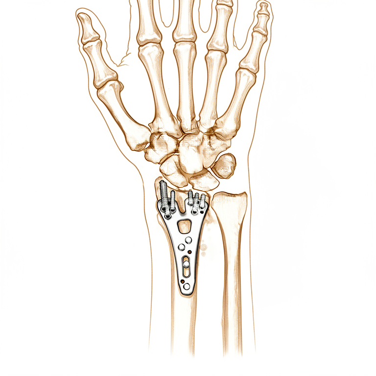
Open reduction and internal fixation (ORIF) is a valuable treatment modality for unstable distal radius fractures in both young and elderly patients, with the anterior approach currently serving as the gold standard [1, 14]. While the best method of treatment has yet to be definitively established due to a multitude of options with varying degrees of evidence [70], ORIF yields significantly better subjective outcomes (DASH scores), restoration of anatomic volar tilt, and forearm flexion and extension compared to external fixation at the end of the follow-up period [11]. In very old patients with distal radius fractures accompanied by ulnar styloid fractures, ORIF may benefit optimal long-term functional recovery [3], and surgical fixation in appropriately selected superelderly patients yields good functional outcomes [17].
External fixation remains a valid treatment option, particularly for all but the most stable extra-articular fractures [28, 37]. Three years after surgery for unstable dorsally displaced fractures, clinical and radiological results for volar locking plates and external fixation were comparable [4], and external fixation provides the same functional recovery after one year as volar locking plate systems [22]. However, external fixation is associated with higher rates of minor and major complications that do not require surgery compared with volar locking plate systems [22]. Current literature offers no evidence to support the use of internal fixation over external fixation for unstable distal radius fractures [5], and complication frequencies did not differ among different treatment modalities in comparative observational studies, though these findings were less robust against potential unmeasured confounders [38].
Early mobilization for patients treated with ORIF may have a beneficial functional effect compared to late mobilization at earlier stages, with mean differences in DASH scores at 6 weeks surpassing the minimal clinically important difference [19]. Routine early post-operative radiographs, particularly for extra-articular fractures, are questioned in terms of value [7]. Substantial variations in surgical direct costs exist, with implant choice being the predominant driver [9], and the factor of the plate should be considered when evaluating surgical treatment outcome [15]. Findings regarding perioperative proton pump inhibitors versus H2-receptor antagonists do not definitively determine whether temporary discontinuation of chronic PPIs alters risks after distal radius ORIF [10]. There is substantial heterogeneity and lack of standardization in the collection of objective and patient-reported outcome measures in level I and II RCTs [8].
Anatomy & Pathophysiology
Bony Anatomy
The carpus comprises two rows of eight bones bridging the forearm and hand [39]. The proximal row, acting as the key intercalated segment, includes the scaphoid, lunate, and triquetrum from radial to ulnar [39]. The distal row consists of the trapezium, trapezoid, capitate, and hamate [39]. The pisiform is a sesamoid bone within the flexor carpi ulnaris tendon sheath and is not theoretically part of the proximal row [39]. The scaphoid is a small, irregular S-shaped tubular bone on the radial aspect, lying at a 45-degree plane to the longitudinal and horizontal axes of the wrist [84]. It functions as a midcarpal joint "bridge" linking and synchronizing the motions of the proximal and distal carpal rows [84].
The distal radius articular surface is biconcave with scaphoid and lunate facets, while the distal radioulnar joint (DRUJ) articulates with the ulna at the sigmoid notch [90]. Lister tubercle is a dorsal prominence serving as a landmark for the dorsal approach and a cause of attritional extensor pollicis longus (EPL) rupture after fracture [90]. The distal radial metaphysis has thin cortex and is vulnerable to bending forces [90]. In a normal wrist with neutral ulnar variance, the distal radius bears 80% of the axial load [90]. The forearm functions as a ring, making isolated radial shaft fractures uncommon in adults [24]. Fractures within 7.5 cm of the radiocarpal joint are more likely to represent a Galeazzi fracture, defined as a radial shaft fracture with associated DRUJ injury [24].
Ligamentous Anatomy
Extrinsic ligaments connect carpal bones to the forearm proximally and metacarpals distally [87]. The extrinsic palmar radiocarpal ligaments include the transverse carpal, radioscaphocapitate, radioscapholunate, radial collateral, long radiolunate, and short radiolunate ligaments [87]. Strong oblique extrinsic palmar radial ligaments prevent medial carpal translation on the angulated slope of the distal radius through two V-shaped bands [87]. The space of Poirier is a V-shaped interligamentous sulcus over the capitolunate articulation representing capsular weakness [87]. The arcuate ligament, located in the central third of the palmar joint capsule, forms a support sling for the midcarpal region, particularly the capitate head [87]. The extrinsic ulnocarpal ligaments include the ulnotriquetral, ulnolunate, and ulnocapitate ligaments [87]. The extrinsic dorsal carpal ligaments include the dorsal radiocarpal and dorsal intercarpal ligaments, forming a V-shaped configuration [87].
Intrinsic ligaments connect individual carpal bones and include palmar midcarpal, proximal interosseous, and distal interosseous ligaments [87]. The scapholunate ligament consists of dorsal, palmar, and proximal portions, with the proximal portion being the widest, thinnest (1 mm), and weakest section [97]. The lunotriquetral ligament also consists of dorsal, palmar, and proximal portions [97]. The radioscaphocapitate ligament does not attach to the scaphoid itself but crosses its waist, acting as a sling that allows rotation [84]. There are no tendon attachments to the scaphoid [84].
Neurovascular Anatomy
Neurovascular supply to the carpus is via the anterior and posterior interosseous nerves [93]. Circulation comprises extraosseous and intraosseous vasculature via dorsal and palmar systems branching from the radial, ulnar, anterior interosseous, and deep palmar arch arteries [93]. The scaphoid is supplied by a single vessel, increasing its risk of avascular necrosis [93]. The dorsal branch enters via foramina along the spiral groove and dorsal ridge, supplying 70% to 80% of the scaphoid proximally [84]. The volar branch enters via the scaphoid tubercle and supplies the remaining 20% to 30% of the distal scaphoid [84]. The waist has minimal or no perforating vasculature, and no vessels perforate the proximal dorsal cartilaginous area or through the scapholunate ligament [84].
Pathophysiology and Deformity
The brachioradialis insertion on the radial styloid acts as a deforming force in distal radius fractures [90]. Fracture patterns include: Colles fracture: Volar angulation, dorsal displacement, loss of radial inclination, and resultant radial shortening [41]. Smith fracture: Dorsally angulated fracture with the hand and wrist displaced volarly with respect to the forearm [41]. Barton fracture: Fracture-dislocation with an intraarticular fracture in which the carpus and a rim of the distal radius are displaced together [41]. Chauffeur's fracture: Radial styloid fracture [41].
Instability patterns include: Dorsal intercalated segmental instability (DISI): Lunate torn from the scaphoid and tilts backwards [40]. Volar intercalated segment instability (VISI): Lunate torn from the triquetrum and tilts forwards [40].
In a normal lateral wrist radiograph, the axes of the radius, lunate, capitate, and third metacarpal are co-linear, with the scaphoid projecting at an angle of about 45 degrees to this line [40]. With traumatic instability, linked carpal segments collapse like the buckled carriages of a derailed train [40]. Injuries to adjacent soft-tissue structures occur in approximately one-half of distal radius fractures and in almost all intra-articular fractures [102]. The most common associated soft-tissue injuries are the triangular fibrocartilage complex (TFCC), scapholunate ligament, and lunotriquetral ligament, in descending order of frequency [102]. All stages of radioulnar instability demonstrate a decrease in the lunate contact area in positions with the forearm in supination [112]. Substantial increases in tension across the wrist do not develop until 6 mm of applied distraction, suggesting carpal ligaments are not substantially loaded at levels below 6 mm [117]. Baseline forces and forces created by active and passive motion of the wrist do not exceed 60 N to 80 N [83].
Classification
Historical Context: Eponymous descriptions such as Colles and Smith have served as guidelines for 150 years [48]. Renewed enthusiasm for defining fracture morphology is driven by increased prevalence in young adults, better understanding of joint kinematics, and the need for objective outcome measures [48].
Volar/Dorsal Classification: A new intra-articular distal radius fractures classification was proposed based on the affection condition of volar or dorsal side [141].
DREAD Classification: The DREAD classification organizes and describes articular fragment escape patterns following distal radius fracture fixation to raise awareness and facilitate decision-making [52].
Other Considerations: A metaphyseal fracture entering the distal radio-ulnar joint (M') affects functional outcome and must be included in the classification [164]. Similarly, a displaced ulnar fracture (U > 1) affects functional outcome and must be included in the classification [164]. Extraarticular and/or intra-articular scores exceeding 8 points on a 12-points scale were statistically associated with more complications, irrespective of the patient’s overall health and age [68]. A minority of fractures are resistant to mal-union in dorsal angulation and may be identified by a radiographically intact dorsal radial cortex [32]. Complete intra-articular distal radius fractures in young active patients must be considered as a generally multi-tissue traumatic entity [29].
Clinical Presentation
Epidemiology and Mechanism
Distal radius fractures are among the most common fracture types, predominantly affecting pediatric and elderly adult populations [20]. The incidence of these injuries appears to be increasing [20]. Mechanisms of injury most commonly involve ground-level falls, falls from heights, or falling on an outstretched hand [20]. In a broader cohort, the overall incidence of distal radial and ulnar metaphyseal fractures was 5.6% of all patients requiring either manipulation or surgical treatment [65].
Associated Injuries
Open fractures of the distal radius frequently present with associated injuries, often necessitating multiple surgical procedures for management [16]. While rare, flexor carpi radialis (FCR) rupture can occur in the context of distal radius fracture and may be difficult to diagnose preoperatively [140]. Osteochondral fractures of the distal radius are also rare, though any joint is susceptible to this type of injury [56]. Fractures located within 7.5 cm of the radiocarpal joint are more likely to represent a Galeazzi fracture, defined as a radial shaft fracture with associated distal radioulnar joint (DRUJ) injury [24]. Isolated radial shaft fractures are uncommon in adults because the forearm functions as a ring [24].
Radiographic and Clinical Assessment
Accurate assessment of DRUJ alignment requires a true lateral view of the wrist, positioned such that the pisiform overlies the volar third of the scaphoid [24]. Radiographic parameters indicating DRUJ injury include: * Fracture of the base of the ulnar styloid * Widening of the DRUJ on the AP wrist radiograph * Dislocation of the ulna relative to the radius on a true lateral wrist radiograph * More than 5 mm of shortening of the radius relative to the ulna (ulnar positive variance) compared with the contralateral wrist [24]
A radial shaft fracture line obliquity greater than 30° is a statistically significant predictor of DRUJ instability [24]. Clinical examination may reveal a prominent ulnar head in cases of dorsal DRUJ subluxation or dislocation, warranting suspicion of DRUJ injury [24]. The surgeon should always examine the contralateral side, as some patients may have symmetrical dorsal prominence of the distal ulna secondary to a distal ulnar bow [24]. Intraoperative radiographic evaluation is insufficient to determine whether an articular step-off has been reduced, as it underestimates the size of the step-off and gap between fragments [50]. CT scan and radiography demonstrate similar levels of interobserver agreement for the classification and treatment indication of distal radius fractures [51].
Patient Factors and Risk Stratification
Many patient-specific factors associated with prolonged opioid consumption after distal radius fracture fixation can be identified prior to prescribing postoperative pain management [27]. Smoking is associated with a statistically significant higher rate of postoperative distal radius tenderness, wrist stiffness, nonunion, hardware removal, and revision procedures compared with nonsmokers [125]. In a review of 417 patients undergoing surgical fixation for distal radius fractures, the overall complication rate for smokers was 9.8% compared with 5.6% in nonsmokers [125]. Substantial variations in surgical direct costs for distal radius ORIF exist, with implant choice being the predominant driver [9]. There is substantial heterogeneity and lack of standardization in the collection of both objective outcome measures and patient-reported outcome measures (PROMs) in level I and II randomized controlled trials for the treatment of distal radius fractures [8].
Investigations
Plain radiography: The standard radiographic series for distal radius fractures comprises posteroanterior (PA), lateral, and oblique views [108]. All measurements must be taken on a true lateral view with the radius and ulna superimposed, as forearm rotation significantly affects accuracy; a 5-degree rotational change results in a 1.6-degree change in volar tilt [108]. Specific parameters include radial inclination (normal 22–23 degrees, range 12–30 degrees), volar tilt (normal 11–12 degrees, range 0–28 degrees), and radial height (normal 11–12 mm, range 8–18 mm) [108]. Radial inclination is measured on the PA view as the angle between a line perpendicular to the radial shaft and a line connecting the distal end of the distal radioulnar joint (DRUJ) to the radial styloid [108]. Volar tilt is measured on a true lateral view as the angle between a line perpendicular to the radial shaft and a line connecting the volar and dorsal rims of the distal radius [108]. Radial height is measured on the PA view as the distance in millimeters between two parallel lines drawn perpendicular to the shaft of the radius, one at the distal end of the ulnar head and the other at the radial styloid [108]. Ulnar variance is defined as the vertical distance between two lines drawn perpendicular to the long axis of the radius, one parallel to the medial corner of the articular surface of the radius and the other parallel to the most distal aspect of the articular surface of the ulnar head [108]. Carpal malalignment is assessed by drawing a line down the long axis of the capitate and a separate line down the long axis of the radius, which should overlap or intersect within the carpus in normal alignment [108]. The teardrop angle, measured on the lateral view as the angle between the radial shaft and the central axis of the teardrop, averages 70 degrees in normal radii; a value less than 45 degrees is associated with articular gap and step-off on CT [108]. Anteroposterior (AP) distance is measured on the lateral view as the distance between the apices of the dorsal and volar rims of the lunate facet [108].
CT: Computed tomography (CT) is useful for preoperative planning of intraarticular fractures [41]. It serves as a useful imaging method alongside radiography for initial evaluation and treatment indication [135]. In cases of complex injury patterns involving the distal radius, CT may be necessary [24]. Additionally, CT may be required to better define associated bony injuries in radiocarpal instability [88].
MRI: Magnetic resonance imaging (MRI) can be used to rule out injuries to the carpal ligaments, such as lunotriquetral and scapholunate ligaments, or to the triangular fibrocartilage complex (TFCC) [41]. MRI also determines the extent of ligamentous disruption in radiocarpal instability [88].
Arthroscopy: Wrist arthroscopy allows satisfactory anatomical reduction and facilitates fixation of fragments by enabling a stable construct under traction [49]. In a non-randomized study, residual articular step-off was significantly smaller in the arthroscopy group compared to the non-arthroscopy group after open reduction and internal fixation (ORIF) [50]. However, arthroscopic reduction conferred no advantage over conventional fluoroscopic guidance in achieving anatomical reduction of intraarticular distal radius fractures when using a volar locking plate [50]. Arthroscopy is an interesting tool for specialized treatment of articular distal radius fractures in specific cases considering displacement and functional needs [53]. Arthroscopic-assisted distal radius fracture fixation (AADRF) is an effective adjunct, offering direct visualization for reduction of articular fragments [178].
Other Considerations: Ulnar translation is a commonly overlooked and unrecognized deformity of distal radius fractures [2]. Simple techniques to correct ulnar translation have not been found to add any morbidity or complications to the procedures of external fixation and/or ORIF of a distal radius fracture [2]. Ulnar carpal translation can occur following reduction and fixation, particularly in cases with a more severe fracture pattern combined with ulnar minus variance [72]. In fresh distal radius fractures, associated lesions of the ulnar head are frequent [171]. Radiographic parameters associated with injury to the distal radioulnar joint (DRUJ) include fracture of the base of the ulnar styloid, widening of the DRUJ on the AP wrist radiograph, dislocation of the ulna relative to the radius on a true lateral wrist radiograph, and more than 5 mm of shortening of the radius relative to the ulna (ulnar positive variance) [24]. Radial shaft fracture line obliquity greater than 30 degrees is a statistically significant predictor of DRUJ instability [24]. Routine early post-operative radiographs are questioned in value for the management of distal radius fracture fixations, particularly if the fracture is extra-articular [7]. An intraoperative radiograph counter is associated with reduced fluoroscopic utilization during ORIF of distal radius fractures without reducing reduction quality [159].
Treatment
Non-Operative
Non-surgical treatment is indicated only when the distal radius fracture is minimally or not displaced [61].
Operative
Indications: Volar fixation is a valuable treatment modality for unstable distal radius fractures in both young and elderly patients [1]. Surgical fixation in appropriately selected superelderly patients yields good functional outcomes [17]. Very old patients with distal radius fractures accompanied by ulnar styloid fractures may benefit from ORIF to achieve optimal long-term functional recovery [3].
Surgical Approach / Technique: A brachioradialis splitting approach that spares the pronator quadratus has been utilized for over 6 years in the treatment of all distal radius fractures requiring volar plating without the requirement for further incisions [21]. A single volar approach using mediolateral windows allows for the fixation of both-bone forearm fractures with only a single incision in accurately selected cases [136]. A modified single volar approach with two windows is indicated for complex articular distal radius fractures associated with ulnar comminution to reduce tension on flexor tendons and the median nerve [151].
Implant Selection: Volar locking plate (VLP) fixation is commonly effective in treating distal radius fractures accompanied by a dorsoulnar fragment involving over 15% of the articular surface [59]. Dorsal plating is an effective and safe method for treating dorsally displaced, unstable distal radius fractures [64]. Intramedullary implants for dorsally displaced extra-articular or simple intra-articular distal radius fractures may offer good radiological and functional outcomes without hardware irritation in elderly patients [77]. Internal radiocarpal distraction plating is a versatile tool for treating distal radius fractures not amenable to standard fixation, offering acceptable radiographic results and functional outcomes while avoiding complications associated with external fixation [67]. Cement augmentation improves biomechanical properties in volar plating of the distal radius by increasing load and cycles until failure, construct stiffness, and decreasing screw subsidence [158]. The use of plate and screw fixation techniques for distal radius fractures increased in all age groups between 1997 and 2009 [57].
Comparison of Fixation Methods: ORIF yields significantly better subjective outcome (DASH scores) in the first year after operation, restoration of anatomic volar tilt, and forearm flexion and extension at the end of the follow-up period compared to external fixation [11]. ORIF with plate fixation resulted in significantly better functional outcomes as measured by DASH scores compared to external fixation [12]. Conversely, the current literature offers no evidence to support the use of internal fixation over external fixation for unstable distal radius fractures [5]. Outcomes for patients with complex unstable intraarticular fractures of the distal radius are similar regardless of whether they are treated with a static bridging external fixator or a dynamic non-bridging external fixator [63]. Non-bridging external fixation offers a reliable method of maintaining radiological reduction of Older type II/III fractures of the distal radius and gives a good functional outcome after 1 year [134]. External fixation is an effective method of treating complex fractures of the distal radius that are not amenable to closed reduction or open reduction and internal fixation [138]. Distraction by an external fixator is not advised because of the high incidence of complex regional pain syndrome and the destabilization of bone and ligaments surrounding the intra-articular fracture site [182]. Complication frequencies did not differ among different distal radius fracture treatment modalities, but observed complication frequencies from most comparative observational studies were less robust against potential unmeasured confounders [38].
Bone Grafting and Augmentation: Defects in AO type C3.2 or C3.3 fractures require bone grafting to provide stability and prevent collapse of the articular surface [62]. Bone grafting is the mainstay of treatment in comminuted distal radius fractures along with fracture stabilisation [71]. External fixators preserved radial length both with and without bone grafts, and with all types of graft material [137].
Postoperative Management and Rehabilitation: Early mobilization for patients with distal radius fractures treated with ORIF may have a beneficial functional effect compared to late mobilization, with mean differences in DASH scores at 6 weeks surpassing the minimal clinically important difference [19]. Simple techniques for ulnar translation correction have not been found to add any morbidity or complications to the procedures of external fixation and/or ORIF of a distal radius fracture and would not alter the postoperative rehabilitation routine [2]. Routine early post-operative radiographs in the management of distal radius fracture fixations, particularly if the fracture is extra-articular, may not be necessary [7]. When evaluating the surgical treatment outcome of distal radius fracture, the factor of the plate should be considered [15].
Complications and Risks: Screw loosening and migration to the palm is a possible rare complication that can occur following the treatment of distal radius fractures with locking-plates [80]. A minor injury occurring in the postoperative period may lead to plate failure in patients treated with a volar PEEK plate [184]. Patients who underwent distal radius fracture fixation were at greater odds for prolonged opioid use after surgery in the presence of comorbid cardiovascular, renal, metabolic, and mental health illnesses and postoperative medical and surgical complications [165]. Findings regarding the effect of temporary discontinuation of chronic PPIs on risks after distal radius ORIF should be interpreted cautiously and do not definitively determine whether such discontinuation alters risks [10].
Adjunctive Procedures: Routine first dorsal compartment release during distal radius fracture fixation may expedite symptom relief in patients with de Quervain disease [183]. Non-operative management is safe and allows good restoration of pain-free movement and function in the majority of patients with concomitant ulna fracture, provided the distal radioulnar joint is congruent after fixation of the radius [152]. Untreated ulnar styloid fractures do not compromise wrist function in patients with distal radius fractures, regardless of the ulnar styloid fracture type [6].
Complications
General Complication Rates and Risk Factors: Extraarticular and/or intra-articular scores exceeding 8 points on a 12-points scale are statistically associated with increased complications following volar locking plate fixation, irrespective of patient health or age [68]. Nontobacco nicotine dependence significantly elevates the risk of postoperative infection, hardware loosening, and nonunion [191], though it is associated with a lower risk of wrist stiffness [191]. Surgical timing influences outcomes; delays greater than two weeks may lead to inferior patient-reported outcomes [30], while early surgery is associated with improved long-term Disabilities of the Arm, Shoulder, and Hand scores [30]. Surgeon experience affects complication rates, with the highest number occurring within the first 10 cases for a single surgeon [142]. All complications in distal radius fixation by a single surgeon diminished to zero after 75 to 80 cases [142]. Radiological outcomes and complication rates appear unaffected by the number of surgeons performing fixation [197].
Infection and Wound Complications: External fixation for unstable dorsally displaced fractures carries higher rates of minor and major complications not requiring surgery compared to volar locking plate systems [22]. Volar locking plate fixation reduces the risk of postoperative superficial infection compared to K-wire fixation over a 1-year follow-up [190]. Type I open distal radius fractures are at low risk for infection with immediate internal plate and screw fixation [194]. The timing of initial debridement for Type I open fractures is likely less critical than for grade 3 injuries [194].
Hardware and Implant Complications: Extensor pollicis longus rupture after volar plating occurs in 0% to 1% of cases [144]. This rupture usually manifests approximately 3 months after fixation [144].
Nerve and Soft Tissue Complications: External fixation introduces risks of pin tract infection, radial neuralgia, extrinsic extensor tightness, and wrist capsular stiffness [69]. In one series, 18.7% of patients (3/16) exhibited signs of median nerve compression while in an external fixator for unstable distal forearm fractures [133]. Additionally, 25% of patients (4/16) lacked full flexion of their index fingers at review following treatment with a small AO external fixator [133].
Fracture Healing Complications: Nonunion after distal radius fracture is rare, serious, and unpredictable [31]. Closed reduction and external fixation of severely comminuted unstable fractures in the elderly may result in significant secondary displacement, which occurred in 11 of 16 patients in one study [196]. Bone unions occurred in all patients treated with a dual plating technique for volar rim fractures after an average follow-up of 3.7 months [195]. All distal radial fractures treated with the primary Sauve–Kapandji procedure united without major complications [36].
Functional and Long-term Outcomes: At 7 years after radius fracture, the cumulative estimated survival in a cohort of elderly patients was 57%, compared with an expected value of 71% for the US population [81].
Recovery
Light activity (weeks): The evidence does not specify a precise week range for the resumption of desk work, driving, or light activities of daily living. However, early surgery is associated with improved long-term Disabilities of the Arm, Shoulder, and Hand scores [30], and patients with articular distal radius fractures treated with a volar locking screw plate system and arthroscopic assistance show less time to labor reinsertion compared with those treated without arthroscopic assistance [146]. The Primary Sauve–Kapandji procedure for comminuted distal radius and ulnar fractures resulted in all patients returning to their work or daily activities within a short time period without any additional surgical treatment, except for removal of implants in three patients [36].
Full activity (months): Distal radius fracture fixation with an intramedullary nail can quickly get patients back on the road of recovery [34]. Volar plating allows for stable fixation and earlier mobilization compared with nonoperative management or alternative fixation techniques [20]. Metaphyseal external fixation provides a reliable method of maintaining fracture reduction whilst allowing early return of function for intraarticular fractures [149].
Complete recovery / outcome plateau (months): Three years after surgery for unstable dorsally displaced distal radius fractures, the clinical and radiological results for volar locking plate (VLP) and external fixation (EF) were comparable [4]. For distal radius fractures, external fixation provides the same functional recovery after one year compared with volar locking plate systems [22]. ORIF yields significantly better subjective outcome (DASH scores) in the first year after operation compared to external fixation [11]. ORIF results in restoration of anatomic volar tilt and forearm flexion and extension at the end of the follow-up period compared to external fixation [11].
Rehabilitation protocol: Early mobilization for patients with distal radius fractures treated with ORIF may have a beneficial effect compared to late mobilization at earlier stages [19]. The absence of sustained radiocarpal traction during healing of the distal radius fractures may promote prehensile functioning of the hand, potentially facilitating postoperative rehabilitation [129]. The simple techniques described for ulnar translation assessment have not been found to add any morbidity or complications to the procedures of external fixation and/or ORIF of a distal radius fracture [2]. These assessment techniques would not alter the postoperative rehabilitation routine [2].
Functional milestones: A network meta-analysis of randomized trials revealed that open reduction and internal fixation with a plate offers the best results for adult patients with a distal radius fracture, in terms of early and sustained functional recovery [75]. Open reduction and internal fixation with a plate offers a reduction in fracture healing complications compared to other interventions for adult patients with a distal radius fracture [75]. Function and radiographic results are excellent, with articular congruency restored in 88% of cases for comminuted, intraarticular fractures treated with ORIF [186]. 2.4 mm variable angle LCP volar rim distal radius plates provide reproducible satisfactory outcome with exercise-stable fixation and rapid recovery as standard volar locking plates [188]. The π plate has been verified as a utility and safe option in the treatment of complex fractures of the distal radius based on early results from a multicenter prospective trial [74]. Volar fixation represents a valuable treatment modality for the most frequent types of unstable fractures of the distal radius in young and elderly patients [1].
Other Considerations: There remains sparse literature outlining standardized, patient-centered guidance regarding postoperative recovery milestones for distal radius fractures treated with volar plate fixation [20]. Patients frequently seek clear expectations related to pain resolution, range of motion, strength of recovery, return to work, and resumption of daily activities [20]. Patient expectations regarding pain, functional return, and timeline to recovery can affect patient outcomes [20]. Patient recovery including expectations regarding pain, functional return, and timeline to recovery is often variable despite volar plating allowing for stable fixation and earlier mobilization [20]. Routine early post-operative radiographs in the management of distal radius fracture fixations may not be valuable, in particular if the fracture is extra-articular [7]. Patients with articular distal radius fractures with workers compensation treated with a volar locking screw plate system and arthroscopic assistance show less number of reoperation compared with patients who underwent the same surgery without arthroscopic assistance [146]. Untreated ulnar styloid fractures do not compromise wrist function in patients with distal radius fractures, regardless of the USF fracture type [6]. External fixation was less cost-effective than volar locking plate treatment for distal radial fractures from a societal perspective, primarily because patients managed with external fixation had a longer absence from work [162].
Key Evidence
- [L3] This method represents a valuable treatment modality for the most frequent types of unstable fractures of the distal radius in young and elderly patients. [1] (10.1053/jhsu.2002.32081)
- [Paper] The simple techniques described here have not been found to add any morbidity or complications to the procedures of external fixation and/or ORIF of a distal radius fracture and would not alter the postoperative rehabilitation routine. [2] (10.1097/bth.0b013e31817d54e8)
- [L3] Very old patients with distal radius fracture accompanied by ulnar styloid fractures may benefit from ORIF to achieve optimal long-term functional recovery. [3] (10.1186/s12891-024-07964-3)
- [L2] Three years after surgery for unstable dorsally displaced distal radius fractures, the clinical and radiological results for VLP and EF were comparable. [4] (10.1016/j.jhsa.2018.09.015)
- [L1] The current literature offers no evidence to support the use of internal fixation over external fixation for unstable distal radius fractures. [5] (10.1016/j.jhsa.2005.08.009)
- [L1] Untreated USF do not compromise wrist function in patients with DRF, regardless of the USF fracture type. [6] (10.1016/j.injury.2026.113479)
- [L4] Our study questions the value of routine early post-operative radiographs in the management of distal radius fracture fixations, in particular if the fracture is extra-articular. [7] (10.1142/s2424835519500553)
- [L1] There is substantial heterogeneity and lack of standardization in the collection of both objective outcome measures and PROMs in level I and II RCTs for the treatment of distal radius fractures. [8] (10.1177/15589447211028919)
- [L3] Substantial variations in surgical direct costs for distal radius ORIF exist, and implant choice is the predominant driver. [9] (10.1016/j.jhsa.2018.04.015)
- [L3] These findings should be interpreted cautiously and do not definitively determine whether temporary discontinuation of chronic PPIs alters risks after distal radius ORIF. [10] (10.1016/j.jhsg.2026.101039)
- [L2] ORIF yields significantly better subjective outcome (DASH scores) the first year after operation, restoration of anatomic volar tilt, and forearm flexion and extension at the end of the follow-up period. [11] (10.1016/j.otsr.2012.11.018)
- [L1] ORIF with plate fixation resulted in significantly better functional outcomes as measured by DASH scores compared to external fixation. [12] (10.1016/j.injury.2012.12.003)
- [L1] These results for functional recovery after distal radius surgery offer insight into treatment decisions and interpretations of treatment outcomes for patients with comminuted intra-articular distal radius fractures. [13] (10.1016/j.jhsa.2014.09.025)
- [L5] The anterior approach is currently the gold standard for ORIF of distal radius fractures. [14] (10.1016/j.hansur.2016.03.007)
- [L4] Thus, when we evaluate the surgical treatment outcome of distal radius fracture, the factor of the plate should be considered. [15] (10.1177/1558944716660555ba)
- [L4] Open fractures of the distal radius commonly have associated injuries, and their treatment often requires multiple surgical procedures. [16] (10.1053/jhsu.2002.30073)
- [L4] Surgical fixation of distal radius fractures in appropriately selected patients in the superelderly population yields good functional outcomes. [17] (10.1142/s2424835520500034)
- [L1] Functionally, at earlier stages, early mobilization for patients with distal radius fractures treated with ORIF may have a beneficial effect compared to late mobilization, with mean differences in DASH scores at 6 weeks surpassing the minimal clinically important difference. [19] (10.1186/s13018-021-02837-0)
- [L4] They have utilized this approach for over 6 years in the treatment of all fractures of the distal radius requiring volar plating without the requirement for further incisions. [21] (10.1097/bth.0000000000000104)
- [L4] For distal radius fractures, external fixation provides the same functional recovery after one year compared with volar locking plate systems, although it is associated with higher rates of minor and major complications that do not require surgery. [22] (10.1016/s0020-1383(15)30004-8)
- [L3] Many patient-specific factors for prolonged opioid consumption after distal radius fracture fixation can be identified prior to prescribing postoperative pain management. [27] (10.1177/2325967123s00196)
- [L5] External fixation remains a valid treatment option for distal radius fractures, with indications largely unchanged despite the popularity of volar plate fixation. [28] (10.1016/j.jhsa.2007.09.009)
- [Paper] Complete intra-articular distal radius fractures in young active patients must be considered as a generally multi-tissue traumatic entity. [29] (10.1016/j.hansur.2016.09.007)
- [L4] A delay in time-to-surgery greater than two weeks for distal radius fractures may be associated with inferior patient-reported outcomes, with early surgery associated with improved long-term Disabilities of the Arm, Shoulder, and Hand scores. [30] (10.1016/j.jhsa.2022.12.018)
- [L4] Nonunion after distal radius fracture is rare, serious and unpredictable. [31] (10.1016/j.hansur.2016.03.012)
- [L3] A minority of fractures are resistant to mal-union in dorsal angulation and may be identified by a radiographically intact dorsal radial cortex. [32] (10.1016/0266-7681_89_90116-2)
- [L4] The authors believe that this system is a valuable addition to the arsenal of distal radius fracture treatment options and can quickly get patients back on the road of recovery. [34] (10.1097/01.bth.0000190818.66285.b6)
- [L4] All the distal radial fractures united without major complications, and all patients returned to their work or daily activities within a short time period without any additional surgical treatment, except for removal of implants in three patients. [36] (10.1016/j.jhsb.2004.09.004)
- [L5] External fixation is best for all but the most stable of (extra-articular) distal radius fractures. [37] (10.1016/s0363-5023(98)80030-0)
- [L2] Complication frequencies did not differ among different distal radius fracture treatment modalities, but the observed complication frequencies from most comparative observational studies were less robust against potential unmeasured confounders. [38] (10.1097/corr.0000000000002528)
- [L5] The authors note that while eponymous descriptions like Colles and Smith have served as guidelines for 150 years, renewed enthusiasm for defining fracture morphology is driven by increased prevalence in young adults, better understanding of joint kinematics, and the need for objective outcome measures. [48] (10.1016/s0363-5023(97)80110-4)
- [L4] The use of wrist arthroscopy allowed satisfactory anatomical reduction in all cases and facilitated the fixation of fragments by enabling a stable construct under traction. [49] (10.1016/s1297-3203(01)00057-9)
- [L3] [50] (10.1016/j.hansur.2017.03.003)
- [L4] [51] (10.1177/1558944716660555bk)
- [L5] The DREAD classification organizes and describes articular fragment escape patterns following distal radius fracture fixation to raise awareness and facilitate decision-making. [52] (10.1016/j.jhsa.2024.07.018)
- [L3] The arthroscopy is an interesting tool for specialized treatment of articular distal radius fractures in specific cases considering displacement and functional needs. [53] (10.1016/j.otsr.2017.08.021)
- [L5] Although osteochondral fracture of the distal end of the radius is rare, we should, for the sake of early diagnosis and therapy, consider that any joint might sustain this kind of injury. [56] (10.1016/0363-5023(93)90097-m)
- [L3] The use of plate and screw fixation techniques for distal radius fractures increased in all age groups. [57] (10.1016/j.injury.2012.10.025)
- [L3] The volar locking plate (VLP) fixation was commonly effective in treating distal radius fractures accompanied by a dorsoulnar fragment involving over 15 % of the articular surface. [59] (10.1016/j.injury.2024.111513)
- [Paper] Non-surgical treatment is only indicated when the fracture is minimally or not displaced. [61] (10.1016/j.hansur.2016.03.005)
- [L4] [62] (10.1016/j.jhsa.2006.01.007)
- [L1] This study demonstrated that the outcomes of patients with complex unstable intraarticular fractures of the distal radius are similar, regardless of whether they are treated with a static bridging external fixator or a dynamic non-bridging external fixator. [63] (10.1016/s0266-7681(03)00083-4)
- [L4] Dorsal plating of the distal radius is an effective and safe way of treating dorsally displaced, unstable distal radius fractures. [64] (10.1097/01.bth.0000248361.91577.fc)
- [L4] [65] (10.1016/s0266-7681(05)80094-4)
- [L4] Internal radiocarpal distraction plating is a versatile tool for treating distal radius fractures not amenable to standard fixation, offering acceptable radiographic results and functional outcomes while avoiding complications associated with external fixation. [67] (10.1177/1558944718787877)
- [L3] [68] (10.1016/j.hansur.2016.03.003)
- [L5] [69] (10.1097/00130911-200103000-00005)
- [Paper] The best method of treatment for distal radius fractures has yet to be found, as there is a multitude of treatment options available with varying degrees of evidence to support their use. [70] (10.1016/j.injury.2015.09.030)
- [L4] Bone grafting is the mainstay of treatment in comminuted distal radius fractures along with fracture stabilisation. [71] (10.1186/1749-799x-6-23)
- [L3] This study highlights the potential for ulnar carpal translation to occur following reduction and fixation of distal radius fractures, particularly in cases with a more severe fracture pattern and combined with ulnar minus variance. [72] (10.1186/s13018-024-04689-w)
- [Paper] Early results from an ongoing multicenter prospective trial have verified the utility and safety of the π plate in the treatment of complex fractures of the distal radius. [74] (10.1097/00130911-199706000-00009)
- [L1] A network meta-analysis of randomized trials revealed that open reduction and internal fixation with a plate offers the best results for adult patients with a distal radius fracture, in terms of early and sustained functional recovery and a reduction in fracture healing complications. [75] (10.5435/jaaos-d-18-00424)
- [L4] The findings of this study indicate that intramedullary implants for dorsally displaced extra-articular or simple intra-articular distal radius fractures may offer good radiological and functional outcomes without hardware irritation in elderly patients. [77] (10.1142/s2424835516500089)
- [L4] The best indication for double dorsal plates on distal radius fractures is for metaphyso-diaphyseal fractures with dorsal displacement, comminution and articular involvement. [78] (10.1016/j.main.2005.12.006)
- [L5] Screw loosening and migration to the palm is a possible rare complication that can occur following the treatment of distal radius fractures with locking-plates. [80] (10.1142/s0218810417720248)
- [L3] At 7 years after radius fracture, the cumulative estimated survival in the cohort under study was 57% compared with an expected value of 71% for the US population. [81] (10.1053/jhsu.2002.36995)
- [L5] They report that baseline forces and forces created by active and passive motion of the wrist do not exceed 60 N to 80 N, based on preliminary data from an ongoing study. [83] (10.1053/jhsu.2000.9437)
- [L5] All stages of radioulnar instability demonstrated a decrease in the lunate contact area in positions with the forearm in supination. [112] (10.1016/0363-5023(90)90144-g)
- [L5] Substantial increases in tension across the wrist did not develop until 6 mm of applied distraction, suggesting that carpal ligaments were not substantially loaded at levels below 6 mm. [117] (10.1016/s0363-5023(97)80048-2)
- [L3] [125] (10.1177/1558944718810882)
- [L5] The absence of sustained radiocarpal traction during healing of the distal radius fractures may promote prehensile functioning of the hand, potentially facilitating postoperative rehabilitation. [129] (10.1097/00130911-200212000-00011)
- [L4] [133] (10.1016/0266-7681(88)90091-5)
- [L3] Non-bridging external fixation offers a reliable method of maintaining radiological reduction of Older type II/III fractures of the distal radius and gives a good functional outcome after 1 year. [134] (10.1177/1753193409102457)
- [L4] CT scan showed to be a useful imaging method, besides the use of radiography, for initial evaluation and also for indication of the treatment of fractures of the distal end of the radius. [135] (10.1177/1558944716660555bj)
- [L4] In accurately selected cases, this new single volar approach allows the surgeon to fix both-bone fractures of the forearm with only a single incision. [136] (10.1097/bth.0000000000000277)
- [L4] External fixators preserved radial length both with and without bone grafts, and with all types of graft material. [137] (10.1016/s1297-3203(01)00071-3)
- [L4] External fixation is an effective method of treating complex fractures of the distal radius that are not amenable to closed reduction or open reduction and internal fixation. [138] (10.1016/0363-5023(91)90002-s)
- [L4] FCR rupture in the context of distal radius fracture is a rare primary event that may be difficult to diagnose preoperatively. [140] (10.1016/j.jhsg.2021.06.005)
- [L3] A new intra-articular distal radius fractures classification was proposed based on the affection condition of volar or dorsal side. [141] (10.1186/s12891-024-08215-1)
- [L3] [142] (10.1177/15589447241265518)
- [L4] The incidence of EPL rupture after volar plating of DRF is between 0% and 1% and usually occurs about 3 months after fixation. [144] (10.1177/15589447241233763)
- [L3] Patients with articular distal radius fractures with workers compensation treated with a volar locking screw plate system and arthroscopic assistance show less number of reoperation and less time to labor reinsertion compared with patients who underwent the same surgery without arthroscopic assistance. [146] (10.1177/1558944716660555bl)
- [L3] This method of external fixation provides a reliable method of maintaining fracture reduction whilst allowing early return of function. [149] (10.1016/s0266-7681(98)80066-1)
- [L4] [151] (10.1097/bth.0b013e3182351dfc)
- [L4] Non-operative management is safe and allows good restoration of pain free movement and function in the majority of patients with concomitant ulna fracture, providing the DRUJ is congruent after fixation of radius. [152] (10.1177/17531934231166247)
- [L5] Cement augmentation improves biomechanical properties in volar plating of the distal radius by increasing load and cycles until failure, construct stiffness, and decreasing screw subsidence. [158] (10.1007/s00402-012-1628-y)
- [L3] An intraoperative radiograph counter is associated with reduced fluoroscopic utilization during open reduction and internal fixation of distal radius fracture without reducing reduction quality. [159] (10.1016/j.jhsa.2025.02.016)
- [L1] External fixation was less cost-effective than volar locking plate treatment for distal radial fractures from a societal perspective, primarily because patients managed with external fixation had a longer absence from work. [162] (10.2106/jbjs.19.01288)
- [L3] Additionally, a metaphyseal fracture entering the distal radio-ulnar joint (M') and a displaced ulnar fracture (U > 1) affect functional outcome and must be included in the classification. [164] (10.1016/j.hansur.2016.03.008)
- [L3] Patients who underwent distal radius fracture fixation were at greater odds for prolonged opioid use after surgery in the presence of comorbid cardiovascular, renal, metabolic, and mental health illnesses and postoperative medical and surgical complications. [165] (10.1016/j.jhsg.2023.03.003)
- [L4] Dans les fractures fraîches du radius distal, les lésions associées de la tête ulnaire sont fréquentes. [171] (10.1016/j.main.2012.10.027)
- [L4] AADRF is an effective adjunct, offering direct visualization for reduction of articular fragments. [178] (10.1016/j.eats.2024.103033)
- [Paper] Distraction by an external fixator is not advised because of the high incidence of complex regional pain syndrome and the destabilization of bone and ligaments surrounding the intra-articular fracture site. [182] (10.1016/j.hansur.2016.03.014)
- [L1] This indicates that routine first dorsal compartment release during distal radius fracture fixation may expedite symptom relief in patients with de Quervain disease. [183] (10.1016/j.jhsg.2024.03.009)
- [L4] A minor injury occurring in the postoperative period may lead to plate failure. [184] (10.1177/1753193419865124)
- [L4] Function and radiographic results are excellent, with articular congruency restored in 88% of cases. [186] (10.1016/s0363-5023(09)91097-8)
- [L4] Our results demonstrate that 2.4 mm variable angle LCP volar rim distal radius plates provides reproduceable satisfactory outcome with exercise-stable fixation and rapid recovery as standard volar locking plates. [188] (10.1142/s0218810417500228)
- [L1] ORIF with VLP fixation provided lower DASH scores and reduced total postoperative complications, most specifically lowering the risk for postoperative superficial infection compared to K-wire fixation over a 1-year follow-up period. [190] (10.1186/s13018-015-0252-2)
- [L2] Nontobacco nicotine dependence is associated with significantly higher risk of postoperative complications following distal radius ORIF, including infection, loosening of ORIF hardware, and nonunion, but a lower risk of wrist stiffness. [191] (10.1016/j.jhsa.2025.08.003)
- [L4] Grade 1 open distal radius fractures are at low risk for infection or nonunion with immediate internal plate and screw fixation, and the timing of initial debridement is likely less important than for grade 3 injuries. [194] (10.1016/j.jhsa.2010.06.008)
- [L4] Bone unions occurred in all patients as assessed after an average follow-up period of 3.7 months. [195] (10.1142/s0218810417500447)
- [L4] Closed reduction and external fixation of severely comminuted unstable distal radial fractures in the elderly may result in an acceptable functional outcome in the majority of cases, although significant secondary displacement occurred in eleven of the sixteen patients. [196] (10.1016/j.injury.2004.08.004)
- [L3] Furthermore, radiological outcomes and complication rates appear unaffected by the number of surgeons. [197] (10.1186/s12891-025-09359-4)
See Also
References
[1] Volar fixation for dorsally displaced fractures of the distal radius: A preliminary report. The Journal of Hand Surgery. 2002. DOI: 10.1053/jhsu.2002.32081
[2] Ulnar Translation, A Commonly Overlooked, Unrecognized Deformity of Distal Radius Fractures. Techniques in Hand & Upper Extremity Surgery. 2008. DOI: 10.1097/bth.0b013e31817d54e8
[3] Surgical intervention for distal radial fractures with concurrent ulnar styloid fractures may be beneficial for the elderly patients: a retrospective study. BMC Musculoskeletal Disorders. 2024. DOI: 10.1186/s12891-024-07964-3
[4] External Fixation Versus Volar Locking Plate for Unstable Dorsally Displaced Distal Radius Fractures—A 3-Year Follow-Up of a Randomized Controlled Study. The Journal of Hand Surgery. 2019. DOI: 10.1016/j.jhsa.2018.09.015
[5] A Meta-Analysis of Outcomes of External Fixation Versus Plate Osteosynthesis for Unstable Distal Radius Fractures. The Journal of Hand Surgery. 2005. DOI: 10.1016/j.jhsa.2005.08.009
[6] Untreated ulnar styloid fractures do not compromise wrist function in patients with distal radius fractures: A systematic review and meta-analysis. Injury. 2026. DOI: 10.1016/j.injury.2026.113479
[7] The Role of Early Post-Operative Radiographs Following Distal Radius Fracture Fixation with a Volar Locking Plate: Time for Change?. The Journal of Hand Surgery (Asian-Pacific Volume). 2019. DOI: 10.1142/s2424835519500553
[8] Outcome Metrics in the Treatment of Distal Radius Fractures in Patients Aged Above 50 Years: A Systematic Review. HAND. 2021. DOI: 10.1177/15589447211028919
[9] Evaluation of Factors Driving Cost Variation for Distal Radius Fracture Open Reduction Internal Fixation. The Journal of Hand Surgery. 2018. DOI: 10.1016/j.jhsa.2018.04.015
[10] Perioperative Proton Pump Inhibitors Versus H2-Receptor Antagonists and 1-Year Complications After Distal Radius ORIF: A Propensity-Matched Cohort Study. Journal of Hand Surgery Global Online. 2026. DOI: 10.1016/j.jhsg.2026.101039
[11] Open reduction and internal fixation versus external fixation for unstable distal radial fractures: A meta-analysis. Orthopaedics & Traumatology: Surgery & Research. 2013. DOI: 10.1016/j.otsr.2012.11.018
[12] External fixation versus open reduction with plate fixation for distal radius fractures: A meta-analysis of randomised controlled trials. Injury. 2013. DOI: 10.1016/j.injury.2012.12.003
[13] A Randomized Comparison of Volar Plate and External Fixation for Intra-Articular Distal Radius Fractures. The Journal of Hand Surgery. 2015. DOI: 10.1016/j.jhsa.2014.09.025
[14] Les voies d’abord des fractures du radius distal : une mise au point. Hand Surgery and Rehabilitation. 2016. DOI: 10.1016/j.hansur.2016.03.007
[15] Functional Outcome Following Removal of Locked Volar Distal Radius Plates. HAND. 2016. DOI: 10.1177/1558944716660555ba
[16] Open fractures of the distal radius. The Journal of Hand Surgery. 2002. DOI: 10.1053/jhsu.2002.30073
[17] Epidemiology of Distal Radius Fixations and Functional Outcomes in the Superelderly Population. The Journal of Hand Surgery (Asian-Pacific Volume). 2020. DOI: 10.1142/s2424835520500034
[19] In adults, early mobilization may be beneficial for distal radius fractures treated with open reduction and internal fixation: a systematic review and meta-analysis. Journal of Orthopaedic Surgery and Research. 2021. DOI: 10.1186/s13018-021-02837-0
[20] 10.1177-15589447261483202. n.d..
[21] A Brachioradialis Splitting Approach Sparing the Pronator Quadratus for Volar Plating of the Distal Radius. Techniques in Hand & Upper Extremity Surgery. 2015. DOI: 10.1097/bth.0000000000000104
[22] External fixation in the elderly. Injury. 2015. DOI: 10.1016/s0020-1383(15)30004-8
[24] Orthopaedic Knowledge Update Trauma. Fractures of the Forearm and Distal Radius > Forearm Fractures > Galeazzi Fractures.
[27] Poster 211: Prolonged Opioid Use Following Distal Radius Fracture Fixation: Who is at Risk?. Orthopaedic Journal of Sports Medicine. 2023. DOI: 10.1177/2325967123s00196
[28] External Fixation of Distal Radius Fractures. The Journal of Hand Surgery. 2007. DOI: 10.1016/j.jhsa.2007.09.009
[29] Fractures articulaires complètes de l’extrémité distale du radius chez le sujet jeune actif. Hand Surgery and Rehabilitation. 2016. DOI: 10.1016/j.hansur.2016.09.007
[30] Effect of Time-To-Surgery on Distal Radius Fracture Outcomes: A Systematic Review. The Journal of Hand Surgery. 2023. DOI: 10.1016/j.jhsa.2022.12.018
[31] Les pseudarthroses après fracture de l’extrémité distale du radius : mise au point. Hand Surgery and Rehabilitation. 2016. DOI: 10.1016/j.hansur.2016.03.012
[32] The Unstable Colles’ Fracture. Journal of Hand Surgery. 1989. DOI: 10.1016/0266-7681_89_90116-2
[34] Distal Radius Fracture Fixation with an Intramedullary Nail. Techniques in Hand & Upper Extremity Surgery. 2005. DOI: 10.1097/01.bth.0000190818.66285.b6
[36] The Primary Sauve–Kapandji Procedure—For Treatment of Comminuted Distal Radius and Ulnar Fractures. Journal of Hand Surgery. 2005. DOI: 10.1016/j.jhsb.2004.09.004
[37] Distal radius fractures: External fixation proves best. The Journal of Hand Surgery. 1998. DOI: 10.1016/s0363-5023(98)80030-0
[38] Are Observational Studies on Distal Radius Fracture Treatment Robust? An E-value Approach to Analysis. Clinical Orthopaedics & Related Research. 2023. DOI: 10.1097/corr.0000000000002528
[39] Rockwood And Green S Fractures In Adults. 42: Fractures of the Distal Radius and Ulna > Pathoanatomy and Applied Anatomy Relating to Carpal Fractures and Dislocations.
[40] Apley And Solomon S Concise System Of Orthopaedics And Trauma. FRACTURES OF THE DISTAL RADIUS IN CHILDREN > Imaging.
[41] A Lange Medical Book Current Diagnosis Treatment In Orthopedics Fifth Edition. 2Musculoskeletal Trauma Surgery > 1. Distal Radius and Ulna Fracture (ICD-9:813.40).
[48] Comparative classification for fractures of the distal end of the radius. The Journal of Hand Surgery. 1997. DOI: 10.1016/s0363-5023(97)80110-4
[49] Intérêt de l'arthroscopie du poignet dans le traitement des fractures articulaires du inférieur du radius : à propos de 27 cas. Chirurgie de la Main. 2001. DOI: 10.1016/s1297-3203(01)00057-9
[50] Contribution of arthroscopy to the treatment of intraarticular fracture of the distal radius: Retrospective study of 40 cases. Hand Surgery and Rehabilitation. 2017. DOI: 10.1016/j.hansur.2017.03.003
[51] Tendon Rupture After Volar Locking Plate Fixation of Distal Radius Fractures. HAND. 2016. DOI: 10.1177/1558944716660555bk
[52] Articular Fragment Escape and Carpal Subluxation Following Distal Radius Fracture Fixation. The Journal of Hand Surgery. 2024. DOI: 10.1016/j.jhsa.2024.07.018
[53] Treatment of intra-articular fracture of distal radius fractures with fluoroscopic only or combined with arthroscopic control: A prospective tomodensitometric comparative study of 40 patients. Orthopaedics & Traumatology: Surgery & Research. 2018. DOI: 10.1016/j.otsr.2017.08.021
[56] Osteochondral fracture in the distal end of the radius. The Journal of Hand Surgery. 1993. DOI: 10.1016/0363-5023(93)90097-m
[57] Epidemiology and health-care utilisation of wrist fractures in older adults in The Netherlands, 1997–2009. Injury. 2013. DOI: 10.1016/j.injury.2012.10.025
[59] Size and fixation options of dorsoulnar fragments in distal radius fractures. Injury. 2024. DOI: 10.1016/j.injury.2024.111513
[61] Fractures extra-articulaires de l’extrémité distale du radius chez l’adulte jeune. Hand Surgery and Rehabilitation. 2016. DOI: 10.1016/j.hansur.2016.03.005
[62] The Use of Tricorticocancellous Bone Graft in Severely Comminuted Intra-Articular Fractures of the Distal Radius. The Journal of Hand Surgery. 2006. DOI: 10.1016/j.jhsa.2006.01.007
[63] Intra-Articular Fractures of the Distal Radius: A Prospective Randomised Controlled Trial Comparing Static Bridging and Dynamic Non-Bridging External Fixation. Journal of Hand Surgery. 2003. DOI: 10.1016/s0266-7681(03)00083-4
[64] Low-profile Dorsal Plating for Dorsally Angulated Distal Radius Fractures. Techniques in Hand & Upper Extremity Surgery. 2007. DOI: 10.1097/01.bth.0000248361.91577.fc
[65] Fractures of the Distal Radius and Ulna. Journal of Hand Surgery. 1995. DOI: 10.1016/s0266-7681(05)80094-4
[67] Review of Internal Radiocarpal Distraction Plating for Distal Radius Fracture Fixation. HAND. 2018. DOI: 10.1177/1558944718787877
[68] Complications des plaques antérieures verrouillées pour fractures fraîches de l’extrémité distale du radius. Hand Surgery and Rehabilitation. 2016. DOI: 10.1016/j.hansur.2016.03.003
[69] Volar Approach to Dorsal Displaced Fractures of the Distal Radius. Techniques in Hand & Upper Extremity Surgery. 2001. DOI: 10.1097/00130911-200103000-00005
[70] Stabilisation of distal radius fractures: Lessons learned and future directions. Injury. 2016. DOI: 10.1016/j.injury.2015.09.030
[71] Loss of correction in unstable comminuted distal radius fractures with external fixation and bone grafting -a long term followup study. Journal of Orthopaedic Surgery and Research. 2011. DOI: 10.1186/1749-799x-6-23
[72] Ulnar carpal translation following palmar locking plate fixation for distal radius fractures: a retrospective analysis. Journal of Orthopaedic Surgery and Research. 2024. DOI: 10.1186/s13018-024-04689-w
[74] Operative Fixation of Fractures of the Distal Radius Using the π Plate. Techniques in Hand & Upper Extremity Surgery. 1997. DOI: 10.1097/00130911-199706000-00009
[75] Interventions for Distal Radius Fractures: A Network Meta-analysis of Randomized Trials. Journal of the American Academy of Orthopaedic Surgeons. 2019. DOI: 10.5435/jaaos-d-18-00424
[77] Intramedullary Nail Fixation for Displaced and Unstable Distal Radial Fractures in Patients Aged 65 Years or Older. The Journal of Hand Surgery (Asian-Pacific Volume). 2016. DOI: 10.1142/s2424835516500089
[78] Traitement des fractures du radius distal à déplacement postérieur avec double plaque dorsale : à propos de 12 cas. Chirurgie de la Main. 2006. DOI: 10.1016/j.main.2005.12.006
[80] Locking Screw Migration to the Palm Four Years Following Surgical Implantation of Distal Radius Locking Plate. The Journal of Hand Surgery (Asian-Pacific Volume). 2017. DOI: 10.1142/s0218810417720248
[81] Survival among elderly patients after fractures of the distal radius. The Journal of Hand Surgery. 2002. DOI: 10.1053/jhsu.2002.36995
[83] Plating techniques for the dorsum of the distal radius. The Journal of Hand Surgery. 2000. DOI: 10.1053/jhsu.2000.9437
[84] Rockwood And Green S Fractures In Adults. 42: Fractures of the Distal Radius and Ulna > Pathoanatomy and Applied Anatomy Related to Scaphoid Fractures.
[87] Rockwood And Green S Fractures In Adults. 42: Fractures of the Distal Radius and Ulna > Extrinsic Ligaments.
[88] Rockwood And Green S Fractures In Adults. 42: Fractures of the Distal Radius and Ulna > Assessment of Radiocarpal Instability.
[90] Miller S Review Of Orthopaedics. DISTAL RADIUS FRACTURES.
[93] Rockwood And Green S Fractures In Adults. 42: Fractures of the Distal Radius and Ulna > Neurovascular Anatomy of Carpal Fractures and Dislocations.
[97] Rockwood And Green S Fractures In Adults. 42: Fractures of the Distal Radius and Ulna > Extrinsic Ligaments of the Carpus.
[102] Orthopaedic Knowledge Update Trauma. Fractures of the Forearm and Distal Radius > Distal Radius Fractures > Associated Injuries.
[108] Rockwood And Green S Fractures In Adults. 42: Fractures of the Distal Radius and Ulna > Imaging and Other Diagnostic Studies for Fractures of the Distal Radius and Ulna > Radiography.
[112] Effects of radioulnar instability on the radiocarpal joint: A biomechanical study. The Journal of Hand Surgery. 1990. DOI: 10.1016/0363-5023(90)90144-g
[117] Correlation of wrist ligamentotaxis with carpal distraction: Implications for external fixation. The Journal of Hand Surgery. 1997. DOI: 10.1016/s0363-5023(97)80048-2
[125] Smoking Increases Postoperative Complications After Distal Radius Fracture Fixation: A Review of 417 Patients From a Level 1 Trauma Center. HAND. 2018. DOI: 10.1177/1558944718810882
[129] Treatment of Distal Radius Fractures with External Fixation: Technical Considerations for Rehabilitation. Techniques in Hand and Upper Extremity Surgery. 2002. DOI: 10.1097/00130911-200212000-00011
[133] The small ao external fixator in the treament of unstable distal forearm fractures. The Journal of Hand Surgery: Journal of the British Society for Surgery of the Hand. 1988. DOI: 10.1016/0266-7681(88)90091-5
[134] Colles’ Fracture Treated with Non-Bridging External Fixation: A 1-Year Follow-Up. Journal of Hand Surgery (European Volume). 2009. DOI: 10.1177/1753193409102457
[135] The Role of CT in Changing the Classification and Treatment of the Fractures of the Distal Radius Compared With Conventional Radiography Regarding Interobserver Agreement. HAND. 2016. DOI: 10.1177/1558944716660555bj
[136] A New Single Volar Approach for Both-Bone Fractures of the Forearm: The Mediolateral Windows Approach Extended. Techniques in Hand & Upper Extremity Surgery. 2019. DOI: 10.1097/bth.0000000000000277
[137] De l'utilité des substituts osseux : à propos de 101 fractures du radius distal. Chirurgie de la Main. 2001. DOI: 10.1016/s1297-3203(01)00071-3
[138] External fixation of distal radial fractures: Results and complications. The Journal of Hand Surgery. 1991. DOI: 10.1016/0363-5023(91)90002-s
[140] Concurrent Flexor Carpi Radialis Tendon Rupture and Distal Radius and Ulna Fractures. Journal of Hand Surgery Global Online. 2021. DOI: 10.1016/j.jhsg.2021.06.005
[141] A new distal radius fracture classification depending on the specific fragments through machine learning clustering method. BMC Musculoskeletal Disorders. 2024. DOI: 10.1186/s12891-024-08215-1
[142] Do Social Deprivation and Surgical Volume Influence Outcomes Following Distal Radius Fracture Fixation?. HAND. 2024. DOI: 10.1177/15589447241265518
[144] Cause of Extensor Pollicis Longus Ruptures After Distal Radius Fracture Fixation Using a Volar Plate. HAND. 2024. DOI: 10.1177/15589447241233763
[146] Comparative Study of Surgical Treatment of Articular Fractures of the Distal Radius With Volar Locking Screw Plate System in Patients With Workers Compensation. HAND. 2016. DOI: 10.1177/1558944716660555bl
[149] Intraarticular Fractures of the Distal Radius Treated with Metaphyseal External Fixation. Journal of Hand Surgery. 1998. DOI: 10.1016/s0266-7681(98)80066-1
[151] A New Single Volar Approach for Epiphyseal Ulnar and Radial-sided Comminutive Fracture of the Distal Radius. Techniques in Hand & Upper Extremity Surgery. 2012. DOI: 10.1097/bth.0b013e3182351dfc
[152] Re: Shibata et al. Comparison of surgical treatments for distal ulna fracture when combined with anterior locking plate fixation of the distal radius in the over 70 age group. J Hand Surg Eur. Epub ahead of print 28 January 2023. DOI: 10.1177/17531934221150495. Journal of Hand Surgery (European Volume). 2023. DOI: 10.1177/17531934231166247
[158] Distal radius fracture fixation with volar locking plates and additional bone augmentation in osteoporotic bone: a biomechanical study in a cadaveric model. Archives of Orthopaedic and Trauma Surgery. 2012. DOI: 10.1007/s00402-012-1628-y
[159] Utilization of an Intraoperative Real-Time Radiograph Counter on Radiation Exposure Events for Distal Radius Fracture Fixation: A Follow-Up Investigation. The Journal of Hand Surgery. 2026. DOI: 10.1016/j.jhsa.2025.02.016
[162] Cost-Effectiveness of Volar Locking Plate Compared with Augmented External Fixation for Displaced Intra-Articular Wrist Fractures. Journal of Bone and Joint Surgery. 2020. DOI: 10.2106/jbjs.19.01288
[164] Le système de classification MEU des fractures de l’extrémité distale du radius. Intérêts pronostique et thérapeutique d’une analyse indépendante des différents paramètres de la fracture. Hand Surgery and Rehabilitation. 2016. DOI: 10.1016/j.hansur.2016.03.008
[165] Prolonged Opioid Use Following Distal Radius Fracture Fixation: Who Is at Risk and What are the Consequences?. Journal of Hand Surgery Global Online. 2023. DOI: 10.1016/j.jhsg.2023.03.003
[171] Lésions associées de l’ulna et RUD, dans les fractures de l’extrémité inférieure du radius. À propos d’une série multicentrique de 301 cas. Chirurgie de la Main. 2012. DOI: 10.1016/j.main.2012.10.027
[178] Arthroscopic‐Assisted Distal Radius Fracture Fixation Using the NanoScope System. Arthroscopy Techniques. 2024. DOI: 10.1016/j.eats.2024.103033
[182] Place du traitement par distraction externe ou interne dans les fractures de l’extrémité distale du radius. Hand Surgery and Rehabilitation. 2016. DOI: 10.1016/j.hansur.2016.03.014
[183] First Dorsal Compartment Release During Volar Approach for Distal Radius Fracture Fixation Reduces Symptoms in Patients With Pre-Existing De Quervain Disease. Journal of Hand Surgery Global Online. 2024. DOI: 10.1016/j.jhsg.2024.03.009
[184] Volar PEEK plate for distal radial fracture: analysis of plate ruptures in a group of 120 patients. Journal of Hand Surgery (European Volume). 2019. DOI: 10.1177/1753193419865124
[186] Open reduction and internal fixation of comminuted, intraarticular fractures of the distal radius. The Journal of Hand Surgery. 1990. DOI: 10.1016/s0363-5023(09)91097-8
[188] Midterm Follow-up of Treating Volar Marginal Rim Fractures with Variable Angle Lcp Volar Rim Distal Radius Plates. The Journal of Hand Surgery (Asian-Pacific Volume). 2017. DOI: 10.1142/s0218810417500228
[190] Meta-analysis for dorsally displaced distal radius fracture fixation: volar locking plate versus percutaneous Kirschner wires. Journal of Orthopaedic Surgery and Research. 2015. DOI: 10.1186/s13018-015-0252-2
[191] Association of Nontobacco Nicotine Dependence with Postoperative Complications After Distal Radius ORIF: A Retrospective Analysis. The Journal of Hand Surgery. 2025. DOI: 10.1016/j.jhsa.2025.08.003
[194] Open Fractures of the Distal Radius. The Journal of Hand Surgery. 2010. DOI: 10.1016/j.jhsa.2010.06.008
[195] Dual Plating Technique for Volar Rim Fracture of the Distal Radius Using DePuy-Synthes 2.0 m and 2.4 mm Locking Plates. The Journal of Hand Surgery (Asian-Pacific Volume). 2017. DOI: 10.1142/s0218810417500447
[196] External fixation for unstable intra-articular distal radial fractures in women older than 55 years. Injury. 2005. DOI: 10.1016/j.injury.2004.08.004
[197] Operation time and outcomes in distal radius fracture fixation: a retrospective analysis of single- vs. dual-surgeon procedures in 163 cases. BMC Musculoskeletal Disorders. 2025. DOI: 10.1186/s12891-025-09359-4