Clinicians › Wrist
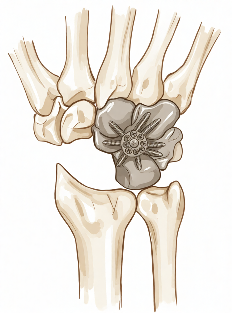
Partial Wrist Fusion (Four-Corner and Capitolunate Arthrodesis)
Limited intercarpal arthrodesis options — four-corner fusion (with scaphoid excision) and capitolunate (two-corner) fusion — for SLAC/SNAC wrist; preserves some midcarpal motion vs total wrist fusion.
For patients: a plain-language version of this topic is available. See the patient guide.
Overview
Partial wrist fusion serves as a salvage operation for patients with clinical and radiographic evidence of wrist arthritis, aiming to restore a pain-free and functional wrist while preserving useful motion [2]. This approach is indicated for specific carpal disorders, including posttraumatic or nonprogressive arthritis, where optimizing postoperative strength, stability, and aesthetic outcome is prioritized over total immobilization [7, 8]. Good long-term results are achievable when indications are accurately respected and technique is well performed to prevent complications [3]. In stage II scapholunate advanced collapse (SLAC) wrists with a preserved capitolunate joint, proximal row carpectomy is preferred due to its lower technical demand and durable results [9]. Conversely, scaphoid excision and four-corner fusion is the choice for stage III SLAC wrist [70]. When capitolunate arthritis is present, four-bone fusion with scaphoid excision is preferred [68].
Capitolunate arthrodesis is a feasible option for scapholunate advanced collapse and scaphoid nonunion advanced collapse wrists, with complication profiles similar to those encountered in four-corner arthrodesis [11]. In a review of 11 patients, this procedure restored a pain-free and functional wrist in eight cases [4]. Union rates for capitolunate arthrodesis improve with the excision of the triquetrum in addition to scaphoid excision [36]. Radioscapholunate arthrodesis with compression screws and local autograft achieves a 100% union rate at a mean follow-up of 12 months in appropriately selected patients with a preserved midcarpal joint, with no reported complications [34]. Both proximal row carpectomy and scaphoid excision with four-corner arthrodesis are motion-preserving options for SLAC arthritis, showing minimal subjective or objective differences in short-term follow-up [21].
Wrist fusion is performed nearly five times more frequently than total wrist arthroplasty, though short-term findings suggest total wrist arthroplasty compares favorably with fusion [5]. Total wrist fusion should only be used in exceptional circumstances [13]. Arthroscopic partial wrist fusion is a viable option for patients seeking to preserve motion with good aesthetic outcomes [7]. Arthroscopic resection arthroplasty may serve as a temporary alternative for younger patients or a definitive procedure for less demanding patients, although results may be short-lived [10]. The use of a pyrocarbon capitate resurfacing implant may represent a good alternative to total and partial wrist arthrodesis [14]. Once disease extends outside of the lunate, reconstruction with proximal row carpectomy or partial or total wrist arthrodesis should be considered based on which articular surfaces are affected [18].
Anatomy & Pathophysiology
Bony Anatomy
The wrist comprises the distal radioulnar, radiocarpal, and ulnocarpal joints, along with the eight carpal bones and their associated ligaments [76]. The proximal carpal row consists of the scaphoid, lunate, triquetrum, and pisiform, while the distal row includes the trapezium, trapezoid, capitate, and hamate [76]. The capitate is the largest carpal bone and articulates with seven other bones [76]. Conversely, the pisiform and trapezoid are the smallest bones; the pisiform articulates solely with the triquetrum [76]. The distal radius features two concave facets for the scaphoid and lunate, separated by the scapholunate ridge, and a sigmoid notch along the ulnar border for the distal radioulnar joint [86]. The distal ulna is covered with hyaline cartilage on its dorsal, lateral, palmar, and distal surfaces, with the ulnar styloid projecting distally to house the fovea for triangular fibrocartilaginous complex insertion [86].
The triquetrum articulates with the hamate distally, the lunate radially, and the pisiform volarly [86]. The hamate consists of a body and a hook (hamulus), which serves as an attachment for the transverse carpal ligament and origins for the flexor digiti minimi and opponens digiti minimi [86]. The capitate head often relies on a retrograde vascular supply, and its distal articular surface is divided by two ridges into three facets for the index, long, and ring finger metacarpals [86]. The trapezoid possesses two distal facets articulating with the index finger metacarpal [86]. The trapezium features a saddle-shaped articulation with the thumb metacarpal base and a palmar groove for the flexor carpi radialis, bordered laterally by a palmar tuberosity and transverse carpal ligament attachment [86]. The pisiform is a sesamoid bone within the flexor carpi ulnaris tendon and serves as the origin for the abductor digiti minimi [86].
The distal radius has three articular components: the scaphoid and lunate fossae, and the sigmoid notch [80]. The radial styloid allows attachment of the brachioradialis tendon and serves as the origin for the radial scapholunate and radial lunocapitate ligaments [80]. The concave elliptical distal radius is oriented in the sagittal plane with an average of 11 degrees of volar tilt [80]. In the frontal plane, the average radial inclination is 23 degrees [80]. Radial length, measured from the tip of the radial styloid to the ulnar articular surface, averages 13 mm [80]. The triquetrum also articulates with the triangular fibrocartilage proximally [67].
Vascular anatomy is critical for surgical planning. The scaphoid’s primary vascular supply is a branch of the radial artery at the dorsal ridge, with smaller vessels entering the palmar tubercle to supply the distal 30% [86]. The transverse carpal ligament attaches to the scaphoid’s palmar tubercle [86]. In 80% of wrists, the lunate has both dorsal and palmar vascular supplies, while in 20%, only a palmar supply is present [86]. The lunate is broader palmarly than dorsally [86]. The triquetrum is stabilized to the fovea of the ulna through the ulnotriquetral ligament [86].
Ligaments
Extrinsic carpal ligaments connect the radius or ulna to the carpus, with volar ligaments generally stronger than dorsal ones [81]. The radioscaphocapitate ligament connects to the scaphoid waist, limiting ulnar translation of the carpus [81]. The long radiolunate ligament helps limit ulnar translocation, while the short radiolunate ligament controls lunate position [81]. The radioscapholunate ligament, also known as the ligament of Testut, is a vascular conduit rather than a true ligament [81]. The ulnolunate ligament attaches to the palmar radioulnar ligament and lunate [81]. The ulnocapitate ligament attaches to the ulnar head, originates from the volar margin of the ulnar fovea, and is the most superficial or palmar [81]. The ulnotriquetral ligament attaches to the palmar radioulnar ligament and triquetrum [81].
The dorsal radiocarpal ligament has a trapezoidal shape, passing from the dorsal rim of the distal radius to the lunate and triquetrum [81]. Fibers of this ligament insert onto the dorsal lunotriquetral interosseous ligament [81]. Damage to the dorsal radiocarpal ligament, when combined with other intrinsic injuries, confers further carpal instability [81]. Intrinsic carpal ligaments originate and insert within the carpus [81]. The scapholunate interosseous ligament is the major stabilizer of the wrist and the most commonly injured wrist ligament [81]. It is C-shaped, consisting of dorsal, palmar, and interosseous portions, with the dorsal portion being the strongest and thickest [81]. This ligament provides a flexion force on the lunate given its attachment to the scaphoid [81].
The lunotriquetral interosseous ligament is also C-shaped, with the volar portion being the thickest and strongest [81]. It provides an extension moment on the lunate given its attachment to the triquetrum [81]. The capitohamate ligament is a thick ligament, 5 × 5 mm in cross section, with extensions to the third or fourth metacarpals [81]. The dorsal intercarpal ligament passes from the dorsal tubercle of the triquetrum to the distal pole of the scaphoid [81]. Along with the dorsal radiocarpal ligament, it reinforces the elastic dorsal wrist capsule and helps stabilize the scapholunate articulation via deep fibers contributing to the dorsal scapholunate interosseous ligament [81].
The space of Poirier is an area adjacent to the proximal capitate without ligamentous attachment, situated ulnar to the radioscaphocapitate ligament and radial to the long radiolunate in the floor of the carpal tunnel [81]. This weak area is vulnerable to instability, and the distal carpal row separates from the lunate through this space during a perilunate dislocation [81]. The triangular fibrocartilage complex is formed by the central meniscus homolog, dorsal and volar radioulnar ligaments, the floor of the extensor carpi ulnaris tendon sheath, and volar ulnocarpal ligaments [86]. It arises from the radial border of the distal radius and inserts into the base of the ulnar styloid and distal ulna through the ligamentum subcruentum [86]. The dorsal and volar radioulnar ligaments are the primary stabilizers of the distal radioulnar joint [86]. Only the peripheral 10% to 40% of the volar, ulnar, and dorsal triangular fibrocartilage complex has a vascular supply [86].
The dorsal radiocarpal ligament originates at the dorsal lip of the distal radius, adjacent to the dorsal radial tubercle (Lister tubercle), and traverses the radiocarpal joint obliquely to insert into the lunate and triquetrum [86]. The dorsal intercarpal ligament arises from the triquetrum [86].
Vascular Supply
The terminal branches of the radial, ulnar, and anterior interosseous arteries provide extraosseous blood supply to the carpus through three dorsal and three palmar transverse arterial arches with longitudinal connections [89]. The dorsal radiocarpal arch is located at the radiocarpal joint and supplies the lunate and triquetrum [89]. The dorsal intercarpal arch is the largest, located between the proximal and distal carpal rows, supplying the distal carpal row and, through anastomoses with the radiocarpal arch, the lunate and triquetrum [89]. The basal metacarpal arch is located at the base of the metacarpals, is the most variable, and supplies the distal carpal row [89]. The palmar radiocarpal arch is located at the level of the radiocarpal joint on the palmar surfaces of the lunate and triquetrum [89]. The intercarpal arch is located between the proximal and distal carpal rows, is the most variable, and does not contribute to nutrient vessels in the carpus [89]. The deep palmar arch is located at the level of the metacarpal bases, is consistent, and communicates with the dorsal basal metacarpal arch and the palmar metacarpal arteries [89].
Kinematics and Biomechanics
The wrist can essentially be considered a two-joint system linking the hand to the forearm around the highly mobile bones of the proximal carpal row [87]. The two principle articulations are the radiocarpal and midcarpal joints, situated proximal and distal to the mobile proximal carpal row [87]. The bones of the wrist are composed of multiple articulations: the radiocarpal joint, midcarpal joint, pisotriquetral joint, trapeziometacarpal joint, and carpometacarpal joints [81]. The wrist joint’s motion planes include flexion, extension, radial deviation, ulnar deviation, and circumduction [81]. There is minimal carpal motion with pronosupination [81].
Approximately 62° of wrist extension occurs through the radiocarpal joint [81]. 62% of wrist flexion occurs through the midcarpal joint [81]. The midcarpal joint is mostly responsible for 20° and 40° of radial and ulnar deviation, respectively [81]. The midcarpal joint is responsible for the “dart thrower’s motion,” which involves moving from radial extension into ulnar flexion positioning of the wrist [81]. The radius bears 80% of the axial load transmitted through the radiocarpal joint, while the ulna bears 20% in neutral ulnar variance [81]. The proximal row of carpal bones forms an intercalated segment between the distal carpal row and the distal radius and is bound into a functional unit by the scapholunate interosseous ligament and lunotriquetral interosseous ligament [81]. The distal row is rigid, with little motion between its bones due to stout intercarpal ligaments, and thus they act as a functional unit with the scaphoid bridging both rows [81].
During wrist flexion from neutral, the distal row flexes and ulnarly deviates slightly while the scaphoid also pronates [81]. The proximal row flexes differentially, with more rotation through the scaphoid, followed by the triquetrum and the lunate [81]. The proximal row translates dorsally [81]. During wrist extension from neutral, the distal row extends and radially deviates slightly while the scaphoid also supinates [81]. The proximal row extends differentially, with more motion in the scaphoid, followed by the triquetrum and then the lunate [81]. The proximal row translates palmarly [81]. The proximal carpal row has no muscular or tendinous attachments and is an intercalary segment [86]. With ulnar deviation, the proximal row extends relative to the forearm/distal row [86]. With radial deviation, the proximal row flexes relative to the forearm/distal row [86].
With axial loading through the neutral wrist, approximately 80% of forces are transmitted through the distal radius (60% scaphoid facet, 40% lunate facet) and 20% through the distal ulna [86]. With wrist flexion, 60% of the motion is midcarpal and 40% is radiocarpal [86]. With wrist extension, 33% of the motion is midcarpal and 66% is radiocarpal [86]. Relative contributions to wrist motion were: wrist flexion: radiocarpal joint 63%, midcarpal joint 36%; wrist extension: radiocarpal joint 53%, midcarpal joint 46% [126]. More than half the motion of the carpus when the wrist was loaded in extension occurred at the midcarpal joint [108]. The 'dart thrower's motion' of the wrist, from radial extension to ulnar flexion, may be a unifying concept of functional wrist motion that is uniquely human [127]. A wide range of dart-throwing planes exists, and midcarpal arthrodesis adversely affects dart-throwing motion compared with radiocarpal arthrodesis [128].
The eight carpal bones that comprise the wrist joint represent the most complex articular system in the human body [77]. Each carpal bone is capable of moving in different degrees or directions dependent on the position, motion, and force generation of the hand in space [77]. The human wrist allows precise positioning of the hand and optimization of power and prehensile tasks throughout a nearly hemispherical arc of wrist motion [77]. Injuries to the wrist mechanism can lead to instability, resulting in a painful lack of motion, strength, and function [77]. A wrist joint should be considered biomechanically unstable when it is not able to bear loads and does not exhibit normal kinematics throughout its arc of motion [74]. The longitudinal 'columnar' concept of wrist kinematics does not fit in with many of the recent findings related to carpal instabilities [75]. Traction (distraction) changes normal carpal kinematics and motion of the wrist combined with traction induces abnormal patterns of motion [90]. Despite complex carpal bone anatomy and kinematics, computed fiber elongations were found to vary linearly with wrist position [85]. During simple unresisted wrist motions, the force in the scapholunate interosseous ligament did not exceed 20 N [93]. The contact areas between the scaphoid and distal radius are maximized during full extension of the wrist, which helps stabilize the radiocarpal joint and potentially reduces the risk of injury to the carpus and the distal radius [113].
Scaphoid nonunions have a dramatic impact on carpal kinematics, partially uncoupling the proximal and distal carpal rows [103]. The kinematic consequences of resecting the distal scaphoid are different: the flexion-extension motion of the proximal row during deviations is diminished at the expense of increased radioulnar translational motion [104]. A pattern of kinematic changes was established after scapholunate ligament injury, despite individual variance [121]. The experimental kinetic behavior of the chronic scapholunate advanced collapse wrist is similar to the one with a recent scapholunate dissociation without carpal collapse associated [124]. Kinematic changes in scapholunate instability may predict the development of radioscaphoid arthritis and help identify a kinematically abnormal wrist [95]. Four-dimensional computed tomography is a promising, non-invasive, and affordable method to assess and quantify wrist kinematics, extending conventional CT by incorporating the temporal dimension [92]. This study describes the effect of lunate morphology on 3-dimensional carpal kinematics during wrist flexion and extension [82]. The complex nature of the wrist has plagued us clinically and hampered our ability to formulate concise, yet thorough, algorithmic approaches to evaluation and treatment [114]. The results highlight the significant effect of rotation on radiographic landmarks at the wrist, indicating that 10° of supination can drastically alter the developed radi
Classification
SLAC/SNAC Wrist Staging: SLAC and SNAC wrist arthritis are staged 1 through 4, with surgical options depending on patient age, expectations, and joint involvement [131]. The modified classification of SLAC/SNAC wrist stages is based on intraoperative findings [48]. Stage I is defined by degenerative changes limited to the radial styloid/scaphoid articulation [48], characterized specifically by radial styloid arthritis [131]. Stage II involves degenerative changes across the entire radioscaphoid joint with mild or no changes at the capitolunate or radiolunate joint [48]. Stage III is subdivided into IIIA and IIIB to describe different patterns of degeneration seen by inspection rather than a sequence of degeneration [48]. Stage IIIA is defined by mild or no changes at the capitolunate joint and moderate to severe changes at the radiolunate joint [48]. Stage IIIB is defined by moderate to severe changes at the capitolunate joint and mild or no changes at the radiolunate joint [48]. Stage IV is defined by moderate to severe changes at both the capitolunate and radiolunate joints [48].
Surgical Indications: Stage IIIA wrists are considered candidates for proximal row carpectomy [48]. Stage IIIB wrists are considered candidates for scaphoid excision and four-corner arthrodesis [48]. In a study of 18 patients using the modified classification, 13 were stage IIIA, 3 were stage IIIB, and 2 were stage IV [48].
Other Considerations: The four-corner arthrodesis is the most common intercarpal arthrodesis procedure [23]. It involves the fusion of the capitate, lunate, hamate, and triquetrum following scaphoidectomy [23]. Triquetro-lunate fusions are the commonest type of fusions in the carpal region [179].
Clinical Presentation
Wrist pain is a common presenting symptom for hand surgeons, yet routine history and physical examination often fail to uncover the underlying cause [109]. When clinical assessment is insufficient, special diagnostic methods including routine x-rays, electrical diagnostic studies, bone scans, wrist arthrograms, and cineradiography may aid in establishing the diagnosis [109]. Despite these adjuncts, a fair percentage of patients remain undiagnosed [109]. Wrist arthroscopy provides direct visualization of the articular cartilage of the distal radius, proximal scaphoid, proximal lunate, and proximal triquetrum, as well as the scapholunate ligament, triquetrolunate ligament, and triangular fibrocartilage [109]. This visualization allows for the observation of proximal pole scaphoid fractures and the judgment of scaphoid nonunion integrity under direct vision [109]. Preoperative arthroscopic assessment of articular cartilage enables specific recommendations to be made to the patient before definitive surgery [109].
Indications for Partial Fusion
Limited wrist fusion is indicated when arthritic changes are confined to either the radiocarpal or intercarpal joints, serving as a useful alternative to panarthrodesis [17]. Radiolunate and radioscapholunate fusions are indicated in painful wrist conditions where the midcarpal joint remains essentially normal [22]. In inflammatory arthritis, radial lunate fusion is often used to control ulnar translation and palmar subluxation of the carpus, preventing further degeneration [22]. The scaphoid can be added to a radial lunate fusion if the radioscaphoid facet is arthritic [22]. Carpal instability with arthritis of the radioscaphoid joint is a specific indication for radioscapholunate fusion [22]. This procedure serves as an alternative to scaphoid excision and four-corner fusion when wear of the proximal scaphoid pole and dorsal margin of the scaphoid facet of the distal radius is significant in scapholunate instability, provided the midcarpal joint is normal [22]. Radioscapholunate fusion is contraindicated if any articular loss of the capitolunate joint exists [22]. The best indication for radioscapholunate arthrodesis is in patients following unsatisfactory results from distal radius fractures where cartilage loss, arthrofibrosis, or joint incongruity exists on the distal radial surface [22]. Radiolunate fusion is sufficient in cases of the lunate facet being involved with a dye punch injury that has gone on to a malunion or cartilage loss [22]. In stage IV Kienböck’s disease, radiolunate fusion is indicated when the distal surface of the lunate is normal [22].
Kienböck disease management depends on the extent of involvement. Once disease extends outside of the lunate, reconstruction with proximal row carpectomy or partial or total wrist arthrodesis should be considered based on which articular surfaces are affected [18]. In adolescents with higher stages of Kienböck disease, radial shortening or partial wrist fusion result in good function [27]. Intercarpal arthrodesis is performed most commonly for the treatment of arthritis and carpal instability, with the objective of minimizing or eliminating pain while maximizing motion and function [23]. These goals are achieved by removing arthritic joint surfaces and altering carpal motion and load transference [23]. While some intercarpal fusion procedures have good, predictable outcomes with maintenance of results over time [23], others are infrequently used or of primarily historical interest owing to unpredictable results and high rates of complications [23]. The most common intercarpal arthrodesis procedure is the scaphoidectomy with fusion of the capitate, lunate, hamate, and triquetrum [23].
Capitolunate arthrodesis is a feasible option for scapholunate advanced collapse (SLAC) and scaphoid nonunion advanced collapse (SNAC) wrists [11]. In a ten-year follow-up series, capitolunate arthrodesis allowed restoring a pain-free and functional wrist in eight out of 11 patients [4]. Scaphoid excision and four-corner fusion remains a viable option for patients with advanced wrist arthritis, providing reliable, resilient functional results that remain stable over time [158]. The outcome of this procedure is favorable at one year and does not deteriorate significantly between one and 10 years [43]. Functional results of four-corner fusion were good at long-term follow-up despite radiographic changes in the radiolunate joint in 73% of patients [41]. However, four-corner arthrodesis and proximal row carpectomy should not always be standard management for advanced radiocarpal arthritis [35]. Total wrist fusion should only be used in exceptional circumstances for the treatment of SNAC and SLAC wrists [13]. When properly executed, lunocapitate arthrodesis with scaphoid excision provides functional and radiological results comparable to four-corner fusion without the deleterious effect on ulnocarpal and proprioceptive dysfunction associated with including the triquetrum in the fusion site [15].
Range of Motion and Functional Outcomes
Postoperative range of motion varies by specific procedure. After radioscapholunate arthrodesis, patients experience less flexion and radial deviation of the wrist [167]. After four-corner fusion, scaphoid excision allowed significantly greater radial deviation compared with scaphoid retention and scaphoid inclusion [168]. The patient will gain approximately 30˚ more flexion and extension if the scaphoid is not included in the fusion [22]. Although some may view motion-preserving procedures as a staged full wrist fusion, good long-term results can be obtained if indications are accurately respected and the technique is well performed to prevent complications [3]. If the basic principles of limited wrist fusions are adhered to, a good outcome can be obtained [6]. The technique of partial intercarpal fusion relieves pain in all but two instances and restores a strong grip in all but one patient, with wrist immobilization required for only two months [32].
Complications and Union Assessment
Complication rates vary significantly among partial fusion techniques. Four of 24 patients developed non-unions after limited wrist arthrodesis for the salvage of SLAC wrist; two of these had successful revision to limited wrist arthrodesis, while the other two required pan-arthrodesis of the wrist [20]. The second most common complication of scaphocapitate arthrodesis is persistent wrist pain despite radiographic evidence of scaphocapitate bony union, occurring in 4/30 patients (13%) [39]. High rates of nonunion have recently been reported for midcarpal fusions [51]. Two-dimensional films are not reliable in determining union of a midcarpal fusion due to curvature and overlap of carpal interfaces [51]. Computed tomography is the most reliable radiographic method of evaluating osseous union for midcarpal fusion [51]. Patients may be reticent to sacrifice motion when selecting fusion as a treatment option [51]. The goal in a midcarpal fusion is to first achieve a solid fusion, and then mobilize the wrist in therapy as soon as possible [51]. It is expected that a patient will achieve better final range of motion with a shorter period of immobilization following surgery [51]. The dilemma in midcarpal fusion management comes in knowing when the fusion has actually achieved union and it is safe to begin full force rehabilitation [51]. Plain films are unreliable in making a definitive assessment of union for midcarpal fusion [51]. A computed tomography scan is more reliable than plain films for assessing union of a midcarpal fusion [51].
While the overall non-union rate for four-corner arthrodesis performed with Kirschner wires is quite low, of the order of 4%, there is wide variation [122]. The non-union rate for lunocapitate fusion is inferior, up to 35% [122]. Better results were obtained for lunocapitate fusion in a more recent report [122]. Specific circular plates for midcarpal fusion are available, with high complication rates reported by some but not others [122]. Scaphoidectomy and midcarpal arthrodesis is a salvage procedure used in a number of conditions, especially SLAC and SNAC [122]. It is feasible as the radiolunate joint is spared degeneration [122]. Most commonly, the scaphoid is excised and the lunate, capitate, hamate, and triquetrum are fused [122]. Some have fused only the lunate and capitate [122]. Kirschner wires, headless screws, or staples have been used for midcarpal fusion [122]. The aim of using a new plate and screw system for midcarpal fusion was to assess efficacy and allow for early postoperative mobilization [122].
Alternative Salvage Procedures
In all cases of ulnar shortening osteotomy for ulnar carpal instability and ulnar carpal impaction, wrist function improved both subjectively and objectively, an outcome independent of the type of osteotomy performed [66]. Good and excellent clinical results in the majority of the patients following radiolunate fusion do not depend on the fixation device [29]. RSL fusion with distal scaphoidectomy and entire triquetrum excision can be a useful alternative to total wrist arthrodesis for patients with an intact midcarpal joint [60]. This surgical procedure may represent a good alternative to total and partial wrist arthrodesis [14]. It may serve as a temporary alternative to partial wrist arthrodesis for younger patients or a definitive procedure for less demanding patients, though results may be short-lived [10]. Although wrist fusion is performed nearly 5 times more frequently than total wrist arthroplasty, short-term findings suggest that total wrist arthroplasty compares favorably with wrist fusion [5]. Total wrist arthrodesis provides reliable pain relief and good functional outcomes with high patient satisfaction, particularly for end-stage arthritis and as a salvage technique [42]. Bilateral total wrist arthrodesis improved pain while enabling patients with severe carpal arthrosis to maintain a satisfactory level of extremity function and quality of life [38]. Total wrist arthrodesis leads to successful relief in pain but limits function [123]. Total wrist arthroplasty attempts to relieve pain and retain some movement but the results can be unpredictable [123]. Recent studies have shown promising results of total wrist arthroplasty in patients with non-rheumatoid arthritis but postoperative function can be quite poor [123]. It remains uncertain whether newer designs of wrist replacement prostheses improve function [123].
Proximal row carpectomy (PRC) is a useful procedure for various conditions including degenerative joint arthritis regardless of cause, be they idiopathic, osteoarthritis, SLAC, or status post-intercarpal pathology [130]. PRC has been used for patients who have Kienböck disease and on patients who have rheumatoid arthritis and osteoarthritis [130]. Preoperative evaluations for PRC are focused on an understanding of the extent of the patient's disability based on the patient's subjective evaluation of pain and loss of function and on objective evaluations of wrist range of motion and grip strength [130]. Pain is the most common reason to pursue PRC [130]. Patients should have had a full complement of conservative treatment measures, including physical therapy modalities, home exercises, intermittent use of mechanical bracing of the wrist, and a trial of anti-inflammatory agents administered orally or by injection [130]. Imaging studies to evaluate the particulars of the intra-articular pathology of the radiocarpal joint can be helpful but generally are not necessary for preoperative planning if the surgeon is prepared to perform PRC or one of the variations [130]. If there is significant intraarticular pathology on the distal radius, imaging studies can be beneficial to assess the appropriateness of a PRC procedure to ameliorate pain and loss of function [130]. PRC has become an established mean of managing radiocarpal arthritis, with the advantage of preserving wrist motion compared with partial wrist fusion procedures and avoiding total wrist fusions [50]. Multiple studies have demonstrated that PRC improves pain levels postoperatively, preserves some wrist motion, and gives high rates of patient satisfaction [50]. In patients with advanced wrist arthrosis, there is debate regarding the indications for salvage procedures, such as PRC, four-corner fusion, or other intercarpal arthrodesis [50]. Recent studies of PRCs have suggested that the loads on the capitate may be excessive and predispose to early arthritic wear [50]. The long-term outcomes of PRC have shown that radiocapitate arthritis develops following the procedure [50]. A direct linkage between radiographic signs of arthritis and patient outcome has yet to be established [50]. Post-operative motion and grip strength values following PRC appear to remain stable over time [52]. Surgical failure rates with conversion to wrist fusion occurred early within the post-operative follow-up [52]. Many patients continued to complain of pain requiring daily medication and were unable to return to manual labor [52]. Radiographic follow-up beyond 2 years revealed joint narrowing and arthritic changes within the radiocapitate joint [52]. Forty-six patients (74%) were not satisfied with the results of their surgery due to persistent pain or inability to return to previous occupational activities [52]. Fifty-two patients required daily pain medication for wrist pain [52]. Twelve patients had undergone a wrist arthrodesis [52].
Classification and Diagnostic Nuances
Our observations call into question the validity of the accepted classification of Krakauer et al. [48]. A modified classification, based on intraoperative findings, would redescribe and subdivide stage III [48]. Stage IIIA wrists, with mild or no degeneration at the capitolunate joint and moderate to severe changes at the radiolunate joint, are considered candidates for PRC [48]. Stage IIIB wrists, with essentially reversed findings, are considered candidates for SEFCA [48]. The separation of stage III into 2 subdivisions is not intended to describe a sequence of degeneration, but rather, different patterns of degeneration seen by inspection [48]. A larger sampling of patients and long-term follow-up will be necessary to validate this classification [48]. The subdivision into stages IIIA and IIIB will aid in comparison of long-term outcomes [48]. Using this classification, none of the patients fit into stage II; 13 were stage IIIA, 3 were stage IIIB, and 2 were stage IV [48]. The 2 stage IV patients, with moderate degenerative changes at the capitolunate joint and severe changes at the radiolunate joint discovered at surgery, underwent PRC [48]. In retrospect, total wrist arthrodesis or arthroplasty would have been alternate options in both stage IV cases [48]. In both stage IV cases, radiographs showed moderate changes at the capitolunate joint [48]. Moderate symptoms do persist after Blatt's dorsal capsulodesis for chronic scapholunate dissociation, becoming in some cases severe enough to necessitate wrist fusion [148]. Forty patients followed for 1 to 3 years fell into two distinct groups according to duration of symptoms and preoperative side-to-side radiographic differences in the scapholunate gap [162].
Investigations
MRI: Magnetic resonance imaging is the modality of choice for imaging radiographically occult fractures of the hand and wrist [84]. A static magnetic field strength of at least 1.5 T using a dedicated wrist coil is recommended for analyzing the interosseous, intrinsic, and extrinsic ligament insertions [98]. The volar extrinsic, SL interosseous, dorsal intercarpal, and LT ligaments are best visualized using 1 mm slices (with no interslice gap) in the coronal plane [98]. The DRC and intercarpal ligaments are best viewed on both the coronal and sagittal images [98]. Oblique axial views along the longitudinal axes of the DRC and intercarpal ligaments allow further analysis, especially when an injury is suspected [98]. Concomitant cartilage-sensitive imaging is integrative to influence the assessment and surgical management, as cartilage integrity will influence clinical and surgical management, especially in the setting of SLAC wrist [98]. Real-time MRI has been used to investigate dynamic instabilities, although its routine use in clinical practice is yet to be further determined [98].
CT: CT scanning enables the 3D analysis of carpal dysfunction [98]. By adding motion in real time (4D CT), this may hold promise in the future to potentially quantify the location and degree of injury noninvasively and help surgeons plan their surgical treatment [98]. CT is more useful than plain X-rays for evaluating partial carpal arthrodesis [164].
Dynamic Imaging: Dynamic fluoroscopy shows abnormal motion between the scaphoid and lunate and changes in the kinematics of the midcarpal joint [98]. Live imaging shows whether the DISI is reducible, giving the physician valuable information as the treatment is planned [98].
Other Considerations: MRI provides earlier detection of synovitis and erosive bone changes associated with rheumatoid arthritis than do radiographs [101]. With proper technique, injuries to the triangular fibrocartilage complex (TFCC) can be demonstrated with MRI [101]. Perforations in the TFCC appear as linear defects or gaps filled with hyperintense fluid on coronal gradient-echo or T2-weighted pulse sequences [101]. The addition of arthrographic contrast improves the visualization of the scapholunate and lunotriquetral ligaments on MR images [101]. MRI is useful in detecting additional marrow abnormalities in osteonecrosis, as seen in the lunate in Kienböck disease or in the scaphoid after fracture [101]. Asymmetry of marrow signal in proximal and distal fragments of a fractured scaphoid is suggestive of proximal pole ischemia [101]. MRI has an expanding role in the evaluation of inflammatory arthritis [101].
Arthroscopy: Arthroscopy is considered by many to be the diagnostic intervention of choice for determining the degree of injury to the wrist and can assess the condition of the cartilage (normal or degenerative), ability to reduce the carpus and any other associated injuries [98]. The degree of intrinsic and extrinsic ligament injury can be identified from the arthroscopic evaluation [98]. Geissler grade II SLIL injuries tend to be isolated, whereas grade IV injuries often involve complete dorsal extrinsic ligament disruption [98].
Treatment
Non-Operative
The provided evidence does not detail specific conservative management protocols such as weight loss, physical therapy, or pharmacological interventions. However, the clinical context implies that surgical intervention is considered when other options fail to provide adequate pain relief or functional preservation [176].
Operative
Indications: Total wrist fusion is reserved for exceptional circumstances, as various surgical techniques can preserve a functional wrist and arthrodesis is no longer the sole solution for arthritic wrists [40, 59]. The ideal surgical treatment prioritizes effective pain relief while balancing mobility [176]. Radioscapholunate (RSL) and radiolunate fusions are indicated in painful wrist conditions where the midcarpal joint remains essentially normal [22]. Specifically, RSL arthrodesis is best indicated following unsatisfactory results from distal radius fractures [22]. If cartilage loss, arthrofibrosis, or joint incongruity exists on the distal radial surface, the scaphoid and lunate are fused to the radius to retain wrist motion; no other intercarpal arthrodesis adequately addresses this specific pathology [22]. Radiolunate fusion is sufficient when the lunate facet is involved with a dye punch injury that has progressed to malunion or cartilage loss, and is indicated in stage IV Kienbock’s disease where the distal surface of the lunate is normal [22]. Capitolunate arthrodesis via the lateral approach is indicated for severe radioscaphoidal degenerative changes not amenable to reconstruction [55]. Conversely, capitolunate fusion is contraindicated if severe degenerative changes are present in the radiolunate joint, as it increases pressure and exacerbates pain [55].
Surgical Approach / Technique: Arthrodesis of the wrist is a well-established procedure performed by a variety of techniques [142]. The goal of a midcarpal fusion is to establish a stable, load-bearing articulation at the congruent radiolunate interface, which requires a broad surface area of contact, ample bone graft, compression, and stable fixation [173]. The capitate must be positioned correctly relative to the lunate to avoid impingement on the dorsal rim of the radius [173]. For capitolunate arthrodesis via the lateral approach, partial or entire resection of the scaphoid is required to visualize the articulation [55]. This approach allows perfect visualization of the capitolunate articulation but makes correction of frontal plane malalignment impossible [55]. Ulnar translation of the lunate, often encountered in later stages, may not be managed by the lateral approach; in cases of advanced ulnar translation, visualization of the articular surfaces is extremely difficult, and the classic dorsal approach is elected instead [55]. The scaphoid can be added to the fusion if the radioscaphoid facet is arthritic [22]. However, this fusion should not be performed if any articular loss of the capitolunate joint exists [22]. A combination of triquetral and distal scaphoid pole excision after RSL fusion improves wrist motion to levels close to normal in cadaver models [61].
Implant Selection: The evidence does not specify prosthesis design choices or implant types for arthrodesis. However, osteochondral autograft remains a good alternative to partial or complete wrist fusion when the proximal pole articular surface cannot be restored [26]. Arthroplasty should be used as an alternative to arthrodesis in the treatment of posttraumatic wrist arthritis, given proper patient selection and indications [160].
Alignment / Balancing Strategy: Nonspanning arthrodesis is advocated as an alternative method for total wrist fusion, offering a high union rate and minimal risk of complications at the carpometacarpal joint [37]. Total wrist arthrodesis combined with proximal row carpectomy (PRC) provides reliable and reproducible benefits [64]. Both PRC and scaphoid excision with four-corner arthrodesis are motion-preserving options for scapholunate advanced collapse (SLAC) arthritis, with minimal subjective or objective differences in short-term follow-up evaluations [21].
Pain Management: Partial wrist denervation is a reliable motion-preserving procedure for patients with chronic wrist pain, with 71% of patients experiencing pain relief and not requiring further salvage procedures at an average of 78 months of follow-up [178]. Total wrist denervation is a reliable and reproducible surgical technique for pain relief and preservation of wrist function in painful osteoarthritis [183].
Adjuncts: The evidence does not detail specific adjuncts such as tranexamic acid or navigation systems. However, a reliable, efficient, and cost-effective technique for wrist fusion has been presented [19].
Setting of Care: The evidence does not specify outpatient versus inpatient settings.
Revision: Conversion to arthrodesis after failed wrist arthroplasty is worthwhile and reliably improves wrist function over failed wrist arthroplasty [28]. In a cohort of 24 patients, four developed non-unions; two had successful revision to limited wrist arthrodesis, while the other two required pan arthrodesis of the wrist [20]. The senior author did not have to revise or convert any patient into a wrist fusion or a total wrist arthroplasty, suggesting that longer survival rates for this procedure may be possible [25].
Other Considerations: Computed tomography is the most reliable radiographic method for evaluating osseous union in midcarpal fusions, whereas plain films are unreliable for making a definitive assessment of union [51]. Before surgery, the patient should be informed about the loss of wrist motion that may occur after capitolunate arthrodesis [55]. Outcomes for RSL arthrodesis in appropriately selected patients with a preserved midcarpal joint show a 100% union rate at a mean follow-up of 12 months with no complications [34]. Midcarpal arthrodesis is a long-lasting treatment option for advanced carpal collapse with good long-term results [186]. In 19 patients, the procedure resulted in increased range of wrist motion in all planes in 85%, grip improved by 134%, and 13 patients experienced complete relief of pain [57]. After surgery, patients demonstrated acceptable levels of function, pain, and wrist range of motion [166]. RSL fusion is a motion-sparing procedure that offers a stable wrist and potential for good grip strength in an active person [22].
Complications
Nonunion and Failure to Fuse
Nonunion remains a significant risk in partial wrist fusion. In a series of 24 patients undergoing limited wrist arthrodesis for SLAC wrist, four developed non-unions, two of which required pan arthrodesis [20]. A study of 55 cases reported a nonunion rate of four cases for the capitolunate arthrodesis group compared with two for the four-corner arthrodesis group [54]. In a surgical treatment series for SLAC wrist, six of 51 motion-sparing cases were converted to total arthrodeses [54]. The Stein and Gill technique achieved an index fusion rate of 87.5% (14 of 16), increasing to 93.75% with reoperation [202]. In distal radius reconstruction, nonunion occurred in one of six (17%) patients with fibular autograft fusion, one of three (33%) with ulnar transposition and fusion, and one of three (33%) with fibular allograft fusion [140]. Careful technique can minimize nonunion in scaphotrapeziotrapezoid arthrodesis [198]. The Xpode plate in partial carpal arthrodesis had a complication rate of 8/12, deemed unacceptable due to increased non-unions and re-operations [204].
Hardware-Related Complications
Hardware issues frequently necessitate secondary intervention. Seventy-five (1.48%) patients required removal of symptomatic fusion hardware after index four-corner fusion [134]. Proximal migration of headless compression screws in midcarpal fusion resulted in one patient undergoing total wrist fusion and one undergoing proximal row carpectomy with interposition arthroplasty [143]. In a four-corner fusion study using a locking plate, one patient developed carpal tunnel syndrome treated by surgical release [132]. Conversely, one patient using K-wires developed a postoperative wound infection treated conservatively [132]. No implant-related complications were observed in a comparison of K-wire and locking plate techniques for four-corner fusion [132]. In a series of 93 modified Clayton-Mannerfelt arthrodeses for rheumatoid arthritis, hardware did not have to be removed in most cases [200]. Complications and reoperations in total wrist arthrodesis occur frequently, most often due to incomplete bone fusion or hardware-related problems [196].
Conversion to Total Wrist Arthrodesis
Conversion to total wrist arthrodesis occurs in a small but notable percentage of patients. Subsequent total wrist arthrodesis was performed in 135 patients (2.67%) with a previous four-corner fusion and in 65 patients (1.79%) with a previous proximal row carpectomy [134]. Including both hardware removal and subsequent total wrist arthrodesis, 220 (4.16%) of four-corner fusion patients required a secondary procedure [134]. PRC and FCA demonstrated similarly low rates of conversion to total wrist arthrodesis [197]. In a long-term follow-up study of proximal row carpectomy, twelve patients had undergone a wrist arthrodesis [52]. One case in the plate group of a four-corner fusion study was converted to a total wrist fusion due to persisting pain without implant-related complications [132].
Pain and Functional Outcomes
Persistent pain and functional dissatisfaction are common complications. The second most common complication in scaphocapitate arthrodesis is persistent wrist pain despite radiographic evidence of bony union, occurring in 4/30 patients (13%) [39]. Forty-six patients (74%) were not satisfied with the results of proximal row carpectomy due to persistent pain or inability to return to previous occupational activities [52]. Fifty-two patients required daily pain medication for wrist pain in a long-term follow-up study of proximal row carpectomy [52]. One patient in a midcarpal fusion hardware migration series reported substantial relief following hardware removal but had persistent stiffness and pain with activities [143]. Panarthrodesis of the wrist may entail a considerable complication rate of 14%-68% [141].
Infection and Other Complications
Common complications of arthroscopic partial wrist fusion are likely to be similar to 4-corner fusion using other methods, including nonunion, infection, decreased range of motion, and dorsal radiocarpal impingement [133]. Procedure-specific complications are more common with four-corner arthrodesis, while joint space narrowing is more common after PRC, particularly in patients under 35 years of age [195]. Four-corner arthrodesis has been associated with an increased risk of complications including nonunion, painful hardware, or implant failure [154]. PRC is thought to have fewer post-operative complications and is easier to carry out than four-corner arthrodesis [154]. A high rate of re-operation was observed in patients receiving radioscapholunate arthrodesis, and the expected benefit of preserving the midcarpal joint was not observed [62]. Awareness of complications associated with wrist arthrodesis and how best to avoid them is essential for the treating physician to appropriately counsel patients [194].
Other Considerations
Total wrist arthroplasty complication rates were higher than wrist fusion, with reports of radiological loosening and osteolysis [189]. The results between total wrist arthrodesis for salvage of failed total wrist arthroplasty and primary wrist arthrodesis were slightly in favour of patients with a primary wrist arthrodesis [31].
Recovery
Rehabilitation protocol: Rehabilitation may be initiated as early as one week post-operatively to mitigate wrist stiffness following capitolunate arthrodesis [144]. Wrist immobilization is required for a duration of only two months following partial intercarpal fusion [32].
Functional milestones: During the first eight weeks of hand therapy, pain and disability in patients referred to hand therapy improved at a faster rate than in the larger group of all distal radius fractures at any given time over a one-year period [136]. Patients treated for scaphoid nonunion demonstrated the capacity to continually improve their wrist function beyond 3 years following their treatment [155].
Key Evidence
- [L4] This technique was used in 17 patients for either limited or complete wrist fusion. [1] (10.1016/s0363-5023(87)80273-3)
- [L5] A salvage operation, such as a partial wrist fusion, should be considered when there is clinical and radiographic evidence of wrist arthritis. [2] (10.1016/j.hcl.2015.04.013)
- [Paper] Although some may see the motion preserving procedures of the wrist as a staged full wrist fusion, if the indications are accurately respected and the technique is well performed to prevent complications, then good long-term results can be obtained. [3] (10.1055/s-0032-1330070)
- [L4] Capitolunate arthrodesis allowed restoring a pain free and functional wrist in eight out of the 11 patients reviewed. [4] (10.1016/j.main.2013.07.002)
- [L4] Although wrist fusion is performed nearly 5 times more frequently than total wrist arthroplasty, short-term findings suggest that total wrist arthroplasty compares favorably with wrist fusion. [5] (10.1177/1558944716668846)
- [L5] If the basic principles of limited wrist fusions are adhered to, a good outcome can be obtained. [6] (10.1055/s-0032-1329548)
- [L4] Arthroscopic partial wrist fusion is a viable option for patients who have posttraumatic or nonprogressive wrist arthritis who would like to preserve useful wrist motion with good aesthetic outcome. [7] (10.1097/bth.0b013e318190244b)
- [L4] Partial carpal fusions are effective methods of treating specific carpal disorders, optimizing postoperative wrist motion, strength, and stability while reducing pain. [8] (10.1055/s-0032-1330071)
- [L4] For stage II SLAC wrist with a preserved capitolunate joint, proximal row carpectomy is preferred because it is technically less demanding and yields durable results. [9] (10.5435/00124635-200307000-00007)
- [L4] It may serve as a temporary alternative to partial wrist arthrodesis for younger patients or a definitive procedure for less demanding patients, though results may be short-lived. [10] (10.1016/j.jhsa.2012.08.039)
- [L4] Capitolunate arthrodesis is a feasible option for scapholunate advanced collapse and scaphoid nonunion advanced collapse wrists with complications similar to those encountered in 4-corner arthrodesis. [11] (10.1016/j.jhsa.2019.10.007)
- [L4] A series of 28 selective limited wrist arthrodeses was presented, with most being intercarpal fusions and some including the radius or index metacarpal. [12] (10.1016/s0363-5023(81)80074-3)
- [L4] Total wrist fusion should only be used in exceptional circumstances. [13] (10.1097/01.sap.0000194245.94684.54)
- [L4] This surgical procedure may represent a good alternative to total and partial wrist arthrodesis. [14] (10.1177/1753193413501730)
- [L4] When properly executed, lunocapitate arthrodesis with scaphoid excision provides functional and radiological results comparable to four-corner fusion without the deleterious effect on ulnocarpal and proprioceptive dysfunction associated with including the triquetrum in the fusion site. [15] (10.1177/1753193409105683)
- [L4] Limited wrist fusion is a useful procedure rather than panarthrodesis of the wrist when the arthritic changes are limited to either the radiocarpal or intercarpal joints. [17] (10.1016/s0363-5023(88)80119-9)
- [L4] Once disease extends outside of the lunate, reconstruction with proximal row carpectomy or partial or total wrist arthrodesis should be considered on the basis of which articular surfaces are affected. [18] (10.2106/jbjs.24.01090)
- [L5] We present a reliable, efficient, and cost-effective technique for wrist fusion. [19] (10.1097/bth.0000000000000093)
- [L4] Four of the 24 patients developed non-unions, two of which had successful revision to limited wrist arthrodesis, while the other two required pan arthrodesis of the wrist. [20] (10.1016/s0266-7681(97)80267-7)
- [L3] Both PRC and scaphoid excision and 4-corner arthrodesis are motion-preserving options for the treatment of scapholunate advanced collapse arthritis with minimal subjective or objective differences in short-term follow-up evaluations. [21] (10.1053/jhsu.2001.20160)
- [Paper] [22] (10.1097/00130911-200306000-00004)
- [L5] [23] (10.1016/j.jhsa.2013.09.014)
- [L4] The senior author did not have to revise or convert any patient into a wrist fusion or a total wrist arthroplasty, suggesting that longer survival rate of this procedure may be possible. [25] (10.1055/s-0032-1329592)
- [L4] Where the proximal pole articular surface cannot be restored, osteochondral autograft remains a good alternative to partial or complete wrist fusion. [26] (10.1097/00130911-200109000-00008)
- [Paper] In adolescents, for higher stages radial shortening or partial wrist fusion result in good function. [27] (10.1177/175319340703201s18)
- [L4] Conversion to arthrodesis after failed wrist arthroplasty is worthwhile and reliably improves wrist function over failed wrist arthroplasty. [28] (10.1177/1753193416674929)
- [L4] Good and excellent clinical results in the majority of the patients following radiolunate fusion do not depend on the fixation device. [29] (10.1177/1753193409342054)
- [L3] The results between the two groups were slightly in favour of patients with a primary wrist arthrodesis. [31] (10.1177/17531934211057389)
- [L4] The technique of partial intercarpal fusion relieves pain in all but two instances and restores a strong grip in all but one patient, with wrist immobilization required for only two months. [32] (10.2106/00004623-196648040-00008)
- [L4] This technique is an effective method to perform radioscapholunate arthrodesis in appropriately selected patients with a preserved midcarpal joint, achieving a 100% union rate at mean follow-up of 12 months with no complications. [34] (10.1016/j.jhsa.2013.01.026)
- [L4] Four corner arthrodesis and proximal row carpectomy should not always be standard management for advanced radiocarpal arthritis. [35] (10.1177/1753193420973322)
- [L4] Capitolunate arthrodesis rates were improved by excision of the triquetrum in addition to excision of the scaphoid. [36] (10.1053/jhsu.2000.16364)
- [L4] They advocate nonspanning arthrodesis as an alternative method for total wrist fusion with a high union rate and minimal risk of complications at the carpometacarpal joint. [37] (10.1055/s-0037-1606257)
- [L3] Bilateral total wrist arthrodesis improved pain while enabling patients with severe carpal arthrosis to maintain a satisfactory level of extremity function and quality of life. [38] (10.1016/j.jhsa.2014.10.032)
- [Paper] The second most common complication is persistent wrist pain despite radiographic evidence of scaphocapitate bony union, occurring in 4/30 patients (13%). [39] (10.1097/00130911-200206000-00003)
- [L4] Total wrist fusion should only be used for exceptional circumstances. [40] (10.1054/jhsb.2000.0434)
- [L4] Functional results were good at long-term follow-up despite radiographic changes in the radiolunate joint in 73% of patients. [41] (10.1177/1558944716681949)
- [L4] Total wrist arthrodesis provides reliable pain relief and good functional outcomes with high patient satisfaction, particularly for end-stage arthritis and as a salvage technique. [42] (10.5435/jaaos-d-15-00424)
- [L4] The outcome of scaphoid excision and 4-corner arthrodesis is favorable at one year and does not deteriorate significantly between one and 10 years. [43] (10.1016/j.jhsa.2010.01.025)
- [L4] [48] (10.1016/j.jhsa.2014.03.032)
- [L3] [50] (10.1177/1753193415597096)
- [Paper] [51] (10.1007/s12593-011-0030-2)
- [L3] [52] (10.1007/s11552-011-9368-y)
- [L4] [54] (10.1016/0363-5023(94)90178-3)
- [L4] [55] (10.1097/00130911-200112000-00005)
- [L4] The results of this procedure in 19 patients revealed increased range of wrist motion in all planes in 85%; grip improved by 134%, and 13 patients experienced complete relief of pain. [57] (10.1097/00130911-200206000-00009)
- [L5] Various surgical techniques can preserve a functional wrist, and wrist arthrodesis is no longer the only solution for arthritic wrists. [59] (10.1016/j.otsr.2013.06.015)
- [L5] RSL fusion with distal scaphoidectomy and entire triquetrum excision can be a useful alternative to total wrist arthrodesis for patients with an intact midcarpal joint. [60] (10.1055/s-0033-1364095)
- [L5] The combination of triquetral and distal scaphoid pole excision after RSL fusion improves wrist motion to levels close to normal in the cadaver model. [61] (10.1016/j.jhsa.2009.02.007)
- [L1] A high rate of re-operation was observed in patients receiving radioscapholunate arthrodesis, and the expected benefit of preserving the midcarpal joint was not observed. [62] (10.1177/1753193418778471)
- [Paper] Total wrist arthrodesis combined with PRC provides reliable and reproducible benefits. [64] (10.1016/j.otsr.2015.09.032)
- [L4] In all cases, wrist function improved both subjectively and objectively, and this outcome was independent of the type of osteotomy performed. [66] (10.1016/s0266-7681(97)80265-3)
- [L5] [67] (10.1016/s0749-0712(21)00119-0)
- [L5] When capitolunate arthritis is present, the author prefers four-bone fusion with scaphoid excision. [68] (10.1016/s0749-0712(21)01451-7)
- [L4] The procedure is the choice for stage III SLAC wrist, whereas proximal row carpectomy is indicated in stage II SLAC where the capitolunate joint is intact. [70] (10.1053/otor.2003.36321)
- [L2] Total wrist arthroplasty and total wrist arthrodesis are both extremely cost-effective procedures. [71] (10.1016/j.jhsa.2009.12.013)
- [L5] A wrist joint should be considered biomechanically unstable when it is not able to bear loads and does not exhibit normal kinematics throughout its arc of motion. [74] (10.1053/jhsu.1999.0866)
- [L5] The longitudinal 'columnar' concept of wrist kinematics does not fit in with many of the recent findings related to carpal instabilities. [75] (10.1016/s0363-5023(83)80025-2)
- [L5] This study describes the effect of lunate morphology on 3-dimensional carpal kinematics during wrist flexion and extension. [82] (10.1016/j.jhsa.2014.09.019)
- [L5] Despite complex carpal bone anatomy and kinematics, computed fiber elongations were found to vary linearly with wrist position. [85] (10.1016/j.jhsa.2012.04.025)
- [L5] Traction (distraction) changes normal carpal kinematics and motion of the wrist combined with traction induces abnormal patterns of motion. [90] (10.1053/jhsu.1999.jhsu24a0113)
- [L5] Four-dimensional computed tomography (4DCT) is a promising, non-invasive, and affordable method to assess and quantify wrist kinematics, extending conventional CT by incorporating the temporal dimension. [92] (10.1177/17531934251326028)
- [L5] However, during simple unresisted wrist motions, the force did not exceed 20 N. [93] (10.1016/j.jhsa.2015.04.007)
- [L3] These kinematic changes may predict the development of radioscaphoid arthritis and help identify a kinematically abnormal wrist. [95] (10.1177/17531934241242676)
- [L4] Scaphoid nonunions have a dramatic impact on carpal kinematics, partially uncoupling the proximal and distal carpal rows. [103] (10.1016/j.jhsa.2008.03.008)
- [L4] The kinematic consequences of resecting the distal scaphoid are different: the flexion-extension motion of the proximal row during deviations is diminished at the expense of increased radioulnar translational motion. [104] (10.1097/00130911-199909000-00004)
- [L4] More than half the motion of the carpus when the wrist was loaded in extension occurred at the midcarpal joint. [108] (10.1016/j.jhsa.2012.10.035)
- [Paper] [109] (10.1016/s0363-5023(85)80133-7)
- [L5] The contact areas between the scaphoid and distal radius are maximized during full extension of the wrist, which helps stabilize the radiocarpal joint and potentially reduces the risk of injury to the carpus and the distal radius. [113] (10.1177/1753193413507810)
- [Paper] The complex nature of the wrist has plagued us clinically and hampered our ability to formulate concise, yet thorough, algorithmic approaches to evaluation and treatment. [114] (10.1016/s0894-1130(96)80065-2)
- [L5] Despite individual variance, a pattern of kinematic changes was established after scapholunate ligament injury. [121] (10.1177/1753193415600669)
- [L4] [122] (10.1177/1753193410395357)
- [L4] [123] (10.1177/1753193417690472)
- [L5] The experimental kinetic behavior of the chronic SLAC wrist is similar to the one with a recent scapholunate dissociation without carpal collapse associated. [124] (10.1177/1558944716660555w)
- [L5] Relative contributions to wrist motion were: wrist flexion: RC joint 63%, MC joint 36%; wrist extension: RC joint 53%, MC joint 46%. [126] (10.1016/s0363-5023(88)80013-3)
- [L5] The 'dart thrower's motion' of the wrist, from radial extension to ulnar flexion, may be a unifying concept of functional wrist motion that is uniquely human. [127] (10.5435/00124635-201001000-00007)
- [L5] This report updates information on wrist dart-throwing motion based on recent research regarding its kinematics, kinetics, and clinical applications, noting that a wide range of DT planes exists and that midcarpal arthrodesis adversely affects DT motion compared with radiocarpal arthrodesis. [128] (10.1016/j.jhsa.2014.02.035)
- [L4] [130] (10.1016/j.hcl.2005.08.006)
- [L5] [131] (10.1016/j.jht.2022.01.001)
- [L3] [132] (10.1007/s00402-016-2416-x)
- [L5] [133] (10.1097/bth.0b013e3182688c6a)
- [L3] [134] (10.1016/j.jhsa.2019.12.010)
- [L3] During the first eight weeks of hand therapy, pain and disability in the patients referred to hand therapy improved at a faster rate than in the larger group of all distal radius fractures at any given time over a one-year period. [136] (10.1197/j.jht.2006.07.012)
- [L3] [140] (10.1007/s11999-014-4054-3)
- [L4] [141] (10.1016/j.hcl.2005.03.005)
- [L5] Arthrodesis of the wrist is a well established procedure that may be performed by a variety of techniques. [142] (10.1016/s0363-5023(86)80223-4)
- [L4] [143] (10.1055/s-0034-1384750)
- [L4] Rehabilitation may be started after only 1 week to decrease wrist stiffness. 12 of 13 patients achieved capitolunate fusion in an average of 9 weeks, and no radiolunate arthritis was noted. [144] (10.1097/bth.0b013e31802caa87)
- [L4] Moderate symptoms do persist, becoming in some cases severe enough to necessitate wrist fusion. [148] (10.1016/s0363-5023(03)80342-8)
- [L1] [154] (10.1177/1753193414554359)
- [L4] These patients also had the capacity to continually improve their wrist function beyond 3 years following their treatment. [155] (10.1016/j.injury.2013.01.016)
- [L4] Scaphoid excision and four-corner fusion remains a viable option for patients with advanced wrist arthritis with reliable, resilient functional results that remain stable over time. [158] (10.1016/j.jhsa.2014.06.118)
- [L3] Arthroplasty should be used as an alternative to arthrodesis in the treatment of posttraumatic wrist arthritis, given the proper patient selection and indications. [160] (10.1016/j.jhsa.2013.02.013)
- [L4] Forty patients followed for 1 to 3 years fell into two distinct groups according to duration of symptoms and preoperative side-to-side radiographic differences in the scapholunate gap. [162] (10.1016/s0749-0712(21)00024-x)
- [L4] We conclude that CT is more useful than plain X-rays for evaluating partial carpal arthrodesis. [164] (10.1016/s0266-7681(97)80367-1)
- [L4] After surgery, patients demonstrated acceptable levels of function, pain, and wrist range of motion. [166] (10.1016/j.jhsa.2019.12.008)
- [L5] After radioscapholunate arthrodesis, patients experience less flexion and radial deviation of the wrist. [167] (10.1097/bth.0b013e3181638215)
- [L5] After 4-corner fusion scaphoid excision allowed significantly greater radial deviation compared with scaphoid retention and scaphoid inclusion. [168] (10.1016/s0363-5023(03)00372-1)
- [L5] [173] (10.1007/s12593-009-0008-5)
- [L5] The ideal wrist balances mobility and pain relief, with the optimal surgical treatment often prioritizing effective pain relief. [176] (10.1016/j.otsr.2011.03.007)
- [L4] Partial wrist denervation is a reliable motion preserving procedure for patients with chronic wrist pain, with 71% of patients experiencing pain relief and not requiring further salvage procedures at an average of 78 months of follow up. [178] (10.1016/j.jhsa.2015.06.059)
- [L5] Triquetro-lunate fusions are the commonest type of fusions in the carpal region. [179] (10.1016/0020-1383(95)00192-1)
- [Paper] Total wrist denervation is a reliable and reproducible surgical technique for pain relief and preservation of wrist function in painful osteoarthritis. [183] (10.1016/j.otsr.2019.04.024)
- [L4] The midcarpal arthrodesis is a long-lasting treatment option for advanced carpal collapse and has good long-term results. [186] (10.1055/s-0032-1329616)
- [L3] Complication rates were higher than wrist fusion, with reports of radiological loosening and osteolysis. [189] (10.1177/1753193414539796)
- [L4] Awareness of complications associated with wrist arthrodesis and how best to avoid them is essential for the treating physician to appropriately counsel patients on different arthrodesis options and to inform them on what to expect from the procedure. [194] (10.1016/j.hcl.2009.11.003)
- [L4] Procedure-specific complications are more common with four-corner arthrodesis, while joint space narrowing is more common after PRC, particularly in patients under 35 years of age. [195] (10.1016/j.jhsa.2009.06.020)
- [L5] Complications and reoperations occur frequently, most often due to incomplete bone fusion or hardware-related problems, and it remains unknown which implant type is best or if the carpometacarpal joint should be included. [196] (10.1177/17531934241295343)
- [L3] PRC and FCA demonstrated similarly low rates of conversion to total wrist arthrodesis. [197] (10.2106/jbjs.19.00965)
- [L4] Complications, specifically nonunion, can be minimized with careful technique. [198] (10.1053/jhsu.2003.50072)
- [L4] The complication rate was low, and the hardware did not have to be removed in most cases. [200] (10.1016/j.jhsa.2013.02.029)
- [L4] The index fusion rate was 87.5% (14 of 16), increasing to 93.75% with reoperation, which is lower than rates reported for plate fixation. [202] (10.1097/00130911-199909000-00006)
- [L4] However, the complication rate (8/12) was unacceptable with more non-unions and re-operations. [204] (10.1016/j.main.2012.10.075)
See Also
- Proximal Row Carpectomy
- Total Wrist Fusion
- Scapholunate Ligament Injury
- SLAC and SNAC Wrist
- Wrist Arthroscopy
- Carpal Tunnel Syndrome
References
[1] Wrist arthrodesis using dorsal radial bone graft. The Journal of Hand Surgery. 1987. DOI: 10.1016/s0363-5023(87)80273-3
[2] Chronic Scapholunate Ligament Injury. Hand Clinics. 2015. DOI: 10.1016/j.hcl.2015.04.013
[3] Motion Preserving Procedures of the Wrist. Journal of Wrist Surgery. 2012. DOI: 10.1055/s-0032-1330070
[4] Résultats cliniques et radiologiques à dix ans de recul moyen de l’arthrodèse capito-lunaire. Série monocentrique de 12 patients. Chirurgie de la Main. 2013. DOI: 10.1016/j.main.2013.07.002
[5] Total Wrist Arthroplasty Versus Wrist Fusion: Utilization and Complication Rates as Reported by ABOS Part II Candidates. HAND. 2016. DOI: 10.1177/1558944716668846
[6] Decision Making for Partial Carpal Fusions. Journal of Wrist Surgery. 2012. DOI: 10.1055/s-0032-1329548
[7] Arthroscopic Partial Wrist Fusion. Techniques in Hand & Upper Extremity Surgery. 2008. DOI: 10.1097/bth.0b013e318190244b
[8] Radioscapholunate Fusions. Journal of Wrist Surgery. 2012. DOI: 10.1055/s-0032-1330071
[9] Proximal Row Carpectomy and Intercarpal Arthrodesis for the Management of Wrist Arthritis. Journal of the American Academy of Orthopaedic Surgeons. 2003. DOI: 10.5435/00124635-200307000-00007
[10] Arthroscopic Resection Arthroplasty for Malunited Intra-Articular Distal Radius Fractures. The Journal of Hand Surgery. 2012. DOI: 10.1016/j.jhsa.2012.08.039
[11] Capitolunate Arthrodesis: A Systematic Review. The Journal of Hand Surgery. 2020. DOI: 10.1016/j.jhsa.2019.10.007
[12] Limited wrist arthrodesis. Part II: Intercarpal and radiocarpal combinations. The Journal of Hand Surgery. 1981. DOI: 10.1016/s0363-5023(81)80074-3
[13] Long-Term Results of Midcarpal Arthrodesis in the Treatment of Scaphoid Nonunion Advanced Collapse (SNAC-Wrist) and Scapholunate Advanced Collapse (SLAC-Wrist). Annals of Plastic Surgery. 2006. DOI: 10.1097/01.sap.0000194245.94684.54
[14] The use of a pyrocarbon capitate resurfacing implant in chronic wrist disorders. Journal of Hand Surgery (European Volume). 2013. DOI: 10.1177/1753193413501730
[15] Long-term results of lunocapitate arthrodesis with scaphoid excision for SLAC and SNAC wrists. Journal of Hand Surgery (European Volume). 2009. DOI: 10.1177/1753193409105683
[17] Limited wrist fusions. The Journal of Hand Surgery. 1988. DOI: 10.1016/s0363-5023(88)80119-9
[18] Kienböck Disease: Recent Advances in Understanding and Management. Journal of Bone and Joint Surgery. 2025. DOI: 10.2106/jbjs.24.01090
[19] Total Wrist Fusion Using an Intramedullary Rod Technique With Proximal Row Carpectomy. Techniques in Hand & Upper Extremity Surgery. 2015. DOI: 10.1097/bth.0000000000000093
[20] Limited Wrist Arthrodesis for the Salvage of SLAC Wrist. Journal of Hand Surgery. 1997. DOI: 10.1016/s0266-7681(97)80267-7
[21] Degenerative arthritis of the wrist: Proximal row carpectomy versus scaphoid excision and four-corner arthrodesis. The Journal of Hand Surgery. 2001. DOI: 10.1053/jhsu.2001.20160
[22] Radiocarpal Fusion. Techniques in Hand and Upper Extremity Surgery. 2003. DOI: 10.1097/00130911-200306000-00004
[23] Intercarpal Arthrodeses. The Journal of Hand Surgery. 2014. DOI: 10.1016/j.jhsa.2013.09.014
[25] Scaphocapitolunate Arthrodesis and Radial Styloidectomy: A Treatment Option for Posttraumatic Degenerative Wrist Disease. Journal of Wrist Surgery. 2012. DOI: 10.1055/s-0032-1329592
[26] Costo-Osteochondral Grafts in the Wrist. Techniques in Hand & Upper Extremity Surgery. 2001. DOI: 10.1097/00130911-200109000-00008
[27] Session 18: Scaphoid and Lunate. Journal of Hand Surgery (European Volume). 2007. DOI: 10.1177/175319340703201s18
[28] Successful conversion of wrist prosthesis to arthrodesis in 11 patients. Journal of Hand Surgery (European Volume). 2016. DOI: 10.1177/1753193416674929
[29] Radiolunate fusion in the rheumatoid wrist with Shapiro staples: clinical and radiological results of 22 cases. Journal of Hand Surgery (European Volume). 2009. DOI: 10.1177/1753193409342054
[31] Comparative outcomes of total wrist arthrodesis for salvage of failed total wrist arthroplasty and primary wrist arthrodesis. Journal of Hand Surgery (European Volume). 2021. DOI: 10.1177/17531934211057389
[32] Arthrodesis of the Carpal Bones in the Treatment of Kienböckʼs Disease, Painful Ununited Fractures of the Navicular and Lunate Bones with Avascular Necrosis, and Old Fracture-Dislocations of Carpal Bones. The Journal of Bone & Joint Surgery. 1966. DOI: 10.2106/00004623-196648040-00008
[34] Radioscapholunate Arthrodesis With Compression Screws and Local Autograft. The Journal of Hand Surgery. 2013. DOI: 10.1016/j.jhsa.2013.01.026
[35] Management of scapholunate advanced collapse and scaphoid nonunion advanced collapse without proximal row carpectomy or four corner fusion. Journal of Hand Surgery (European Volume). 2020. DOI: 10.1177/1753193420973322
[36] Capitolunate arthrodesis with scaphoid and triquetrum excision. The Journal of Hand Surgery. 2000. DOI: 10.1053/jhsu.2000.16364
[37] Nonspanning Total Wrist Arthrodesis with a Low-Profile Locking Plate. Journal of Wrist Surgery. 2017. DOI: 10.1055/s-0037-1606257
[38] Long-Term Functional Outcomes After Bilateral Total Wrist Arthrodesis. The Journal of Hand Surgery. 2015. DOI: 10.1016/j.jhsa.2014.10.032
[39] Scaphocapitate Arthrodesis. Techniques in Hand and Upper Extremity Surgery. 2002. DOI: 10.1097/00130911-200206000-00003
[40] Midcarpal Arthrodesis with Complete Scaphoid Excision and Interposition Bone Graft in the Treatment of Advanced Carpal Collapse (SNAC/SLAC Wrist): Operative Technique and Outcome Assessment. Journal of Hand Surgery. 2000. DOI: 10.1054/jhsb.2000.0434
[41] Ten-Year Minimum Follow-Up of 4-Corner Fusion for SLAC and SNAC Wrist. HAND. 2016. DOI: 10.1177/1558944716681949
[42] Total Wrist Arthrodesis: Indications and Clinical Outcomes. Journal of the American Academy of Orthopaedic Surgeons. 2017. DOI: 10.5435/jaaos-d-15-00424
[43] The Outcome of Scaphoid Excision and Four-Corner Arthrodesis for Advanced Carpal Collapse at a Minimum of Ten Years. The Journal of Hand Surgery. 2010. DOI: 10.1016/j.jhsa.2010.01.025
[48] Proximal Row Carpectomy Versus Scaphoid Excision and Intercarpal Arthrodesis: Intraoperative Assessment and Procedure Selection. The Journal of Hand Surgery. 2014. DOI: 10.1016/j.jhsa.2014.03.032
[50] Factors associated with improved outcomes following proximal row carpectomy: a long-term outcome study of 144 patients. Journal of Hand Surgery (European Volume). 2015. DOI: 10.1177/1753193415597096
[51] Reliability of the 8 Week Time Point for Single Assessment of Midcarpal Fusion by CT Scan. Journal of Hand and Microsurgery. 2011. DOI: 10.1007/s12593-011-0030-2
[52] Long-Term Outcomes of Proximal Row Carpectomy: A Minimum of 15-Year Follow-up. HAND. 2011. DOI: 10.1007/s11552-011-9368-y
[54] Surgical treatment of scapholunate advanced collapse. The Journal of Hand Surgery. 1994. DOI: 10.1016/0363-5023(94)90178-3
[55] Capitolunate Arthrodesis: The Lateral Approach. Techniques in Hand and Upper Extremity Surgery. 2001. DOI: 10.1097/00130911-200112000-00005
[57] Distal Scaphoid Resection Arthroplasty for the Treatment of Patients With Degenerative Arthritis Secondary to Scaphoid Nonunion. Techniques in Hand and Upper Extremity Surgery. 2002. DOI: 10.1097/00130911-200206000-00009
[59] Post-traumatic carpal instability. Orthopaedics & Traumatology: Surgery & Research. 2014. DOI: 10.1016/j.otsr.2013.06.015
[60] RSL Fusion with Excision of Distal Scaphoid and Triquetrum: A Cadaveric Study. Journal of Wrist Surgery. 2014. DOI: 10.1055/s-0033-1364095
[61] Range of Motion Effects of Distal Pole Scaphoid Excision and Triquetral Excision After Radioscapholunate Fusion: A Cadaver Study. The Journal of Hand Surgery. 2009. DOI: 10.1016/j.jhsa.2009.02.007
[62] A blinded, randomized trial comparing bicolumnar arthrodesis to radioscapholunate arthrodesis in scapholunate advanced collapse II arthritis: a pilot study. Journal of Hand Surgery (European Volume). 2018. DOI: 10.1177/1753193418778471
[64] Proximal row carpectomy in total arthrodesis of the rheumatoid wrist. Orthopaedics & Traumatology: Surgery & Research. 2015. DOI: 10.1016/j.otsr.2015.09.032
[66] Ulnar Shortening Osteotomy for Ulnar Carpal Instability and Ulnar Carpal Impaction. Journal of Hand Surgery. 1997. DOI: 10.1016/s0266-7681(97)80265-3
[67] FRACTURES OF THE CARPAL BONES. Hand Clinics. 1997. DOI: 10.1016/s0749-0712(21)00119-0
[68] INTERCARPAL FUSION FOR THE TREATMENT OF SCAPHOID NONUNION. Hand Clinics. 2001. DOI: 10.1016/s0749-0712(21)01451-7
[70] Scaphoid excision and four-corner fusion in the SLAC/SNAC wrist. Operative Techniques in Orthopaedics. 2003. DOI: 10.1053/otor.2003.36321
[71] A Cost-Utility Analysis of Nonsurgical Management, Total Wrist Arthroplasty, and Total Wrist Arthrodesis in Rheumatoid Arthritis. The Journal of Hand Surgery. 2010. DOI: 10.1016/j.jhsa.2009.12.013
[74] Position Statement: Definition of Carpal Instability. The Journal of Hand Surgery. 1999. DOI: 10.1053/jhsu.1999.0866
[75] Management of chronic rotary subluxation of the scaphoid by scapho-trapezio-trapezoid arthrodesis. The Journal of Hand Surgery. 1983. DOI: 10.1016/s0363-5023(83)80025-2
[76] Campbell S Operative Orthopaedics 4 Volume Set. NERVE INJURIES AT THE LEVEL OF THE HAND AND WRIST > ANATOMY.
[77] Green S Operative Hand Surgery. INTRODUCTION.
[80] A Lange Medical Book Current Diagnosis Treatment In Orthopedics Fifth Edition. 2Musculoskeletal Trauma Surgery > FRACTURES AND DISLOCATIONS OF THE DISTAL AND MID-FOREARM.
[81] Aaos Comprehensive Orthopaedic Review 3. Carpal Instability* > II. Anatomy and Biomechanics (See Chapter 92).
[82] The Effect of Lunate Morphology on the 3-Dimensional Kinematics of the Carpus. The Journal of Hand Surgery. 2015. DOI: 10.1016/j.jhsa.2014.09.019
[84] Orthopaedic Knowledge Update 13 Ebook Without Multimedia. Anatomy, Evaluation, Clinical Examination, and Imaging > Imaging: Advances in Imaging of the Hand and Upper Extremity > Magnetic Resonance Imaging.
[85] Elongation of the Dorsal Carpal Ligaments: A Computational Study of In Vivo Carpal Kinematics. The Journal of Hand Surgery. 2012. DOI: 10.1016/j.jhsa.2012.04.025
[86] Aaos Comprehensive Orthopaedic Review 3. Anatomy of the Hand and Wrist > VII. The Wrist.
[87] Green S Operative Hand Surgery. WRIST BIOMECHANICS > Carpal Kinematics.
[89] Campbell S Operative Orthopaedics 4 Volume Set. NERVE INJURIES AT THE LEVEL OF THE HAND AND WRIST > CIRCULATION.
[90] The effects of wrist distraction on carpal kinematics. The Journal of Hand Surgery. 1999. DOI: 10.1053/jhsu.1999.jhsu24a0113
[92] Dynamic wrist imaging: How it works and how to assess kinematic changes in wrists with scapholunate instability. Journal of Hand Surgery (European Volume). 2025. DOI: 10.1177/17531934251326028
[93] Force in the Scapholunate Interosseous Ligament During Active Wrist Motion. The Journal of Hand Surgery. 2015. DOI: 10.1016/j.jhsa.2015.04.007
[95] Radiocarpal and midcarpal kinematics in scapholunate instability: a four-dimensional CT study in vivo. Journal of Hand Surgery (European Volume). 2024. DOI: 10.1177/17531934241242676
[98] Green S Operative Hand Surgery. Advanced Imaging.
[101] Campbell S Operative Orthopaedics 4 Volume Set. WRIST AND ELBOW.
[103] Interfragmentary Motion in Patients With Scaphoid Nonunion. The Journal of Hand Surgery. 2008. DOI: 10.1016/j.jhsa.2008.03.008
[104] Distal Scaphoid Excision in Scaphoid-Trapezium-Trapezoid Arthritis. Techniques in Hand & Upper Extremity Surgery. 1999. DOI: 10.1097/00130911-199909000-00004
[108] In Vivo Kinematics of the Scaphoid, Lunate, Capitate, and Third Metacarpal in Extreme Wrist Flexion and Extension. The Journal of Hand Surgery. 2013. DOI: 10.1016/j.jhsa.2012.10.035
[109] Wrist arthroscope. The Journal of Hand Surgery. 1985. DOI: 10.1016/s0363-5023(85)80133-7
[113] Contact areas of the scaphoid and lunate with the distal radius in neutral and extension: correlation of falling strategies and distal radial anatomy. Journal of Hand Surgery (European Volume). 2013. DOI: 10.1177/1753193413507810
[114] The Mysterious Wrist. Journal of Hand Therapy. 1996. DOI: 10.1016/s0894-1130(96)80065-2
[121] Impact of scapholunate dissociation on human wrist kinematics. Journal of Hand Surgery (European Volume). 2015. DOI: 10.1177/1753193415600669
[122] A new plate for partial wrist fusions: results in midcarpal arthrodesis. Journal of Hand Surgery (European Volume). 2011. DOI: 10.1177/1753193410395357
[123] Dynamic assessment of the wrist after total wrist arthroplasty. Journal of Hand Surgery (European Volume). 2017. DOI: 10.1177/1753193417690472
[124] Kinetics of the Wrist in Scapholunate Advanced Collapse. HAND. 2016. DOI: 10.1177/1558944716660555w
[126] An in vitro analysis of wrist motion: The effect of limited intercarpal arthrodesis and the contributions of the radiocarpal and midcarpal joints. The Journal of Hand Surgery. 1988. DOI: 10.1016/s0363-5023(88)80013-3
[127] The Advantage of Throwing the First Stone: How Understanding the Evolutionary Demands of Homo sapiens Is Helping Us Understand Carpal Motion. Journal of the American Academy of Orthopaedic Surgeons. 2010. DOI: 10.5435/00124635-201001000-00007
[128] International Federation of Societies for Surgery of the Hand 2013 Committee's Report on Wrist Dart-Throwing Motion. The Journal of Hand Surgery. 2014. DOI: 10.1016/j.jhsa.2014.02.035
[130] Proximal Row Carpectomy. Hand Clinics. 2005. DOI: 10.1016/j.hcl.2005.08.006
[131] Surgical management of osteoarthritis of the hand and wrist. Journal of Hand Therapy. 2022. DOI: 10.1016/j.jht.2022.01.001
[132] Four-corner fusion: comparison of patient satisfaction and functional outcome of conventional K-wire technique vs. a new locking plate. Archives of Orthopaedic and Trauma Surgery. 2016. DOI: 10.1007/s00402-016-2416-x
[133] Scaphoid Excision and 4-Corner Fusion Using Retrograde Headless Compression Screws. Techniques in Hand & Upper Extremity Surgery. 2012. DOI: 10.1097/bth.0b013e3182688c6a
[134] Proximal Row Carpectomy Versus 4-Corner Fusion: Incidence, Conversion to Fusion, and Cost. The Journal of Hand Surgery. 2020. DOI: 10.1016/j.jhsa.2019.12.010
[136] Prospective Outcome Study of 83 Distal Radius Fractures Treated by Hand Therapists. Journal of Hand Therapy. 2006. DOI: 10.1197/j.jht.2006.07.012
[140] Is Intralesional Treatment of Giant Cell Tumor of the Distal Radius Comparable to Resection With Respect to Local Control and Functional Outcome?. Clinical Orthopaedics & Related Research. 2015. DOI: 10.1007/s11999-014-4054-3
[141] Salvage of Post-Traumatic Arthritis Following Distal Radius Fracture. Hand Clinics. 2005. DOI: 10.1016/j.hcl.2005.03.005
[142] Arthrodesis of the wrist: Past and present. The Journal of Hand Surgery. 1986. DOI: 10.1016/s0363-5023(86)80223-4
[143] Proximal Migration of Hardware in Patients Undergoing Midcarpal Fusion with Headless Compression Screws. Journal of Wrist Surgery. 2014. DOI: 10.1055/s-0034-1384750
[144] Capitolunate Arthrodesis With Compression Screws. Techniques in Hand & Upper Extremity Surgery. 2007. DOI: 10.1097/bth.0b013e31802caa87
[148] Long-term results of blatt's dorsal capsulodesis for chronic scapholunate dissociation. The Journal of Hand Surgery. 2003. DOI: 10.1016/s0363-5023(03)80342-8
[154] Clinical outcomes of proximal row carpectomy versus four-corner arthrodesis for post-traumatic wrist arthropathy: a systematic review. Journal of Hand Surgery (European Volume). 2014. DOI: 10.1177/1753193414554359
[155] Ilizarov treatment of scaphoid nonunion. Injury. 2013. DOI: 10.1016/j.injury.2013.01.016
[158] Resilience of SLAC 4-Corner Fusion: Long-Term Follow-Up. The Journal of Hand Surgery. 2014. DOI: 10.1016/j.jhsa.2014.06.118
[160] Clinical Outcomes of Arthrodesis and Arthroplasty for the Treatment of Posttraumatic Wrist Arthritis. The Journal of Hand Surgery. 2013. DOI: 10.1016/j.jhsa.2013.02.013
[162] THE ROLE OF ARTHROSCOPY IN THE TREATMENT OF SCAPHOLUNATE INSTABILITY. Hand Clinics. 1995. DOI: 10.1016/s0749-0712(21)00024-x
[164] Computed Tomography in Partial Carpal Arthrodesis. Journal of Hand Surgery. 1997. DOI: 10.1016/s0266-7681(97)80367-1
[166] Preliminary Clinical, Radiographic, and Patient-Reported Outcomes of the Medial Femoral Trochlea Osteochondral Free Flap for Lunate Reconstruction in Advanced Kienböck Disease. The Journal of Hand Surgery. 2020. DOI: 10.1016/j.jhsa.2019.12.008
[167] Radioscapholunate Arthrodesis for Advanced Degenerative Radiocarpal Osteoarthritis. Techniques in Hand & Upper Extremity Surgery. 2008. DOI: 10.1097/bth.0b013e3181638215
[168] Management of the scaphoid during four-corner fusion—a cadaveric study. The Journal of Hand Surgery. 2003. DOI: 10.1016/s0363-5023(03)00372-1
[173] Internal headless compression screw method for 4-corner fusion. Journal of Hand and Microsurgery. 2009. DOI: 10.1007/s12593-009-0008-5
[176] The arthritic wrist. II - The degenerative wrist: Indications for different surgical treatments. Orthopaedics & Traumatology: Surgery & Research. 2011. DOI: 10.1016/j.otsr.2011.03.007
[178] Buying Time: Long-term Results of Wrist Denervation and Need for Revision Surgery. The Journal of Hand Surgery. 2015. DOI: 10.1016/j.jhsa.2015.06.059
[179] An atypical fracture of a triquetral-lunate synostosis. Injury. 1996. DOI: 10.1016/0020-1383(95)00192-1
[183] Total wrist denervation: Retrospective study of 39 wrists with 56 months’ follow-up. Orthopaedics & Traumatology: Surgery & Research. 2019. DOI: 10.1016/j.otsr.2019.04.024
[186] Long-Term Results after Midcarpal Arthrodesis. Journal of Wrist Surgery. 2012. DOI: 10.1055/s-0032-1329616
[189] Total wrist arthroplasty: a systematic review of the evidence from the last 5 years. Journal of Hand Surgery (European Volume). 2014. DOI: 10.1177/1753193414539796
[194] Complications of Limited and Total Wrist Arthrodesis. Hand Clinics. 2010. DOI: 10.1016/j.hcl.2009.11.003
[195] Management of Scapholunate Advanced Collapse Pattern of Degenerative Arthritis of the Wrist. The Journal of Hand Surgery. 2009. DOI: 10.1016/j.jhsa.2009.06.020
[196] Total wrist arthrodesis in patients with advanced osteoarthritis: current implants and outcomes. Journal of Hand Surgery (European Volume). 2025. DOI: 10.1177/17531934241295343
[197] Risk of Total Wrist Arthrodesis or Reoperation Following 4-Corner Arthrodesis or Proximal Row Carpectomy for Stage-II SLAC/SNAC Arthritis. Journal of Bone and Joint Surgery. 2020. DOI: 10.2106/jbjs.19.00965
[198] Scaphotrapeziotrapezoid arthrodesis: A follow-up study. The Journal of Hand Surgery. 2003. DOI: 10.1053/jhsu.2003.50072
[200] The Modified Clayton-Mannerfelt Arthrodesis of the Wrist in Rheumatoid Arthritis: Operative Technique and Report on 93 Cases. The Journal of Hand Surgery. 2013. DOI: 10.1016/j.jhsa.2013.02.029
[202] The Stein and Gill Technique for Wrist Arthrodesis. Techniques in Hand & Upper Extremity Surgery. 1999. DOI: 10.1097/00130911-199909000-00006
[204] La plaque Xpode : une avancée dans l’arthrodèse partielle des os du carpe ?. Chirurgie de la Main. 2012. DOI: 10.1016/j.main.2012.10.075