Clinicians › Shoulder
Acromioclavicular Joint Injury (Shoulder Separation)
Acromioclavicular separation: Rockwood grading and CC reconstruction (corpus-synthesised).
For patients: a plain-language version of this topic is available. See the patient guide.
Overview
Acromioclavicular (AC) joint separations are common athletic injuries typically caused by a direct blow to the shoulder, specifically a fall on the point of the shoulder [1, 8, 18]. The mechanism involves progressive disruption of ligamentous support, beginning with capsular ligaments and extending to coracoclavicular (CC) ligaments [8]. While the exact incidence is unknown [8], these injuries are classified into six types based on the degree of ligamentous disruption and clavicular displacement [18]. Type I and II injuries involve sprains or ruptures of the AC ligament with intact CC ligaments, resulting in a normal CC distance on radiographs [8]. Type III injuries feature disruption of both AC and CC ligaments with superior clavicular displacement up to 100% of the clavicle width [8]. Higher-grade injuries (Types IV, V, and VI) involve posterior, greater than 100% superior, or inferior displacement of the clavicle, respectively [8].
Clinical evaluation includes assessing for distal clavicle prominence, localized bruising, and tenderness, alongside a complete neurologic examination to rule out brachial plexus injuries [8]. Imaging relies on AP, caudal tilt, and axillary views, with the axillary view essential for detecting posterior translation [8]. The normal CC distance is 11 to 13 mm [8]. Nonsurgical management is recommended for Type I and II injuries, yielding good functional outcomes with most patients regaining full function within 4 to 6 weeks [8]. For Type III separations, data support both surgical and nonoperative management, with meta-analyses demonstrating similar outcomes for both approaches [2, 8]. Surgical treatment is indicated for most Type IV, V, and VI injuries, as nonsurgical management of these high-grade injuries likely results in substantial residual pain and limited function [8].
Concomitant glenohumeral pathologies are frequently associated with AC separations, with diagnosis rates reaching 57.3% in patients over 35 years with high-grade injuries [4]. Given this association, shoulder arthroscopy is recommended at the time of AC surgery to identify and address these epiphenomena [60]. No single surgical technique has been proven superior for all AC separations, and the optimal technique remains undetermined [9, 15, 23]. Emerging research supports anatomic reconstructions, which may offer better cosmesis and functional outcomes compared with nonanatomic techniques [186, 23].
Anatomy & Pathophysiology
Joint Anatomy
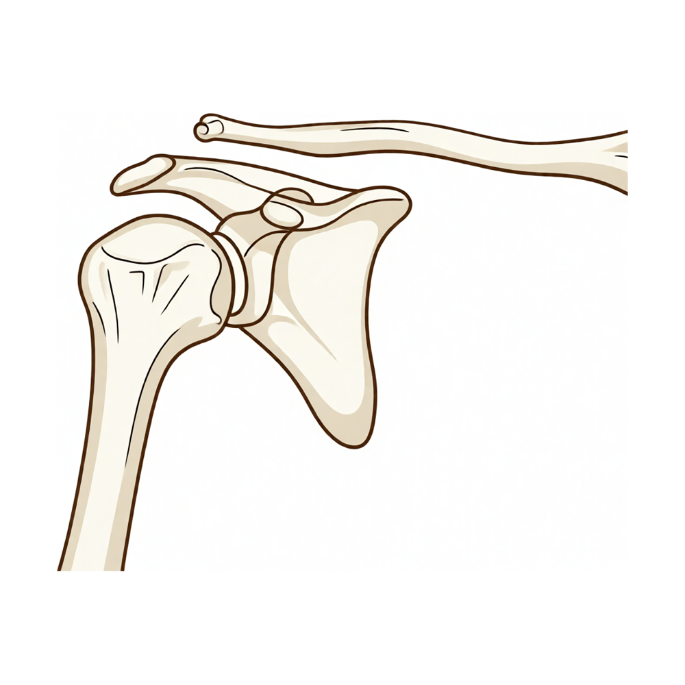
The acromioclavicular (AC) joint is a synovial diarthrodial joint located between the medial margin of the acromion and the lateral end of the clavicle [102]. It serves as the only articulation between the clavicle and scapula, although up to 1% of individuals possess a coracoclavicular bar or joint [105]. The joint contains a fibrocartilaginous disk that separates the articular surfaces; this disk degenerates with age and becomes essentially non-functional beyond the fourth decade [80, 102]. The angle of inclination of the distal clavicle and acromial facet articular surfaces is highly variable, ranging from near vertical to 50° angulation [80]. Approximately 50% of individuals have incongruent articular surfaces where the clavicle overrides the acromion [102]. The distal clavicle epiphysis fuses with the clavicle late, between the ages of 18 and 22 years [80].
The AC joint capsule is reinforced on all sides by the AC joint capsular ligaments, which include superior, inferior, anterior, and posterior components [80, 102]. The superior AC ligament is the strongest of the capsular ligaments and blends with the fibers of the deltoid and trapezius muscles [102]. The joint is innervated by branches of the lateral pectoral, suprascapular, and axillary nerves [80, 102].
Ligamentous Anatomy
The coracoclavicular (CC) ligament consists of two components: the conoid ligament and the trapezoid ligament [102]. The conoid ligament is cone-shaped, attaching to the posteromedial base of the coracoid process and the conoid tubercle on the posterior undersurface of the clavicle [102]. The conoid tubercle is located at the apex of the posterior clavicular curve, at the junction of the lateral third and medial two-thirds of the clavicle [102]. The trapezoid ligament arises from the coracoid process anterior and lateral to the conoid ligament attachment and extends to a rough line on the undersurface of the clavicle [102].
Conoid ligament dimensions: The conoid ligament varies from 0.7 to 2.5 cm in length and 0.4 to 0.95 cm in width [102]. Trapezoid ligament dimensions: The trapezoid ligament measures 0.8 to 2.5 cm in length and 0.8 to 2.5 cm in width [102].
The distance from the lateral clavicle to the insertion of the superior AC ligament/capsule ranges from 5.2 to 7 mm in women and approximately 8 mm in men [102]. The distance from the lateral clavicle to the most lateral fibers of the trapezoid ligament may measure as little as 10 mm [102].
Biomechanics and Stability
The AC joint ligaments provide horizontal stability to the clavicle, with the superior ligament contributing 50% and the posterior ligament contributing 25% [80]. Vertical stability of the clavicle is provided by the AC joint ligaments at physiologic loads and by the coracoclavicular ligaments at higher loads [80]. The conoid ligament is the strongest and most important vertical stabilizer of the AC joint at higher loads [80]. The trapezoid ligament provides restraint against lateral translation of the clavicle in relation to the coracoid as well as vertical stability [80]. The deltotrapezial fascia plays an important but unquantified role in AC joint stability [80]. The CC ligament is the prime suspensory ligament of the upper extremity, suspending the upper extremities from the distal clavicles [102].
At small displacements, the AC ligaments are the primary restraint to posterior (89%) and superior (68%) translation of the clavicle [102]. At large displacements, the conoid ligament provides the primary restraint (62%) to superior translation, while the AC ligaments remain the primary restraint (90%) to posterior translation [102]. The normal AC joint is capable of translating 4 to 6 mm in the anterior, posterior, and superior planes under 70-N loads [187]. The AC joint accommodates rotary motion of 5° to 8° during scapulothoracic motion and 40° to 45° with shoulder abduction and elevation [187]. The clavicle rotates posteriorly 40° to 45° with full elevation of the shoulder, but only 5° to 8° of rotation occurs at the AC joint due to synchronous scapular and sternoclavicular motion [80]. Minimal resection of the distal clavicle (5- and 10-mm) increases the horizontal motion of the clavicle in cadaveric models [80].
Injury Mechanisms
The most common mechanism of AC injury is a direct blow to the superior acromion with the arm adducted at the side [33]. This injury typically results from a fall onto the shoulder during contact sports, skiing, or cycling [33]. The force drives the acromion and clavicle inferiorly, but the sternoclavicular joint restricts clavicular movement, resulting in dissociation between the acromion and clavicle [33]. Indirect trauma from a fall onto the outstretched hand or elbow can drive the humerus against the acromion, disrupting AC ligaments but typically sparing CC ligaments [33]. A medially directed force to the lateral shoulder that drives the acromion into and underneath the distal clavicle can result in higher degrees of injury and displacement [46].
The injury force produces a progressive pattern: first disruption of AC ligaments, followed by CC ligaments, and finally disruption of the deltotrapezial fascia [46]. The characteristic clinical deformity in complete AC dislocation is caused by inferior displacement of the shoulder and arm, not superior elevation of the clavicle [46]. Associated injuries to the clavicle, glenohumeral joint, rotator cuff, and superior labrum occur in 20% of AC separations [33].
Pathoanatomy of Injury
AC joint separation results in progressive disruption of the ligamentous support, beginning with the capsular ligaments and progressing to the CC ligaments [8].
Type I: Minimal to moderate tenderness, mild swelling, and minimal pain with arm movements, with no significant radiographic displacement [52]. Type II: Disruption of AC ligaments presenting with moderate to severe pain, slight superior clavicle prominence, and ecchymosis; bilateral Zanca views demonstrate partial (<50% clavicle width) superior clavicle subluxation [52]. Type III: Disruption of the AC joint capsule and ligaments with significant injury to CC ligaments allowing inferior translation of the limb; produces a characteristic shoulder droop sign and may cause the clavicle to tent the skin; reducible with upward pressure or active shrugging [52]. Type IV: Posterior displacement of the distal clavicle into the trapezius muscle [26]. Type V: Superior displacement of the clavicle greater than 100% compared to the contralateral shoulder due to disruption of the deltotrapezial fascia; not reducible due to deltotrapezial fascia interposition [26, 52]. Type VI: Inferior displacement of the clavicle into the subcoracoid space [26].
In children, type III AC injuries may present as pseudodislocations where the clavicle is displaced superiorly through a rent in the periosteal sleeve while CC ligaments remain intact [94].
Classification
Rockwood Classification System
The Rockwood classification system for acromioclavicular joint injuries was initially established in 1963 with types I, II, and III [26]. It was expanded in 1984 to include types IV, V, and VI [26]. Displacement in this classification is compared with that of the contralateral shoulder and not determined according to clavicular diameter [26].
Type I: Involves AC ligament sprain with intact CC ligaments and normal radiographic CC distance [8]. The AC joint is not applicable for reducibility assessment, and the radiographic CC distance is normal (1.1–1.3 cm) [26].
Type II: Involves AC ligament rupture with sprained but intact CC ligaments and normal radiographic CC distance [8]. This consists of complete rupture of the AC ligament, CC ligament sprain, widened AC joint, and an increase in CC distance by less than 25% compared with the contralateral shoulder [26]. The injury is reducible [26].
Type III: Involves disruption of both AC and CC ligaments, increased CC distance, superior clavicle displacement of up to 100% of the clavicle width, and a reducible deformity [8]. It is characterized by disrupted AC and CC ligaments, widened AC joint, and an increase in CC distance by 25% to 100% compared with the contralateral shoulder [26]. Grade III separations are described as those with 100% displacement of the clavicle on radiographs compared with the contralateral side [17].
Type IV: Involves disruption of AC and CC ligaments with posterior displacement of the clavicle into or through the deltotrapezial fascia and a non-reducible deformity [8]. It is diagnosed when the distal clavicle is displaced posteriorly into the trapezius muscle [26]. The injury is not reducible [26].
Type V: Involves disruption of AC and CC ligaments with greater than 100% superior displacement of the clavicle, markedly increased CC distance, and a deformity that is usually not reducible due to herniation through the deltotrapezial fascia [8]. It is characterized by a CC distance increased by more than 100% compared with the contralateral shoulder due to disruption of the deltotrapezial fascia, which can result in tenting of the overlying skin [26]. Grade V separations are described as exaggerated superior dislocation of the AC joint between 100% and 300% as seen radiographically [17].
Type VI: Involves disruption of AC and CC ligaments with inferior displacement of the clavicle under the acromion or coracoid process [8]. It involves inferior displacement of the clavicle into the subcoracoid space [26]. The injury is not reducible [26]. Type VI subacromial supracoracoid dislocation of the acromioclavicular joint is a rare injury and should be subclassified as type VIa [155].
The differentiation between grade III and grade V separations can be made during physical examination using the shrug test, where the AC joint of type III injuries may reduce but type V injuries cannot reduce due to tearing of the deltotrapezial fascia [17].
Pathoanatomy and Mechanism
The first ligament injured in AC joint trauma is the AC ligament, followed by the CC ligaments, and then the deltotrapezial fascia as severity increases [57]. Trauma with intact coracoclavicular ligaments did not result in acromioclavicular joint lesions higher than Rockwood type I and II [156].
Imaging and Diagnostic Reliability
Plain radiographs for AC joint evaluation include an AP view of the clavicle, a caudal tilt view, and an axillary view [8]. A Zanca view provides excellent detail of the distal clavicle [8]. Rockwood’s original classification was based on findings observed on plain radiographs [137]. Accurate classification of acromioclavicular joint separation requires standard AP films plus an additional modified axial or axillary view to demonstrate antero-posterior displacement, as single-plane imaging may misdiagnose Rockwood grade IV injuries [169].
Interobserver reliability for Rockwood classification among eight shoulder surgeons was moderate with an intraclass correlation coefficient of 0.602 [137]. Intraobserver reliability for Rockwood classification among eight shoulder surgeons was moderate with a Spearman’s rank correlation coefficient of 0.694 [137]. Interobserver reliability for the decision to operate based on Rockwood classification was moderate with an intraclass correlation coefficient of 0.469 [137]. Intraobserver reliability for the decision to operate based on radiographs alone showed only slight agreement with a kappa statistic of 0.366 [137].
Interobserver reliability of the Rockwood classification using plain radiographs alone was fair with a correlation coefficient of 0.214 [137]. Intraobserver reliability of the Rockwood classification using plain radiographs alone was moderate with a correlation coefficient of 0.474 [137]. Interobserver reliability of treatment decisions using plain radiographs alone was fair with a correlation coefficient of 0.213 [137]. Intraobserver reliability of treatment decisions using plain radiographs alone was fair with a correlation coefficient of 0.399 [137].
MR imaging provides exquisite visualization of the soft-tissue structures of the shoulder girdle [10]. In a comparison of 47 cases, ultrasound, MRI, and radiography results were in agreement in 24 cases [42]. In the same comparison of 47 cases, ultrasound was wrong in six cases and radiography was wrong in 24 cases regarding grade classification [42]. Comparison of both sides of the acromioclavicular joint could help inform physicians in predicting the need for additional evaluations in skeletally immature patients [30].
Associated Injuries
Glenohumeral injuries are a much more common epiphenomenon during acromioclavicular separation than previously ascertained, with a diagnosis rate rising to 57.3% in patients above 35 years [4]. Concomitant injuries to the shoulder girdle obtained during traumatic AC joint separation may be more frequent than previously thought [28]. High-grade AC-separations may be associated with traumatic concomitant glenohumeral pathologies resulting from the same trauma impact to the shoulder girdle [37].
Clinical Presentation
Epidemiology and Mechanism
The incidence of acromioclavicular joint separation is unknown [8]. These injuries occur most commonly in men with an 8:1 ratio in their third decade of life [89]. They are frequently associated with contact sports or athletic activity involving a direct blow to the lateral aspect of the shoulder in male patients less than 30 years of age [27]. The most common mechanism is a direct blow to the superior acromion with the arm adducted at the side, typically due to a fall onto the shoulder during contact sports, skiing, or cycling [33]. Indirect injuries from a fall onto the outstretched hand or elbow are rare and typically result in lower-grade injuries by disrupting the acromioclavicular ligaments while sparing the coracoclavicular ligaments [8, 33]. Because the clavicle suspends the shoulder away from the axial skeleton, injuries to the acromioclavicular joint can result in a wide range of shoulder dysfunction [27].
Physical Examination
Inspection and Palpation: Localized bruising, swelling, and tenderness are present in acute acromioclavicular injuries [8]. Higher grade injuries result in prominence of the distal clavicle [8]. Point tenderness at the acromioclavicular joint with or without deformity is a key finding [38]. Diffuse shoulder pain is located in the anterolateral neck, acromioclavicular joint, and anterolateral deltoid [38]. The sternoclavicular joint should be evaluated for swelling, deformity, and tenderness [8]. In type III injuries, the entire length of the clavicular shaft should be palpated to detect an associated clavicle shaft fracture [52].
Range of Motion: Range of motion and rotator cuff strength are typically normal in chronic injuries but may be limited in acute injuries secondary to pain [8].
Stability and Special Tests: The ability to reduce the deformity with manual pressure is important and can help differentiate nonsurgical versus surgical treatment for higher grade injuries [8]. The cross-body adduction test is used as a decision tool where superior displacement of the distal clavicle with pain on adduction of the arm across the body may indicate better outcomes with operative management [17]. A positive cross-arm adduction test (arm flexed 90 degrees, adducted across chest) produces compression pain localized to the acromioclavicular joint [38]. O'Brien's active compression test with localized pain over the acromioclavicular joint is a physical examination finding [38]. Paxino's test involves thumb pressure directed anterior at the posterior acromioclavicular joint [38]. Diagnostic analgesic injection resulting in positive relief of pain and symptoms is a physical examination finding [38].
Injury-Specific Findings
Type I: There is minimal to moderate tenderness to palpation over the acromioclavicular joint, mild swelling may be present, and minimal pain occurs with arm movements including adduction across the body [52]. Patients respond very well to local anesthetic/corticosteroid injections for reduction of inflammation and acute pain relief [52].
Type II: The distal end of the clavicle may be noted to be slightly superior to the acromion, and ecchymosis may be present [52]. Adduction motion of the shoulder typically produces pain in the acromioclavicular joint [52]. A common complaint is difficulty sleeping [52]. Anterior–posterior motion of the clavicle in the horizontal plane can be evaluated after acute inflammation has decreased, with little to no instability in the vertical plane [52].
Type III: The patient characteristically presents with the upper extremity held adducted close to the body and supported in an elevated position to relieve pain [52]. Significant injury to the coracoclavicular ligaments allows inferior translation of the limb and produces the characteristic shoulder droop sign [52]. The clavicle may be prominent enough to tent the skin [52]. Moderate pain is the rule, and any motion of the arm, particularly abduction, increases the pain [52]. Tenderness is noted at the acromioclavicular joint, the coracoclavicular interspace, and along the superior aspect of the lateral clavicle [52]. The acromioclavicular joint can be reduced with upward pressure under the elbow or by having the patient actively shrug in a type III injury, a maneuver known as the "shrug test" [52].
Differentiation of Higher Grades: A type III injury is differentiated from a type IV or V injury by the ability to reduce the joint, as type IV or V injuries cannot be reduced if the deltotrapezial fascia is interposed [52]. The differentiation between grade III and grade V separations can be made during physical examination by use of the shrug test, where the acromioclavicular joint of type III injuries may reduce but type V injuries cannot [17].
Imaging
Radiographic Technique: Plain radiographs for acromioclavicular joint evaluation include an AP view of the clavicle, a caudal tilt view, and an axillary view [8]. Good-quality radiographs require one-third to one-half the beam penetration required to image the glenohumeral joint [38]. Radiographs taken using routine shoulder technique will be overpenetrated and small fractures may be overlooked [38]. The radiographic technician must be specifically requested to take radiographs of the "acromioclavicular joint" rather than the "shoulder" [38]. The Zanca view is a modified, underpenetrated AP view with a cephalic tilt of 10° to 15° that gives excellent detail of the distal clavicle [8]. It involves placing the beam 10–15 degrees cephalad and using 50% of the AP penetration strength to determine displacement with comparison to the contralateral acromioclavicular joint and coracoclavicular distance [38]. The axillary view is used to determine the anteroposterior position of the distal clavicle in relation to the acromion [38].
Interpretation and Stress Views: The anterior aspect of the clavicle should lie in the same plane as the anterior aspect of the acromion on radiographs [8]. Normal coracoclavicular distance between the superior aspect of the coracoid and the inferior clavicle should be between 11 to 13 mm [8]. Fracture of the base of the coracoid process should be ruled out as it can result in superior displacement of the clavicle with an intact coracoclavicular distance, creating a functionally equivalent acromioclavicular joint separation [8]. Weighted views include bilateral AP views with a weight tied to the wrists in relaxed standing and help distinguish between type II and type III separations, though they are rarely indicated and often not clinically helpful [8]. Cross-arm stress views, also known as Basmania views, are AP views with the arm adducted [38]. Reducibility stress views include an active shrug maneuver with an AP of the shoulder or with the patient applying an upward directed load on the elbow while lying on a table during radiography of the acromioclavicular joint to determine whether the deltotrapezial fascia is interposed to prevent reduction [38]. Comparison of both sides of the acromioclavicular joint could help to inform physicians in predicting the need for additional evaluations in the skeletally immature patient [30].
Advanced Imaging: Ultrasound, MRI, and radiography results were in agreement in 24 of 47 cases for acromioclavicular joint injury grading [42]. Ultrasound was wrong in six cases and radiography in 24 cases when compared to MRI for grading acromioclavicular joint injuries [42]. Wide field of view CT provides dynamic information not available through other imaging modalities for acromioclavicular joint instability [63]. 4D CT has provided information that altered the clinical management of a patient with acromioclavicular joint injury [63].
Classification
Type I: Involves AC ligament sprain with intact CC ligaments and normal radiographic CC distance [8]. It involves AC ligament sprain without injury to the CC ligaments, no AC joint widening, and no clavicular displacement [26]. It results from minor strains of the acromioclavicular ligament and joint capsule, with a stable joint and minimal pain [51]. It involves a sprain or partial tear of the acromioclavicular ligament with a normal acromioclavicular interval, which is occult on nonstress radiographs [89]. Periosteal calcification at the distal end of the clavicle may be apparent later in type I injuries even if initial radiographs are negative [51].
Type II: Involves AC ligament rupture with sprained but intact CC ligaments and normal radiographic CC distance [8]. It consists of complete rupture of the AC ligament, CC ligament sprain, widening of the AC joint, and an increase in the CC distance by less than 25% compared with the contralateral shoulder [26]. It involves rupture of the acromioclavicular ligament and joint capsule with intact coracoclavicular ligaments, resulting in an unstable joint [51]. The lateral end of the clavicle may ride higher than the acromion by less than the thickness of the clavicle even when stress is applied to the joint [51]. Considerable pain and tenderness are present over the acromioclavicular joint [51]. Stress radiographs are necessary to assess the degree of instability after type II injuries [51]. It has torn acromioclavicular and intact coracoclavicular ligaments with less than 100% elevation of the clavicle in relation to the acromion [89].
Type III: Involves disruption of the AC and CC ligaments, increased CC distance, superior displacement of the clavicle of up to 100% of the clavicle width, and a reducible deformity [8]. It involves disruption of the AC and CC ligaments, widened AC joint, and an increase in the CC distance by 25% to 100% compared with the contralateral shoulder [26]. It results from a force sufficient to rupture the acromioclavicular and coracoclavicular ligaments [51]. It shows complete disruption of both the acromioclavicular and coracoclavicular ligaments, with greater superior displacement of the distal clavicle [89].
Type IV: Involves disruption of the AC and CC ligaments with posterior displacement of the clavicle, increased CC distance, distal clavicle herniated into or through the deltotrapezial fascia, and a non-reducible deformity [8]. It is diagnosed when the distal clavicle is displaced posteriorly into the trapezius muscle [26]. It involves ligament disruption with the distal end of the clavicle displaced posteriorly into or through the trapezius muscle [51]. It occurs when the distal clavicle moves posteriorly into the trapezius muscle and is often missed on frontal radiographs [89]. It can be associated with concomitant anterior dislocation of the proximal clavicle at the sternoclavicular joint [89].
Type V: Involves disruption of the AC and CC ligaments with greater than 100% displacement of the clavicle superiorly, markedly increased CC distance, and a deformity usually not reducible because of herniation through the deltotrapezial fascia [8]. It is similar to type III, except the CC distance is increased by more than 100% compared with the contralateral shoulder because of disruption of the deltotrapezial fascia, and tenting of the overlying skin can result [26]. It involves ligament and muscle attachment disruption with the clavicle and acromion widely separated [51]. It is a more severe form of type III with a more superiorly displaced clavicle [89]. It is defined by a coracoclavicular distance that is 100% greater than that of the opposite side, requiring bilateral AC views [18]. The diagnosis is established if a marked superior displacement of the distal clavicle and a 100% to 300% increase in the coracoclavicular distance compared with the normal side is found [100].
Type VI: Involves disruption of the AC and CC ligaments with inferior clavicle displacement, resulting in the distal clavicle lying under the acromion or coracoid process [8]. It is rare and involves inferior displacement of the clavicle into the subcoracoid space [26]. It involves ligament disruption with the distal clavicle dislocated inferior to the coracoid process and posterior to the biceps and coracobrachialis tendons [51]. It is rare and is characterized by inferior displacement of the distal clavicle with respect to the acromion [89].
MRI Findings: MR images show discontinuity of normally low-signal-intensity acromioclavicular and coracoclavicular ligaments, surrounding soft-tissue edema, and other features in acromioclavicular joint separation [89].
Associated Pathologies
Concomitant injuries to the shoulder girdle obtained during traumatic acromioclavicular joint separation may be more frequent than previously thought [28]. High-grade acromioclavicular separations may be associated with traumatic concomitant glenohumeral pathologies resulting from the same trauma impact to the shoulder girdle [37]. Associated injuries to the clavicle, glenohumeral joint, rotator cuff, and superior labrum occur in 20% of acromioclavicular separations [33]. Acromioclavicular joint dislocation coupled with ipsilateral mid-shaft clavicle fracture is a rare dual injury of the shoulder joint that is often overlooked in poly-trauma cases [146]. Combined acromial fracture and acromioclavicular dislocation can occur following minor trauma [145].
Investigations
Physical Examination
The mechanism of injury for acromioclavicular joint separations is usually direct trauma resulting from a fall on the point of the shoulder [8]. Indirect injuries to the acromioclavicular joint are rare [8]. During physical examination, the sternoclavicular joint should be evaluated for swelling, deformity, and tenderness [8]. Range of motion and rotator cuff strength are typically normal in chronic acromioclavicular injuries but may be limited in acute injuries secondary to pain [8]. Scapular motion should be carefully assessed as scapular dyskinesis can be seen with acromioclavicular injury [8].
Specific provocative tests aid in diagnosis and management. The cross-arm adduction test produces compression pain localized to the acromioclavicular joint [38]. O'Brien's active compression test with localized pain over the acromioclavicular joint is suggestive of acromioclavicular pathology [38]. A diagnostic analgesic injection providing positive relief in pain and symptoms supports the diagnosis of acromioclavicular pathology [38]. The shrug test differentiates grade III from grade V separations, as grade III injuries may reduce during the test while grade V injuries cannot due to deltotrapezial fascia tearing [17]. The cross-body adduction test can serve as a decision tool for operative management if the distal clavicle displaces superiorly with pain on adduction of the arm across the body [17]. The ability to reduce the deformity with manual pressure can help differentiate nonsurgical versus surgical treatment for higher grade acromioclavicular injuries [8].
Radiography
Plain radiography: The radiographic technician must be specifically requested to take radiographs of the “AC joint” rather than the “shoulder” [38]. On the axillary view, the anterior aspect of the clavicle should lie in the same plane as the anterior aspect of the acromion [8]. This view determines the anteroposterior position of the distal clavicle in relation to the acromion [38]. The normal coracoclavicular distance between the superior aspect of the coracoid and the inferior clavicle should be between 11 and 13 mm [8]. Fracture of the base of the coracoid process should be ruled out on radiographs, as it can result in superior displacement of the clavicle with an intact coracoclavicular distance [8]. Type V injury is defined by a coracoclavicular distance that is 100% greater than that of the opposite side, requiring bilateral acromioclavicular views [18].
Weighted views include bilateral AP views with a weight tied to the wrists in relaxed standing and help distinguish between type II and type III separations [8]. However, weighted views are rarely indicated and often are not clinically helpful [8]. Reducibility stress views include an active shrug maneuver with an AP of the shoulder or with the patient applying an upward directed load on the elbow while lying on a table to determine whether the deltotrapezial fascia is interposed to prevent reduction [38].
Advanced Imaging
MRI: MR imaging performed in the coronal oblique plane parallel to the distal clavicle enables distinction between grade 2 and grade 3 injuries and diagnosis of grade 1 injuries by demonstrating a ruptured superior acromioclavicular ligament [184].
CT: 4D CT has provided information that altered the clinical management of acromioclavicular joint injury [63].
Other Considerations: In a comparison of imaging modalities for acute acromioclavicular dislocation, ultrasound, MRI, and radiography results were in agreement in 24 of 47 cases [42]. In this comparison, ultrasound was wrong in six cases and radiography was wrong in 24 cases [42].
Treatment
Non-Operative
Nonsurgical management is the standard of care for Rockwood type I and type II acromioclavicular joint injuries [8]. Treatment consists of sling immobilization followed by gradual active range of motion exercises, stretching, and strengthening as tolerated [8]. Most patients regain full shoulder function within 4 to 6 weeks [8]. Immobilization duration varies by injury grade; type I injuries typically require one week of immobilization, while type II injuries may require 2 to 3 weeks [92, 167]. Although most patients recover well, those with type I and II injuries face an increased risk of painful acromioclavicular joint arthritis [8]. Between 30% and 50% of young, very active patients experience mild to moderate residual pain at the acromioclavicular joint [8]. Pain may persist for as long as six months, with up to 33% of patients reporting continued pain and instability at longer-term follow-up [92]. Retrospective studies report persistent symptoms in up to 40% to 50% of patients at 1, 6, and 10 years post-injury [167]. In one study, 27% of patients with type I or II injuries underwent surgical intervention at a mean of 26 months after injury [167]. Distal clavicle resection, either arthroscopic or open, can address persistent acromioclavicular joint inflammation, osteoarthritis, or distal clavicle osteolysis when nonsurgical treatment fails [167]. Aggressive treatment and rehabilitation are indicated in acute acromioclavicular injuries to avoid unnecessary residual symptoms [191].
For type III injuries, the general consensus is to utilize nonoperative treatment for 8 to 12 weeks followed by reexamination to evaluate residual pain and/or dysfunction [17]. The primary indicator for operative treatment is pain, with secondary factors including weakness and loss of motion [17]. Multiple retrospective comparative studies show good clinical results and return to sport with nonsurgical treatment of type III injuries, with no advantage from surgery [8]. Nonsurgical treatment results in quicker recovery and return to work compared to surgical treatment, while surgical treatment increases complications [8]. A 2007 survey found that 86.3% of American Orthopaedic Society for Sports Medicine members and residency directors preferred an initial trial of nonsurgical management for uncomplicated type III injuries [167]. In a prospective randomized study, the nonoperatively treated group demonstrated quicker regain of movement, quicker return to work and sports, and fewer poor results compared to the operatively treated group [54]. However, subgroup analysis showed better results in the operatively treated group for acromioclavicular dislocations with >2 cm of displacement [54]. Another prospective randomized study comparing nonoperative treatment with the Phemister procedure found shorter rehabilitation time in the nonoperative group and a higher amount of complications in the operative group, with no difference in clinical results [54]. Authors recommend that grade III acromioclavicular dislocations be treated nonoperatively [69]. Non-operative treatment can be expected to give a good long-term outcome and should remain the treatment of choice until an operative procedure is shown to give better results consistently [61]. Conservative treatment is adequate in the great majority of patients [194]. A majority of patients with untreated acute grade III separation do well without formal treatment, though a small percentage may require delayed surgical intervention [5].
For higher-grade injuries, nonsurgical treatment likely results in substantial residual pain and limited function for type IV, V, and VI injuries, although outcome studies are limited [8]. Less favorable outcomes occur when grade IV, V, and VI injuries are treated non-operatively [36]. In a retrospective study of conservative treatment of type V separations in active-duty service members, 11 patients (61%) returned to full duty without surgery at an average of 97.8 days [14]. In the same study, 6 patients (75%) in the acute surgical group returned to full duty at an average of 169.3 days [14]. These findings suggest that conservative management may be a viable alternative to surgery for high-grade dislocations in patients without specific clinical indications for immediate surgical intervention [172]. In a prospective randomized clinical trial comparing surgical treatment with nonsurgical management of acute type III and IV separations, there were no statistical differences in validated outcome scores at 1 year follow-up, but faster recovery occurred in the nonsurgical group [167]. Five patients (16%) in the nonsurgical group required surgery for persistent symptoms at a mean of 8.7 months [167]. A short period of immobilization followed by a graduated rehabilitation program is important for the success of non-operative treatment of grades I to III injuries and for higher grade injuries in patients not candidates for operative management [36].
Operative
Indications: Surgical treatment is recommended for most patients with type IV, V, and VI acromioclavicular joint injuries [8]. Surgery is also often recommended for patients with type IIIB (horizontally unstable) separations [8]. Operative treatment is generally the accepted method for active healthy patients with complete acromioclavicular joint injuries (types IV, V, and VI) due to significant morbidity associated with the injury pattern, which can lead to a persistently dislocated, unstable acromioclavicular joint, altered scapular kinematics, and shoulder dysfunction [162]. Surgical management is usually indicated for type IV and V separations given the high likelihood of persistent shoulder pain, dysfunction, and substantial deformity [167]. In chronic acromioclavicular joint instability, surgery is indicated after failed conservative therapy of 3 to 6 months [177]. If secondary arthritis is the main problem in cases of low-grade instability, a lateral clavicle resection may be sufficient [177]. Biologic augmentation should be considered for chronic high-grade instabilities, accounting for the lack of intrinsic healing and scar-forming potential of the ligamentous tissue [177]. One-directional vertical high-grade instabilities can be treated in an all-arthroscopic coracoacromial ligament transfer [177].
Surgical Approach / Technique: No single surgical technique has been proven clearly superior to all others for acromioclavicular joint separations, though emerging research supports more anatomic reconstructions [23]. A lack of consensus exists on the optimal surgical technique, leading to unique complication profiles based on the technique used [9]. It is clear that a gold standard for surgical stabilization of acute, painful acromioclavicular joint dislocations has yet to be established [27]. There is insufficient evidence from randomised controlled trials to determine when surgical treatment is indicated for acromioclavicular dislocation in adults in current practice [112]. The literature highlights serious weaknesses, particularly the lack of compelling evidence supporting surgery over nonoperative management for many cases, especially type III injuries [188]. Early surgical repair of type III injuries with or without augmentation seems to result in better patient satisfaction and clinical outcomes compared with delayed reconstruction [167]. A 2016 systematic review comparing early and delayed surgical intervention in complete dislocations involving mostly type III injuries found superior functional outcomes in the early surgical group (P < 0.05) compared with delayed surgery [167]. Partial dislocations or redislocations were found in 26% of cases in the early treatment group compared with 38.1% of cases in the delayed group (P < 0.05) [167]. Complication rates were 12.5% in the early surgical group and 17.7% in the delayed surgical group, although differences did not reach statistical significance [167]. Another review confirmed that early surgery results in better functional outcomes and fewer resubluxations/redislocations, with overall complication rates comparable between early and delayed management [14]. In a multicenter randomized clinical trial of nonoperative versus operative treatment of acute dislocation, operative repair with hook-plate fixation improved radiographic but not clinical outcomes compared with nonoperative treatment [88]. Both nonoperative and operative groups had very good restoration of shoulder function and patient satisfaction at 24 months in a randomized controlled trial of type III and V dislocation, and operative treatment did not lead to better outcomes compared with nonoperative treatment [174].
Implant Selection: In a meta-analysis comparing suture button fixation with hook plate fixation in acute unstable dislocations, outcomes favored the suture button technique in terms of outcome scores and pain scores [14]. Subgroup analysis showed better outcome scores in the arthroscopic suture button group compared with the hook plate group, with no differences between the open suture button group and the hook plate group [14]. Considering the nearly anatomical reconstruction, avoidance of hardware complications, and low rate of recurrence, double-loop suture repair may be an attractive alternative for acute separations [13]. Shoulder complications were observed in 0 patients who underwent double-loop anatomic acromioclavicular reconstruction [6]. TACCR represents a safe, reliable and creative surgical technique that yields good to excellent clinical and radiologic outcomes in the treatment of severe acromioclavicular separation [62]. The described procedure aims to treat subacute high-grade injuries by restoring normal acromioclavicular joint kinematics while ensuring an adequate biological environment [20]. This method provides a reliable surgical option for the treatment of acromioclavicular joint separation [21]. The acute acromioclavicular joint dislocation clinical outcome will be achieved if you perform both procedures [11]. The results confirm the operative regime for separations of the acromioclavicular joint [12]. Operative management was associated with a higher rate of complications than nonoperative treatment, and reduction of the acromioclavicular joint was not necessary to obtain consistently good results [35]. The acromioclavicular harness has been used with good results in treating twenty cases, maintaining reduction and bringing about permanent healing [71]. Satisfactory outcome depends upon restoring the stability of the clavicle as well as the acromioclavicular joint [64]. Single coracoclavicular suture fixation with Mersilene tape provided early recovery of shoulder motion and avoided further morbidity of the acromion [176]. This technique is very effective for symptomatic acromioclavicular arthritis of the shoulder [150]. For appropriate clinical indications, beveling the inferior 20% to 25% of the clavicle to make it co-planar with the decompressed acromion is safe and is not an etiologic factor in acromioclavicular joint pain or instability [160]. Surgical treatment based on the exclusive extirpation of the ganglion is ineffective if there is not an effective treatment of the associated injuries of the rotator cuff and the acromioclavicular joint [151].
Other Considerations: Operative intervention should be clearly discussed with each patient, as the objectives are different for each patient [162]. No overall consensus exists on treatment for type III dislocations although a trend toward initial nonoperative treatment is presently favored in most cases [162]. A proportion of conservatively treated patients with type III injuries will have persistent pain and an inability to return to their sport or job, and subsequent surgical stabilization, albeit delayed, has allowed return to sport or work in such cases [162]. To date no high-quality prospective study has demonstrated surgical benefit for the treatment for type III injuries [162]. At the institution described in Rockwood and Green, acute type I, II, III, and V injuries are managed with a 6-week course of supervised rehabilitation to maximize rotator cuff strength and the stabilizing function of the periscapular muscles [162]. Patients who have failed a minimum 6 weeks of shoulder stabilization-directed physical therapy have had good to excellent results with delayed surgical reconstruction using a tendon graft [162]. Altered shoulder biomechanics secondary to complete acromioclavicular joint displacement was shown in a recent study of patients with type III injuries to result in scapular dyskinesis and, eventually, SICK scapula syndrome [162]. SICK scapula syndrome developed in 54% of patients with chronic type III acromioclavicular dislocations in another study [162]. Ipsilateral os acromiale may be a relative contraindication to the clavicle hook plate [159]. Excessive medialization of the clavicular tunnels has been associated with a higher rate of failure in coracoclavicular ligament reconstruction [18]. Backup coracoclavicular stabilization is usually required for a successful outcome [18]. A modification of the all-arthroscopic coracoacromial ligament transfer includes an additional synthetic augmentation with a transclavicular-transcoracoidal stabilization with a
Complications
Associated Injuries
The provided evidence base does not contain specific data regarding associated injuries for this section.
Nonoperative Management
Despite the frequent occurrence of radiographic changes, long-term functional outcome after Rockwood I and II acromioclavicular joint injuries is good, with only clinically nonrelevant functional differences between the injured and contralateral shoulders [7].
Operative Management
Complications of the acromioclavicular (AC) and sternoclavicular (SC) joint injuries can occur as a result of the injury itself, nonoperative management, or operative treatment [32]. Surgical treatment may offer early benefits in pain relief and coracoclavicular distance improvement but does not enhance long-term functional outcomes and is associated with higher specific complication rates [74]. Considering the nearly anatomical reconstruction, the avoidance of hardware complications, and the low rate of recurrence, this technique may be an attractive alternative to the management of acute acromioclavicular joint separations [13].
Instability: In a series of 35 dislocations operated on with open reduction and percutaneous Kirschner wire fixation, complications included broken wire, inadequate fixation, and late recurrence of deformity [181]. In one patient treated with open reduction and percutaneous Kirschner wire fixation, redislocation had occurred 4 years after operation [181]. Three patients with old AC separations had prominence of the distal clavicle or symptoms due to residual hypermobility of the clavicle in spite of the CC stabilization [163].
Wound complications: In a series of 35 dislocations operated on with open reduction and percutaneous Kirschner wire fixation, complications included superficial wound infection [181].
Other Considerations: Most patients were ultimately treated surgically, although complications such as anterior shoulder pain, AC joint asymmetry, and activity-related weakness were common sequelae resulting in physical limitations and separation from military service [55]. In a series of 35 dislocations operated on with open reduction and percutaneous Kirschner wire fixation, complications included osteoarthrosis without symptoms [181]. The patient who had not an anterior acromioplasty in spite of subacromial impingement had unchanged impingement symptoms [163].
Recovery
Non-Operative Management Outcomes: Long-term functional outcomes following Rockwood I and II acromioclavicular joint injuries are good, characterized by only clinically nonrelevant functional differences between the injured and contralateral shoulders despite frequent radiographic changes [7]. More than 50% of patients with Grade I and II acromioclavicular dislocations reported having a good or excellent shoulder 6 years after the injury [200]. Non-operative treatment of acromioclavicular dislocations can be expected to give a good long-term outcome [61]. A majority of patients with untreated acute grade III acromioclavicular separation do well without any formal treatment, though a small percentage may require delayed surgical intervention [5]. In a retrospective study of conservative treatment for type V acromioclavicular separations in active-duty service members, 11 patients (61%) returned to full duty without surgery at an average of 97.8 days [21]. The acromioclavicular ligament exhibits early and dynamic healing responses following acute traumatic rupture [198].
Operative Management Outcomes: Surgical treatment for Rockwood type III acromioclavicular joint dislocation may offer early benefits in pain relief and coracoclavicular distance improvement but does not enhance long-term functional outcomes and is associated with higher specific complication rates [74]. In a prospective randomized controlled trial comparing surgical treatment using tunneled suspensory fixation with nonsurgical treatment of type III and IV acromioclavicular joint dislocations, there was no difference in outcome measures between both groups at 1-year follow-up [18]. In the same trial, faster recovery was observed in the nonsurgical group [18]. Additionally, five patients in the nonsurgical group were later treated surgically because of dissatisfaction with nonsurgical treatment [18]. A systematic review and meta-analysis comparing surgical and nonsurgical management of acute high-grade acromioclavicular dislocations noted no difference in outcome scores between both groups [14]. However, the surgical group showed better cosmetic outcomes [14], while the nonsurgical group showed faster return to work [14]. Fifteen years postoperatively after arthroscopically assisted 2-bundle anatomic reduction of acute acromioclavicular joint separations, good clinical results persisted and anatomic reduction was overall maintained [201]. In the same cohort, asymptomatic ossification of the coracoclavicular ligaments was often observed [201]. Short-term follow-up of 15 patients treated with minimally invasive coracoclavicular ligament augmentation with a flip button/polydioxanone repair for total acromioclavicular joint dislocation revealed excellent radiologic and clinical results, with no subluxations or dislocations of the acromioclavicular joint noted [66]. Late reconstruction of the ligaments in young patients with complete acromioclavicular separations can yield better results than excision of the lateral clavicle, allowing patients to return to strenuous sports or heavy labor [31]. In a comparison of early and delayed surgical management of acromioclavicular joint separations, early surgery resulted in better functional outcomes and fewer resubluxations/redislocations [20]. Overall complication rates were comparable between the two groups [20]. In the retrospective study of active-duty service members with type V separations, 6 patients (75%) in the acute surgical group returned to full duty at an average of 169.3 days [21]. Good functional and long-lasting recovery without pain and limitation of shoulder movement can be expected for inferior subcoracoid acromioclavicular dislocation [189].
Associated Pathologies and Kinematics: Type III acromioclavicular separations result in a significant change in the shoulder's motion and may warrant surgical reconstruction to restore normal function [70]. Satisfactory outcome for fracture clavicle with acromioclavicular dislocation depends upon restoring the stability of the clavicle as well as the acromioclavicular joint [64].
Key Evidence
- [L5] Acromioclavicular joint separations are common injuries, with low-grade injuries frequently managed successfully using non-surgical measures and high-grade injuries often warranting surgical intervention to minimize pain and maximize shoulder function. [1] (10.1007/s12178-012-9144-9)
- [L5] Data support both surgical and nonoperative management for grade III acromioclavicular joint separations. [2] (10.1249/jsr.0000000000000530)
- [L4] Type I and II acromioclavicular joint disruptions impair long-term shoulder function in about half of patients 10 years after injury. [3] (10.1177/0363546508319047)
- [L4] Glenohumeral injuries are a much more common epiphenomenon during acromioclavicular separation than previously ascertained, with a diagnosis rate rising to 57.3% in patients above 35 years. [4] (10.1186/s12891-017-1803-y)
- [L2] A majority of patients with untreated acute grade III acromioclavicular separation will do well without any formal treatment, though a small percentage may require delayed surgical intervention. [5] (10.1177/03635465010290060401)
- [L4] Shoulder complications were observed in 0 patients who underwent double-loop anatomic acromioclavicular reconstruction. [6] (10.1097/bte.0000000000000021)
- [L3] Despite the frequent occurrence of radiographic changes, long-term functional outcome after Rockwood I and II acromioclavicular joint injuries is good, with only clinically nonrelevant functional differences between the injured and contralateral shoulders. [7] (10.1177/0363546520981993)
- [L4] A lack of consensus exists on the optimal surgical technique for acromioclavicular joint injuries, leading to unique complication profiles based on the technique used. [9] (10.5435/jaaos-d-24-00696)
- [L5] MR imaging provides exquisite visualization of the soft-tissue structures of the shoulder girdle. [10] (10.2214/ajr.180.4.1801103)
- [L5] The acute acromioclavicular joint dislocation clinical outcome will be achieved if you perform both procedures. [11] (10.1016/j.eats.2022.05.012)
- [L4] The results confirm the operative regime for separations of the acromioclavicular joint. [12] (10.1007/s001130050403)
- [L4] Considering the nearly anatomical reconstruction, the avoidance of hardware complications, and the low rate of recurrence, this technique may be an attractive alternative to the management of acute acromioclavicular joint separations. [13] (10.1177/0363546505284187)
- [L4] The optimal technique for treating acromioclavicular separations has yet to be determined. [15] (10.1177/0363546516651613)
- [L5] The described procedure aims to treat subacute high-grade injuries by restoring normal acromioclavicular joint kinematics while ensuring an adequate biological environment. [20] (10.1016/j.eats.2021.12.025)
- [L5] This method provides a reliable surgical option for the treatment of acromioclavicular joint separation. [21] (10.1016/j.eats.2025.103903)
- [L3] Reconstruction of chronic acromioclavicular joint dislocations restores good shoulder function and results in satisfied patients. [22] (10.1016/j.jse.2010.02.006)
- [L5] No single surgical technique has been proven clearly superior to all others for acromioclavicular joint separations, though emerging research supports more anatomic reconstructions. [23] (10.1016/j.csm.2014.06.009)
- [L4] Concomitant injuries to the shoulder girdle obtained during traumatic AC joint separation may be more frequent than previously thought. [28] (10.1177/0363546508322891)
- [L3] Comparison of both sides of the acromioclavicular joint could help to inform physicians in predicting the need for additional evaluations. [30] (10.1007/s11999-013-3242-x)
- [L4] Late reconstruction of the ligaments in young patients with complete acromioclavicular separations can yield better results than excision of the lateral clavicle, allowing patients to return to strenuous sports or heavy labor. [31] (10.2106/00004623-197658060-00008)
- [L5] Complications of the acromioclavicular (AC) and sternoclavicular (SC) joint injuries can occur as a result of the injury itself, nonoperative management, or operative treatment. [32] (10.1016/s0278-5919(02)00102-3)
- [L5] [33] (10.1148/rg.2020200039)
- [L3] Operative management was associated with a higher rate of complications than nonoperative treatment, and reduction of the acromioclavicular joint was not necessary to obtain consistently good results. [35] (10.2106/00004623-198769070-00013)
- [L4] High-grade AC-separations may be associated with traumatic concomitant glenohumeral pathologies resulting from the same trauma impact to the shoulder girdle. [37] (10.1007/s00167-008-0666-z)
- [L2] [42] (10.1007/s00330-016-4413-4)
- [L4] Most patients were ultimately treated surgically, although complications such as anterior shoulder pain, AC joint asymmetry, and activity-related weakness were common sequelae resulting in physical limitations and separation from military service. [55] (10.1177/03635465211036713)
- [L4] The authors recommend that all patients undergo shoulder arthroscopy at the time of acromioclavicular surgery. [60] (10.1016/s1058-2746(00)90051-x)
- [L4] This study shows that non-operative treatment of acromioclavicular dislocations can be expected to give a good long-term outcome and that until an operative procedure is shown to give better results consistently, non-operative treatment should remain the treatment of choice. [61] (10.1016/0020-1383(95)90030-6)
- [L4] TACCR represents a safe, reliable and creative surgical technique that yields good to excellent clinical and radiologic outcomes in the treatment of severe acromioclavicular separation. [62] (10.1016/j.jse.2017.12.015)
- [L5] It provides dynamic information not available through other imaging modalities. 4D CT has provided information that altered the clinical management of this patient, showing future benefits for clinical approach to diagnosis and management of acromioclavicular joint injury. [63] (10.1111/1754-9485.12283)
- [L4] Satisfactory outcome depends upon restoring the stability of the clavicle as well as the acromioclavicular joint. [64] (10.1111/j.1758-5740.2010.00102.x)
- [L5] Postoperative radiographs confirmed preservation of reduction at the acromioclavicular joint. [65] (10.1097/bth.0b013e318159076a)
- [L4] The short-term follow-up of 15 recently operated patients reveals excellent radiologic and clinical results, with no subluxations or dislocations of the acromioclavicular joint noted. [66] (10.1016/j.arthro.2006.12.015)
- [L5] Studies have shown no distinct advantage for surgical reconstruction over nonoperative treatment for type III acromioclavicular injuries. [68] (10.5435/00124635-199701000-00002)
- [L4] The authors recommend that grade III acromioclavicular dislocations be treated nonoperatively. [69] (10.1177/036354659202000316)
- [L5] Type III AC separations result in a significant change in the shoulder's motion and may warrant surgical reconstruction to restore normal function. [70] (10.1186/s12891-016-1330-2)
- [L4] The acromioclavicular harness has been used with good results in treating twenty cases, maintaining reduction and bringing about permanent healing. [71] (10.2106/00004623-195234010-00032)
- [L1] Surgical treatment may offer early benefits in pain relief and coracoclavicular distance improvement but does not enhance long-term functional outcomes and is associated with higher specific complication rates. [74] (10.1186/s12891-024-08100-x)
- [L1] [88] (10.2106/jbjs.16.00582)
- [L5] [89] (10.2214/ajr.13.11460)
- [L4] [92] (10.2106/jbjs.l.00734)
- [L4] [100] (10.1177/0363546511399379)
- [L1] There is insufficient evidence from randomised controlled trials to determine when surgical treatment is indicated for acromioclavicular dislocation in adults in current practice. [112] (10.1002/14651858.cd007429.pub2)
- [L5] [137] (10.1007/s11999-016-5079-6)
- [L5] This case demonstrates that combined acromial fracture and acromioclavicular dislocation can occur following minor trauma and that the injury is compatible with good outcome. [145] (10.1016/0020-1383(95)00117-r)
- [L5] Acromioclavicular joint dislocation coupled with ipsilateral mid-shaft clavicle fracture is a rare dual injury of the shoulder joint that is often overlooked in poly-trauma cases. [146] (10.1136/bcr-2015-213254)
- [L4] This technique is very effective for symptomatic acromioclavicular arthritis of the shoulder. [150] (10.1016/s1058-2746(98)90091-x)
- [L5] Surgical treatment based on the exclusive extirpation of the ganglion is ineffective if there is not an effective treatment of the associated injuries of the rotator cuff and the acromioclavicular joint. [151] (10.1016/j.ejrex.2007.04.007)
- [L5] Type VI subacromial supracoracoid dislocation of the acromioclavicular joint is a rare injury and should be subclassified as type VIa. [155] (10.1177/2054270414527281)
- [L5] Trauma with intact coracoclavicular ligaments did not result in acromioclavicular joint lesions higher than Rockwood type I and II. [156] (10.1007/s00402-014-2045-1)
- [L4] Ipsilateral os acromiale may be a relative contraindication to the clavicle hook plate. [159] (10.1186/s12891-021-04841-1)
- [L4] We conclude that for appropriate clinical indications, beveling the inferior 20% to 25% of the clavicle to make it co-planar with the decompressed acromion is safe and is not an etiologic factor in acromioclavicular joint pain or instability. [160] (10.1067/mse.2000.109560)
- [L4] [163] (10.1016/s1058-2746(96)80520-9)
- [L5] The authors suggest that accurate classification of acromioclavicular joint separation requires standard AP films plus an additional modified axial or axillary view to demonstrate antero-posterior displacement of the clavicle, as single-plane imaging may misdiagnose Rockwood grade IV injuries. [169] (10.1016/j.injury.2016.06.031)
- [L3] These findings suggest that conservative management may be a viable alternative to surgery for high-grade acromioclavicular dislocations in patients without specific clinical indications for immediate surgical intervention. [172] (10.1016/j.jseint.2026.101722)
- [L1] Both the nonoperative and operative treatment groups had very good restoration of shoulder function and patient satisfaction at 24 months, and operative treatment did not lead to better outcomes compared with nonoperative treatment. [174] (10.1016/j.jse.2021.12.003)
- [L3] However, single coracoclavicular suture fixation with Mersilene tape provided early recovery of shoulder motion and avoided further morbidity of the acromion. [176] (10.1186/1749-799x-9-42)
- [L4] [181] (10.1016/0020-1383(82)90325-4)
- [L5] MR imaging performed in the coronal oblique plane parallel to the distal clavicle enables distinction between grade 2 and grade 3 injuries and diagnosis of grade 1 injuries by demonstrating a ruptured superior acromioclavicular ligament. [184] (10.1148/rg.282075714)
- [Paper] The authors describe a preferred surgical technique for anatomic repair of acromioclavicular joint separation using a semitendinosus allograft, noting that anatomic reconstruction allows for better cosmesis and functional outcomes compared with nonanatomic techniques. [186] (10.1016/j.eats.2017.06.045)
- [L4] [187] (10.1302/2058-5241.3.170027)
- [L5] The literature on unstable acromioclavicular joint injuries highlights serious weaknesses, particularly the lack of compelling evidence supporting surgery over nonoperative management for many cases, especially type III injuries. [188] (10.1016/j.arthro.2018.03.022)
- [L5] Good functional and long-lasting recovery without pain and limitation of shoulder movement can be expected. [189] (10.1007/s00068-003-1254-z)
- [L3] Aggressive treatment and rehabilitation are indicated in acute acromioclavicular injuries to avoid unnecessary residual symptoms. [191] (10.1177/036354658100900111)
- [L1] Both open and arthroscopic repair of acute acromioclavicular joint dislocation yielded good clinical results, yet the arthroscopic technique is more expensive and has a longer surgical time. [192] (10.1016/j.jse.2019.06.007)
- [L4] Considering its less morbidity, excellent cosmesis, no need of hardware removal, and minimal complications from breakage or migration of metal implants, this new technique offers an attractive alternative in acromioclavicular joint stabilization. [193] (10.1007/s00402-005-0073-6)
- [L4] Conservative treatment of the acromioclavicular injury is adequate in the great majority of patients and can properly be recommended and applied. [194] (10.1016/s0020-1383(73)80003-8)
- [L4] The acromioclavicular ligament exhibits early and dynamic healing responses following acute traumatic rupture. [198] (10.1186/s12891-020-03614-6)
- [L4] More than 50% of the patients reported having a good or excellent shoulder 6 years after the injury. [200] (10.1016/s1058-2746(03)00215-5)
- [L3] Fifteen years postoperatively, good clinical results persisted and anatomic reduction was overall maintained, often with asymptomatic ossification of the coracoclavicular ligaments. [201] (10.1177/03635465251355958)
See Also
- Shoulder Arthroscopy
- Rotator Cuff
- Fractures
- Clavicle Fracture
- Distal Clavicle Osteolysis
- Os Acromiale
References
[1] Acromioclavicular joint separations. Current Reviews in Musculoskeletal Medicine. 2012. DOI: 10.1007/s12178-012-9144-9
[2] Evaluation and Management of Grade III Acromioclavicular Joint Separations. Current Sports Medicine Reports. 2018. DOI: 10.1249/jsr.0000000000000530
[3] Long-Term Shoulder Function after Type I and II Acromioclavicular Joint Disruption. The American Journal of Sports Medicine. 2008. DOI: 10.1177/0363546508319047
[4] Concomitant glenohumeral pathologies in high-grade acromioclavicular separation (type III – V). BMC Musculoskeletal Disorders. 2017. DOI: 10.1186/s12891-017-1803-y
[5] A Prospective Evaluation of Untreated Acute Grade III Acromioclavicular Separations. The American Journal of Sports Medicine. 2001. DOI: 10.1177/03635465010290060401
[6] Double-loop Anatomic Acromioclavicular Reconstruction. Techniques in Shoulder & Elbow Surgery. 2014. DOI: 10.1097/bte.0000000000000021
[7] Long-term Outcome After Nonoperative Treatment for Rockwood I and II Acromioclavicular Joint Injuries. The American Journal of Sports Medicine. 2021. DOI: 10.1177/0363546520981993
[8] Aaos Comprehensive Orthopaedic Review 3. Disorders of the Acromioclavicular Joint > II. Traumatic Conditions of the AC Joint.
[9] Acromioclavicular Separations: Complications and How to Avoid Them. Journal of the American Academy of Orthopaedic Surgeons. 2025. DOI: 10.5435/jaaos-d-24-00696
[10] MR Imaging Appearance and Classification of Acromioclavicular Joint Injury. American Journal of Roentgenology. 2003. DOI: 10.2214/ajr.180.4.1801103
[11] Arthroscopic‐Assisted Coracoclavicular Stabilization With Anchorless Transosseous Double‐Row Acromioclavicular Ligament Complex Repair: The Acute Acromioclavicular Joint Dislocation. Arthroscopy Techniques. 2022. DOI: 10.1016/j.eats.2022.05.012
[12] Central K-wire and PDS-Cord for Acromioclavicular separations - a successful treatment?. Der Unfallchirurg. 1999. DOI: 10.1007/s001130050403
[13] Double-Loop Suture Repair for Acute Acromioclavicular Joint Disruption. The American Journal of Sports Medicine. 2006. DOI: 10.1177/0363546505284187
[14] Orthopaedic Knowledge Update Sports Medicine 6. Disorders of the Acromioclavicular Joint, Sternoclavicular Joint, and Clavicle > Annotated References.
[15] Medialized Clavicular Bone Tunnel Position Predicts Failure After Anatomic Coracoclavicular Ligament Reconstruction in Young, Active Male Patients. The American Journal of Sports Medicine. 2016. DOI: 10.1177/0363546516651613
[17] Rockwood And Matsen S The Shoulder. Arthroscopic Treatment of Acromioclavicular Joint Osteolysis, Arthritis, and Instability > Acromioclavicular and Coracoclavicular Instability.
[18] Miller S Review Of Orthopaedics. SECTION 16 PATELLAR TRACKING IN TOTAL KNEE ARTHROPLASTY > ACROMIOCLAVICULAR AND STERNOCLAVICULAR INJURIES.
[20] All‐Arthroscopic Coracoacromial Ligament Transfer: The Modified Neviaser Procedure for Acromioclavicular Dislocations. Arthroscopy Techniques. 2022. DOI: 10.1016/j.eats.2021.12.025
[21] Arthroscopic Suspensory Construct for Acute Acromioclavicular Joint Separation. Arthroscopy Techniques. 2025. DOI: 10.1016/j.eats.2025.103903
[22] Surgical treatment of chronic acromioclavicular dislocations: A comparative study of Weaver-Dunn augmented with PDS-braid or hook plate. Journal of Shoulder and Elbow Surgery. 2010. DOI: 10.1016/j.jse.2010.02.006
[23] Surgical Management of Acromioclavicular Dislocations. Clinics in Sports Medicine. 2014. DOI: 10.1016/j.csm.2014.06.009
[26] Orthopaedic Knowledge Update Sports Medicine 6. Disorders of the Acromioclavicular Joint, Sternoclavicular Joint, and Clavicle > AC Joint Injuries > Classification.
[27] Rockwood And Green S Fractures In Adults. 29: Principles of Nonunion and Bone Defect Treatment > Introduction to Acromioclavicular and Sternoclavicular Joint Injuries.
[28] Incidence of Associated Injuries with Acute Acromioclavicular Joint Dislocations Types III through V. The American Journal of Sports Medicine. 2008. DOI: 10.1177/0363546508322891
[30] What Role Do Plain Radiographs Have in Assessing the Skeletally Immature Acromioclavicular Joint?. Clinical Orthopaedics & Related Research. 2014. DOI: 10.1007/s11999-013-3242-x
[31] Late reconstruction of the ligaments following acromioclavicular separation. The Journal of Bone & Joint Surgery. 1976. DOI: 10.2106/00004623-197658060-00008
[32] Complications of the treatment of the acromioclavicular and sternoclavicular joint injuries, including instability. Clinics in Sports Medicine. 2003. DOI: 10.1016/s0278-5919(02)00102-3
[33] Imaging of the Acromioclavicular Joint: Anatomy, Function, Pathologic Features, and Treatment. RadioGraphics. 2020. DOI: 10.1148/rg.2020200039
[35] Dislocation of the acromioclavicular joint. An end-result study.. The Journal of Bone & Joint Surgery. 1987. DOI: 10.2106/00004623-198769070-00013
[36] 16. Non-operative Management of Acromioclavicular Joint Injuries: Indications and Outcomes. 2010.
[37] Prevalence of concomitant intraarticular lesions in patients treated operatively for high‐grade acromioclavicular joint separations. Knee Surgery, Sports Traumatology, Arthroscopy. 2008. DOI: 10.1007/s00167-008-0666-z
[38] Rockwood And Green S Fractures In Adults. 29: Principles of Nonunion and Bone Defect Treatment > Imaging and Other Diagnostic Studies for Acromioclavicular and Sternoclavicular Joint Injuries > Imaging for Acromioclavicular Joint Injuries.
[42] Ultrasound of the coracoclavicular ligaments in the acute phase of an acromioclavicular disjonction: Comparison of radiographic, ultrasound and MRI findings. European Radiology. 2016. DOI: 10.1007/s00330-016-4413-4
[46] Rockwood And Green S Fractures In Adults. 29: Principles of Nonunion and Bone Defect Treatment > Assessment of Acromioclavicular and Sternoclavicular Joint Injuries > Mechanisms of Injury for Acromioclavicular and Sternoclavicular Joint Injuries.
[51] Campbell S Operative Orthopaedics 4 Volume Set. OPEN REDUCTION AND REPAIR OF PATELLAR DISLOCATION > ACROMIOCLAVICULAR JOINT.
[52] Rockwood And Green S Fractures In Adults. 29: Principles of Nonunion and Bone Defect Treatment > Examination Findings Based on Grade of Acromioclavicular Joint Injury.
[54] Rockwood And Green S Fractures In Adults. 29: Principles of Nonunion and Bone Defect Treatment > Treatment Options for Acromioclavicular and Sternoclavicular Joint Injuries > Nonoperative Treatment of Acromioclavicular Joint Injuries.
[55] Coracoid or Clavicle Fractures Associated With Coracoclavicular Ligament Reconstruction. The American Journal of Sports Medicine. 2021. DOI: 10.1177/03635465211036713
[57] Chapter 2 Disorders of the Acromioclavicular Joint, Sternoclavicular Joint, and Clavicle. 2019.
[60] Shoulder pathology associated with symptomatic acromioclavicular joint degeneration. Journal of Shoulder and Elbow Surgery. 2000. DOI: 10.1016/s1058-2746(00)90051-x
[61] The long-term results of non-operative treatment of acromioclavicular dislocation. Injury. 1995. DOI: 10.1016/0020-1383(95)90030-6
[62] Truly anatomic coracoclavicular ligament reconstruction with 2 Endobutton devices for acute Rockwood type V acromioclavicular joint dislocations. Journal of Shoulder and Elbow Surgery. 2018. DOI: 10.1016/j.jse.2017.12.015
[63] Wide field of view
[64] Fracture Clavicle with Acromioclavicular Dislocation: A Complex Injury. Shoulder & Elbow. 2011. DOI: 10.1111/j.1758-5740.2010.00102.x
[65] Treatment of Complete Acromioclavicular Separations With a Modified Weaver and Dunn Technique. Techniques in Hand & Upper Extremity Surgery. 2008. DOI: 10.1097/bth.0b013e318159076a
[66] Minimally Invasive Coracoclavicular Ligament Augmentation With a Flip Button/Polydioxanone Repair for Treatment of Total Acromioclavicular Joint Dislocation. Arthroscopy. 2007. DOI: 10.1016/j.arthro.2006.12.015
[68] Acromioclavicular Joint Injuries and Distal Clavicle Fractures. Journal of the American Academy of Orthopaedic Surgeons. 1997. DOI: 10.5435/00124635-199701000-00002
[69] Strength testing after third-degree acromioclavicular dislocations. The American Journal of Sports Medicine. 2020. DOI: 10.1177/036354659202000316
[70] Influence of disruption of the acromioclavicular and coracoclavicular ligaments on glenohumeral motion: a kinematic evaluation. BMC Musculoskeletal Disorders. 2016. DOI: 10.1186/s12891-016-1330-2
[71] TREATMENT OF ACROMIOCLAVICULAR DISLOCATION BY MEANS OF A HARNESS. The Journal of Bone & Joint Surgery. 1952. DOI: 10.2106/00004623-195234010-00032
[74] Comparative efficacy of operative versus conservative treatment for Rockwood type III acromioclavicular joint dislocation: a systematic review and meta-analysis of randomized controlled trials. BMC Musculoskeletal Disorders. 2024. DOI: 10.1186/s12891-024-08100-x
[80] Aaos Comprehensive Orthopaedic Review 3. Disorders of the Acromioclavicular Joint > I. Acromioclavicular Joint Anatomy and Biomechanics.
[88] Hook-Plate Fixation in Patients with Acute Acromioclavicular Joint Dislocation Improved Radiographic But Not Clinical Outcomes Compared with Nonoperative Treatment. Journal of Bone and Joint Surgery. 2016. DOI: 10.2106/jbjs.16.00582
[89] Acromioclavicular Joint: The Other Joint in the Shoulder. American Journal of Roentgenology. 2014. DOI: 10.2214/ajr.13.11460
[92] Management of Acromioclavicular Joint Injuries. Journal of Bone and Joint Surgery. 2014. DOI: 10.2106/jbjs.l.00734
[94] Campbell S Operative Orthopaedics 4 Volume Set. RADICAL PLANTAR-MEDIAL RELEASE AND DORSAL CLOSING WEDGE OSTEOTOMY > ACROMIOCLAVICULAR DISLOCATIONS.
[100] Arthroscopically Assisted Stabilization of Acute High-Grade Acromioclavicular Joint Separations. The American Journal of Sports Medicine. 2011. DOI: 10.1177/0363546511399379
[102] Rockwood And Green S Fractures In Adults. 29: Principles of Nonunion and Bone Defect Treatment > Pathoanatomy and Applied Anatomy Relating to Acromioclavicular and Sternoclavicular Joint Injuries > Anatomy of the Acromioclavicular Joint.
[105] Rockwood And Matsen S The Shoulder. Developmental Anatomy of the Shoulder and Anatomy of the Glenohumeral Joint > Acromioclavicular Joint.
[112] Surgical versus conservative interventions for treating acromioclavicular dislocation of the shoulder in adults. Cochrane Database of Systematic Reviews. 2010. DOI: 10.1002/14651858.cd007429.pub2
[137] Classifications in Brief: Rockwood Classification of Acromioclavicular Joint Separations. Clinical Orthopaedics & Related Research. 2017. DOI: 10.1007/s11999-016-5079-6
[145] Fracture of the acromion associated with acromioclavicular dislocation. Injury. 1995. DOI: 10.1016/0020-1383(95)00117-r
[146] A dual injury of the shoulder: acromioclavicular joint dislocation (type IV) coupled with ipsilateral mid-shaft clavicle fracture. BMJ Case Reports. 2015. DOI: 10.1136/bcr-2015-213254
[150] 31 A modified arthroscopic acromioplasty for acromioclavicular arthritis. Journal of Shoulder and Elbow Surgery. 1998. DOI: 10.1016/s1058-2746(98)90091-x
[151] Acromioclavicular joint ganglion. European Journal of Radiology Extra. 2007. DOI: 10.1016/j.ejrex.2007.04.007
[155] Subacromial, supracoracoid dislocation of the acromioclavicular joint with ipsilateral clavicle fracture: a case report with review of the literature and classification. JRSM Open. 2014. DOI: 10.1177/2054270414527281
[156] Acromioclavicular joint dislocations: radiological correlation between Rockwood classification system and injury patterns in human cadaver species. Archives of Orthopaedic and Trauma Surgery. 2014. DOI: 10.1007/s00402-014-2045-1
[159] Os acromiale may be a contraindication of the clavicle hook plate: case reports and literature review. BMC Musculoskeletal Disorders. 2021. DOI: 10.1186/s12891-021-04841-1
[160] Midterm results of arthroscopic co-planing of the acromioclavicular joint. Journal of Shoulder and Elbow Surgery. 2000. DOI: 10.1067/mse.2000.109560
[162] Rockwood And Green S Fractures In Adults. 29: Principles of Nonunion and Bone Defect Treatment > Operative Treatment of Acromioclavicular Joint Injuries.
[163] The usefulness of distal clavicle resection for acromioclavicular joint disease. Journal of Shoulder and Elbow Surgery. 1996. DOI: 10.1016/s1058-2746(96)80520-9
[167] Orthopaedic Knowledge Update Sports Medicine 6. Disorders of the Acromioclavicular Joint, Sternoclavicular Joint, and Clavicle > AC Joint Injuries > Management.
[169] Addition notes on the 3-dimensional assessment of acromioclavicular joint injuries. Injury. 2016. DOI: 10.1016/j.injury.2016.06.031
[172] Is surgery always warranted for high-grade acromioclavicular dislocations?. JSES International. 2026. DOI: 10.1016/j.jseint.2026.101722
[174] No difference in clinical outcome at 2-year follow-up in patients with type III and V acromioclavicular joint dislocation treated with hook plate or physiotherapy: a randomized controlled trial. Journal of Shoulder and Elbow Surgery. 2022. DOI: 10.1016/j.jse.2021.12.003
[176] Comparison of single coracoclavicular suture fixation and hook plate for the treatment of acute unstable distal clavicle fractures. Journal of Orthopaedic Surgery and Research. 2014. DOI: 10.1186/1749-799x-9-42
[177] Rockwood And Matsen S The Shoulder. Shoulder and Elbow Specialty Clinic Workers’ Survey > Other Disorders of the Acromioclavicular Joint > Treatment Considerations and Selection Decision.
[181] Open reduction and percutaneous Kirschner wire fixation in complete disruption of the acromioclavicular joint. Injury. 1982. DOI: 10.1016/0020-1383(82)90325-4
[184] MR Imaging Appearances of Acromioclavicular Joint Dislocation. RadioGraphics. 2008. DOI: 10.1148/rg.282075714
[186] Anatomic Acromioclavicular Joint Reconstruction With Semitendinosus Allograft: Surgical Technique. Arthroscopy Techniques. 2017. DOI: 10.1016/j.eats.2017.06.045
[187] Acromioclavicular joint injuries: diagnosis, classification and ligamentoplasty procedures. EFORT Open Reviews. 2018. DOI: 10.1302/2058-5241.3.170027
[188] Editorial Commentary: Similar Outcomes Among Surgical Techniques for Unstable Acromioclavicular Injuries: Is There More to the Story?. Arthroscopy. 2018. DOI: 10.1016/j.arthro.2018.03.022
[189] Inferior Subcoracoid Acromioclavicular Dislocation. European Journal of Trauma. 2003. DOI: 10.1007/s00068-003-1254-z
[191] The fate of the acromioclavicular joint in athletic injuries. The American Journal of Sports Medicine. 1981. DOI: 10.1177/036354658100900111
[192] Open versus modified arthroscopic treatment of acute acromioclavicular dislocation using a single tight rope: randomized comparative study of clinical outcome and cost-effectiveness. Journal of Shoulder and Elbow Surgery. 2019. DOI: 10.1016/j.jse.2019.06.007
[193] Arthroscopic reconstruction of the acromioclavicular joint disruption: surgical technique and preliminary results. Archives of Orthopaedic and Trauma Surgery. 2005. DOI: 10.1007/s00402-005-0073-6
[194] Injuries to the acromioclavicular joint. Injury. 1973. DOI: 10.1016/s0020-1383(73)80003-8
[198] The acromioclavicular ligament shows an early and dynamic healing response following acute traumatic rupture. BMC Musculoskeletal Disorders. 2020. DOI: 10.1186/s12891-020-03614-6
[200] Grade I and II acromioclavicular dislocations: results of conservative treatment. Journal of Shoulder and Elbow Surgery. 2003. DOI: 10.1016/s1058-2746(03)00215-5
[201] Long-term Follow-up After Arthroscopically Assisted 2-Bundle Anatomic Reduction of Acute Acromioclavicular Joint Separations. The American Journal of Sports Medicine. 2025. DOI: 10.1177/03635465251355958