Clinicians › Hand
Flexor tendon repair
For patients: a plain-language version of this topic is available. See the patient guide.
Overview
Primary flexor tendon repair is the procedure of choice for most flexor tendon lacerations [3]. While no gold standard has been determined for the optimal repair algorithm, with choices often driven by familiarity and technical difficulty [27], the majority of surgeons perform zone II repairs in accordance with current evidence, though variability persists regarding suture material, technique, and rehabilitation [162]. The procedure is technically demanding, particularly in Zone II, yet outcomes have become more predictable and satisfying [16]. In pediatric patients, outcomes are superior to those in adults due to rapid tendon healing [2], and a good outcome can be expected after repair of zone I or II injuries in this population [10].
Indications for primary repair have expanded, limiting secondary repairs to cases of neglected initial wound care or deficiencies in surgical technique or rehabilitation [7]. Tendon grafting remains the treatment of choice for flexor tendon injuries in zones I and II when direct repair is not possible or is delayed [25]. Regarding operative decision-making, no repair should be carried out to flexor tendon injuries involving 60% or less of their cross-sectional area [20]. Conversely, authors recommend attempting to systematically repair both flexor tendons when they are damaged in daily surgical practice [8]. A four-strand modification of the Tang method is recommended for clinical flexor tendon repairs [9].
Complications may be reduced by appropriate timing of the surgery [17]. Continued therapy utilization after primary repair is an independent predictor of reoperation need [4]. An appropriate treatment protocol includes attention to maintenance of the integrity of the flexor tendon repair while avoiding secondary complications due to reduced gliding amplitude [11]. The decrease in repair strength should be considered in planning a tendon suture to tolerate active finger flexion and a tendon motion protocol after primary tendon repair [35]. Better evidence is required in many domains of flexor tendon repair and rehabilitation, with hopes that current RCTs will provide this data [12].
Anatomy & Pathophysiology
Extrinsic Flexor Muscles
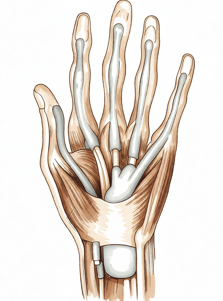
The extrinsic finger flexors comprise the flexor digitorum profundus (FDP) and the flexor digitorum superficialis (FDS) [39]. The FDP originates from the proximal ulna and interosseous membrane, dividing in the forearm into a radial component for the index finger and an ulnar component for the middle, ring, and little fingers [93]. It inserts on the proximal volar aspect of the distal phalanx and flexes the distal interphalangeal, proximal interphalangeal, and metacarpophalangeal joints [39]. Innervation of the FDP to the index and middle fingers is via the anterior interosseous branch of the median nerve, while the ring and little fingers receive ulnar nerve innervation [93].
The FDS possesses a radial head from the proximal radius and a humeral ulnar head from the medial humeral epicondyle and coronoid process [93]. Its tendon bifurcates around the FDP at the A2 pulley, with slips reuniting distally at the Camper chiasm; approximately half of the fibers remain ipsilateral while half cross contralaterally [93]. The FDS inserts via radial and ulnar slips into the proximal metaphysis of the middle phalanx and is entirely innervated by the median nerve [93]. Its primary function is digital flexion at the proximal interphalangeal joint [93].
The flexor pollicis longus (FPL) originates from a radial head on the proximal radius and interosseous membrane, plus an accessory head on the coronoid process and medial humeral epicondyle [93]. It inserts into the proximal base of the thumb distal phalanx, is innervated by the anterior interosseous branch of the median nerve, and flexes both the interphalangeal and metacarpophalangeal joints of the thumb [93].
Flexor Tendon Sheath and Pulleys
Distal to the metacarpal neck, flexor tendons enter the fibroosseous tunnel, or digital flexor sheath, which extends to the proximal aspect of the distal phalanx [93]. This sheath contains annular pulleys for mechanical stability and cruciate pulleys for flexibility [93]. The A1, A3, and A5 pulleys are located over the metacarpophalangeal, proximal interphalangeal, and distal interphalangeal joints, respectively, while the A2 and A4 pulleys sit over the middle portions of the proximal and middle phalanges [93]. The A2 and A4 pulleys are most essential for maintaining the mechanical advantage of the flexor tendons [93].
The tenosynovium lining the tunnel supplies nutrition and lubrication to the poorly vascularized tendons within the sheath [93]. Proximal to the sheath, tendons are well vascularized by the peritenon, whereas within the sheath, vascularity is supplied by the vincula system (vinculum longus and brevis) [93]. The FPL tenosynovial sheath is continuous with the radial bursa, and the little finger sheath is continuous with the ulnar digital bursa [93]. In some patients, these bursae communicate, allowing a horseshoe abscess to spread between the thumb and little finger if infection occurs in either flexor tendon sheath [93].
Zone 2 is the most complicated anatomical area of the flexor mechanism due to the presence of both FDS and FDP tendons within the digital flexor sheath [192]. In zone 2C, the FDS tendon is located ventral, medial, lateral, and dorsal to the FDP tendon, and neither tendon glides parallel to the long axis of the gliding tunnel [192]. During finger flexion in this zone, the FDP moves dorsal proximally while the FDS moves palmar proximally [192]. The midportion of zone 2C is the narrowest part of the gliding tunnel compared with its distal or proximal parts [192]. In the proximal portion of zone 2C, the FDP glides under the FDS tendon rather than the A2 pulley, with the FDS acting as a second sheath for the FDP to create a "double sheath" system [192]. Thumb pulley anatomy is more complex than initially described, with significant morphological variability of the A1 pulley [152].
Tendon Healing and Pathophysiology
Flexor tendon healing proceeds through both extrinsic and intrinsic mechanisms [93]. Extrinsic healing involves cells brought to the repair site by ingrowth of capillaries and fibroblasts, which leads to adhesion formation at the repair site [93]. Intrinsic healing occurs from tenocytes within the tendon [93]. The goal of repair and postoperative care is to encourage both healing pathways without forming thick adhesions, which limit tendon excursion and restrict finger motion [93].
Healing occurs via two simultaneous pathways: intrinsic healing involving tenocyte proliferation and extracellular matrix production, and extrinsic healing involving cell invasion from the surrounding sheath and synovium [203]. Extrinsic healing contributes to adhesion formation [203]. Tendons have minimal capacity for intrinsic healing due to poor cell density [203]. Up to 30% to 40% of flexor tendon injuries result in postsurgical adhesion formation between the tendon and surrounding tissues [203]. Adhesion formation results from disruption of the gliding mechanism due to puncture, laceration, or compression of the tendon [203].
Inserting a grasping suture into tendon tissue causes adverse changes at the suture-to-tendon interface, including a reduction in ultimate tensile strength beginning soon after insertion [205]. Suture insertion also causes cell proliferation and lysis of types I and III collagen by collagenase, as well as the presence of gelatinase at the interface [205]. No histologic or biochemical differences were detected between Kessler and Savage suture patterns regarding these adverse changes [205]. Epitenon fibroblasts proliferate and migrate into laceration sites to form collagen fibers bridging the defect; after the smooth tendon surface is restored, this cellular response diminishes [208].
Biomechanics and Kinematics
Flexor tendons function as cables transmitting forces to move and stabilize joints [127]. Forces up to 34 N have been measured in vivo during unresisted active finger motion [112]. Biomechanical analysis confirmed that the atypical 'hook grip' was likely the origin of FDS tendon rupture in a climber, as FDS tendon force for this grip is greater than in other climbing grip techniques [123]. During grip, the ring fingertip becomes 5 mm more prominent than any other digit in 90% of subjects, and the ring finger absorbs more force than any other finger during pull-away testing [153].
The thumb tendon has a greater tendency to rupture than finger tendons because it provides about 50% of hand function and cannot be spared from use [151]. Motion of the digits for several cycles greatly reduces the resistance to subsequent movement [132]. The index finger is characterized by lower amplitude and higher frequency, while the middle finger exhibits greater spectral complexity, consistent with known anatomical and kinematic differences [102]. In the supple hand, the ability of border digits to move laterally is critical to functional grasping activities involving all four fingers [135]. Functional prehension requires a stable wrist and at least two sensate digits that can oppose with some power [148].
Classification
Closed Avulsion Injuries: A new classification scheme for closed avulsion injuries of the flexor digitorum profundus tendon was proposed to guide surgeons toward the best treatment option [184]. The presence of a fracture in a profundus tendon avulsion with simultaneous intraarticular fracture of the distal phalanx excludes types I or II classifications [183].
Boyes’ Classification: The IFSSH Flexor Tendon Committee report proposes a vascularized transfer of a sliding unit composed of a flexor tendon and its surrounding sheaths in one single operation for secondary flexor tendon repair in clinical cases of grades III and IV severity according to Boyes’ classification [177]. The report emphasizes the tendon–sheath coupled unit and the major role of tendon vascularization and peripheral collagen organization in reconstruction [177].
Two-Stage Reconstruction Grading: In two-stage flexor tendon reconstruction, fingers classified as grade I (good soft tissues, supple joints, and no significant scarring) are treated with a one-stage flexor tendon autograft [169]. Fingers considered grades II through V are reconstructed using a two-stage allograft to replace the profundus tendon [169]. One-stage tendon autografts are not used in fingers graded II through V in the practice described by the authors of the refrigerated flexor tendon allograft study [169].
Clinical Presentation
A digit with a complete flexor tendon laceration typically displays a loss of the normal partially flexed digital resting posture, a loss of active finger flexion, and an absent tenodesis effect [58]. Spontaneous flexor tendon ruptures of the hand occur more often than generally recognized, with the majority involving the profundus tendon of the small finger in the palm [108]. Spontaneous rupture of the flexor digitorum superficialis tendon of the index and middle fingers is associated with "The pen sign" [8]. Simultaneous dislocation of both interphalangeal joints and flexor tendon tear can occur in a finger [1].
After blunt or penetrating trauma to the digits, accurate assessment of tendon integrity is essential to determine if surgical repair is required for lacerated tendons or conservative management for intact tendons [66]. Pediatric flexor tendon injuries often require surgical exploration due to uncooperative patients [43].
Imaging Modalities: Magnetic resonance imaging can be used for evaluation when the clinical diagnosis of rupture of a digital flexor tendon is uncertain 10 days after primary repair [45]. MRI is recommended for evaluation of gaps after flexor tendon repair, as 1.5T MRI has slightly better sensitivity and specificity for distinguishing clinically intact from clinically impaired repairs than 3T MRI [48]. Ultrasound can help evaluate the preoperative status of injured flexor tendons and, in cases of completely lacerated tendons, can help identify the location of the proximal tendon stump [58]. Sonography can differentiate digital tendon rupture from adhesive scarring after primary surgical repair [66]. A grading system based on clear differences in ultrasound findings (groups 1 to 5) has been proposed for assessing results of flexor tendon repairs [24].
Adhesive scarring between tendon, synovial sheath, and adjacent ligaments after primary flexor tendon repair is reported to occur in 20% of patients [66]. The outcome of a flexor tendon repair is influenced by many factors that cannot be controlled intraoperatively [15]. One year after flexor tendon repair, patients continued to report difficulties, particularly with symptoms (stiffness, weakness, pain) and social interaction, while activity limitations were less prominent [115].
Investigations
Clinical Examination and History: A careful physical examination is essential to direct care and future testing if indicated [38]. Patients often have difficulty accurately describing their symptoms and may incorrectly attribute pathology to a perceived deficit, whether real or imagined [38].
Ultrasound: Ultrasound can help to evaluate the preoperative status of injured flexor tendons and, in cases of completely lacerated tendons, can help identify the location of the proximal tendon stump [58]. In a study of 13 injured digits in 10 patients with 20 potentially injured flexor tendons, ultrasonography accurately identified the status of the flexor tendon in 11 of 13 digits and in 18 of 20 flexor tendons [58]. With complete tendon lacerations, the location of the proximal tendon stump was accurately identified by ultrasonography in 5 of 6 digits [58]. A new method of assessing results of flexor tendon repairs using ultrasound has been proposed, describing a grading system, groups 1 to 5, based on clear differences in findings on ultrasound [24]. Sonographic differentiation of digital tendon rupture from adhesive scarring after primary surgical repair is possible [66]. Adhesive scarring between tendon, synovial sheath, and adjacent ligaments after primary flexor and extensor tendon repair is reported to occur in 20% and 17% of patients, respectively [66]. In the diagnosis and follow-up of flexor pulley injuries, movement of the tendon in a plane perpendicular to that of the tendon movement during flexion distorts the colour Doppler image, giving inaccurate assessments [222].
Magnetic Resonance Imaging (MRI): Magnetic resonance imaging was used for evaluation when the clinical diagnosis of rupture of a digital flexor tendon was uncertain 10 days after primary repair, correlating precisely with the surgical pathology [45]. MRI is recommended for evaluation of gaps after flexor tendon repair, as the 1.5T has slightly better sensitivity and specificity for distinguishing clinically intact from clinically impaired repairs than the 3T [48]. Magnetic resonance imaging and carpal tunnel view x-ray films revealed the site and etiology of rupture in a case of flexor digitorum profundus rupture secondary to nonunion of the hook of hamate, clarifying treatment options [186].
Plain radiography: Patients with avulsion injuries of the flexor digitorum profundus should have exploration and repair as soon as possible because of the difficulty of accurately predicting the level of flexor tendon retraction preoperatively on the basis of x-ray films [219].
Treatment
Non-Operative
The provided evidence does not support conservative management as a primary treatment for acute flexor tendon injuries; surgical intervention is the standard of care for repair and reconstruction.
Operative
Indications: Flexor tendon injuries are complex, requiring consideration of surgical timing, injury location, approach, and soft tissue handling [30]. The concept that flexor tendon repair constitutes a surgical emergency has been refuted by studies demonstrating that delayed primary suture yields equal or better results [52]. Secondary reconstruction remains an important technique for complicated injuries or those that have failed primary repair [21]. Traumatic sequelae represent the main indications for two-stage grafts in old flexor tendon lesions [32]. Identification of factors associated with tenolysis allows surgeons to risk-stratify patients prior to surgery and guide postoperative expectations [14].
Surgical Approach / Technique: The surgeon approaches the flexor tendon using a combination of volar zigzag incisions and midaxial incisions to expose the flexor sheath [168]. Care is taken to preserve the neurovascular bundles on the radial and ulnar sides [168]. To prevent adhesions, the peritenon should be handled as little as possible [168]. Preservation of A2 and A4 pulleys is critical to prevent bowstringing [168]. A new and simple approach via strategically placed transverse incisions in the flexor tendon sheath has been described [176]. This technique has been effectively applied for secondary suturing of flexor tendons that ruptured following primary repair [53]. A technique for flexor tendon retrieval that eliminates the need for proximal incisions has been used successfully over 3 years on over 20 injuries [49, 101].
Implant Selection: The 6-strand double-loop suture technique simplifies flexor tendon repair [110]. A four-strand modification of the Tang method is recommended for clinical repairs [9]. Tensile strength increases with suture purchase, likely due to the mechanical characteristics of the suture polymer rather than improved grip [70, 215]. The knot technique offers an advantage when barbed sutures are not available [106]. Barbed sutures may have appealing attributes for reconstruction, but further investigation is required to evaluate and optimize these materials [189]. Novel repair techniques, including barbed suture, should be tested cyclically in a manner that closely mimics in vivo conditions during postoperative rehabilitation before clinical adoption [77]. A novel flexor tendon repair technique with improved biomechanical performance may be available for use [56]. Further developments in suture materials are important for advancements in repair strength [200]. Polyvinylidene fluoride sutures offer a possible alternative to polypropylene sutures [216]. Flexor tendon repair can be achieved using suture anchors placed in the distal phalanx, avoiding the morbidity associated with the pullout button technique [55]. A method of repair that gives sufficient strength to allow early active motion has been described [26]. Currently, the basis for the best results is a strong gap-resistant suture technique and early controlled motion in therapy [195]. The surgical techniques of hand surgeons and rehabilitation protocols of hand therapists have improved enormously, making results much more reliable [105]. Repairs achieved with all suture techniques were considerably weaker than those achieved when comparable techniques were used on flexor tendons [104].
Pain Management: The authors no longer perform flexor tendon repair with tourniquet, sedation, or muscle paralysis [175]. Tendon surgery can be performed under local anesthesia without a tourniquet by injecting epinephrine mixed with lidocaine to achieve vasoconstriction [229]. Postoperative pain is well controlled without a "pain rebound" at the end of the local anesthetic's duration of action [218]. Despite a decline in opioid quantity per patient, most patients still receive opioids after zone II flexor tendon repair [210].
Adjuncts: In the finger and palm, a volar zigzag incision is preferred for staged flexor tendon reconstruction [80]. This incision spares the deep vascular connections to the tendon bed and permits complete exposure of the tendon bed [80]. All undamaged segments of the fibro-osseous pulley system and the transverse digital arteries are spared during staged reconstruction [80]. A generous segment of the distal profundus tendon, at least 1 cm, is preserved distally, and the joint capsule is left intact [80]. It will probably be necessary to sacrifice the A5 pulley to perform this adequately [80]. If resection of scarred tendons and deep connective tissue fails to release the contracture, scarring of the joint capsule should be suspected and a capsulotomy may be necessary [80]. In severe cases, a compromise in ultimate function may become necessary, and a reasonable way to proceed is to create a "superficialis finger," i.e., the distal attachment of the implant is placed on the middle phalanx [80]. Unless the basic anatomy of the flexor pulley system can be restored at stage I surgery, the functional arc of motion for grip cannot be restored despite good hand therapy programs [80]. Either a flexor digitorum superficialis or profundus tendon that is to be discarded provides excellent material for pulley reconstruction [80].
Other Considerations: Although repair of both flexor digitorum profundus and flexor digitorum superficialis tendons is slightly more preferable based on increased grip strength, the repair of the flexor digitorum superficialis together with flexor digitorum profundus is not mandatory [159]. A flexor tendon graft through an intact functioning superficialis tendon in the ring finger seldom was indicated [209]. Nonrheumatoid ruptures of the flexor tendons in the palm are rare [170]. This problem was successfully managed by surgical tenolysis under local anesthesia [214]. The outcome after flexor tendon repair in children is better than in adults because rapid healing of tendons occurs in children [2]. Pediatric flexor tendon injuries differ from adults in diagnosis and rehabilitation, often requiring surgical exploration due to uncooperative patients [43].
Secondary Reconstruction and Grafting: This procedure cannot be advocated as a common method of secondary flexor tendon repair, and it cannot replace established techniques of reconstruction by one or two-stage grafting [42]. This is a procedure that has limited indication in a cooperative patient with a good tendon bed who reports late with both flexor tendons cut in zone 3 [36]. Single-stage tendon grafting for reconstruction of zone I and II flexor tendon injuries is a challenging procedure in hand surgery [164]. Careful patient selection, strict indications, and adherence to sound surgical principles are mandatory for return of digital motion after single-stage tendon grafting [164]. Tendon grafts will tolerate early motion therapy if the proximal and distal tenorrhaphy junctures are strong enough to withstand these forces of active finger motion [164]. The active tendon implant is a reliable, biologically acceptable temporary tendon implant, with the major advantage being the ability to use the digit for extended periods between stage I and stage II [79]. This technique should be reserved for motivated patients, and palliative solutions should be discussed if a functional superficial flexor tendon is present [82]. The incidence of rupture following staged flexor tendon reconstruction varies considerably from 0% to 14% in a series comprising a minimum of 25 cases [33]. In the series of 155 cases reported by Wehbe et al, the ruptures all occurred between 2 and 60 weeks [33]. The longest interval between tendon graft and rupture is reported to be 4.5 years [33]. When the sites of rupture were documented, they were either distal at the insertion of the graft into the distal phalanx or at its proximal attachment to the motor [33]. Poor surgical technique, compromised vascularity, patient noncompliance, and trauma have been cited as etiologies for rupture following staged flexor tendon reconstruction [33]. Tendon grafts always remain vulnerable to rupture [33]. The need for a tendon graft is noted during flexor tendon reconstruction [129]. Graft selection is determined by the donor’s presence, the anticipated length of graft material necessary to secure the graft, and the demands of the procedure [129]. The presence of the palmaris longus tendon may be accurately determined using the Schaeffer test of opposition of the thumb to the little finger during resisted flexion [129]. The most common donor tendon for palm-to-fingertip reconstruction is the palmaris longus tendon, and the most common donor tendon for forearm-to-fingertip reconstruction is the plantaris tendon [129]. Prior to use of either tendon, consider preoperative ultrasound imaging to determine its presence and dimensions [129]. Other graft options are long extensors of the three middle toes, toe flexors, extensor indicis proprius tendon, and extensor digiti minimi tendon [129]. Intrasynovial grafts are associated with fewer adhesions in animal models [129]. Keep harvested graft material in a moist saline sponge and handle it with the utmost care [129]. Identify and protect the median nerve at the time of palmaris longus tendon graft harvest [129]. The palmaris longus tendon is present in only 75% to 85% of people [129]. The plantaris tendon is present in about 80% of people [129]. If enough profundus stump is available, the graft can be sutured to the profundus stump or woven through the stump as in an end-weave juncture [129]. When there is not sufficient profundus tendon available for primary repair, the traditional method is a modification of the classic Bunnell tendon-to-bone pull-out technique [129]. Suture anchors are commonly used in patients with good bone quality and may be combined with a pull-out suture for the increased strength of a multistrand repair [129]. Indications and advantages of toe-to-finger free flexor tendon transfer are discussed [68].
Rehabilitation and Postoperative Management: Patients who had had flexor tendon repairs and who were referred for therapeutic intervention within the first week postoperatively had better outcomes than did patients who were referred for treatment after the first postoperative week [23]. These results confirm the safety of the controlled active motion regimen as an alternative to other regimens of postoperative flexor tendon repair mobilization in zone 1 and 2 finger injuries [60]. Using the described régime, full activity can be encouraged 8 weeks after flexor tendon repair in zone 2 of the hand without adverse effects on functional results or increased risk of rupture of the repair [62]. Continuous Passive Motion following flexor tendon repair shows early promise and should be studied further [67]. Based on a lack of superior benefits following true active motion regimens, there is not sufficient evidence to support true active motion as an effective or preferable choice for flexor tendon rehabilitation at this time [193]. The postoperative management of zone 2 flexor tendon repairs in Canada is variable, both within and between therapists [220].
Complications and Adhesion Prevention: Despite improvements in surgical technique and rehabilitation, functional outcomes for flexor tendon injuries remain unreliable, with adhesion formation and joint contractures being the most common complications [31]. Overall, a single intraoperative application of 5-FU at concentrations of 25 mg/mL appears to be an effective mechanism for reducing postoperative flexor tendon adhesions [227]. The article outlines preoperative, operative, and postoperative considerations for flexor tenolysis, emphasizing that patient selection, cooperation, and a rational goal are as key to success as the operative procedure itself [163]. Preservation of the digital pulley system during tenolysis is critical for a successful result [199]. The design of the currently available surgical instruments and blades is such that multiple transverse openings in the flexor sheath may be necessary to fully mobilize the tendon, which may lead to weakening or perforation of the sheath and destruction of important pulleys with resultant loss of tendon excursion and weakening of flexor power [199]. A modified 69 Beaver blade allows up to 30 mm between openings in the sheath during tenolysis [199].
Complications
Adhesions and Stiffness
Adhesion formation and joint contractures represent the most common complications following flexor tendon injuries [31]. The reported rate of tendon adhesions is 4% [172]. In a systematic review comparing 2-strand versus multistrand core suture techniques, standards for outcomes and rupture rates in digital flexor tendon repair were presented [18]. Experimental data indicate that adhesion formation can be mitigated; in a study of lubricant and autologous bone marrow stromal cell augmentation, the total number of tendons with adhesions was significantly higher in the untreated group than the treated group at both 21 and 42 days [99]. Gross observation in treated tendons revealed less adhesion formation and smoother tendon surfaces compared to untreated repaired tendons at these time points [99]. Furthermore, normalized work of flexion and repaired tendon gliding resistance studies showed improved digit function and gliding ability in treated tendons [99].
Rupture and Reoperation
The overall rate of reoperation after flexor tendon repair in all zones is 6% [172], with a reported repair rupture rate of 4% [172]. In New York state, the rate of reoperation for repair rupture is 2.3%, and the median time to reoperation was 140 days [172]. Literature documents repair rupture rates ranging from 4% to 10% in finger flexors and 3% to 17% in the flexor pollicis longus of the thumb [234]. A multicentre cohort study of 218 primary single-finger flexor tendon repairs found that most ruptures occurred in zone 2 (n = 17), with two in zone 1 and none in zone 3 [113]. All ruptures in this cohort happened after a six-strand (n = 13) or four-strand (n = 6) suture [113]. Eleven ruptures occurred in the little finger, including the two ruptures in zone 1 [113]. Poor results and second ruptures were particularly common after re-repair of ruptured tendon repairs in the little finger [247]. The rate of tenolysis performed after flexor tendon repair is 3.6% in New York state [172]. Although complication rates are low, data suggest that there has been no definitive improvement in reported complications before and after 2000 [204].
Other Considerations
Immobilization had a higher complication rate, including rupture or the need for secondary procedures, which increased total costs by 57% [244]. In a study comparing early motion protocols, the incidence of early rupture was the same in both groups at 4.9% and 4.3% respectively, and no patients required tenolysis [240]. In a study of immediate constrained digital motion in chickens, an initial loss of flexor tendon repair strength is not inevitable [75]. One of the main causes of flexor tendon repair failure in two common repair techniques is knot unravelling [78]. All tendons failed due to suture pullout in a study of symmetric versus asymmetric peripheral running sutures [213]. The safety factor achieved with three pullout suture techniques for flexor digitorum profundus tendon-to-bone repair is lower than that achieved with modern tendon-to-tendon repair methods [236]. With failure occurring late after repair, the capacity of the tendon to heal at the site of rupture was impaired [250].
In a study of 46 consecutive patients requiring flexor tendon grafts, 53 tendon grafts were evaluated [111]. Patients were assigned to groups based on whether the recipient bed permitted single-procedure grafting or required silicone rubber rod preparation [111]. All patients in both groups had sustained tendon lacerations in Zone II of the hand [111]. Twenty-one patients were assigned to Group I, characterized by a favorable recipient bed [111]. Thirteen patients in Group I had injuries limited to the flexor tendons and had formed a soft scar limited to the site of injury [111]. Eight patients in Group I had severed both flexor tendons and either one or both neurovascular bundles [111]. In Group I, the initial injury had healed with scarring limited to the site of skin, fibrous sheath, and tendon laceration [111]. The scar was soft and pliable [111]. The graft bed consisted of soft fatty tissues and unscarred fibrous sheath after resection of the scar [111].
Patients assigned to Group II had sustained multiple injuries to the finger, including fracture of the proximal or middle phalanges, power saw injuries of the tendon and bony phalanx, failed tendon repairs, and nerve injury requiring nerve grafts [111]. All patients in Group II demonstrated extensive scarring in the finger which was confluent with the periosteum of the phalanges [111]. The initial injury had healed with extensive scarring which was confluent with the periosteum of the proximal phalanx [111]. The fatty tissues overlying the fibrous sheath were scarred [111]. The scar was mature but not soft [111]. The graft bed consisted of scar tissue either on the volar or dorsal surface or both after resection of the scar [111].
Recovery
Full activity (months): Full activity can be encouraged 8 weeks after flexor tendon repair in zone 2 of the hand without adverse effects on functional results or increased risk of rupture of the repair [62].
Rehabilitation protocol: The ultimate goal of surgical intervention is to achieve enough strength to allow early motion, prevent adhesions within the tendon sheath, and restore the finger to normal range of motion and function [59]. Specifically, the goal of surgical treatment for intrasynovial flexor tendon lacerations is the achievement of a primary tendon repair of tensile strength sufficient to allow the application of a postoperative motion rehabilitation protocol to inhibit the formation of intrasynovial adhesions, facilitate restoration of the gliding surface, and stimulate the accrual of strength at the repair site [131]. The effects of an early active motion protocol may improve the grip strength and upper limb mobility of individuals after zone II flexor tendon repair [124]. High-force rehabilitation does not stimulate accelerated healing after intrasynovial flexor tendon repair [141].
Other Considerations: Despite early flexor tendon rehabilitation, some degree of dysfunction and loss of active ability for finger flexion persisted after 12 weeks [140]. Flexor tenolysis can be effective for improving motion following tendon repair, particularly in primary procedures and isolated tendon injuries [139]. Delayed primary flexor tendon repair under WALANT can achieve excellent functional outcomes, although longer follow-up is needed for extension deficit recovery [167]. Outcomes were worse if there was an accompanying volar plate injury in zone 2 flexor tendon repairs [230]. The use of patient-reported outcomes, in addition to clinician-reported outcomes, provided deeper insight into patients' perceptions of their recovery after flexor tendon injury [47].
Key Evidence
- [L5] Flexor tendon repair was followed by good function. [1] (10.1016/s0020-1383(97)00202-7)
- [L4] The outcome after flexor tendon repair in children is better than in adults in our hands because rapid healing of tendons occurs in children. [2] (10.1016/0266-7681(94)90237-2)
- [Paper] Primary flexor tendon repair is the procedure of choice for most flexor tendon lacerations. [3] (10.1016/s0363-5023(03)00381-2)
- [L3] Continued therapy utilization after primary flexor tendon repair is an independent predictor of reoperation need. [4] (10.1016/j.jht.2021.04.015)
- [L5] The improvement of primary flexor tendon repair has limited the indications of secondary repairs to neglected initial wound care and deficiencies in the surgical technique or rehabilitation. [7] (10.1016/j.main.2014.05.007)
- [L5] The authors recommend attempting to systematically repair both flexor tendons when they are damaged in daily surgical practice. [8] (10.1016/j.main.2009.05.002)
- [Paper] We recommend this new and simplified technique for clinical flexor tendon repairs. [9] (10.1016/j.jhsb.2005.04.003)
- [L3] A good outcome can be expected after repair of zone I or II flexor tendon injuries in children. [10] (10.1016/j.jhsa.2006.09.003)
- [Paper] An appropriate treatment protocol includes attention to maintenance of integrity of the flexor tendon repair, while avoiding secondary complications due to reduced gliding amplitude. [11] (10.1197/j.jht.2006.07.015)
- [L5] The authors conclude that better evidence is required in many domains of flexor tendon repair and rehabilitation, hoping that current RCTs will provide this data. [12] (10.1177/17531934261445904)
- [L3] Identification of factors associated with tenolysis following flexor tendon repair allows surgeons to risk-stratify patients prior to surgery and help guide postoperative expectations if complications arise. [14] (10.1177/15589447221142890)
- [L4] The outcome of a flexor tendon repair is influenced by many factors that cannot be controlled intraoperatively. [15] (10.1016/j.jhsa.2022.01.015)
- [L5] Flexor tendon repair in Zone II is a technically demanding procedure, but outcomes have become more predictable and satisfying. [16] (10.1016/j.hcl.2004.11.001)
- [L3] We conclude that complications after flexor tendon repair may be reduced by appropriate timing of the surgery. [17] (10.1142/s2424835519500218)
- [L1] We present the standards for outcomes as well as rupture rate for digital flexor tendon repair. [18] (10.1016/j.jhsa.2013.12.037)
- [L4] We recommend that no repair should be carried out to flexor tendon injuries involving 60% or less of their cross-sectional area. [20] (10.1016/0266-7681(92)90083-e)
- [L5] Secondary reconstruction remains an important and useful technique for complicated flexor tendon injuries or those that have failed primary repair. [21] (10.1016/j.jhsa.2007.08.018)
- [L4] Patients who had had flexor tendon repairs and who were referred for therapeutic intervention within the first week postoperatively had better outcomes than did patients who were referred for treatment after the first postoperative week. [23] (10.1016/s0894-1130(12)80152-9)
- [L4] We propose a new method of assessing results of flexor tendon repairs using ultrasound, describing a grading system, groups 1 to 5, based on clear differences in findings on ultrasound. [24] (10.1016/0266-7681(94)90055-8)
- [L5] Tendon grafting is the treatment of choice for flexor tendon injuries in zones I and II when direct repair is not possible or delayed. [25] (10.1016/j.hcl.2004.12.003)
- [L4] A method of flexor tendon repair is described which gives sufficient strength to allow early active motion. [26] (10.1016/s0363-5023(79)80043-x)
- [L5] No gold standard has been determined for the optimal flexor tendon repair algorithm, and repairs are usually chosen based on familiarity, popularity, and technical difficulty. [27] (10.1016/j.jhsa.2014.06.025)
- [L5] Flexor tendon injuries are complex, and management requires consideration of surgical timing, injury location, approach, and soft tissue handling. [30] (10.1016/j.jhsa.2024.05.013)
- [L5] Despite improvements in surgical technique and rehabilitation, functional outcomes for flexor tendon injuries remain unreliable, with adhesion formation and joint contractures being the most common complications. [31] (10.1016/j.hcl.2009.11.004)
- [L5] Traumatic sequelae represent the main indications for two-stage grafts in old flexor tendon lesions. [32] (10.1016/j.hansur.2016.10.147)
- [L4] [33] (10.1053/jhsu.2000.18489)
- [L5] The decrease in repair strength should therefore be considered in planning a tendon suture to tolerate active finger flexion and a tendon motion protocol after primary tendon repair. [35] (10.1053/jhsu.2001.28425)
- [L4] This is a procedure that has limited indication in a cooperative patient with a good tendon bed who reports late with both flexor tendons cut in zone 3. [36] (10.1016/s0363-5023(88)80054-6)
- [L4] This procedure cannot be advocated as a common method of secondary flexor tendon repair, and it cannot replace established techniques of reconstruction by one or two-stage grafting. [42] (10.1016/s0266-7681(96)80018-0)
- [L5] Pediatric flexor tendon injuries differ from adults in diagnosis and rehabilitation, often requiring surgical exploration due to uncooperative patients. [43] (10.1016/j.hcl.2004.11.004)
- [L5] Magnetic resonance imaging was used for evaluation when the clinical diagnosis of rupture of a digital flexor tendon was uncertain 10 days after primary repair, correlating precisely with the surgical pathology. [45] (10.1016/s0266-7681(05)80080-4)
- [L3] The use of patient-reported outcomes, in addition to clinician-reported outcomes, provided deeper insight into patients' perceptions of their recovery after flexor tendon injury. [47] (10.1016/j.jht.2024.12.011)
- [L5] We recommend MRI for evaluation of gaps after flexor tendon repair, as the 1.5T has slightly better sensitivity and specificity for distinguishing clinically intact from clinically impaired repairs than the 3T. [48] (10.1016/j.jhsa.2020.10.031)
- [L4] The technique has been used successfully over a period of 3 years on over 20 flexor tendon injuries. [49] (10.1016/j.jhse.2007.03.011)
- [L5] The concept that the flexor tendon repair should be considered as a surgical emergency has been effectively refuted by several studies showing that equal or better results can be achieved from delayed primary flexor tendon suture. [52] (10.1016/0266-7681(89)90079-x)
- [L5] This technique has been effectively applied for the secondary suturing of flexor tendons that have ruptured following primary repair. [53] (10.1016/s0266-7681(03)00007-x)
- [L3] Flexor tendon repair can be achieved using suture anchors placed in the distal phalanx, thereby avoiding the potential morbidity associated with the pullout button technique. [55] (10.1016/j.jhsa.2005.10.020)
- [L5] A novel flexor tendon repair technique with improved biomechanical performance may be available for use in flexor tendon repairs. [56] (10.1177/1558944716685829)
- [L4] [58] (10.1053/jhsu.2000.jhsu25a0236)
- [L5] The ultimate goal of surgical intervention for flexor tendon repair is to achieve enough strength to allow early motion, prevent adhesions within the tendon sheath, and restore the finger to normal range of motion and function. [59] (10.1016/j.ocl.2015.08.019)
- [L4] These results confirm the safety of this regimen as an alternative to other regimens of postoperative flexor tendon repair mobilization in zone 1 and 2 finger injuries. [60] (10.1016/0266-7681(94)90126-0)
- [L1] Using the described régime, full activity can be encouraged 8 weeks after flexor tendon repair in zone 2 of the hand without adverse effects on functional results or increased risk of rupture of the repair. [62] (10.1016/s0266-7681(96)80016-7)
- [L1] [66] (10.1016/j.jhsa.2006.01.003)
- [L3] Continuous Passive Motion following flexor tendon repair shows early promise and should be studied further. [67] (10.1016/0266-7681(89)90156-3)
- [L4] Indications and advantages of this free tendon transfer are also discussed. [68] (10.1016/s0363-5023(03)80402-1)
- [L5] Tensile strength of a flexor tendon repair increases with suture purchase, likely due to the mechanical characteristics of the suture polymer rather than improved grip. [70] (10.1177/1753193409104555)
- [Paper] An initial loss of flexor tendon repair strength is not inevitable. [75] (10.1016/s0363-5023(87)80213-7)
- [Letter] The authors advocate that novel flexor tendon repair techniques, including barbed suture, should be tested cyclically in a manner that closely mimics in vivo conditions during postoperative rehabilitation before clinical adoption. [77] (10.1016/j.jhsa.2015.02.034)
- [Paper] This study has clearly demonstrated one of the main causes of flexor tendon repair failure in two common repair techniques is knot unravelling. [78] (10.1142/s2424835516500302)
- [L4] The active tendon implant is a reliable, biologically acceptable temporary tendon implant, with the major advantage being the ability to use the digit for extended periods between stage I and stage II. [79] (10.1016/0363-5023(88)90259-6)
- [L4] [80] (10.1016/s0363-5023(83)80274-3)
- [L4] This technique should be reserved for motivated patients, and palliative solutions should be discussed if a functional superficial flexor tendon is present. [82] (10.1016/j.main.2012.10.005)
- [Paper] [99] (10.1002/jor.22980)
- [L4] We describe a technique for flexor tendon retrieval that eliminates the need for proximal incisions. [101] (10.1016/s0363-5023(05)80025-5)
- [L5] Repairs achieved with all suture techniques were considerably weaker than those achieved when comparable techniques were used on flexor tendons. [104] (10.1016/s0363-5023(09)91077-2)
- [L5] The surgical techniques of hand surgeons and the rehabilitation protocols of hand therapists have improved enormously from these scientific efforts and the results of flexor tendon repair have become much more reliable. [105] (10.1197/j.jht.2005.01.013)
- [Paper] The authors conclude that the knot technique offers an advantage for flexor tendon repair when barbed sutures are not available. [106] (10.1142/s2424835520500320)
- [L4] Spontaneous flexor tendon ruptures of the hand occur more often than one might recognize, with the majority involving the profundus tendon of the small finger in the palm. [108] (10.1016/j.jhsa.2007.06.012)
- [L5] The 6-strand double-loop suture technique simplifies flexor tendon repair. [110] (10.1053/jhsu.1999.1315)
- [L3] [111] (10.1016/s0363-5023(76)80035-4)
- [L3] Forces up to 34 N have been measured in vivo during unresisted active finger motion. [112] (10.1053/jhsu.2000.20151)
- [L3] [113] (10.1177/17531934231184737)
- [L1] One year after flexor tendon repair, patients continued to report difficulties, particularly with symptoms (stiffness, weakness, pain) and social interaction, while activity limitations were less prominent. [115] (10.1186/s12891-026-09626-y)
- [L5] The biomechanical analysis confirmed that the atypical ‘‘hook grip’’ was likely at the origin of the rupture, since flexor digitorum superficialis tendon force for this grip is greater than in other climbing grip techniques. [123] (10.1016/j.hansur.2021.04.007)
- [L1] The effects of an early active motion protocol may improve the grip strength and upper limb mobility of individuals after zone II flexor tendon repair. [124] (10.1016/j.jht.2023.05.012)
- [L5] An understanding of the biomechanics of the flexor tendon system is essential to proper evaluation and treatment of disorders of the upper extremity, as the tendons function as cables transmitting forces to move and stabilize joints. [127] (10.1016/j.hcl.2004.11.002)
- [Paper] The goal of surgical treatment of intrasynovial flexor tendon lacerations is the achievement of a primary tendon repair of tensile strength sufficient to allow the application of a postoperative motion rehabilitation protocol to inhibit the formation of intrasynovial adhesions, facilitate restoration of the gliding surface, and stimulate the accrual of strength at the repair site. [131] (10.1197/j.jht.2005.01.009)
- [Paper] Motion of the digits for several cycles greatly reduces the resistance to the subsequent movement. [132] (10.1016/j.jhsa.2006.08.001)
- [L4] In the supple hand this ability of the border digits to move laterally is critical to functional grasping activities that involve all four fingers. [135] (10.1016/s0894-1130(89)80005-5)
- [L4] Flexor tenolysis can be effective for improving motion following tendon repair, particularly in primary procedures and isolated tendon injuries. [139] (10.1016/j.jhsg.2025.100941)
- [L3] Despite early flexor tendon rehabilitation, some degree of dysfunction and loss of active ability for finger flexion persisted after 12 weeks. [140] (10.1258/ht.2012.012005)
- [Paper] These findings suggest that high-force rehabilitation does not stimulate accelerated healing after intrasynovial flexor tendon repair. [141] (10.1053/jhsu.2001.26190)
- [L5] The hand requires a stable wrist and at least two sensate digits that can oppose with some power for functional prehension. [148] (10.1016/s0749-0712(02)00130-0)
- [L5] The author posits that the thumb tendon has a greater tendency to rupture than finger tendons because it provides about 50% of hand function and cannot be spared from use, unlike fingers which can share load. [151] (10.1054/jhsb.2000.0421)
- [L5] The authors state that the anatomical arrangement of thumb pulleys is more complex than initially described, with significant morphological variability of the A1 pulley. [152] (10.1016/j.main.2014.06.005)
- [L4] A laboratory investigation shows that during grip the ring fingertip becomes 5 mm more prominent than any other digit in 90% of subjects and that it absorbs more force than any other finger during pull-away testing. [153] (10.1016/s0363-5023(88)80053-4)
- [L3] Although repair of both flexor digitorum profundus and flexor digitorum superficialis tendons is slightly more preferable based on increased grip strength, the repair of the flexor digitorum superficialis together with flexor digitorum profundus is not mandatory. [159] (10.1177/1753193420932446)
- [L4] This study demonstrates that the majority of respondents are performing zone II flexor tendon repairs in accordance with the best currently available evidence, although there is variability with respect to suture material, surgical technique, and rehabilitation protocols. [162] (10.1016/j.jhsa.2016.11.022)
- [L5] The article outlines preoperative, operative, and postoperative considerations for flexor tenolysis, emphasizing that patient selection, cooperation, and a rational goal are as key to success as the operative procedure itself. [163] (10.1016/j.hcl.2004.11.008)
- [L5] [164] (10.1016/j.jhsa.2015.04.016)
- [L4] Delayed primary flexor tendon repair under WALANT can achieve excellent functional outcomes, although longer follow-up is needed for extension deficit recovery. [167] (10.1177/17531934251315039)
- [Paper] [168] (10.1097/00130911-199712000-00009)
- [L4] [169] (10.1016/s0363-5023(83)80087-2)
- [L5] Nonrheumatoid ruptures of the flexor tendons in the palm are rare. [170] (10.1016/0363-5023(93)90247-z)
- [L5] [172] (10.5435/jaaos-22-12-791)
- [L5] The authors no longer perform flexor tendon repair with tourniquet, sedation, or muscle paralysis. [175] (10.1016/j.hcl.2013.02.009)
- [L5] [176] (10.1016/0266-7681(94)90218-6)
- [Paper] [177] (10.1016/j.jhsb.2004.09.001)
- [L5] The tendon did not have a bony fragment attached, which would have been indicative of type III; the presence of a fracture excluded types I or II classifications. [183] (10.1016/s0363-5023(81)80141-4)
- [L3] Our new classification scheme of FDP tendon avulsion appeared to be comprehensive and useful in guiding the surgeon for the best treatment option. [184] (10.1142/s0218810417500083)
- [L5] Magnetic resonance imaging and carpal tunnel view x-ray films revealed the site and etiology of rupture, clarifying treatment options. [186] (10.1016/s0363-5023(96)80014-1)
- [L5] Barbed sutures may have appealing attributes for flexor tendon reconstruction, and further investigation is required to evaluate and optimize these materials for use in this context. [189] (10.1016/j.jhsa.2011.05.040)
- [L5] [192] (10.1016/s0266-7681(05)80078-6)
- [L1] Based on a lack of superior benefits following true active motion regimens, there is not sufficient evidence to support true active motion as an effective or preferable choice for flexor tendon rehabilitation at this time. [193] (10.1016/j.jht.2018.06.001)
- [Paper] Currently, the basis for the best results from flexor tendon repair are strong gap resistant suture technique and early controlled motion in therapy. [195] (10.1197/j.jht.2005.02.015)
- [L5] [199] (10.1016/s0363-5023(83)80138-5)
- [L3] Further developments in suture materials are important for advancements in flexor tendon repair strength. [200] (10.1016/j.jhsa.2005.03.011)
- [L5] [203] (10.1016/j.jhsa.2017.06.013)
- [L1] Although complication rates are low, our data suggest that there has been no definitive improvement in reported complications before and after 2000. [204] (10.1016/j.jhsa.2011.11.006)
- [L5] [205] (10.1053/jhsu.2002.34006)
- [L5] [208] (10.1016/s0363-5023(85)80198-2)
- [L4] A flexor tendon graft through an intact functioning superficialis tendon in the ring finger seldom was indicated. [209] (10.1016/s0363-5023(77)80012-9)
- [L2] Despite a decline in opioid quantity per patient, most patients still receive opioids after zone II flexor tendon repair. [210] (10.1016/j.jhsg.2026.100971)
- [L5] [213] (10.1142/s0218810417500265)
- [L5] This problem was successfully managed by surgical tenolysis under local anesthesia. [214] (10.1016/s0363-5023(87)80015-1)
- [L5] Tensile strength of a flexor tendon repair, in this test system, increases with suture purchase. [215] (10.1177/1753193408104555)
- [L5] Polyvinylidene fluoride sutures offer a possible alternative to polypropylene sutures for flexor tendon repair. [216] (10.1054/jhsb.2000.0508)
- [L4] Postoperative pain is well controlled without a 'pain rebound' at the end of the local anesthetic's duration of action. [218] (10.1016/j.hansur.2018.10.120)
- [L4] We recommend that patients with avulsion injuries of the flexor digitorum profundus have exploration and repair as soon as possible because of the difficulty of accurately predicting the level of flexor tendon retraction preoperatively on the basis of x-ray films. [219] (10.1016/0363-5023(92)90465-2)
- [L4] The postoperative management of zone 2 flexor tendon repairs in Canada is variable, both within and between therapists. [220] (10.1016/s0894-1130(04)00201-7)
- [L5] However, in the diagnosis and follow-up of flexor pulley injuries, movement of the tendon in a plane perpendicular to that of the tendon movement during flexion distorts the CDI image, giving inaccurate assessments. [222] (10.1016/s0266-7681(98)80021-1)
- [L3] Overall, a single intraoperative application of 5-FU at concentrations of 25 mg/mL appears to be an effective mechanism for reducing postoperative flexor tendon adhesions. [227] (10.1053/jhsu.2000.jhsu25a0242)
- [L5] Tendon surgery can be performed under local anesthesia without a tourniquet by injecting epinephrine mixed with lidocaine to achieve vasoconstriction. [229] (10.4055/cios.2015.7.3.275)
- [L4] The study aims to evaluate the contribution of palmar plate injury to postoperative recovery in zone 2 flexor tendon repairs, noting that outcomes were worse if there was an accompanying volar plate injury. [230] (10.1177/17531934241300510)
- [L4] Repair ruptures were documented in most reports with rates ranging from 4%-10% in finger flexors and 3%-17% in FPL of thumbs. [234] (10.1016/j.hcl.2004.11.005)
- [L3] The data indicated that the safety factor achieved with these repair methods is lower than that achieved with modern tendon-to-tendon repair methods. [236] (10.1016/s0363-5023(98)80099-3)
- [L3] The incidence of early rupture was the same in both groups at 4.9% and 4.3% respectively, and no patients required tenolysis. [240] (10.1016/s0266-7681(96)80145-8)
- [L4] Immobilization had a higher complication rate (rupture or need for secondary procedures), which in itself increased total costs by 57%. [244] (10.1016/s0266-7681(02)00352-2)
- [L4] Poor results and second ruptures were particularly common after re-repair of ruptured tendon repairs in the little finger. [247] (10.1016/j.jhsb.2006.06.006)
- [L5] With failure occurring late after repair, it would appear that the capacity of the tendon to heal at the site of rupture was impaired. [250] (10.1016/s0363-5023(05)80024-3)
See Also
References
[1] Simultaneous dislocation of both interphalangeal joints and flexor tendon tear in a finger. Injury. 1998. DOI: 10.1016/s0020-1383(97)00202-7
[2] Flexor Tendon Injuries in Children. Journal of Hand Surgery. 1994. DOI: 10.1016/0266-7681(94)90237-2
[3] No man’s land revisited: the primary flexor tendon repair controversy. The Journal of Hand Surgery. 2004. DOI: 10.1016/s0363-5023(03)00381-2
[4] Patient outcomes and costs after isolated flexor tendon repairs of the hand. Journal of Hand Therapy. 2022. DOI: 10.1016/j.jht.2021.04.015
[7] Reconstruction par greffe des tendons fléchisseurs. Chirurgie de la Main. 2014. DOI: 10.1016/j.main.2014.05.007
[8] Spontaneous rupture of the flexor digitorum superficialis tendon of the index and middle fingers: “The pen sign”. Chirurgie de la Main. 2009. DOI: 10.1016/j.main.2009.05.002
[9] Biomechanical Evaluation of a Four-Strand Modification of the Tang Method of Tendon Repair. Journal of Hand Surgery. 2005. DOI: 10.1016/j.jhsb.2005.04.003
[10] Factors That Influence the Outcome of Zone I and Zone II Flexor Tendon Repairs in Children. The Journal of Hand Surgery. 2006. DOI: 10.1016/j.jhsa.2006.09.003
[11] The Pathomechanics and Management of Secondary Complications following Flexor Tendon Repair. Journal of Hand Therapy. 2006. DOI: 10.1197/j.jht.2006.07.015
[12] Re: Tang et al. The IFSSH consensus and current guidelines on flexor tendon repairs and reconstruction. Journal of Hand Surgery (European Volume). 2026. DOI: 10.1177/17531934261445904
[14] Revision of Flexor Tendon Repair: Factors Associated With Flexor Tenolysis. HAND. 2022. DOI: 10.1177/15589447221142890
[15] Outcomes of Wide-Awake Flexor Tendon Repairs in 58 Fingers and 9 Thumbs. The Journal of Hand Surgery. 2023. DOI: 10.1016/j.jhsa.2022.01.015
[16] Acute Flexor Tendon Repairs in Zone II. Hand Clinics. 2005. DOI: 10.1016/j.hcl.2004.11.001
[17] The Effect of Delay to Surgery on Major Complications after Primary Flexor Tendon Repair. The Journal of Hand Surgery (Asian-Pacific Volume). 2019. DOI: 10.1142/s2424835519500218
[18] A Systematic Review of 2-Strand Versus Multistrand Core Suture Techniques and Functional Outcome After Digital Flexor Tendon Repair. The Journal of Hand Surgery. 2014. DOI: 10.1016/j.jhsa.2013.12.037
[20] Partial Flexor Tendon Injuries: To Repair or Not. Journal of Hand Surgery. 1992. DOI: 10.1016/0266-7681(92)90083-e
[21] Secondary Flexor Tendon Reconstruction, A Review. The Journal of Hand Surgery. 2007. DOI: 10.1016/j.jhsa.2007.08.018
[23] Effects of Delayed Therapeutic Intervention following Zone II Flexor Tendon Repair. Journal of Hand Therapy. 1995. DOI: 10.1016/s0894-1130(12)80152-9
[24] The Role of Ultrasound in the Management of Zone 1 Flexor Tendon Injuries. Journal of Hand Surgery. 1994. DOI: 10.1016/0266-7681(94)90055-8
[25] Delayed Treatment of Flexor Tendon Injuries Including Grafting. Hand Clinics. 2005. DOI: 10.1016/j.hcl.2004.12.003
[26] Early active motion following a beveled technique of flexor tendon repair: Report on fifty cases. The Journal of Hand Surgery. 1979. DOI: 10.1016/s0363-5023(79)80043-x
[27] Flexor Tendon Repairs: Techniques, Eponyms, and Evidence. The Journal of Hand Surgery. 2014. DOI: 10.1016/j.jhsa.2014.06.025
[30] Flexor Tendon Injuries. The Journal of Hand Surgery. 2024. DOI: 10.1016/j.jhsa.2024.05.013
[31] Complications After Flexor Tendon Injuries. Hand Clinics. 2010. DOI: 10.1016/j.hcl.2009.11.004
[32] Plaie palmaire négligée en zone 2 de l’annulaire datant de 14 ans - la chirurgie de reconstruction en deux temps selon la technique de hunter. Hand Surgery and Rehabilitation. 2016. DOI: 10.1016/j.hansur.2016.10.147
[33] Intratendinous rupture of a flexor tendon graft many years after staged reconstruction: A report of three cases. The Journal of Hand Surgery. 2000. DOI: 10.1053/jhsu.2000.18489
[35] Effects of tension direction on strength of tendon repair. The Journal of Hand Surgery. 2001. DOI: 10.1053/jhsu.2001.28425
[36] Delayed repair of flexor profundus tendon in the palm (zone 3) with superficialis transfer. The Journal of Hand Surgery. 1988. DOI: 10.1016/s0363-5023(88)80054-6
[38] Orthopaedic Knowledge Update 13 Ebook Without Multimedia. Anatomy, Evaluation, Clinical Examination, and Imaging > Evaluation and Clinical Examination: Current Concepts.
[39] A Lange Medical Book Current Diagnosis Treatment In Orthopedics Fifth Edition. 9Hand Surgery > Image DISORDERS OF THE MUSCULATURE OF THE HAND.
[42] Human Composite Flexor Tendon Allografts. Journal of Hand Surgery. 1996. DOI: 10.1016/s0266-7681(96)80018-0
[43] Pediatric Flexor Tendon Injuries. Hand Clinics. 2005. DOI: 10.1016/j.hcl.2004.11.004
[45] Magnetic Resonance Imaging for Diagnosis of Digital Flexor Tendon Rupture After Primary Repair. Journal of Hand Surgery. 1995. DOI: 10.1016/s0266-7681(05)80080-4
[47] Finger flexor tendon injuries repaired surgically followed by an early active motion program: A prospective cohort study of clinician- and patient-reported outcomes. Journal of Hand Therapy. 2026. DOI: 10.1016/j.jht.2024.12.011
[48] Comparative Accuracy of 1.5T MRI, 3T MRI, and Static Ultrasound in Diagnosis of Small Gaps in Repaired Flexor Tendons: A Cadaveric Study. The Journal of Hand Surgery. 2021. DOI: 10.1016/j.jhsa.2020.10.031
[49] Flexor Tendon Retrieval—Another Way. Journal of Hand Surgery (European Volume). 2007. DOI: 10.1016/j.jhse.2007.03.011
[52] Flexor tendon surgery Part 1: Primary flexor tendon repair. The Journal of Hand Surgery: Journal of the British Society for Surgery of the Hand. 1989. DOI: 10.1016/0266-7681(89)90079-x
[53] No more Frayed Ends – A Technique for Trimming the Uneven Ends of Ruptured Flexor Tendons Prior to Repair. Journal of Hand Surgery. 2003. DOI: 10.1016/s0266-7681(03)00007-x
[55] Comparison of Pullout Button Versus Suture Anchor for Zone I Flexor Tendon Repair. The Journal of Hand Surgery. 2006. DOI: 10.1016/j.jhsa.2005.10.020
[56] A Biomechanical Study of a Novel Asymmetric 6-Strand Flexor Tendon Repair Using Porcine Tendons. HAND. 2017. DOI: 10.1177/1558944716685829
[58] Ultrasound evaluation of flexor tendon lacerations. The Journal of Hand Surgery. 2000. DOI: 10.1053/jhsu.2000.jhsu25a0236
[59] Flexor Tendon Repair. Orthopedic Clinics of North America. 2016. DOI: 10.1016/j.ocl.2015.08.019
[60] The Rupture Rate of Acute Flexor Tendon Repairs Mobilized by the Controlled Active Motion Regimen. Journal of Hand Surgery. 1994. DOI: 10.1016/0266-7681(94)90126-0
[62] The Effects of a Shortened Postoperative Mobilization Programme after Flexor Tendon Repair in Zone 2. Journal of Hand Surgery. 1996. DOI: 10.1016/s0266-7681(96)80016-7
[66] Sonographic Differentiation of Digital Tendon Rupture From Adhesive Scarring After Primary Surgical Repair. The Journal of Hand Surgery. 2006. DOI: 10.1016/j.jhsa.2006.01.003
[67] Continuous passive motion following flexor tendon repair. The Journal of Hand Surgery: Journal of the British Society for Surgery of the Hand. 1989. DOI: 10.1016/0266-7681(89)90156-3
[68] Toe-to-finger free flexor tendon transfer for digital flexion reconstruction. The Journal of Hand Surgery. 2003. DOI: 10.1016/s0363-5023(03)80402-1
[70] What is the Significance of Tendon Suture Purchase?. Journal of Hand Surgery (European Volume). 2009. DOI: 10.1177/1753193409104555
[75] The effect of immediate constrained digital motion on the strength of flexor tendon repairs in chickens. The Journal of Hand Surgery. 1987. DOI: 10.1016/s0363-5023(87)80213-7
[77] Letter Regarding “Biomechanical Analysis of Flexor Tendon Repair Using Knotted Kessler and Bunnell Techniques and the Knotless Bunnell Technique”. The Journal of Hand Surgery. 2015. DOI: 10.1016/j.jhsa.2015.02.034
[78] Causes of Flexor Tendon Repair Failures in Two Common Repair Techniques: A Cadaver Study. The Journal of Hand Surgery (Asian-Pacific Volume). 2016. DOI: 10.1142/s2424835516500302
[79] Active tendon implants in flexor tendon reconstruction. The Journal of Hand Surgery. 1988. DOI: 10.1016/0363-5023(88)90259-6
[80] Staged flexor tendon reconstruction. The Journal of Hand Surgery. 1983. DOI: 10.1016/s0363-5023(83)80274-3
[82] Résultats des reconstructions tendineuses en deux temps de Hunter des lésions du tendon fléchisseur profond en zone 2 : à propos de 59 cas. Chirurgie de la Main. 2012. DOI: 10.1016/j.main.2012.10.005
[93] A Lange Medical Book Current Diagnosis Treatment In Orthopedics Fifth Edition. 9Hand Surgery > FLEXOR TENDON INJURY.
[99] Effects of lubricant and autologous bone marrow stromal cell augmentation on immobilized flexor tendon repairs. Journal of Orthopaedic Research. 2015. DOI: 10.1002/jor.22980
[101] Endoscopic retrieval of severed flexor tendons. The Journal of Hand Surgery. 1995. DOI: 10.1016/s0363-5023(05)80025-5
[102] 10.1177-15589447261483211. n.d..
[104] Biomechanical characteristics of extensor tendon suture techniques. The Journal of Hand Surgery. 1992. DOI: 10.1016/s0363-5023(09)91077-2
[105] The Scientific Basis for Advances in Flexor Tendon Surgery. Journal of Hand Therapy. 2005. DOI: 10.1197/j.jht.2005.01.013
[106] Biomechanics Study of the Holding Power between Tendon and Suture Using Different Techniques. The Journal of Hand Surgery (Asian-Pacific Volume). 2020. DOI: 10.1142/s2424835520500320
[108] Spontaneous Flexor Tendon Ruptures of the Hand: Case Series and Review of the Literature. The Journal of Hand Surgery. 2007. DOI: 10.1016/j.jhsa.2007.06.012
[110] A comparative analysis of the six-strand double-loop flexor tendon repair and three other techniques: A human cadaveric study. The Journal of Hand Surgery. 1999. DOI: 10.1053/jhsu.1999.1315
[111] Rate and extent of functional recovery after flexor tendon grafting with and without silicone rod preparation. The Journal of Hand Surgery. 1976. DOI: 10.1016/s0363-5023(76)80035-4
[112] A comparison of four repair techniques for Champer's chiasma flexor digitorium superficialis lacerations: Tested in an in vitro model. The Journal of Hand Surgery. 2000. DOI: 10.1053/jhsu.2000.20151
[113] Outcomes of 218 primary single-finger flexor tendon repairs up to 1 year after surgery: a multicentre cohort study. Journal of Hand Surgery (European Volume). 2023. DOI: 10.1177/17531934231184737
[115] Item-level reanalysis of DASH outcomes after flexor tendon repair using Svensson’s non-parametric method. BMC Musculoskeletal Disorders. 2026. DOI: 10.1186/s12891-026-09626-y
[123] Unusual rupture of the middle finger flexor digitorum superficialis tendon in a climber: Biomechanical analysis. Hand Surgery and Rehabilitation. 2021. DOI: 10.1016/j.hansur.2021.04.007
[124] Safety and efficacy of ultrasonography of tension after zone II flexor tendon repair: A randomized controlled trial. Journal of Hand Therapy. 2023. DOI: 10.1016/j.jht.2023.05.012
[127] Biomechanics of the Flexor Tendons. Hand Clinics. 2005. DOI: 10.1016/j.hcl.2004.11.002
[129] Green S Operative Hand Surgery. Obtaining Tendon Grafts.
[131] Recent Progress in Flexor Tendon Healing. Journal of Hand Therapy. 2005. DOI: 10.1197/j.jht.2005.01.009
[132] Resistance to Motion of Flexor Tendons and Digital Edema: An In Vivo Study in a Chicken Model. The Journal of Hand Surgery. 2006. DOI: 10.1016/j.jhsa.2006.08.001
[135] Convergence points of normal fingers in individual flexion and simultaneous flexion. Journal of Hand Therapy. 1989. DOI: 10.1016/s0894-1130(89)80005-5
[139] Clinical Outcomes of Flexor Tenolysis Following Zone 2-3 Flexor Tendon Repair: A Retrospective Review. Journal of Hand Surgery Global Online. 2026. DOI: 10.1016/j.jhsg.2025.100941
[140] Is there a correlation between patient-reported outcome assessed by the Disabilities of the Arm, Shoulder and Hand Questionnaire and total active motion after flexor tendon repair?. Hand Therapy. 2012. DOI: 10.1258/ht.2012.012005
[141] The effect of variations in applied rehabilitation force on collagen concentration and maturation at the intrasynovial flexor tendon repair site. The Journal of Hand Surgery. 2001. DOI: 10.1053/jhsu.2001.26190
[148] Biomechanics and hand trauma: what you need. Hand Clinics. 2003. DOI: 10.1016/s0749-0712(02)00130-0
[151] Letters to the Editor. Journal of Hand Surgery. 2000. DOI: 10.1054/jhsb.2000.0421
[152] Lettre à la rédaction à propos de « Reconstruction of closed rupture of thumb flexor tendon pulleys with a single free palmaris longus tendon graft: A case report and review of literature » de F. Fazilleau, D. Cheval, J. Richou et D. Le Nen. Chir Main 2014;33:51–4. Chirurgie de la Main. 2014. DOI: 10.1016/j.main.2014.06.005
[153] Avulsion of the flexor digitorum profundus: Anatomic and biomechanical considerations. The Journal of Hand Surgery. 1988. DOI: 10.1016/s0363-5023(88)80053-4
[159] Flexor digitorum profundus with or without flexor digitorum superficialis tendon repair in acute Zone 2B injuries. Journal of Hand Surgery (European Volume). 2020. DOI: 10.1177/1753193420932446
[162] Zone II Flexor Tendon Repairs in the United States: Trends in Current Management. The Journal of Hand Surgery. 2017. DOI: 10.1016/j.jhsa.2016.11.022
[163] Flexor Tenolysis. Hand Clinics. 2005. DOI: 10.1016/j.hcl.2004.11.008
[164] Single-Stage Flexor Tendon Grafting: Refining the Steps. The Journal of Hand Surgery. 2015. DOI: 10.1016/j.jhsa.2015.04.016
[167] Delayed primary flexor tendon repair in zone II injuries: results of using WALANT and controlled true active motion. Journal of Hand Surgery (European Volume). 2025. DOI: 10.1177/17531934251315039
[168] Flexor Tendon Repairs. Techniques in Hand & Upper Extremity Surgery. 1997. DOI: 10.1097/00130911-199712000-00009
[169] Clinical use of refrigerated flexor tendon allografts to replace a silicone rubber rod. The Journal of Hand Surgery. 1983. DOI: 10.1016/s0363-5023(83)80087-2
[170] Rupture of the flexor digitorum profundus tendon in the palm caused by repeated, chronic direct trauma. The Journal of Hand Surgery. 1993. DOI: 10.1016/0363-5023(93)90247-z
[172] Update on Zone II Flexor Tendon Injuries. Journal of the American Academy of Orthopaedic Surgeons. 2014. DOI: 10.5435/jaaos-22-12-791
[175] Wide-awake Flexor Tendon Repair and Early Tendon Mobilization in Zones 1 and 2. Hand Clinics. 2013. DOI: 10.1016/j.hcl.2013.02.009
[176] A New Approach to Flexor Tendon Repair. Journal of Hand Surgery. 1994. DOI: 10.1016/0266-7681(94)90218-6
[177] Ifssh Flexor Tendon Committee Report. Journal of Hand Surgery. 2005. DOI: 10.1016/j.jhsb.2004.09.001
[183] Avulsion of a profundus tendon with simultaneous intraarticular fracture of the distal phalanx—Case report. The Journal of Hand Surgery. 1981. DOI: 10.1016/s0363-5023(81)80141-4
[184] A New Classification Scheme for Closed Avulsion Injuries of the Flexor Digitorum Profundus Tendon. The Journal of Hand Surgery (Asian-Pacific Volume). 2017. DOI: 10.1142/s0218810417500083
[186] Flexor digitorum profundus rupture of the small finger secondary to nonunion of the hook of hamate: A case report. The Journal of Hand Surgery. 1996. DOI: 10.1016/s0363-5023(96)80014-1
[189] Biomechanical Evaluation of Flexor Tendon Repair Using Barbed Suture Material: A Comparative ex vivo Study. The Journal of Hand Surgery. 2011. DOI: 10.1016/j.jhsa.2011.05.040
[192] The Double Sheath System and Tendon Gliding in Zone 2C. Journal of Hand Surgery. 1995. DOI: 10.1016/s0266-7681(05)80078-6
[193] Flexor tendon rehabilitation in the 21st century: A systematic review. Journal of Hand Therapy. 2019. DOI: 10.1016/j.jht.2018.06.001
[195] JHT Read for Credit. Journal of Hand Therapy. 2005. DOI: 10.1197/j.jht.2005.02.015
[199] Modified 69 blade for tenolysis. The Journal of Hand Surgery. 1983. DOI: 10.1016/s0363-5023(83)80138-5
[200] A Biomechanical Analysis of Suture Materials and Their Influence on a Four-Strand Flexor Tendon Repair. The Journal of Hand Surgery. 2005. DOI: 10.1016/j.jhsa.2005.03.011
[203] Molecular Biology of Flexor Tendon Healing in Relation to Reduction of Tendon Adhesions. The Journal of Hand Surgery. 2017. DOI: 10.1016/j.jhsa.2017.06.013
[204] Complications After Flexor Tendon Repair: A Systematic Review and Meta-Analysis. The Journal of Hand Surgery. 2012. DOI: 10.1016/j.jhsa.2011.11.006
[205] Characterization of the tensile properties and histologic/biochemical changes in normal chicken tendon at the site of suture insertion. The Journal of Hand Surgery. 2002. DOI: 10.1053/jhsu.2002.34006
[208] Intrinsic restoration of the flexor tendon surface in the nonhuman primate. The Journal of Hand Surgery. 1985. DOI: 10.1016/s0363-5023(85)80198-2
[209] Avulsion of the profundus tendon insertion in athletes. The Journal of Hand Surgery. 1977. DOI: 10.1016/s0363-5023(77)80012-9
[210] Opioid Prescription Patterns Following Zone II Flexor Tendon Repairs: A National Database Study. Journal of Hand Surgery Global Online. 2026. DOI: 10.1016/j.jhsg.2026.100971
[213] Symmetric Peripheral Running Sutures are Superior to Asymmetric Peripheral Running Sutures for Increasing the Tendon Strength in Flexor Tendon Repair. The Journal of Hand Surgery (Asian-Pacific Volume). 2017. DOI: 10.1142/s0218810417500265
[214] Flexor digitorum profundus entrapment after closed treatment of a displaced Colles' fracture. The Journal of Hand Surgery. 1987. DOI: 10.1016/s0363-5023(87)80015-1
[215] What is the Significance of Tendon Suture Purchase?. Journal of Hand Surgery (European Volume). 2009. DOI: 10.1177/1753193408104555
[216] Comparison of Mechanical Properties of Polyvinylidene Fluoride and Polypropylene Monofilament Sutures Used for Flexor Tendon Repair. Journal of Hand Surgery. 2001. DOI: 10.1054/jhsb.2000.0508
[218] Walant échoguidee pour chirurgie de la main — description de la technique et premiers résultats. Hand Surgery and Rehabilitation. 2018. DOI: 10.1016/j.hansur.2018.10.120
[219] Misleading fractures after profundus tendon avulsions: A report of six cases. The Journal of Hand Surgery. 1992. DOI: 10.1016/0363-5023(92)90465-2
[220] Current trends in the therapy management of zone 2 flexor tendon repairs: A survey of Canadian CHTs. Journal of Hand Therapy. 2004. DOI: 10.1016/s0894-1130(04)00201-7
[222] Colour Doppler Ultrasound in the Diagnosis, Management and Follow-Up of a Digital Flexor Pulley Injury. Journal of Hand Surgery. 1998. DOI: 10.1016/s0266-7681(98)80021-1
[227] Effects of 5-fluorouracil on flexor tendon repair. The Journal of Hand Surgery. 2000. DOI: 10.1053/jhsu.2000.jhsu25a0242
[229] Wide-Awake Primary Flexor Tendon Repair, Tenolysis, and Tendon Transfer. Clinics in Orthopedic Surgery. 2015. DOI: 10.4055/cios.2015.7.3.275
[230] Influence of palmar plate injury on outcomes of zone 2 flexor tendon repair. Journal of Hand Surgery (European Volume). 2024. DOI: 10.1177/17531934241300510
[234] Clinical Outcomes Associated with Flexor Tendon Repair. Hand Clinics. 2005. DOI: 10.1016/j.hcl.2004.11.005
[236] Flexor digitorum profundus tendon-to-bone repair: An ex vivo biomechanical analysis of 3 pullout suture techniques. The Journal of Hand Surgery. 1998. DOI: 10.1016/s0363-5023(98)80099-3
[240] Flexor Tendon Repair in Zone 2 Followed by Early Active Mobilization. Journal of Hand Surgery. 1996. DOI: 10.1016/s0266-7681(96)80145-8
[244] What Determines the Costs of Repair and Rehabilitation of Flexor Tendon Injuries in Zone II? A Multiple Regression Analysis of Data from Southern Sweden. Journal of Hand Surgery. 2003. DOI: 10.1016/s0266-7681(02)00352-2
[247] The Results of Immediate Re-Repair of Zone 1 and 2 Primary Flexor Tendon Repairs which Rupture. Journal of Hand Surgery. 2006. DOI: 10.1016/j.jhsb.2006.06.006
[250] Flexor pollicis longus rupture in a trigger thumb: A case report. The Journal of Hand Surgery. 1995. DOI: 10.1016/s0363-5023(05)80024-3