Clinicians › Wrist
Internal Fixation and Implants
Internal fixation of the wrist, focusing on volar locking plates for distal radius fractures and the management of associated complications.
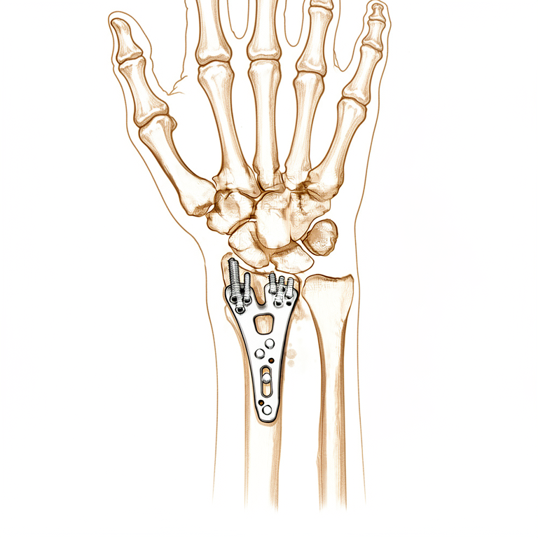
Overview
Internal fixation serves as an effective treatment modality for a broad spectrum of orthopaedic trauma, with successful healing and the avoidance of complications largely determined by surgical technique [3]. The locked internal fixator technique aims at simple and safe handling, optimizing biological conditions for soft and hard tissues, and being universally applicable [4]. The Locking Compression Plate (LCP) represents a new implant revolutionizing internal fixation that requires adapted surgical techniques and new thinking about commonly used concepts of interventional fixation to avoid failures and complications [5]. In pediatric trauma, indications for implant removal include wire or pin fixation when substantial growth remains and infection [1]. For distal radius fractures, external fixation and volar locking plate (VLP) are both good options, and treatment preference should often follow the patient's choice [2].
Clinical outcomes vary by specific application. Internal fixation of displaced distal radial fractures with implants featuring locking screw fixation can result in good-to-excellent outcomes with a limited number of complications [18]. Stable internal fixation with standard techniques allowed an early return to functional activities in step-cut ulnar shortening osteotomy for ulnar impaction syndrome [10]. A novel technique for posterior internal fixation of acetabular fractures offers superior outcomes and fewer complications compared to similar internal fixation techniques [12]. For humeral shaft nonunions, bone grafting is a proven necessary adjunct to internal fixation [26], and transplantation of autogenous bone is the elective procedure in cases requiring bone grafting, usually carried out in combination with stable internal fixation [6].
While locking plate fixation has yet to prove clinical superiority in any anatomic site for which good-quality comparative analyses are available [60], it presented an incremental cost effectiveness ratio of £89,322 per QALY within the first 12 months of treatment for adult patients with a dorsally displaced fracture of the distal radius [58]. Submuscular internal fixation might be an interesting alternative to external fixation in clinical practice for osteoporotic pelvic fractures because of better biomechanical properties as well as several advantages in clinical use [173]. External fixation facilitates safe and accurate reduction without major surgical complications and may offer surgeons an additional option for the treatment of unstable pelvic fractures [29]. The intended advantages of increased fixation accuracy and versatility for the extended use of the K-wire and the orthopaedic screw have been realised [32].
Anatomy & Pathophysiology
Bony Anatomy
The wrist constitutes the anatomic region between the forearm and the hand, encompassing the distal radioulnar, radiocarpal, and ulnocarpal joints along with the eight carpal bones [73]. These carpal bones are organized into a proximal row comprising the scaphoid, lunate, triquetrum, and pisiform, and a distal row consisting of the trapezium, trapezoid, capitate, and hamate [73]. The radiocarpal joints form through the articulation of the distal radius with the scaphoid and lunate via their respective concave facets, while the triquetrum articulates on the triangular fibrocartilage [73]. The distal radius features three articular components: the scaphoid and lunate fossae and the sigmoid notch [77]. Its concave elliptical surface is oriented in the sagittal plane with an average volar tilt of 11 degrees, while the frontal plane exhibits an average radial inclination of 23 degrees [77]. Radial length, measured from the tip of the radial styloid to the ulnar articular surface, averages 13 mm [77]. The articular surface of the distal radius contains two concave facets, the scaphoid and lunate facets, separated by the scapholunate ridge [80]. The sigmoid notch along the ulnar border serves as a shallow concavity for the articulating ulnar head at the distal radioulnar joint [80].
The distal ulnar convexity articulates at the lesser sigmoid notch of the distal radius, accommodating the ulnar head through two thirds of its arc [73]. There is approximately a 20-degree inclination of the distal ulna at its articulation with the radius [73]. The distal ulna is covered with hyaline cartilage on its dorsal, lateral, palmar, and distal surfaces [80]. The ulnar styloid projects distally, and at its base, the fovea serves as the insertion site for the triangular fibrocartilaginous complex (TFCC) [80]. Vascular supply to the carpal bones varies significantly; the scaphoid’s primary supply is a branch of the radial artery at the dorsal ridge, with smaller vessels entering the palmar tubercle to supply the distal 30% [80]. In 80% of wrists, the lunate receives both dorsal and palmar vascular supply, whereas only a palmar supply is present in 20% [80]. The capitate head often relies on a retrograde vascular supply [80]. The pisiform functions as a sesamoid bone within the flexor carpi ulnaris tendon and serves as the origin for the abductor digiti minimi [80].
Ligaments and Soft Tissue
The triangular fibrocartilage complex (TFCC) attaches to the ulnar margin of the lunate fossa of the radius and includes the ulnar collateral ligament, dorsal and volar radioulnar ligaments, articular disc, meniscal homologue, extensor carpi ulnaris sheath, and ulnolunate and ulnotriquetral ligaments [73]. Specifically, the TFCC is formed by the central meniscus homolog, the dorsal and volar radioulnar ligaments, the floor of the extensor carpi ulnaris tendon sheath, and the volar ulnocarpal ligaments [80]. The dorsal and volar radioulnar ligaments act as the primary stabilizers of the distal radioulnar joint [80]. Vascular supply to the TFCC is limited, with only the peripheral 10% to 40% of the volar, ulnar, and dorsal regions receiving blood supply [80]. Extrinsic carpal ligaments connect the radius or ulna to the carpus, with volar ligaments generally being stronger than dorsal ligaments [78]. The scapholunate interosseous ligament (SLIL) is a major stabilizer of the wrist and the most commonly injured wrist ligament [78]. It is C-shaped, consisting of dorsal, palmar, and interosseous portions, with the dorsal portion being the strongest and thickest [78]. The lunotriquetral interosseous ligament (LTIL) is also C-shaped, with the volar portion being the thickest and strongest [78].
The space of Poirier is a weak area adjacent to the proximal capitate without ligamentous attachment, situated ulnar to the radioscaphocapitate ligament and radial to the long radiolunate ligament [78]. During a perilunate dislocation, the distal carpal row separates from the lunate through this space [78]. The volar wrist ligaments insert quite near the distal end of the pronator quadratus [129].
Vascular Supply
The terminal branches of the radial, ulnar, and anterior interosseous arteries provide extraosseous blood supply to the carpus through three dorsal and three palmar transverse arterial arches with longitudinal connections [83]. The dorsal intercarpal arch is the largest of the dorsal arches and supplies the distal carpal row and, through anastomoses with the radiocarpal arch, the lunate and triquetrum [83]. The palmar intercarpal arch is the most variable and does not contribute to nutrient vessels in the carpus [83].
Biomechanics and Kinematics
The wrist functions as a two-joint system linking the hand to the forearm around the highly mobile bones of the proximal carpal row [81]. The two principle articulations are the radiocarpal and midcarpal joints, situated proximal and distal to the mobile proximal carpal row [81]. The proximal carpal row has no muscular or tendinous attachments and is an intercalary segment [80]. With ulnar deviation, the proximal row extends relative to the forearm/distal row, while with radial deviation, the proximal row flexes relative to the forearm/distal row [80]. With axial loading through the neutral wrist, approximately 80% of forces are transmitted through the distal radius (60% scaphoid facet, 40% lunate facet) and 20% through the distal ulna [80]. The radius bears 80% of the axial load transmitted through the radiocarpal joint, while the ulna bears 20% in neutral ulnar variance [78].
With wrist flexion, 60% of the motion is midcarpal and 40% is radiocarpal [80]. With wrist extension, 33% of the motion is midcarpal and 66% is radiocarpal [80]. Approximately 62° of wrist extension occurs through the radiocarpal joint and 62% of wrist flexion occurs through the midcarpal joint [78]. The midcarpal joint is mostly responsible for 20° and 40° of radial and ulnar deviation, respectively [78]. It is also responsible for the "dart thrower's motion," which involves moving from radial extension into ulnar flexion positioning of the wrist [78]. During wrist flexion from neutral, the distal row flexes and ulnarly deviates slightly while the scaphoid pronates [78]. During wrist extension from neutral, the distal row extends and radially deviates slightly while the scaphoid supinates [78]. The proximal row translates dorsally during wrist flexion and palmarly during wrist extension [78]. Baseline forces and forces created by active and passive motion of the wrist do not exceed 60 N to 80 N [85].
Implant Biomechanics and Healing
Surgical implantation frequently requires the introduction of an inanimate foreign body into a living host, which may be temporary for fracture treatment or permanent for skeletal substitution [16]. Open operation may create conditions that delay or even prevent completion of healing [16]. The effect of all variables determines the extent of the host's biological tolerance of the implant [16]. Conventional fracture treatment implants rely on a mixed mode of load transfer between the bone segments and the implant, with bending moments and transverse forces balanced by reactive forces created by compression in areas of contact [192]. Vascular damage in areas of direct contact between the implant and bone has been shown to account for most of the implant-related damage to the bone [192]. The Point Contact Fixator (PC-Fix) eliminates mixed mode load transfer in favor of screw-only transfer, made possible by locking of the screw head into the plate [192]. Locking of the screw head in the PC-Fix permits transfer of moments between the screw and the implant, allowing the use of monocortical screws to reduce damage to the bone [192].
Perren first described the fracture strain theory in 1979, explaining how fractures heal based on the stability of the local mechanical environment required to resist bending or torsional forces [190]. Relative stability constructs require just enough fracture micromotion to create an environment suitable for callus formation [190]. The process of endochondral ossification requires a small amount of motion to form bone [190]. If a plate construct is too stiff, not enough motion occurs, and callus formation cannot develop, leading to nonunion and eventual implant fatigue failure [190]. A construct that is too flexible predisposes the bone to inadequate callus formation, generating a hypertrophic nonunion and plastic deformation of the implant, with resulting malunion [190]. Plate constructs that are too flexible fail under earlier cyclic loading compared with stiffer constructs [190].
The ability to measure biomechanical parameters facilitates the objective comparison of fracture stability for different distal radius fracture patterns and the stabilizing effect of different fixation methods [106]. The yield loads of fixed-angle implants for dorsally unstable extra-articular distal radius fractures are similar and much higher than previously described loads for active wrist and finger motion [102]. Internal fixation of a dorsally displaced distal radius fracture can be achieved by smaller implants than commonly used [91]. Internal fixation using plates showed superior biomechanic stability for dorsally comminuted intra-articular distal radius fractures in a cadaver model compared to percutaneous pins [114]. Fragment-specific fixation had improved stiffness characteristics only with respect to the smaller ulnar-sided fragment compared to a fixed-angle volar plate [126]. A variable-angle construct exhibited a distinct mechanical advantage over fixed-angle fixation in the setting of a smaller radial styloid fragment [128]. The influence of distal screw length on the primary stability of volar plate osteosynthesis provides a biomechanical basis to choose distal screws significantly shorter than measured [99]. Well-conducted clinical studies are necessary to determine the optimal number of screws necessary to achieve reproducibly good results in the treatment of distal radius fractures [110]. Although there was no complete congruency between plates and the radial cortex, all distal palmar radius plates investigated presented a reasonable anatomical shape [121].
Fragment motion measured while simulating forearm rotation was much larger than that detected with simulated finger motion, indicating forearm rotation is a more provocative motion [90]. The distal fragment motion with a hybrid fixator applied was comparable to or statistically less than with an AO plate applied during simulated finger and wrist motions [116]. The metacarpal sled demonstrated intermediate biomechanical properties between two standard methods for metacarpal neck fracture fixation [88]. Midcarpal alignment in distal radial fractures and malunions demonstrates a measurable relationship with dorsal tilt of the radius [123]. The presence of an associated ulnar styloid fracture results in a slower recovery of grip strength and wrist flexion, though no long-term differences in measured impairments were observed [124]. Distal radioulnar joint instability in adolescents is often preceded by fracture of the distal radius and is often not an isolated pathoanatomical problem [125]. The modification of the standard mounting of the small external fixator offers greater possibilities compared to traditional application [25]. Bicortical purchase can enable bone growth that seizes the locking head screw thread at the far cortex, making the screw difficult to remove [193]. Younger patients may form more callus, requiring more torque to remove implants and creating risks of complicated removal [193].
Classification
Biological and Mechanical Principles: The focus of internal fixation is shifting from mechanics to biology, where the real determinant of outcome is the biology of the bone itself [7]. The pattern of the external ossific mass represents the most economical structure serving as a rigid line of transmission for stresses until osseous continuity is restored [50]. These callus patterns are characteristic and vary only with fragment displacement in cases where internal fixation is efficient [50]. Ordinary surgical fixation and replacement devices have in all instances been far weaker than the structure of normal bone [14].
Mason Classification: The Mason classification and fracture complexity should guide treatment selection for radial head and neck fractures [174].
Distal Radius Classifications: Contemporary classification systems for distal radius fractures have not achieved impressive interobserver agreement and should not be overly relied upon in dictating treatment plans [163]. The Doi classification system, based on preoperative 3D CT scanning, simply and accurately describes the status of the joint surface, providing an intuitive and practical guideline for arthroscopy and reduction-fixation procedures [168]. The Melone classification system is not suitable for characterizing all C3 fractures [179]. The authors note that while eponymous descriptions like Colles and Smith have served as guidelines for 150 years, renewed enthusiasm for defining fracture morphology is driven by increased prevalence in young adults, better understanding of joint kinematics, and the need for objective outcome measures [56].
Poller Screw Classification: The authors suggest a more organised approach to classify the use of poller screws due to current confusion in the field [69].
Periprosthetic Joint Infection (PJI) Classification: The authors propose introducing a topographic principle into PJI classification, suggesting that identifying the exact location of bacterial colonization (e.g., joint space vs. bone-prosthetic interface) can guide treatment strategy [157]. This topographic approach potentially allows implant retention in cases where the interface is not invaded and necessitates radical intervention otherwise [157].
Pin Site Infection (PSI) Classification: Pin Site Infection (PSI) is recognized as the most common complication of external fixation treatment [35]. Several classifications and diagnostic approaches have been used for PSI with reported incidences varying widely from 1 to 100 % [35]. A Pin Site Consensus Group was established in Cape Town, South Africa in 2018 [35]. The lack of a definition for PSI has impaired understanding of the condition and knowledge about its true incidence [35]. The Checketts and Otterburn classification was used to assess the clinical appearance of pin tracks in a systematic review on hydroxyapatite coating [39]. Pizaet al. used an extraction torque of less than 150 N mm per degree as a definition for loosening [<a class="ow-cite" href="#ref-39">39</a>]. Piza et al. considered a pin track to be infected only when microbiology swabs were positive and an organism was identified [39].
AO/OTA Classification: The AO/OTA fracture classification was used to record fracture types in a study on aseptic subtrochanteric nonunion [42]. The AO classification was used to classify subtrochanteric fractures in a study on condylar plate fixation [170]. The fracture pattern according to the AO classification was used in a study comparing unreamed nailing and external fixation for open tibial fractures [67].
Gustilo Classification: The fracture wound was graded according to Gustilo et al. in a study comparing unreamed nailing and external fixation for open tibial fractures [67].
Scaphoid Fracture Classification: The modified Filan and Herbert classification (1996) was used to classify scaphoid fractures in a comparative study of internal fixation methods [202].
Other Considerations: Atypical femoral fracture was diagnosed using initial plain radiographs according to the American Society of Bone and Mineral Research criteria [42]. Fracture types were classified as 15 burst fractures, three wedge fractures, and one Chance fracture and one unifacet fracture dislocation in a study on transcondylar rod treatment [66]. The Frankel grade was used to assess pre-operative and post-operative neurological status in a study on transcondylar rod treatment [66]. The authors note that there were three types of non-union, such as atrophic, oligotrophic and hypertrophic, and that classification of non-union was a prerequisite for comparing implants [205].
Outcome and Complication Definitions: Delayed union was defined as lack of bridging callus at 5 months in the study comparing unreamed nailing and external fixation [67]. Unsatisfactory fracture alignment, reduction or malunion was defined as an abnormal varus valgus or angle over 5°, antero-posterior angle over 10°, shortening of more than 1.5 cm or more than a 0.5 cm gap at the fracture site [67]. 'Superficial' infection was defined as reddening of the wound which resolved with the administration of antibiotics [67]. 'Deep' infection was defined by the presence of a positive culture and the need for surgical debridement [67]. Severe pin track infection was defined by the presence of drainage, reddening around the pins, a positive culture, treatment with antibiotics or the need for revision surgery [67].
Clinical Presentation
The clinical assessment of post-operative fractures and non-unions requires a high index of suspicion, particularly when the fracture history is unknown or infection is clinically silent [38]. Not all cases of infected non-union present with typical signs of inflammation, active or healed sinus tracts, clear history of wound healing problems, or systemic symptoms [38]. The rate and manner of fracture healing may be modified by open operation, which can create conditions that delay or prevent completion of healing [16]. In cases where internal fixation is efficient, the pattern of the external ossific mass in fractures of the long bones is characteristic and varies only with fragment displacement [50].
Complications and Implant-Related Issues
Pin site infection is recognized as the most common complication of external fixation treatment [35]. Most pin site infections start superficially at the level of the skin and can be resolved with antibiotic treatment and local wound care without further surgical treatment [35]. Complications arose as a result of implant retention in 7.5% of patients, with infection being the most common complication [53]. In a retrospective case series of 157 patients, reasons for titanium plate and screw implant removal included local irritation for 66%, infection for 20%, failure of fixation or revision procedure for 10%, and other for 4% [45]. The mean time between implant placement and subsequent removal in this series was 467 days (1.3 years) [45].
Functional Outcomes and Fixation Failure
Clinical outcomes improved significantly in the short-term and remained excellent in the long-term in patients with a stable implant, but deteriorated clearly in case of loosening [23]. Failure of locking plate fixation is a new phenomenon often brought on by inadequate planning, incorrect reduction, or inappropriate implant selection, leading to malunion, implant failure, or compromised healing [40]. Significant difficulties may be encountered if removal or revision of the LISS implant is required [104].
Investigations
Plain radiography: Two-plane radiographs provide an accurate presentation of jig placement and eventual screw position for Herbert screw insertion in fractured scaphoids [181]. Preoperative CT scans may improve surgical planning by identifying secondary fracture lines poorly visualized on radiographs [203]. The presence of residual radiolucency is an important contraindication to removing a compression plate [199]. Xerograms can confirm the diagnosis of a fracture of a silastic radial-head prosthesis and help localize the positions of fragments within the joint [188].
MRI: MRI is the modality of choice for imaging radiographically occult fractures of the hand and wrist [79]. Its primary advantages over CT and radiography are improved tissue characterization, especially of soft tissues such as ligamentous structures in the wrist and synovium in the hand, and the lack of ionizing radiation [79]. Modern MRI for hand and wrist imaging is generally at 1.5T or 3T, with 3T preferred for imaging small fields of view [79]. A static magnetic field strength of at least 1.5 T using a dedicated wrist coil is recommended for analyzing the interosseous, intrinsic, and extrinsic ligament insertions [86]. The volar extrinsic, SL interosseous, dorsal intercarpal, and LT ligaments are best visualized using 1 mm slices with no interslice gap in the coronal plane [86]. The DRC and intercarpal ligaments are best viewed on both the coronal and sagittal images [86]. Oblique axial views along the longitudinal axes of ligaments allow further analysis, especially when an injury is suspected [86]. Concomitant cartilage-sensitive imaging is integrative to influence the assessment and surgical management, particularly in the setting of SLAC wrist [86].
CT: CT scanning enables the 3D analysis of carpal dysfunction [86]. Dual-energy CT can be used to decrease metal artifact in areas of previous internal fixation, facilitating recognition of pathology such as giant cell tumor [169]. The diagnosis of volar plate impingement based on CT scans is common after volar locked plating and occurs along a spectrum of findings ranging from minimal bony change to articular wear or block to motion [211].
Ultrasound and Intraoperative Imaging: Ultrasound imaging may be useful in cases where intra-articular and/or comminuted fractures require distal plate placement and engagement of screws in the dorsal cortex [177]. Advanced intraoperative imaging helps to identify screws which have penetrated the dorsal compartments of the wrist [210].
Arthroscopy: Arthroscopy is considered by many to be the diagnostic intervention of choice for determining the degree of injury to the wrist and can assess the condition of the cartilage, ability to reduce the carpus, and any other associated injuries [86]. Geissler grade II SLIL injuries tend to be isolated, whereas grade IV injuries often involve complete dorsal extrinsic ligament disruption [86].
Other Considerations: Dynamic fluoroscopy shows abnormal motion between the scaphoid and lunate and changes in the kinematics of the midcarpal joint [86]. MRI with contrast enhancement is most commonly used to determine whether soft-tissue lesions are solid or cystic or, in the case of rheumatologic imaging, to better visualize erosions and synovial burden [79]. Dynamic contrast enhancement has been used with inconsistent results to assess for the presence of avascular necrosis in the lunate or scaphoid after injury [79]. MR angiography of the hand and wrist can be helpful in situations such as diagnosis of the hypothenar hammer syndrome [79]. MR arthrography can be performed for evaluation of the triangular fibrocartilage and intercarpal ligament tears, but this is generally unnecessary with the increasing availability of high field MRI [79]. With proper technique, injuries to the triangular fibrocartilage complex (TFCC) can be demonstrated with MRI, where perforations appear as linear defects or gaps filled with hyperintense fluid on coronal gradient-echo or T2-weighted pulse sequences [89]. The integrity of the scapholunate and lunotriquetral ligaments can be consistently assessed with optimal MRI technique and equipment [89]. The addition of arthrographic contrast improves the visualization of the scapholunate and lunotriquetral ligaments on MR images [89]. MRI provides earlier detection of synovitis and erosive bone changes associated with rheumatoid arthritis than do radiographs [89]. Bone marrow edema on MRI may reveal fractures of the carpal bones or distal radius that are radiographically occult [89]. Asymmetry of marrow signal in proximal and distal fragments of a fractured scaphoid on MRI is suggestive of proximal pole ischemia [89]. MRI has a limited role in the evaluation of carpal tunnel syndrome, which remains a clinical diagnosis, though axial imaging with T2 weighting can display masses within the carpal tunnel and edema or swelling of the median nerve [89]. Clinical tests and magnetic resonance imaging have limited diagnostic value for triangular fibrocartilaginous complex lesions [84]. High-resolution MRI (3T-MRI) in diagnosis of wrist pain raises the question of whether diagnostic arthroscopy is still necessary [84].
Treatment
General Principles and Biomechanics
Operative fracture fixation relies on three primary construct types: plates, intramedullary nails, and external fixators [93]. These constructs function by either splinting the fracture to provide relative stability or compressing fragments to achieve absolute stability [93]. Absolute stability is typically obtained using compression plates and lag screws [93]. When sufficient compression between fragments is maintained throughout fracture healing, direct bone healing without callus formation is expected [93]. The clinical success of minimally invasive internal fixation has resulted in early and reliable solid union, while the severity of complications diminished as there was a shift from biological complications due to necrosis towards possible but infrequent and rather harmless mechanical complications [19].
Distal Radius Fractures
Fractures of the distal radius are among the most common fractures seen in the emergency department [59, 94]. Patients of advanced age with osteoporosis have an increased fracture risk during low-energy falls [59, 94]. The goals of all treatment for distal radius fractures are to optimize comfort and function [59, 94]. Options for managing distal radius fractures include closed reduction and cast immobilization, closed reduction and percutaneous pinning with or without external fixation, and ORIF [59, 94]. Most open fractures and volar shearing fractures are best treated operatively [59, 94]. Surgical treatment indications relate to infirmity, functional demands, tolerance of deformity, and personal preferences [59, 94].
Indications for Surgical Treatment: Loss of reduction including ulnar variance 5 mm or more positive is a characteristic meriting discussion of surgical treatment for distal radius fractures [59, 94]. Loss of reduction including dorsal articular tilt ≥15° (volar apex angulation) is a characteristic meriting discussion of surgical treatment for distal radius fractures [59, 94]. Loss of reduction including loss of radial inclination >10° is a characteristic meriting discussion of surgical treatment for distal radius fractures [59, 94]. An articular gap or step of 2 mm or more is a characteristic meriting discussion of surgical treatment for distal radius fractures [59, 94]. Unstable volar extra-articular fractures (Smith fracture) are a characteristic meriting discussion of surgical treatment for distal radius fractures [59, 94]. Fractures with associated neurovascular injuries are a characteristic meriting discussion of surgical treatment for distal radius fractures [59, 94]. Fractures with associated intercarpal ligament injuries are a characteristic meriting discussion of surgical treatment for distal radius fractures [59, 94]. Multiple trauma, such as bilateral distal radius fractures or the need to use crutches for a leg injury, is a relative indication for surgical treatment of distal radius fractures [59, 94].
Non-Operative Management: Current best evidence suggests initial displacement determines the final alignment regardless of the time of immobilization for distal radius fractures [59, 94]. Wrist splints or short arm casts are usually used for distal radius fractures, and the elbow and forearm are usually left free unless there is severe radioulnar joint injury/disruption [59, 94]. Displaced distal radius fractures are immobilized for 4 to 6 weeks after acceptable closed reduction [94]. Nondisplaced distal radius fractures are associated with occasional extensor pollicis longus rupture, usually about 4 to 6 weeks after injury [94].
Surgical Approach / Technique: Closed reduction and percutaneous pinning for distal radius fractures uses 0.62-inch or 1.6-mm K-wires [94]. Bridging external fixation can be used to protect pin fixation or to provide ligamentotaxis for distal radius fractures [94]. Full incisions over the radius and index metacarpal at the time of fixator pin placement minimize the risk of iatrogenic injury to the superficial branch of the radial nerve or tethering of the first dorsal interosseous muscle [94]. The external fixator and pins typically remain in place for 6 to 8 weeks for distal radius fractures [94]. Bone graft or bone void fillers can be used to structurally support bone defects and perhaps allow earlier removal of the fixator for distal radius fractures [94]. Volar locking plates make it possible to stabilize dorsally displaced distal radius fractures from through the volar Henry approach (through the sheath of the flexor carpi radialis tendon) [94]. Potential pitfalls of volar locking plates include intra-articular screw placement and application to inappropriate fracture patterns with prominent implant placement which may lead to tendon rupture [94]. The most common tendon to rupture following application of a volar plate is the flexor pollicis longus, due to volar extension of the plate beyond the so-called watershed line [94]. Dorsal tendons such as the extensor pollicis longus and extensor digitorum communis can fray and rupture from prominent screw tips following volar insertion [94]. Dorsal plates or constructs are now preferred for dorsal shearing fractures and complex articular fractures (in combination with volar plates) [94]. Distraction (or bridge) plate fixation is increasingly utilized for complex articular fracture, those with complex metaphyseal or diaphyseal fragmentation in particular [94]. A distraction plate is applied between the index or long finger metacarpal and the shaft of the radius (as with external fixation), applied with distraction, and removed about 3 months after injury [94]. Application of the bridge/distraction plate should not be a substitute for accurate ORIF [94]. Volarly displaced extra-articular fractures (Smith fractures) can be treated with reduction and casting if no comminution is present and a good reduction is obtained, but these relatively uncommon injuries are usually treated surgically with a volar plate and screws [94]. Fractures of the radial styloid (chauffeur fractures) may be associated with SL ligament injuries because the intra-articular fracture line extends into the joint at that level [94]. In the setting of isolated radial styloid fractures, intercarpal ligament injuries must be suspected [94]. Nonsurgical treatment is an option for radial styloid fractures if the fracture is nondisplaced or minimally displaced [94]. Intra-articular displacement (or diastasis) greater than 2 mm is an indication for surgery for radial styloid fractures [94]. Compression screw fixation with partially threaded 3.5- or 4.0-mm cancellous screws can effectively compress the fragments and maintain the reduction for radial styloid fractures [94]. Alternative fixation options for radial styloid fractures include K-wires and fragment-specific pin plate and screw fixation [94].
Other Considerations: The distal radioulnar joint is assessed following stabilization of the radius [94]. Slightly greater laxity than the opposite uninjured wrist (based on preoperative examination) is to be expected for the distal radioulnar joint [94]. Only frank dislocation with forearm rotation—very uncommon—merits surgery to stabilize the distal radioulnar joint [94]. The presence of a displaced fracture at the base of the ulnar styloid is not in itself an indication for surgical fixation, and clinical stability of the DRUJ must be elucidated and compared with the normal contralateral side when possible [94]. External fixation and VLP are both good options, and treatment preference should often follow the patient's choice for distal radius fractures [2]. External fixation maintained radial length more effectively (significantly in those patients followed for 2 years) but was associated with higher initial costs (20 times) and a greater number of minor complications compared to pins and plaster [31]. Locking-plate fixation presented an incremental cost effectiveness ratio of £89,322 per QALY within the first 12 months of treatment compared to percutaneous Kirschner wires [58]. The primary aim of the UK DRAFFT trial is to determine if there is a difference in the Patient-Reported Wrist Evaluation one year following K-wire fixation versus locking-plate fixation [52]. Non-locking, bicortically placed shaft screws provide adequate stability to allow for early range of motion without loss of reduction or hardware failure in volar plate fixation of distal radius fractures [135]. Surgeons should retain a flexible approach to treatment choice and master non-operative management, as well as both external and internal skeletal fixation techniques, due to the complexity of distal radial fractures [161]. This demanding procedure (posterior plating) must be compared with other reported procedures (pining and external fixator) to define the advantages and disadvantages [28]. Although alternative fixation methods have demonstrated promising outcomes with high rates of union and few complications for the volar lunate facet fracture of the distal radius, the existing studies have small sample sizes [33]. A prospective randomized trial comparing non-operative with both static and dynamic external fixation is necessary to draw definite conclusions about the value of this treatment modality for severe distal radial fractures [9].
Other Upper Extremity Fractures
Scaphoid and Carpal Fractures: Internal fixation combined with limited cancellous bone grafting produces very satisfactory results in terms of pain relief and function for acute fractures and nonunions of the proximal pole of the scaphoid [184]. Clinical results for internal fixation combined with limited cancellous bone grafting for proximal pole scaphoid fractures are as good as those reported for more complex procedures involving vascularized grafts and prolonged cast immobilization [184]. Treatment for nondisplaced carpal fractures (other than scaphoid) is a short arm cast for 4-6 weeks [154]. Treatment for displaced carpal fractures (other than scaphoid) is ORIF [154]. After anatomical repair and stable fixation, a satisfactory clinical outcome can be achieved for radiocarpal fracture-dislocation [30].
Phalangeal and Elbow Fractures: Percutaneous fixation with K-wires was often sufficient and associated with better aesthetic outcome than open reduction and internal fixation for proximal phalanx fractures [150]. There are very few contraindications to performing percutaneous or open reduction and internal fixation in the properly selected patient for lateral condyle fractures of the humerus [136]. The nondisplaced, stable fracture of the lateral condyle of the humerus does not require surgical treatment; cast immobilization is sufficient [136]. When an underlying medical condition prevents surgery or an anesthetic risk, then either nonsurgical treatment is required or the medical condition must be managed before undertaking a surgical procedure for lateral condyle fractures of the humerus [136].
Proximal Humerus Fractures: Indications for operative treatment of proximal humerus fractures include varus displacement of > 20° [156]. Indications for operative treatment of proximal humerus fractures include valgus displacement of > 40° [156]. Indications for operative treatment of proximal humerus fractures include increased reclination > 30° [156]. Indications for operative treatment of proximal humerus fractures include lateral displacement of > ½ diaphyseal diameter [156]. Indications for operative treatment of proximal humerus fractures include displacement of the major and/or minor tubercle of > 5-10 mm [156].
Humeral Shaft Fractures: The authors question the use of percutaneous fixation in humeral shaft fractures and suggest that complications could have been potentially avoidable with conventional debridement, lavage, and stabilisation with methods such as external fixation [61]. The procedure of interlocking nailing should be used with caution when managing acute non-pathological humeral shaft fractures as there is a high incidence of non-union [127]. Non-operative treatment is considered by many surgeons to be the gold standard for the management of humeral shaft fractures [142]. The functional brace introduced by Sarmiento in 1977 uses incompressible fluids of soft tissue around the fracture site to create a rigid envelope around the fragments, preventing angular and rotational deforming forces [142]. With muscles contracting inside the rigid plastic valve of a functional brace, the bony alignment usually improves [142]. Shortening is more dependent on the type of fracture and cannot be corrected fully using functional bracing [142]. Conservative treatment for humeral shaft fractures is a stepwise process [142]. At the time of the fracture, the upper limb is immobilized in an above-elbow hanging cast or U-shaped coaptation splint, for a duration of 1–2 weeks [142]. Further treatment proceeds with immobilization in a prefabricated functional brace consisting of a part around the arm, possibly over the shoulder and straps to tighten on a daily basis to accommodate the decreasing soft tissue swelling and muscle atrophy encountered during the course of treatment [142]. The patient is asked to mobilize their shoulder and elbow to help align the fractures further [142]. Physical therapy might be beneficial in the beginning to aid with shoulder and elbow motion [142]. Active and active assisted mobilization of both elbow and shoulder according to pain tolerance is recommended from the beginning of treatment [142]. Plain radiographs (AP and lateral) should be obtained one week after brace application to make sure the alignment is correct or even improving [142]. Subsequent radiographs should be obtained every two to six weeks, depending on the fracture evolution [142]. An algorithm of management of the humeral shaft non-unions following a rational approach is suggested [141]. The indications for use of the Silastic radial-head prosthesis after fracture are extremely limited, and its routine use cannot be justified [143].
Lower Extremity and Pelvic Fractures
Femoral and Forearm Fractures: The skeletally mature teenager is treated with rigid intramedullary fixation for pediatric femoral shaft fractures [64]. Invasive primary treatment seemed to prevent re-displacement and the need for re-operation of severe fractures was less common in the invasive treatment group than in the non-invasive treatment group for children's both-bone diaphyseal forearm fractures [151].
Ulnar and Acetabular Fractures: Stable internal fixation with standard techniques allowed an early return to functional activities for ulnar impaction syndrome treated with step-cut ulnar shortening osteotomy [10]. The study presents an alternative method for treating symptomatic ulnar styloid non-unions that provides stable fixation with low risk of implant removal [146]. External fixation represents a validated, minimally-invasive treatment opportunity for geriatric trauma patients, particularly when reduced bone quality or comorbidities preclude standard surgical stabilisation [24]. Compared to similar internal fixation techniques, a novel technique for posterior internal fixation of acetabular fractures offers superior outcomes and fewer complications [12].
Tibial and Pathological Fractures: This minimally invasive surgery (percutaneous plating) has the obvious advantages to the traditional open reduction and internal fixation and the non-operative management by plaster immobilisation for low energy unstable tibial plateau fractures [131]. Intramedullary nailing has a role in the management of established non-union [134]. Interlocking intramedullary nailing with reaming offers clear advantages in patients with aseptic tibial nonunion, providing stable fixation, good rotational control, adequate alignment, early weight-bearing and a high rate of union [158]. No other supplemental stabilisation of nearby joints is necessary and osteopenia, joint stiffness and muscle atrophy are eliminated with interlocking intramedullary nailing for aseptic tibial nonunion [158]. Reamed intramedullary nailing results in a 100% rate of union within a period of 6 months for aseptic tibial nonunion [158]. Bone grafting from the iliac crest combined with static locking is only recommended to promote union in cases in which there is bone loss at the nonunion site with bony contact less than 50% [158]. Change from static to dynamic nailing (dynamization) is not needed in all patients but only in those with delay in callus formation [158]. Fibular osteotomy should be performed routinely to facilitate anatomical alignment, and transferring loads from the fibula to tibia [158]. The use of methylmethacrylate allows secure fixation of extensive osseous lesions where standard techniques would fail, and bone destruction is no longer a contraindication to surgery for pathological fractures [130]. Open reduction and internal fixation with intramedullary devices is a simple method that provides excellent results in terms of healing and function without undue risk of non-union or postoperative infection when indicated for clavicular fractures.
Complications
Implant-Related Complications
Infection: In a series of 418 tibial shaft fractures treated with dynamic compression plates, the complication rate was higher with open fractures than with closed fractures [65]. Factors influencing infection include primary skin damage, initial bacterial invasion, the type and location of a fracture, operative handling of tissues, the quality and rigidity of internal fixation, and the surgeon's experience [65]. In a study of 142 fresh tibial shaft fractures treated with the AO method, the infection rate for the open fracture group was 14 per cent, with only 7 per cent falling into the major infection category and only 1 case severe enough to warrant removal of the plate [189]. The presence of a metal implant does not in itself lead to infection [189]. Provided that fixation is rigid, infection is not a serious difficulty [189]. The infection rate for open fractures of the tibial shaft treated by rigid fixation is of the order of 6–7 per cent [189]. In a retrospective case series of 157 patients undergoing titanium plate and screw implant removal, infection was the reason for removal in 32 patients (20 %) [45]. Complications arose as a result of implant retention in 7.5% of patients after internal fixation of the symphysis pubis, with infection the most common complication [53].
Implant Failure and Loosening: In a series of supracondylar femur fractures treated with indirect reduction and bridge plating, there were two implant failures (12.5%), both of which were due to premature full weight bearing before union of the fracture was obtained [21]. Both patients with implant failure went on to union after a revision plating and bone grafting [21]. In a biomechanical study of plates for the distal radius, 8 of 10 constructs failed with plate bending and screw loosening, while the oldest specimen pair showed primary bone failure [70]. In a long-term follow-up study of the surface replacement trapeziometacarpal joint prosthesis, clinical outcomes improved significantly in the short-term and remained excellent in the long-term in those patients with a stable implant, but deteriorated clearly in case of loosening [23]. In a case report of endoprosthetic diaphyseal replacement for extensive metastatic tumor of the midshaft radius, survivorship of the prosthesis is 2 years without signs of loosening and there are no signs of recurrence [27].
Refracture: One refracture occurred through the proximal screw hole of a still implanted ulna plate that had been inserted 3 years earlier [195]. In a review of 175 patients treated with compression plating of the radius and/or ulna, refracture occurred in 11% of patients after removal of instrumentation [196]. At an average of 6 months postoperatively, refractures in the device removal group occurred at the original site, distal to the plate location, or through a screw hole [196]. Refracture is among the known risks of device removal after plate fixation of forearm fractures [196]. Chapman et al believed the risk of refracture was substantially reduced with the use of 3.5-mm plate and screw constructs rather than the 4.5-mm plates and screws used by Anderson et al [196].
Other Considerations: The Locking Compression Plate (LCP) requires adapted surgical techniques and new thinking about commonly used concepts of interventional fixation to avoid failures and complications [5]. Recent reports have documented favorable outcomes and minimal complications with newer-generation low-profile dorsal plates compared to older implants [149]. In a series of 418 tibial shaft fractures treated with dynamic compression plates, severely comminuted fractures and open fractures with badly damaged soft tissues often present many more complications than simple closed spiral fractures [65]. In a series of 418 tibial shaft fractures treated with dynamic compression plates, no case of infection was observed in the first 100 DCPs from 1966 to 1968, 5 cases were observed in 1969, and 8 cases were observed in 1970 [65]. In a randomized controlled trial comparing two modern total wrist arthroplasties, both implants provided matched function and were stable at short-term follow-up, but with a high complication rate [55]. In a series of intrafocal pin plate fixation of distal ulna fractures associated with distal radius fractures, at a minimum of 1 year after surgery, all fractures united, and no patient developed any symptoms related to the implant or required additional surgery [15]. In a series of internal fixation of children's fractures using the Fixclip system, union occurred in all instances, with the implant enabling small fragments of bone to be captured and the epiphysis secured to the metaphysis without transgression of the physis in growth plate injuries [17].
Non-Union and Healing Complications
Non-Union: In a study of 142 fresh tibial shaft fractures treated with the AO method, the principal factor determining whether non-union would ensue appeared to be the presence of comminution [189]. Where there was no comminution, whether the fracture was closed or open, non-union did not occur [189]. The incidence of non-union in patients with closed comminuted fractures was 11 per cent compared with 23 per cent for patients with open comminuted fractures [189]. Failure of fractures to unite usually could be traced to one or more of the following factors: inadequate initial reduction of the fracture, distraction of the fragments because of improperly applied traction or internal fixation devices, loss of bone, improper operative interference, improper immobilization, impairment of blood supply, infection, metabolic disturbances, and local pathological conditions [187]. Prolonged delay in the fracture-healing process is most common in long-bone fractures that are associated with severe soft-tissue damage [200]. Severe soft-tissue damage associated with non-union may be due to the original injury, to tissue destruction as the result of the treatment program, or to infection or tumor at the fracture site [200]. The well established clinical principles of secure internal fixation, placement of cancellous bone about the site of non-union to promote osteogenesis, and adequate additional external support (when needed) for a sufficient length of time remain the best guidelines for treatment of non-union [200].
Healing Outcomes: In a series of 14 cases of delayed and non-union of humeral shaft fractures treated with compression plating without bone grafts, the average time of union was 8.5 weeks [71]. Callus did not form in any of these cases [71]. Once united no case presented any further problem and the plate and screws did not require removal in any case [71]. In a series of supracondylar femur fractures treated with indirect reduction and bridge plating, the average time to union was 3 and 5 weeks longer than reported by some authors [21]. Avoidance of full weight bearing is necessary before definite union of the fracture [21]. The definition of union was that bridging callus must be identified between all fracture lines on X-rays [21]. In a series of supracondylar femur fractures treated with indirect reduction and bridge plating, seven bone grafting procedures were performed which include one primary allograft, three primary autografts and three secondary autografts [21]. All primary bone grafts were structural bone grafts that were necessary to build up cortical support in the comminuted anterior or lateral aspect of cortex [21]. In a series of supracondylar femur fractures treated with indirect reduction and bridge plating, three secondary autografts were performed in three patients, including two patients with implant failure and one patient with delayed union, because the risk of implant failure is high in these cases [21].
External Fixation Complications
Pin Site Infection: Pin site infection is one of the most common local complications after procedures using the Ilizarov fine wire fixator, with an incidence reported to range from 10% to 100% [208].
Other Considerations: External fixation maintained radial length more effectively than pins and plaster in unstable distal radial fractures, but was associated with higher initial costs (20 times) and a greater number of minor complications [31]. Conversion from external fixation to plating may lead to more soft tissue scarring and complications in open fractures of the distal radius [46]. There was a greater number of complications in the external fixator and K-wire treated patients compared to volar locking plates in arthroscopically assisted intra-articular distal radial fracture fixation [47]. In a series of supracondylar humeral non-union treated by bone autograft and Hoffmann II external fixation, one patient experienced plate infection, removal of internal implants, and re-interventions [152]. One patient experienced breakage of internal implant and spontaneous recovery of neurological lesions [152]. One patient experienced severe CRPS II and required pedicle muscular flap to cover the elbow [152]. One patient was probably septic but had negative peroperative cultures [152].
General Principles and Risks
Implant Removal: In a retrospective case series of 157 patients undergoing titanium plate and screw implant removal, the mean time between implant placement and subsequent removal was 467 days (1.3 years) [45]. 142 patients had their removal conducted as a planned elective procedure (90 %) and 15 (10 %) had it done emergently [45]. Reasons for removal included local irritation for 103 patients (66 %), infection for 32 patients (20 %), failure of fixation or revision procedure for 16 patients (10 %), and other for six patients (4 %) [45]. The mean operation time was 59± 44 min for all patients, which included implant removal and any other indicated procedures [45]. When examining patients who had implant removal due to local irritation only, the mean operative time was 48± 38 min [45]. In a review of 175 patients treated with compression plating of the radius and/or ulna, the overall complication rate in patients who had the plates removed was significantly higher than that in the cohort with retained devices (P < 0.008) [196]. 58% of patients reported no improvement after plate removal and 9% believed they were made worse [196]. Indications for implant removal include wire/pin fixation when substantial growth remains and infection [1]. Several authors recommend against routine removal of forearm plates, particularly removal less than 1 year after fixation [196]. Removal of implants should be predicated on radiographic evidence of union [196].
Other Considerations: Successful fixation requires devices made of tissue-compatible materials with sufficient strength, ease of insertion, and long-term function without deleterious effects [57]. In all cases where autogenous bone transplantation is used, it is usually carried out in combination with stable internal fixation [6]. Although alternative fixation methods for volar lunate facet fractures have demonstrated promising outcomes with high rates of union and few complications, the existing studies have small sample sizes [33].
Recovery
Light activity (weeks): Evidence does not specify a typical week range for desk work, driving, or light ADLs.
Full activity (months): Stable internal fixation with standard techniques for step-cut ulnar shortening osteotomy allows an early return to functional activities [10]. Outcomes for four-corner arthrodesis with a dorsal locking plate show an apparent earlier return to activities compared to other fixation methods [216].
Complete recovery / outcome plateau (months): With rigid internal fixation under compression for humeral shaft non-union, the average time of union in 14 cases was 8.5 weeks [71]. As of 24 months after surgery for an irreducible distal ulna fracture treated with screw fixation and autogenous bone graft, bone fusion has been achieved in both the radius and ulna without DRUJ impairment [217].
Rehabilitation protocol: No specific protocol details regarding PT phasing, immobilisation duration, weight-bearing/ROM progression, or sling/brace removal timing are provided in the evidence.
Functional milestones: Clinical outcomes for surface replacement trapeziometacarpal joint prosthesis improved significantly in the short-term and remained excellent in the long-term in patients with a stable implant [23]. Good and excellent clinical results following radiolunate fusion with Shapiro staples do not depend on the fixation device [22]. After anatomical repair and stable fixation of radiocarpal fracture-dislocation, a satisfactory clinical outcome can be achieved [30].
Other Considerations: Plate and screw fixation maintains alignment and allows immediate mobilization but carries risks of hardware symptoms and refracture after removal [41]. At a minimum of 1 year after surgery for intrafocal pin plate fixation of distal ulna fractures, no patient developed any symptoms related to the implant or required additional surgery [15]. In a series of 14 cases of compression plating for humeral shaft non-union, the plate and screws did not require removal in any case [71]. The clinical success of minimally invasive internal fixation resulted in early and reliable solid union [19]. Callus did not form in any of the 14 cases treated with rigid internal fixation under compression for humeral shaft non-union [71]. Clinical outcomes for surface replacement trapeziometacarpal joint prosthesis deteriorated clearly in cases of loosening [23]. A four-year follow-up of an open ulna fracture treated by bone transport shows good overall function reflecting the durability of the reconstruction [178]. Survivorship of an endoprosthetic diaphyseal replacement for extensive metastatic tumor of the midshaft radius is 2 years without signs of loosening and no signs of recurrence [27]. Clinical studies report successful outcomes for individualized megaimplants in acetabular revision arthroplasty, including increased implant survivorship, lower re-revision rates, and improved functional outcomes [182]. The severity of complications for minimally invasive internal fixation diminished as there was a shift from biological complications due to necrosis towards possible but infrequent and rather harmless mechanical complications [19]. Both modern total wrist arthroplasty implants provided matched function and were stable at short-term follow-up, but with a high complication rate [55]. The long-term consequences of metal-on-cartilage articulation for radial head prosthetic replacement remain incompletely defined [183].
Key Evidence
- [L5] Indications for implant removal include wire/pin fixation when substantial growth remains and infection. [1] (10.5435/jaaosglobal-d-22-00050)
- [L5] External fixation and VLP are both good options, and treatment preference should often follow the patient's choice. [2] (10.1016/j.jhsa.2013.11.039)
- [L5] Internal fixation remains an effective option in select clinical circumstances, with successful healing and avoidance of complications largely determined by surgical technique. [3] (10.5435/jaaos-d-23-01256)
- [Paper] The locked internal fixator technique aims at simple and safe handling, optimizing biological conditions for soft and hard tissues, and being universally applicable. [4] (10.1016/s0020-1383(01)00120-6)
- [L5] The Locking Compression Plate (LCP) is a new implant revolutionizing internal fixation that requires adapted surgical techniques and new thinking about commonly used concepts of interventional fixation to avoid failures and complications. [5] (10.1016/j.injury.2003.09.026)
- [Paper] In all such cases, transplantation of autogenous bone is today the elective procedure and is usually carried out in combination with stable internal fixation. [6] (10.1016/0020-1383(94)90254-2)
- [L5] The focus of internal fixation is shifting from mechanics to biology, where the real determinant of outcome is the biology of the bone itself. [7] (10.1016/s0020-1383(99)90001-3)
- [L3] A prospective randomized trial comparing non-operative with both static and dynamic external fixation is necessary to draw definite conclusions about the value of this treatment modality. [9] (10.1016/0020-1383(95)00151-4)
- [L4] Stable internal fixation with standard techniques allowed an early return to functional activities. [10] (10.2106/jbjs.15.01111)
- [L4] Clinical experience in 66 patients documents a markedly reduced rate of treatment-related complications using the limited open surgical approach for external fixation. [11] (10.1016/0363-5023(90)90111-4)
- [L4] Compared to similar internal fixation techniques, it offers superior outcomes and fewer complications. [12] (10.1186/s13018-025-06049-8)
- [L4] Prospective, high-quality studies are warranted to refine indications, optimize screw placement strategies, and clarify long-term functional outcomes. [13] (10.1016/j.injury.2026.113442)
- [L5] Ordinary surgical fixation and replacement devices have in all instances been far weaker than the structure of normal bone. [14] (10.2106/00004623-195638040-00011)
- [L4] At a minimum of 1 year after surgery, all fractures united, and no patient developed any symptoms related to the implant or required additional surgery. [15] (10.1016/j.jhsa.2011.11.013)
- [L4] Union occurred in all instances, with the implant enabling small fragments of bone to be captured and the epiphysis secured to the metaphysis without transgression of the physis in growth plate injuries. [17] (10.1016/s0020-1383(00)00184-4)
- [L4] Internal fixation of displaced distal radial fractures with implants featuring locking screw fixation can result in good-to-excellent outcomes with a limited number of complications. [18] (10.2106/jbjs.i.01340)
- [Paper] The clinical success of this treatment resulted in early and reliable solid union, while the severity of complications diminished as there was a shift from biological complications due to necrosis towards possible but infrequent and rather harmless mechanical complications. [19] (10.1016/s0020-1383(01)00055-9)
- [L4] Long-term results of prospective studies with larger numbers of patients are needed to show definitively whether percutaneous plate-insertion techniques are an improvement on current biologic plating methods. [20] (10.5435/00124635-200007000-00001)
- [L4] [21] (10.1016/s0020-1383(02)00213-9)
- [L4] Good and excellent clinical results in the majority of the patients following radiolunate fusion do not depend on the fixation device. [22] (10.1177/1753193409342054)
- [L4] However, clinical outcomes improved significantly in the short-term and remained excellent in the long-term in those patients with a stable implant, but deteriorated clearly in case of loosening. [23] (10.1186/s12891-021-03957-8)
- [L4] External fixation represents a validated, minimally-invasive treatment opportunity for geriatric trauma patients, particularly when reduced bone quality or comorbidities preclude standard surgical stabilisation. [24] (10.1016/s0020-1383(15)30004-8)
- [Paper] The modification of the standard mounting of the small external fixator, used since 1988, offers greater possibilities compared to traditional application. [25] (10.1016/0020-1383(95)90164-7)
- [L5] Bone grafting is a proven necessary adjunct to internal fixation. [26] (10.1097/00132589-200609000-00003)
- [L5] Survivorship of the prosthesis is 2 years without signs of loosening and at present there are no signs of recurrence. [27] (10.1097/bth.0b013e3181dc4f91)
- [L4] This demanding procedure must be compared with other reported procedures (pining and external fixator) to define the advantages and disadvantages. [28] (10.1016/s1297-3203(01)00069-5)
- [L4] External fixation facilitates safe and accurate reduction without major surgical complications and may offer surgeons an additional option for the treatment of such fractures. [29] (10.1016/j.otsr.2021.103008)
- [L4] After anatomical repair and stable fixation, a satisfactory clinical outcome can be achieved. [30] (10.1016/s0266-7681(99)90047-5)
- [L1] External fixation maintained radial length more effectively (significantly in those patients followed for 2 years) but was associated with higher initial costs (20 times) and a greater number of minor complications. [31] (10.1016/s0266-7681(05)80095-6)
- [L4] The intended advantages of increased fixation accuracy and versatility have been realised. [32] (10.1016/s0020-1383(00)00055-3)
- [L4] Although alternative fixation methods have demonstrated promising outcomes with high rates of union and few complications, the existing studies have small sample sizes. [33] (10.1016/j.jhsg.2025.100738)
- [L5] [35] (10.1016/j.injury.2023.111230)
- [Paper] [38] (10.1016/j.injury.2015.08.009)
- [L1] [39] (10.1016/j.injury.2009.01.001)
- [L5] Failure of locking plate fixation is a new phenomenon often brought on by inadequate planning, incorrect reduction, or inappropriate implant selection, leading to malunion, implant failure, or compromised healing. [40] (10.1016/j.otsr.2018.04.031)
- [L4] Plate and screw fixation maintains alignment and allows immediate mobilization but carries risks of hardware symptoms and refracture after removal. [41] (10.1016/j.jhsa.2011.03.020)
- [L3] [42] (10.1186/s12891-021-04016-y)
- [L4] [45] (10.1016/j.injury.2024.111824)
- [L4] Conversion from external fixation to plating may lead to more soft tissue scarring and complications. [46] (10.1016/j.jhsa.2010.06.008)
- [L2] There was a greater number of complications in the external fixator and K-wire treated patients. [47] (10.1177/1753193419879567)
- [Paper] Both fixation methods tested (fine wire external fixation and dual plating) performed equally well in vitro, and the choice between them depends on the soft tissue condition and surgeon preference. [51] (10.1002/jor.20270)
- [L1] The primary aim is to determine if there is a difference in the Patient-Reported Wrist Evaluation one year following K-wire fixation versus locking-plate fixation. [52] (10.1186/1471-2474-12-201)
- [L4] Complications arose as a result of implant retention in 7.5% of patients, with infection the most common complication. [53] (10.1016/j.injury.2004.11.012)
- [L1] Both implants provided matched function and were stable at short-term follow-up, but with a high complication rate. [55] (10.1302/0301-620x.104b10.bjj-2022-0201.r2)
- [L5] The authors note that while eponymous descriptions like Colles and Smith have served as guidelines for 150 years, renewed enthusiasm for defining fracture morphology is driven by increased prevalence in young adults, better understanding of joint kinematics, and the need for objective outcome measures. [56] (10.1016/s0363-5023(97)80110-4)
- [L1] Locking-plate fixation presented an incremental cost effectiveness ratio of £89,322 per QALY within the first 12 months of treatment. [58] (10.1302/0301-620x.97b8.35234)
- [L4] Locking plate fixation has yet to prove clinical superiority in any anatomic site for which good-quality comparative analyses are available. [60] (10.1016/j.otsr.2016.11.006)
- [L4] The authors question the use of percutaneous fixation in these types of fractures and suggest that complications could have been potentially avoidable with conventional debridement, lavage, and stabilisation with methods such as external fixation. [61] (10.1016/j.injury.2004.11.013)
- [L5] The skeletally mature teenager is treated with rigid intramedullary fixation. [64] (10.5435/00124635-200409000-00009)
- [L4] [65] (10.1016/s0020-1383(75)80002-7)
- [L4] [66] (10.1016/0020-1383(96)86870-7)
- [L3] [67] (10.1016/s0020-1383(99)00143-6)
- [L5] The authors suggest a more organised approach to classify the use of poller screws due to current confusion in the field. [69] (10.1530/eor-23-0183)
- [L3] The failure mode was similar for both plate types; 8 of 10 constructs failed with plate bending and screw loosening, while the oldest specimen pair showed primary bone failure. [70] (10.1016/s0363-5023(05)80153-4)
- [L4] [71] (10.1016/s0020-1383(73)80253-0)
- [L5] They report that baseline forces and forces created by active and passive motion of the wrist do not exceed 60 N to 80 N, based on preliminary data from an ongoing study. [85] (10.1053/jhsu.2000.9437)
- [L5] The metacarpal sled demonstrated intermediate biomechanical properties between the two standard methods. [88] (10.1007/s11552-015-9744-0)
- [L5] The magnitudes of fragment motion measured while simulating forearm rotation were much larger than those detected with simulated finger motion, indicating that the former is a more provocative motion. [90] (10.1053/jhsu.1999.0992)
- [L5] Our data provide biomechanical evidence that internal fixation of a dorsally displaced distal radius fracture can be achieved by smaller implants than commonly used. [91] (10.1053/jhsu.2000.jhsu025a0029)
- [L5] This study provides the biomechanical basis to choose distal screws significantly shorter than measured. [99] (10.1186/s13018-015-0283-8)
- [L5] The yield loads of all 3 implants were similar and much higher than previously described loads for active wrist and finger motion. [102] (10.1016/j.jhsa.2007.04.016)
- [L5] Significant difficulties may be encountered if removal or revision of the LISS implant is required. [104] (10.1016/s0020-1383(03)00097-4)
- [Paper] The ability to measure this biomechanical parameter facilitates the objective comparison of fracture stability for different distal radius fracture patterns and allows for objective comparison of the stabilizing effect of different methods of internal and external fixation. [106] (10.1016/s0363-5023(97)80111-6)
- [L5] Well conducted clinical studies based on the findings of biomechanical studies are necessary to determine the optimal number of screws necessary to achieve reproducibly good results in the treatment of distal radius fractures. [110] (10.1016/j.injury.2012.10.012)
- [L5] These results show the superior biomechanic stability of internal fixation using plates for dorsally comminuted intra-articular distal radius fractures in this cadaver model. [114] (10.1016/j.jhsa.2007.03.015)
- [L5] During simulated finger and wrist motions with the forearm pronated or supinated, motion of the distal fragment with the hybrid fixator applied was comparable to or statistically less than with the AO plate applied. [116] (10.1053/jhsu.2001.22925)
- [L5] Although there was no complete congruency between the plates and the radial cortex, all distal palmar radius plates investigated presented a reasonable anatomical shape. [121] (10.1007/s00402-014-2072-y)
- [L4] Midcarpal alignment in distal radial fractures and malunions demonstrates a measurable relationship with dorsal tilt of the radius. [123] (10.1177/17531934251385833)
- [L2] No long-term differences in measured impairments were observed, but the presence of an associated ulnar styloid fracture resulted in a slower recovery of grip strength and wrist flexion. [124] (10.1016/j.jhsa.2014.05.032)
- [L4] Distal radioulnar joint instability in adolescents is often preceded by fracture of the distal radius and is often not an isolated pathoanatomical problem. [125] (10.1177/1558944720966707)
- [L3] Fragment-specific fixation had improved stiffness characteristics only with respect to the smaller ulnar-sided fragment. [126] (10.1016/j.jhsa.2005.12.017)
- [L4] The procedure should be used with caution when managing acute non-pathological fractures as there is a high incidence of non-union. [127] (10.1016/0020-1383(96)00056-3)
- [L5] The variable-angle construct exhibited a distinct mechanical advantage over fixed-angle fixation in the setting of a smaller radial styloid fragment. [128] (10.1016/j.jhsa.2012.09.009)
- [L5] The volar wrist ligaments insert quite near the distal end of the pronator quadratus. [129] (10.1177/1558944720906496)
- [L4] The use of methylmethacrylate allows secure fixation of extensive osseous lesions where standard techniques would fail, and bone destruction is no longer a contraindication to surgery. [130] (10.2106/00004623-197456010-00004)
- [L4] This minimally invasive surgery has the obvious advantages to the traditional open reduction and internal fixation and the non-operative management by plaster immobilisation. [131] (10.1016/s0020-1383(00)00118-2)
- [L4] Intramedullary nailing has a role in the management of established non-union. [134] (10.1016/0020-1383(94)90216-x)
- [L4] Non-locking, bicortically placed shaft screws provide adequate stability to allow for early range of motion without loss of reduction or hardware failure. [135] (10.1007/s11552-014-9722-y)
- [L4] [136] (10.5435/00124635-200601000-00010)
- [L4] An algorithm of management of the humeral shaft non-unions following a rational approach is suggested. [141] (10.1016/s0020-1383(07)80008-8)
- [L4] [142] (10.1302/2058-5241.6.200033)
- [L4] The indications for use of the Silastic radial-head prosthesis after fracture are extremely limited, and its routine use cannot be justified. [143] (10.2106/00004623-198163030-00021)
- [L4] The study presents an alternative method for treating symptomatic ulnar styloid non-unions that provides stable fixation with low risk of implant removal. [146] (10.1177/1753193416638483)
- [L5] Recent reports have documented favorable outcomes and minimal complications with newer-generation low-profile dorsal plates compared to older implants. [149] (10.1016/j.jhsa.2013.04.019)
- [L4] Percutaneous fixation with K-wires was often sufficient and associated with better aesthetic outcome than open reduction and internal fixation. [150] (10.1016/j.jhsa.2019.08.010)
- [L3] Invasive primary treatment seemed to prevent re-displacement and the need for re-operation of severe fractures was less common in the invasive treatment group than in the non-invasive treatment group. [151] (10.1016/j.injury.2012.08.032)
- [L4] [152] (10.1016/j.otsr.2019.11.014)
- [L5] [154] (10.5435/jaaos-d-20-00062)
- [Paper] [156] (10.1007/s00402-018-3063-1)
- [L5] The authors propose introducing a topographic principle into PJI classification, suggesting that identifying the exact location of bacterial colonization (e.g., joint space vs. bone-prosthetic interface) can guide treatment strategy, potentially allowing implant retention in cases where the interface is not invaded and necessitating radical intervention otherwise. [157] (10.1007/s00402-018-3058-y)
- [L4] [158] (10.1016/s0020-1383(00)00181-9)
- [L5] Surgeons should retain a flexible approach to treatment choice and master non-operative management, as well as both external and internal skeletal fixation techniques, due to the complexity of distal radial fractures. [161] (10.1054/jhsb.2000.0516)
- [L5] Contemporary classification systems for distal radius fractures have not achieved impressive interobserver agreement and should not be overly relied upon in dictating treatment plans. [163] (10.1016/j.hcl.2007.03.003)
- [L4] The Doi classification system, based on preoperative 3D CT scanning, simply and accurately describes the status of the joint surface, providing an intuitive and practical guideline for arthroscopy and reduction-fixation procedures. [168] (10.1097/01.bth.0000168166.99587.93)
- [L5] Metal artifact in an area of previous internal fixation can make recognition challenging, but dual-energy CT can be used to decrease this artifact. [169] (10.5435/jaaosglobal-d-17-00043)
- [L4] [170] (10.1016/s0020-1383(96)00123-4)
- [L2] Submuscular internal fixation might be an interesting alternative to external fixation in clinical practice because of better biomechanical properties as well as several advantages in clinical use. [173] (10.1016/j.injury.2020.08.017)
- [L5] The Mason classification and fracture complexity should guide treatment selection, and proper surgical technique is critical for achieving optimal long-term function. [174] (10.5435/00124635-200707000-00003)
- [L4] Ultrasound imaging may be useful in cases where intra-articular and/or comminuted fractures require distal plate placement and engagement of screws in the dorsal cortex. [177] (10.1177/1753193410392869)
- [L5] A four year follow-up is provided and the patient has good overall function reflecting the durability of the reconstruction. [178] (10.1016/s0020-1383(99)00098-4)
- [L4] The Melone classification system is not suitable for characterizing all C3 fractures. [179] (10.1186/s13018-020-01739-x)
- [L5] Two plane radiographs will then give an accurate presentation of jig placement and eventual screw position. [181] (10.1016/s0363-5023(09)90036-3)
- [L5] Clinical studies report successful outcomes including increased implant survivorship, lower re-revision rates, and improved functional outcomes. [182] (10.1530/eor-24-0064)
- [L4] While prosthetic replacement restores stability, the long-term consequences of metal-on-cartilage articulation remain incompletely defined. [183] (10.1016/j.jse.2010.11.011)
- [L5] Internal fixation combined with limited cancellous bone grafting produces very satisfactory results in terms of pain relief and function, and clinical results are as good as those reported for more complex procedures involving vascularized grafts and prolonged cast immobilization. [184] (10.1054/jhsb.2001.0736)
- [L5] [187] (10.2106/00004623-196446030-00023)
- [L2] [189] (10.1016/s0020-1383(79)80070-4)
- [L5] [190] (10.5435/jaaos-d-15-00175)
- [L5] [192] (10.1016/0020-1383(95)96892-8)
- [L3] [193] (10.1016/j.injury.2015.03.027)
- [L4] One refracture also occurred through the proximal screw hole of a still implanted ulna plate that had been inserted 3 years earlier. [195] (10.1016/0363-5023(90)90112-5)
- [L4] [196] (10.5435/jaaos-22-07-437)
- [L4] The presence of residual radiolucency is an important contraindication to removing the plate. [199] (10.2106/00004623-199072010-00028)
- [L5] [200] (10.2106/00004623-196547010-00014)
- [L3] [202] (10.1016/s0020-1383(01)00018-3)
- [L5] Preoperative CT scans may improve surgical planning by identifying secondary fracture lines poorly visualized on radiographs. [203] (10.2106/jbjs.20.01478)
- [L5] [205] (10.1007/s00402-014-2073-x)
- [L3] [208] (10.1016/j.injury.2013.07.001)
- [L3] Advanced intraoperative imaging helps to identify screws which have penetrated the dorsal compartments of the wrist. [210] (10.1302/0301-620x.102b7.bjj-2019-1489.r1)
- [L4] The diagnosis of VPI based on CT scans is common after volar locked plating and occurs along a spectrum of findings ranging from minimal bony change to articular wear/block to motion. [211] (10.1016/j.jhsa.2025.04.017)
- [L4] Bone unions occurred in all patients as assessed after an average follow-up period of 3.7 months. [215] (10.1142/s0218810417500447)
- [L4] The outcomes appear not remarkably different from those reported using other fixation methods other than an apparent earlier return to activities. [216] (10.1177/1753193420930587)
- [L5] As of 24 months after surgery, bone fusion has been achieved in both the radius and ulna without DRUJ impairment. [217] (10.1142/s0218810417720145)
See Also
References
[1] Indications for and Risks Associated With Implant Removal After Pediatric Trauma. JAAOS: Global Research and Reviews. 2022. DOI: 10.5435/jaaosglobal-d-22-00050
[2] Distal Radius Fractures: Percutaneous Treatment Versus Open Reduction With Internal Fixation. The Journal of Hand Surgery. 2014. DOI: 10.1016/j.jhsa.2013.11.039
[3] External Fixation Before Planned Conversion to Internal Fixation in Orthopaedic Trauma: Controversies and Current Trends. Journal of the American Academy of Orthopaedic Surgeons. 2024. DOI: 10.5435/jaaos-d-23-01256
[4] Evolution and rationale of locked internal fixator technology. Injury. 2001. DOI: 10.1016/s0020-1383(01)00120-6
[5] Guidelines for the clinical application of the LCP. Injury. 2003. DOI: 10.1016/j.injury.2003.09.026
[6] 1. The history of autogenous bone grafting. Injury. 1994. DOI: 10.1016/0020-1383(94)90254-2
[7] Editorial: Trends in internal fixation potential, limits and requirements. Injury. 1999. DOI: 10.1016/s0020-1383(99)90001-3
[9] Dynamic external fixation versus non-operative treatment of severe distal radial fractures. Injury. 1996. DOI: 10.1016/0020-1383(95)00151-4
[10] Long-Term Outcome of Step-Cut Ulnar Shortening Osteotomy for Ulnar Impaction Syndrome. Journal of Bone and Joint Surgery. 2016. DOI: 10.2106/jbjs.15.01111
[11] Limited open surgical approach for external fixation of distal radius fractures. The Journal of Hand Surgery. 1990. DOI: 10.1016/0363-5023(90)90111-4
[12] A novel technique for posterior internal fixation of acetabular fractures. Journal of Orthopaedic Surgery and Research. 2025. DOI: 10.1186/s13018-025-06049-8
[13] The use of blocking “screws” in retrograde intramedullary nailing for distal femur fractures: A systematic review. Injury. 2026. DOI: 10.1016/j.injury.2026.113442
[14] Stress Tolerance of Bone and Metal. The Journal of Bone & Joint Surgery. 1956. DOI: 10.2106/00004623-195638040-00011
[15] Intrafocal Pin Plate Fixation of Distal Ulna Fractures Associated With Distal Radius Fractures. The Journal of Hand Surgery. 2012. DOI: 10.1016/j.jhsa.2011.11.013
[16] Metallic Surgical Implants: PRINCIPLES AND MECHANICAL FACTORS.. The Journal of Bone and Joint Surgery. American Volume. 1964.
[17] Internal fixation of children's fractures using the Fixclip system. Injury. 2001. DOI: 10.1016/s0020-1383(00)00184-4
[18] Operative Management of Distal Radial Fractures with 2.4-Millimeter Locking Plates: A Multicenter Prospective Case Series. Journal of Bone and Joint Surgery. 2010. DOI: 10.2106/jbjs.i.01340
[19] Minimally invasive internal fixation. Injury. 2001. DOI: 10.1016/s0020-1383(01)00055-9
[20] Percutaneous Plating in the Lower Extremity. Journal of the American Academy of Orthopaedic Surgeons. 2000. DOI: 10.5435/00124635-200007000-00001
[21] Indirect reduction and bridge plating of supracondylar fractures of the femur. Injury. 2003. DOI: 10.1016/s0020-1383(02)00213-9
[22] Radiolunate fusion in the rheumatoid wrist with Shapiro staples: clinical and radiological results of 22 cases. Journal of Hand Surgery (European Volume). 2009. DOI: 10.1177/1753193409342054
[23] A radiostereometric and clinical long-term follow-up study of the surface replacement trapeziometacarpal joint prosthesis. BMC Musculoskeletal Disorders. 2021. DOI: 10.1186/s12891-021-03957-8
[24] External fixation in the elderly. Injury. 2015. DOI: 10.1016/s0020-1383(15)30004-8
[25] Riassunto: Il piccolo fissatore esterno associato al sistema tubolare standard. Injury. 1994. DOI: 10.1016/0020-1383(95)90164-7
[26] Open Reduction and Internal Fixation of Humeral Shaft Nonunions. Techniques in Shoulder and Elbow Surgery. 2006. DOI: 10.1097/00132589-200609000-00003
[27] Use of Endoprosthetic Diaphyseal Replacement: A Novel Approach to Management of Extensive Metastatic Tumor of the Midshaft Radius. Techniques in Hand & Upper Extremity Surgery. 2010. DOI: 10.1097/bth.0b013e3181dc4f91
[28] Ostéosynthèse des fractures du radius distal par plaque postérieure : avantages et inconvénients. Chirurgie de la Main. 2001. DOI: 10.1016/s1297-3203(01)00069-5
[29] Spinopelvic fixation with retention of external fixation in a lateral position for unstable pelvic fracture. Orthopaedics & Traumatology: Surgery & Research. 2021. DOI: 10.1016/j.otsr.2021.103008
[30] Radiocarpal Fracture-Dislocation. Journal of Hand Surgery. 1999. DOI: 10.1016/s0266-7681(99)90047-5
[31] Pins and Plaster vs External Fixation in the Treatment of Unstable Distal Radial Fractures. Journal of Hand Surgery. 1995. DOI: 10.1016/s0266-7681(05)80095-6
[32] Extended use of the k wire and the orthopaedic screw:. Injury. 2000. DOI: 10.1016/s0020-1383(00)00055-3
[33] What Else Can We Use? Alternative Fixation Methods of the Volar Lunate Facet Fracture of the Distal Radius. Journal of Hand Surgery Global Online. 2025. DOI: 10.1016/j.jhsg.2025.100738
[35] A practical definition of pin site infection. Injury. 2024. DOI: 10.1016/j.injury.2023.111230
[38] Surgical management of infected non-unions: An update. Injury. 2015. DOI: 10.1016/j.injury.2015.08.009
[39] The influence of hydroxyapatite coating of external fixator pins on pin loosening and pin track infection: A systematic review. Injury. 2010. DOI: 10.1016/j.injury.2009.01.001
[40] Pitfalls and limits of locking plates. Orthopaedics & Traumatology: Surgery & Research. 2019. DOI: 10.1016/j.otsr.2018.04.031
[41] Adult Diaphyseal Forearm Fractures: Intramedullary Nail Versus Plate Fixation. The Journal of Hand Surgery. 2011. DOI: 10.1016/j.jhsa.2011.03.020
[42] Is open bone graft always necessary when treating aseptic subtrochanteric nonunion with a reamed intramedullary nail?. BMC Musculoskeletal Disorders. 2021. DOI: 10.1186/s12891-021-04016-y
[45] How difficult is titanium plate and screw implant removal? A retrospective case series. Injury. 2024. DOI: 10.1016/j.injury.2024.111824
[46] Open Fractures of the Distal Radius. The Journal of Hand Surgery. 2010. DOI: 10.1016/j.jhsa.2010.06.008
[47] Comparison of volar locking plates with external fixation and K-wires in arthroscopically assisted intra-articular distal radial fracture fixation. Journal of Hand Surgery (European Volume). 2019. DOI: 10.1177/1753193419879567
[50] STRUCTURAL PATTERNS OF CALLUS IN FRACTURES OF THE LONG BONES: I. With Reference to Healing after Internal Fixation.. The Journal of Bone and Joint Surgery. American Volume. 1948.
[51] Influence of bone quality on the strength of internal and external fixation of tibial plateau fractures. Journal of Orthopaedic Research. 2006. DOI: 10.1002/jor.20270
[52] UK DRAFFT - A randomised controlled trial of percutaneous fixation with kirschner wires versus volar locking-plate fixation in the treatment of adult patients with a dorsally displaced fracture of the distal radius. BMC Musculoskeletal Disorders. 2011. DOI: 10.1186/1471-2474-12-201
[53] Implant retention and removal after internal fixation of the symphysis pubis. Injury. 2005. DOI: 10.1016/j.injury.2004.11.012
[55] A randomized controlled trial comparing two modern total wrist arthroplasties. The Bone & Joint Journal. 2022. DOI: 10.1302/0301-620x.104b10.bjj-2022-0201.r2
[56] Comparative classification for fractures of the distal end of the radius. The Journal of Hand Surgery. 1997. DOI: 10.1016/s0363-5023(97)80110-4
[57] WICKSTROM, JACK. The Journal of Bone and Joint Surgery. American Volume. 1964.
[58] Cost effectiveness of treatment with percutaneous Kirschner wiresversusvolar locking plate for adult patients with a dorsally displaced fracture of the distal radius. The Bone & Joint Journal. 2015. DOI: 10.1302/0301-620x.97b8.35234
[59] Chapter 96 Wrist Fractures and Dislocations, Carpal Dissociation, and Distal Radius Fractures. 2019.
[60] Limits of internal fixation in long-bone fracture. Orthopaedics & Traumatology: Surgery & Research. 2017. DOI: 10.1016/j.otsr.2016.11.006
[61] Bridge plate osteosynthesis of humeral shaft fractures. Injury. 2005. DOI: 10.1016/j.injury.2004.11.013
[64] Management of Pediatric Femoral Shaft Fractures. Journal of the American Academy of Orthopaedic Surgeons. 2004. DOI: 10.5435/00124635-200409000-00009
[65] Experience with the dynamic compression plate (DCP) in 418 recent fractures of the tibial shaft. Injury. 1975. DOI: 10.1016/s0020-1383(75)80002-7
[66] The transcondylar rod in the treatment of supracondylar frctures of the adult humerus. Injury. 1996. DOI: 10.1016/0020-1383(96)86870-7
[67] Open tibial fractures: faster union after unreamed nailing than external fixation. Injury. 1999. DOI: 10.1016/s0020-1383(99)00143-6
[69] The evolution of poller screws. EFORT Open Reviews. 2024. DOI: 10.1530/eor-23-0183
[70] Design and biomechanics of a plate for the distal radius. The Journal of Hand Surgery. 1995. DOI: 10.1016/s0363-5023(05)80153-4
[71] Compression plating without bone grafts for delayed and non-union of humeral shaft fractures. Injury. 1973. DOI: 10.1016/s0020-1383(73)80253-0
[73] Campbell S Operative Orthopaedics 4 Volume Set. NERVE INJURIES AT THE LEVEL OF THE HAND AND WRIST > ANATOMY.
[77] A Lange Medical Book Current Diagnosis Treatment In Orthopedics Fifth Edition. 2Musculoskeletal Trauma Surgery > FRACTURES AND DISLOCATIONS OF THE DISTAL AND MID-FOREARM.
[78] Aaos Comprehensive Orthopaedic Review 3. Carpal Instability* > II. Anatomy and Biomechanics (See Chapter 92).
[79] Orthopaedic Knowledge Update 13 Ebook Without Multimedia. Anatomy, Evaluation, Clinical Examination, and Imaging > Imaging: Advances in Imaging of the Hand and Upper Extremity > Magnetic Resonance Imaging.
[80] Aaos Comprehensive Orthopaedic Review 3. Anatomy of the Hand and Wrist > VII. The Wrist.
[81] Green S Operative Hand Surgery. WRIST BIOMECHANICS > Carpal Kinematics.
[83] Campbell S Operative Orthopaedics 4 Volume Set. NERVE INJURIES AT THE LEVEL OF THE HAND AND WRIST > CIRCULATION.
[84] Green S Operative Hand Surgery. AUTHOR'S PREFERRED METHOD OF TREATMENT: ARTHROSCOPIC PARTIAL WRIST FUSION > REFERENCES.
[85] Plating techniques for the dorsum of the distal radius. The Journal of Hand Surgery. 2000. DOI: 10.1053/jhsu.2000.9437
[86] Green S Operative Hand Surgery. Advanced Imaging.
[88] Fixation for Metacarpal Neck Fracture: A Biomechanical Study. HAND. 2015. DOI: 10.1007/s11552-015-9744-0
[89] Campbell S Operative Orthopaedics 4 Volume Set. WRIST AND ELBOW.
[90] Supplemental pinning improves the stability of external fixation in distal radius fractures during simulated finger and forearm motion. The Journal of Hand Surgery. 1999. DOI: 10.1053/jhsu.1999.0992
[91] Comparison of three different plating techniques for the dorsum of the distal radius: A biomechanical study. The Journal of Hand Surgery. 2000. DOI: 10.1053/jhsu.2000.jhsu025a0029
[93] Rockwood And Green S Fractures In Adults. 1: Biomechanics of Fractures and Fracture Fixation > Fixation Constructs.
[94] Aaos Comprehensive Orthopaedic Review 3. Wrist Fractures and Dislocations, Carpal Dissociation, and Distal Radius Fractures > III. Fractures of the Distal Radius.
[99] The influence of distal screw length on the primary stability of volar plate osteosynthesis—a biomechanical study. Journal of Orthopaedic Surgery and Research. 2015. DOI: 10.1186/s13018-015-0283-8
[102] Volar Versus Dorsal Fixed-Angle Fixation of Dorsally Unstable Extra-Articular Distal Radius Fractures: A Biomechanic Study. The Journal of Hand Surgery. 2007. DOI: 10.1016/j.jhsa.2007.04.016
[104] Technical difficulty of metal removal after LISS plating. Injury. 2004. DOI: 10.1016/s0020-1383(03)00097-4
[106] Noninvasive measurement of distal radius instability. The Journal of Hand Surgery. 1997. DOI: 10.1016/s0363-5023(97)80111-6
[110] More is not necessarily better. A biomechanical study on distal screw numbers in volar locking distal radius plates. Injury. 2013. DOI: 10.1016/j.injury.2012.10.012
[114] Percutaneous Pins Versus Volar Plates for Unstable Distal Radius Fractures: A Biomechanic Study Using a Cadaver Model. The Journal of Hand Surgery. 2007. DOI: 10.1016/j.jhsa.2007.03.015
[116] Ilizarov hybrid external fixation for fractures of the distal radius: Part II. Internal fixation versus ilizarov hybrid external fixation: Stability as assessed by cadaveric simulated motion testing. The Journal of Hand Surgery. 2001. DOI: 10.1053/jhsu.2001.22925
[121] Anatomical fit of seven different palmar distal radius plates. Archives of Orthopaedic and Trauma Surgery. 2014. DOI: 10.1007/s00402-014-2072-y
[123] Correlation between dorsal tilt and midcarpal alignment in distal radial fractures or malunions: a radiographic study. Journal of Hand Surgery (European Volume). 2025. DOI: 10.1177/17531934251385833
[124] The Effects of Ulnar Styloid Fractures on Patients Sustaining Distal Radius Fractures. The Journal of Hand Surgery. 2014. DOI: 10.1016/j.jhsa.2014.05.032
[125] Extensor Retinaculum Reconstruction of the Distal Radioulnar Joint in Adolescents. HAND. 2020. DOI: 10.1177/1558944720966707
[126] Biomechanical Stability of a Fixed-Angle Volar Plate Versus Fragment-Specific Fixation System: Cyclic Testing in a C2-Type Distal Radius Cadaver Fracture Model. The Journal of Hand Surgery. 2006. DOI: 10.1016/j.jhsa.2005.12.017
[127] Interlocking nailing of humeral shaft fractures: the Oxford experience 1991 to 1994. Injury. 1996. DOI: 10.1016/0020-1383(96)00056-3
[128] Biomechanical Analysis of a Volar Variable-Angle Locking Plate: The Effect of Capturing a Distal Radial Styloid Fragment. The Journal of Hand Surgery. 2012. DOI: 10.1016/j.jhsa.2012.09.009
[129] How Close Are the Volar Wrist Ligaments to the Distal Edge of the Pronator Quadratus? An Anatomical Study. HAND. 2020. DOI: 10.1177/1558944720906496
[130] The Adjunctive Use of Methylmethacrylate in Fixation of Pathological Fractures. The Journal of Bone & Joint Surgery. 1974. DOI: 10.2106/00004623-197456010-00004
[131] Percutaneous plating of the low energy unstable tibial plateau fractures: a new technique. Injury. 2001. DOI: 10.1016/s0020-1383(00)00118-2
[134] Established non-unions treated with intramedullary nails. Injury. 1994. DOI: 10.1016/0020-1383(94)90216-x
[135] Routine use of Locking Shaft Screws is not Necessary in Volar Plate Fixation of Distal Radius Fractures. HAND. 2015. DOI: 10.1007/s11552-014-9722-y
[136] Fractures of the Lateral Condyle of the Humerus. Journal of the American Academy of Orthopaedic Surgeons. 2006. DOI: 10.5435/00124635-200601000-00010
[141] Humeral diaphyseal aseptic non-unions: An algorithm of management. Injury. 2007. DOI: 10.1016/s0020-1383(07)80008-8
[142] Humeral shaft fractures. EFORT Open Reviews. 2021. DOI: 10.1302/2058-5241.6.200033
[143] Silastic prosthetic replacement for the radial head.. The Journal of Bone & Joint Surgery. 1981. DOI: 10.2106/00004623-198163030-00021
[146] Treatment of symptomatic non-unions of the base of the ulnar styloid with plate osteosynthesis. Journal of Hand Surgery (European Volume). 2016. DOI: 10.1177/1753193416638483
[149] Dorsal Locked Plate Fixation of Distal Radius Fractures. The Journal of Hand Surgery. 2013. DOI: 10.1016/j.jhsa.2013.04.019
[150] Patient-Reported Outcomes and Complications After Surgical Fixation of 143 Proximal Phalanx Fractures. The Journal of Hand Surgery. 2020. DOI: 10.1016/j.jhsa.2019.08.010
[151] Complications and radiographic outcome of children's both-bone diaphyseal forearm fractures after invasive and non-invasive treatment. Injury. 2013. DOI: 10.1016/j.injury.2012.08.032
[152] Treatment of supracondylar humeral non-union by bone autograft and Hoffmann II external fixation. Orthopaedics & Traumatology: Surgery & Research. 2020. DOI: 10.1016/j.otsr.2019.11.014
[154] Evaluation and Management of Carpal Fractures Other Than the Scaphoid. Journal of the American Academy of Orthopaedic Surgeons. 2020. DOI: 10.5435/jaaos-d-20-00062
[156] Long-term follow-up after MIPO Philos plating for proximal humerus fractures. Archives of Orthopaedic and Trauma Surgery. 2018. DOI: 10.1007/s00402-018-3063-1
[157] A new perspective on current prosthetic joint infection classifications: introducing topography as a key factor affecting treatment strategy. Archives of Orthopaedic and Trauma Surgery. 2018. DOI: 10.1007/s00402-018-3058-y
[158] Intramedullary nailing in the treatment of aseptic tibial nonunion. Injury. 2001. DOI: 10.1016/s0020-1383(00)00181-9
[161] Should Anatomic Reduction be Pursued in Distal Radial Fractures?. Journal of Hand Surgery. 2000. DOI: 10.1054/jhsb.2000.0516
[163] Contemporary Evaluation and Treatment of Distal Radius Fractures. Hand Clinics. 2007. DOI: 10.1016/j.hcl.2007.03.003
[168] Arthroscopically Assisted Reduction and Immobilization of Intraarticular Fracture of the Distal End of the Radius: Several Options of Reduction and Immobilization. Techniques in Hand & Upper Extremity Surgery. 2005. DOI: 10.1097/01.bth.0000168166.99587.93
[169] Giant Cell Tumor of Distal Radius After Open Reduction Internal Fixation for Distal Radius Fracture. JAAOS: Global Research and Reviews. 2017. DOI: 10.5435/jaaosglobal-d-17-00043
[170] Condylar plate fixation of subtrochanteric femoral fractures. Injury. 1996. DOI: 10.1016/s0020-1383(96)00123-4
[173] Stability of internal versus external fixation in osteoporotic pelvic fractures – a biomechanical analysis. Injury. 2020. DOI: 10.1016/j.injury.2020.08.017
[174] Fractures of the Radial Head and Neck: Current Concepts in Management. Journal of the American Academy of Orthopaedic Surgeons. 2007. DOI: 10.5435/00124635-200707000-00003
[177] Screw prominences related to palmar locking plating of distal radius. Journal of Hand Surgery (European Volume). 2011. DOI: 10.1177/1753193410392869
[178] An open fracture of the ulna with bone loss, treated by bone transport. Injury. 1999. DOI: 10.1016/s0020-1383(99)00098-4
[179] Melone’s concept revisited in comminuted distal radius fractures: the three-dimensional CT mapping. Journal of Orthopaedic Surgery and Research. 2020. DOI: 10.1186/s13018-020-01739-x
[181] Two modifications for insertion of the Herbert screw in the fractured scaphoid. The Journal of Hand Surgery. 1990. DOI: 10.1016/s0363-5023(09)90036-3
[182] Individualized megaimplants in acetabular revision arthroplasty: what have we learned in the last 15 years?. EFORT Open Reviews. 2024. DOI: 10.1530/eor-24-0064
[183] Radial head fracture: open reduction–internal fixation or prosthetic replacement. Journal of Shoulder and Elbow Surgery. 2011. DOI: 10.1016/j.jse.2010.11.011
[184] Management of Acute Fractures and Nonunions of the Proximal Pole of the Scaphoid. Journal of Hand Surgery. 2002. DOI: 10.1054/jhsb.2001.0736
[187] Delayed Union and Non-Union of Fractures. The Journal of Bone & Joint Surgery. 1964. DOI: 10.2106/00004623-196446030-00023
[188] Fracture of a silastic radial-head prosthesis: diagnosis and localization of fragments by xerography. A case report.. The Journal of Bone and Joint Surgery. American Volume. 1981.
[189] Experience with the AO method in the treatment of 142 cases of fresh fracture of the tibial shaft treated in the UK. Injury. 1979. DOI: 10.1016/s0020-1383(79)80070-4
[190] Stress Modulation of Fracture Fixation Implants. Journal of the American Academy of Orthopaedic Surgeons. 2016. DOI: 10.5435/jaaos-d-15-00175
[192] The biomechanics of the PC-Fix internal fixator. Injury. 1995. DOI: 10.1016/0020-1383(95)96892-8
[193] Possible predictors for difficult removal of locking plates: A case-control study. Injury. 2015. DOI: 10.1016/j.injury.2015.03.027
[195] Forearm fracture plates: To remove or not to remove. The Journal of Hand Surgery. 1990. DOI: 10.1016/0363-5023(90)90112-5
[196] Management of Adult Diaphyseal Both-bone Forearm Fractures. Journal of the American Academy of Orthopaedic Surgeons. 2014. DOI: 10.5435/jaaos-22-07-437
[199] Refracture of bones of the forearm after the removal of compression plates.. The Journal of Bone & Joint Surgery. 1990. DOI: 10.2106/00004623-199072010-00028
[200] Factors in the Pathogenesis of Non-Union. The Journal of Bone & Joint Surgery. 1965. DOI: 10.2106/00004623-196547010-00014
[202] Internal fixation of scaphoid non-union: a comparative study of three methods. Injury. 2001. DOI: 10.1016/s0020-1383(01)00018-3
[203] Patellar Fractures. Journal of Bone and Joint Surgery. 2021. DOI: 10.2106/jbjs.20.01478
[205] Re: Treatment of non-union of humerus diaphyseal fractures. Archives of Orthopaedic and Trauma Surgery. 2014. DOI: 10.1007/s00402-014-2073-x
[208] Ilizarov fixator pin site care: The role of crusts in the prevention of infection. Injury. 2013. DOI: 10.1016/j.injury.2013.07.001
[210] Diagnosis of dorsal screw penetration after volar plating of a distal radial fracture. The Bone & Joint Journal. 2020. DOI: 10.1302/0301-620x.102b7.bjj-2019-1489.r1
[211] Volar Plate Impingement Following Scaphoid Fracture Nonunion Surgery Using a Volar Locking Plate: A Retrospective Case Series. The Journal of Hand Surgery. 2026. DOI: 10.1016/j.jhsa.2025.04.017
[215] Dual Plating Technique for Volar Rim Fracture of the Distal Radius Using DePuy-Synthes 2.0 m and 2.4 mm Locking Plates. The Journal of Hand Surgery (Asian-Pacific Volume). 2017. DOI: 10.1142/s0218810417500447
[216] Four-corner arthrodesis with a dorsal locking plate: 4–9-year follow-up. Journal of Hand Surgery (European Volume). 2020. DOI: 10.1177/1753193420930587
[217] Screw Fixation and Autogenous Bone Graft for an Irreducible Distal Ulna Fracture Associated with Distal Radius Fracture. The Journal of Hand Surgery (Asian-Pacific Volume). 2017. DOI: 10.1142/s0218810417720145