Clinicians › Hand
Trigger finger release
Surgeon-side topic for trigger finger release. Backed by 278 articles from the corpus, retrieved via combined MeSH + title-text matching.
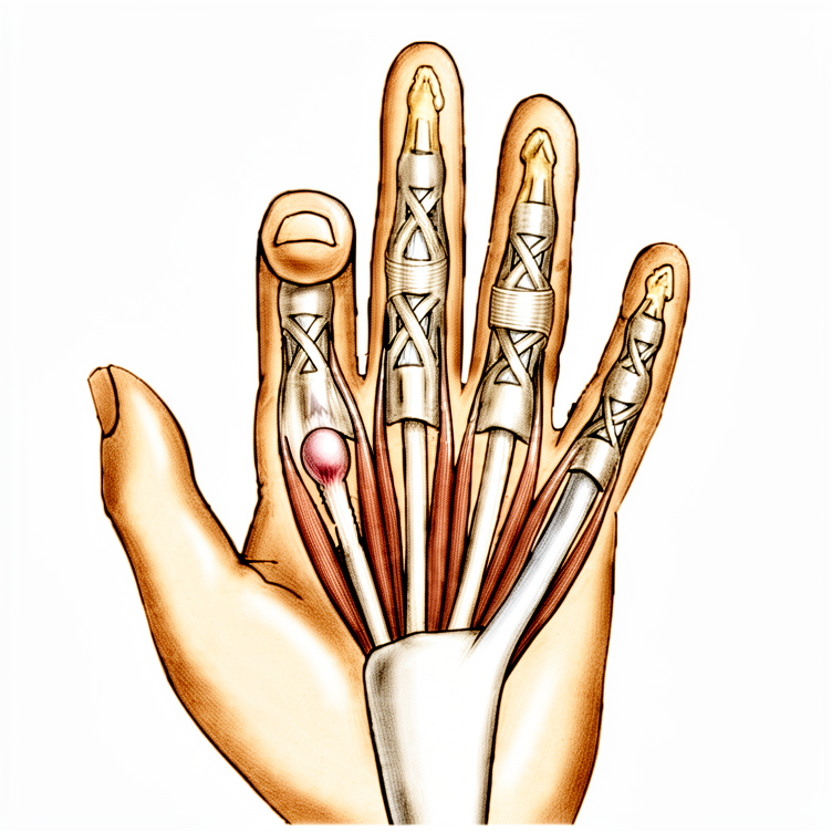
For patients: a plain-language version of this topic is available. See the patient guide.
Overview
Trigger finger release is a surgical intervention for stenosing tenosynovitis, with open release regarded as the gold standard due to its ability to allow careful inspection of the surgical area and its high efficacy with low complication rates [6]. The procedure is generally low-risk, though complications may require reoperation [3], and clinic-based open releases demonstrate complication rates similar to those performed in the operating room [7]. Clinical improvements are reflected in PROMIS PI and UE scores that reach minimum clinically important difference values for hand patients [68], while Michigan Hand Outcomes Questionnaire MIC estimates assist in interpreting outcomes and assessing power in prospective trials [1]. A 2024 narrative review provides an updated practical guide for clinicians to plan tailored rehabilitation management [9].
Operative techniques vary by digit and approach. Open release typically involves local anesthetic infiltration proximal to the incision site [29]. Transverse incisions are placed distal to the distal palmar crease for middle, ring, and small fingers, or distal to the proximal palmar crease for index fingers [29]. Trigger thumbs require release of only the A1 pulley, whereas trigger digits require division of the A1 and A0 or proximal palmar pulley [29]. Percutaneous techniques are safe and effective for all digits [2, 37], with ultrasound guidance proving reliable and efficient without specific morbidity [4], though its clinical significance remains debated [13]. The thread release technique has shown immediate resolution of triggering without complications [15], and percutaneous release using an L15 blade is recommended for lower costs and quicker procedures with equal functional outcomes compared to open surgery [49].
Non-operative management includes corticosteroid injections, where 39% of second and third injections yield long-term relief [17]. Management with two steroid injections before surgery is the least costly strategy [45], while immediate surgical release in the clinic is most cost-effective for diabetic trigger finger assuming a corticosteroid failure rate of at least 34% [23]. Simultaneous steroid injection at the time of percutaneous release provides greater early subjective improvement [5]. A trial found no differences in patient-reported outcomes or severity reduction at 52 weeks among splint alone, steroid alone, and combination treatments, leading authors to recommend splinting alone as the least invasive option [69]. Patients undergoing surgical release have significantly higher odds of developing new-onset Dupuytren disease and undergoing subsequent fasciectomy compared to those managed by other interventions [24].
Anatomy & Pathophysiology
General Etiology and Demographics
Trigger finger is the common term for stenosing tenosynovitis of the flexor tendons, characterized by mechanical impingement at the A1 pulley [51]. It affects 2% to 3% of the general population, with women more commonly affected than men [51]. The condition is associated with diabetes and inflammatory arthropathy [18, 19], and its etiology is possibly linked to repetitive grasping activities [18, 19]. Specifically, repetitive power grip and flexion involved in rowing increase prevalence [130]. Trigger finger is more common in patients with systemic diseases, including diabetes mellitus (10% to 20% lifetime incidence), hypothyroidism, sarcoidosis, rheumatoid arthritis, and septic tenosynovitis [51]. Stenosing tenosynovitis is more common in diabetic patients than in nondiabetic patients [28]; when multiple digits are involved, the possibility of diabetes should be considered [28].
The condition is also associated with gout, calcific tendinitis, pseudogout, and amyloidosis [51]. In gout, monosodium urate precipitation elicits a fulminant inflammatory reaction in the tenosynovium [51]. In calcific tendinitis, calcium salt deposition in the tenosynovium can resemble an infection and result in triggering [51]. In pseudogout, calcium pyrophosphate dihydrate crystal deposition is often localized to the triangular fibrocartilage or within the carpal tunnel [51]. In amyloidosis, beta-2-microglobulin deposits in thick, plaque-like accumulations along the flexor tendons, most commonly seen in patients with renal failure undergoing dialysis [51].
Histology and Pathology
Histology of trigger finger shows fibrocartilaginous metaplasia of the pulley and/or FDS tendon [18, 19]. Pathologic examination of affected pulleys demonstrates a proliferation of chondrocytes and increased type III collagen [51]. The flexor digitorum profundus tendon often demonstrates a pathologic nodule, while the flexor digitorum superficialis is often unaffected [51].
The pathologic anatomy responsible for congenital trigger digits includes narrowing and thickening of the sheath, with occasional formation of a ganglion cyst [98]. An intratendinous nodule may be present proximal to the first annular pulley, often referred to as Notta’s nodule [98]. Chronic inflammation is frequent in congenital trigger digits [98].
Clinical Presentation and Classification
Presentation of trigger finger includes pain and tenderness in the distal palm, progressing to mechanical catching or locking, and may become fixed [18, 19]. A common complaint is referred pain at the dorsal MCP/PIP area [18, 19]. Concomitant trigger finger and carpal tunnel syndrome occurs in 40% to 60% of patients [18, 19]. Triggering is often more pronounced in the morning than later in the day [28]. Patients frequently note catching or triggering of the affected finger or thumb after forceful flexion [28]. In severe cases, the opposite hand must be used to force the finger or thumb passively into extension [28]. In the most severe cases, the finger becomes locked in a flexed position [28].
The Green classification defines severity as follows: * Grade I: Pain and tenderness at the A1 pulley [18, 19]. * Grade II: Catching of the finger [18, 19]. * Grade III: Locking of the finger that is passively correctable [18, 19]. * Grade IV: A fixed, locked finger [18, 19].
Physical examination findings may include tenderness to palpation of the flexor tendon at the level of the A1 pulley [51], palpable triggering or pain with flexion and extension of the finger [51], nodularity of the flexor tendon just proximal to the A1 pulley [51], and the presence of a volar retinacular ganglion cyst between the A1 and A2 pulleys [51]. A fixed flexion deformity of the proximal interphalangeal (PIP) joint may also be present [51].
Patients may note a lump or knot in the palm, which may be the thickened area in the first annular pulley or a nodule or fusiform swelling of the flexor tendon just distal to it [40]. The tendon nodule usually is just proximal to the anulus at the metacarpophalangeal joint level [40]. In a rheumatoid patient, a nodule distal to the metacarpophalangeal joint level may cause triggering [40]. Patients may experience triggering after operative release because of catching of the tendon on the palmar aponeurosis transverse fibers, which usually resolves with time [40]. Occasionally, a partially lacerated flexor tendon at this level heals with a nodule sufficiently large to cause triggering [40]. Local tenderness may be present but is not a prominent complaint in trigger finger [40]. Pressure accentuates the apparent snapping or triggering of the more distal joints [40]. Patients frequently state that the problem is in the proximal interphalangeal joint with trigger finger or in the proximal interphalangeal joint with trigger thumb [40]. Other conditions, such as intraarticular disorders and common extensor tendon subluxation, can cause similar symptoms and must be considered to determine effective treatment for idiopathic trigger finger [40].
Anatomical Variations and Specific Digit Findings
Middle and ring finger involvement is most common in adults [18, 19]. The digits are affected in the following order of decreasing prevalence: thumb, ring, long, little, and index [51]. Newer evidence has found a fourth pulley (variable annular pulley) in 75% of patients with thumb trigger finger, which may contribute to stenosis [18, 19]. The pulley system of the thumb is composed of 4 components, as opposed to the traditional view of only 3 [96]. A1 pulley confluence varies on a digit-to-digit basis, with no observed confluence in the thumb and the most common confluence observed in the middle finger [121]. In the thumb, the flexor tendon and A1 pulley thickened significantly only after patients exhibited triggering [132].
The radial digital nerve is at risk of iatrogenic injury during thumb trigger finger release, given its superficial location [18, 19]. The digital nerves on the thumb are more palmar and closer to the flexor sheath than might be anticipated [29]. The thumb radial digital nerve is especially vulnerable during trigger thumb release [29]. With the little finger held in abduction, the risk of digital nerve injury or inadequate release is reduced during percutaneous A1 pulley release [16].
Pediatric Pathophysiology
Pediatric trigger thumb and trigger finger represent distinct clinical entities and should not be managed like their adult counterparts [12]. Pediatric trigger thumb may spontaneously resolve, although resolution may take several years [12]. Open surgical release of the A1 pulley of the thumb nearly uniformly restores thumb interphalangeal joint motion in pediatric patients [12]. Surgical management of pediatric trigger finger with isolated release of the A1 pulley has been associated with high recurrence rates [12]. Awareness of the anatomic factors that may contribute to triggering in the pediatric finger and willingness to explore and address other involved components of the flexor mechanism can prevent surgical failure [12].
Pediatric trigger thumb presents not at birth but early in childhood [33]. Most evidence suggests that pediatric trigger thumb is caused by a developmental size mismatch between the flexor pollicis longus tendon and its sheath [33]. Patients with pediatric trigger thumb generally present with the thumb interphalangeal joint locked in flexion [33]. Triggering in digits other than the thumb in children is generally associated with an underlying diagnosis including anatomic abnormalities of the tendons, and metabolic, inflammatory, and infectious etiologies [33]. More extensive surgery beyond simple A1 pulley release is often required for pediatric trigger finger, including release of the A3 pulley and resection of a slip of the flexor digitorum superficialis [33].
Pediatric trigger thumbs may be seen in up to 3 per 1,000 children at 1 year of age [127]. Pediatric trigger thumb is an acquired, not congenital, condition [127]. Many patients with pediatric trigger thumb may demonstrate spontaneous resolution of triggering and locking over time [127].
Congenital trigger digit occurs when the normal gliding movement of the flexor tendon is impeded within the digital flexor sheath [98]. In contrast to adults with stenosing tenosynovitis, the congenitally involved finger usually shows a persistent flexion deformity, rather than actual "triggering" [98]. Congenital trigger digit is a relatively rare condition (2.3%) [98]. The condition occurs far more commonly in the thumb and is bilateral in about 25% of patients [98]. It occurs sporadically and is not believed to be an inherited trait [98]. Trigger digits typically occur without other anomalies, but an association with trisomy 13 has been reported [98]. An association with mucopolysaccharidosis has also been described for congenital trigger digits [98]. Trigger digits in children are more commonly acquired, with 25% noted at birth [98]. A prospective study of 5765 newborns did not reveal a single case of congenital trigger thumb [98]. The condition is frequently not noted until age 1 or 2 years, at which time the child has a relatively fixed flexion posture of the interphalangeal joint of the thumb [98]. Even with some force, it may be impossible to extend the interphalangeal joint of the thumb fully in congenital trigger thumb [98]. The abnormal clicking or snapping usually is not the presenting complaint as seen in adults in congenital trigger digit [98].
Fixed contractures are unlikely if the condition resolves or is corrected before the child is 3 years old [98]. Spontaneous resolution occurs in about 30% of children in whom the condition appears within the first year of life [98]. Spontaneous resolution occurs in about 12% of children in whom it appears between 6 months and 2 years of age [98]. Baek et al. noted spontaneous resolution in 63% over a median of 48 months [98]. Trigger finger is not often associated with a fixed flexion deformity in children and may not respond to a simple A-pulley release [98]. When surgical intervention for pediatric trigger finger is undertaken, the surgeon should be prepared for a more extensive exploration of the flexor mechanism, which may include excision of one or both slips of the flexor digitorum superficialis tendon and release of the A3 pulley [98]. Thumbs presenting with an interphalangeal joint angle of 30 degrees or less are more likely to resolve, supporting observation for up to 5 years before offering surgery [75]. Loss of motion is still reversible even after 21 years, and normal thumb function can still be obtained with a very late presentation in pediatric trigger thumb [70]. Extension trigger thumb is a rare manifestation with a low incidence in pediatric trigger thumbs [136]. Closed partial flexor digitorum profundus rupture is an unusual cause of pediatric trigger finger [138].
Associations with Other Conditions
A relationship between carpal tunnel release and trigger finger is possible, related to anatomy and biomechanics modifications after CTR (e.g. possible volar migration of the flexor tendons) and/or to a coexisting disorder [65]. Patients with greater volar migration of the flexor tendons after carpal tunnel release are at a higher risk of developing trigger finger [73]. The data support an association between idiopathic carpal tunnel syndrome and idiopathic trigger finger and lend support to common pathophysiological factors [139]. Carpal tunnel syndrome and trigger digit are 2 of the most common disorders treated by hand surgeons, both due to limited space in an enclosed anatomical region, suggesting a common pathophysiological process [135]. There is sufficient evidence to support a tendency for carpal tunnel syndrome and trigger digit to develop "hand in hand" [135].
The incidence and treatment outcome of cases in which trigger finger occurred in conjunction with Dupuytren’s disease was so different and appeared less predictable than that of ordinary trigger finger [112]. A high percentage of concurrent trigger finger and Dupuytren’s disease was observed in the middle and ring finger (25%) [112]. The percentage of patients with combined trigger finger and Dupuytren’s disease strongly increases with age [112]. 50% in the "80+" age group of trigger finger patients are suffering from both diseases [112]. Smith considers trigger finger as an early indication of Dupuytren’s disease [113]. The deepest layer of the pretendinous fibers (layer three) play a role in causing trigger finger symptoms in Dupuytren’s disease [113]. In the extended finger position, thickening of the pulley wall leads to narrowing of the A1 pulley and synovial congestion [113]. The additional mechanical irritation of the Dupuytren’s cord above the A1 pulley may provoke an aggravation of the irritation [113]. In more progressed stages of Dupuytren’s contracture (Stages II or III), the concomitant appearance of trigger finger and Dupuytren’s contracture is rarely seen [113]. The DD cord that causes a flexed position of the finger is not in such an intimate relation with A1 in advanced stages, resulting in relief of pressure off the affected tendon at the A1 pulley area [113]. Advanced extension deficit reduces the range of motion of the tendon and thus causes less mechanical irritation at A1 [113]. The tendon becomes slightly thinner distal to the chiasm of the deep and superficial flexor tendon in advanced Dupuytren’s contracture [113].
Classification
Trigger finger presentation progresses from pain and tenderness in the distal palm to mechanical catching or locking, which may become fixed [18]. Several classification systems are used to grade the severity of this condition.
Green: The Green classification includes four grades: Grade I (pain and tenderness at the A1 pulley), Grade II (catching of finger), Grade III (locking of finger; passively correctable), and Grade IV (fixed, locked finger) [18].
Froimson: The Froimson classification grades triggering as: Grade I (pre-triggering-pain; tenderness over the A1 pulley; history of catching but not demonstrable on physical examination), Grade II (triggering, active; demonstrable catching, patient can actively extend), and Grade III (triggering, passive; demonstrable catching requiring passive extension or inability to extend) [53].
QuinneII: The QuinneII classification is used to evaluate trigger finger severity [111].
Clinical Presentation
Demographics and Risk Factors
Trigger finger affects 2% to 3% of the general population, with women more commonly affected than men [51]. In adults, the condition is usually seen in individuals older than 45 years of age [40]. The incidence of trigger digit release in Medicare patients is increasing, with the middle digit the most frequently released [31]. When multiple digits are involved, the possibility of diabetes should be considered [28].
Symptoms and Physical Findings
Patients present with pain and tenderness in the palm at the proximal edge of the digital A1 pulley [28]. Pain and tenderness in the distal palm progress to mechanical catching or locking, and may become fixed [18, 19]. A common complaint is referred pain at the dorsal MCP/PIP area [18, 19]. In more severe cases, the opposite hand must be used to force the finger or thumb passively into extension [28].
Patients may note a lump or knot in the palm [40]. This lump may be the thickened area in the first annular pulley or a nodule or fusiform swelling of the flexor tendon just distal to it [40]. The tendon nodule can be palpated by the examiner’s fingertip and moves with the tendon [40]. Local tenderness may be present but is not a prominent complaint [40]. Physical examination findings may include the presence of a fixed flexion deformity of the proximal interphalangeal (PIP) joint [51]. Concomitant trigger finger and carpal tunnel syndrome occur in 40% to 60% of patients [18, 19].
Pediatric Presentation
Pediatric trigger finger is a distinct ailment from adult trigger finger, and a secondary cause must be sought whenever long fingers are affected [80]. The vast majority of patients with bilateral trigger thumbs had bilateral involvement upon initial presentation to the pediatric hand clinic [76].
Investigations
Clinical Assessment and Classification
The clinical presentation of trigger finger typically begins with pain and tenderness in the distal palm, progressing to mechanical catching or locking that may become fixed [18]. Patients frequently report referred pain at the dorsal MCP or PIP area [18]. The Green classification system categorizes the condition into four grades: Grade I involves pain and tenderness at the A1 pulley; Grade II involves catching of the finger; Grade III involves locking of the finger that is passively correctable; and Grade IV involves a fixed, locked finger [18].
Physical examination findings include tenderness to palpation of the flexor tendon at the level of the A1 pulley [51]. Clinicians may also observe palpable triggering or pain with flexion and extension of the finger [51]. Additional signs include nodularity of the flexor tendon just proximal to the A1 pulley [51] and the presence of a volar retinacular ganglion cyst between the A1 and A2 pulleys [51]. A fixed flexion deformity of the proximal interphalangeal (PIP) joint may also be present [51].
Dorsal PIP tenderness is more common in trigger finger than previously thought and is associated with higher and prolonged levels of postoperative pain after A1 pulley release [74]. PIP joint pain results from long symptom duration and consequent joint pathology, and is incompletely resolved after A1 pulley release [30]. Physicians should consider the duration of preoperative symptoms and preoperative flexion contracture of the PIP joint when deciding the timing of surgery [35].
Imaging and Diagnostic Modalities
Ultrasonography: A study of 60 patients with percutaneous trigger finger releases shows no clinical advantage in using pre- or postoperative ultrasonography [11]. However, the P1P can serve as a consistent and readily visible landmark for the distal edge of the A1 pulley to guide accurate distal release during ultrasound-guided percutaneous trigger finger release [137]. While the clinical significance of findings regarding ultrasound-guided percutaneous release is unclear, it raises questions regarding the safety and efficacy of percutaneous trigger finger release, even when adding ultrasound guidance [13].
Corticosteroid Injections: Using ultrasound guidance for corticosteroid injections is more effective for treating trigger fingers than the blinded method, leading to better results and a faster return to work in the early stages of treatment [125].
Systemic and Comorbidity Screening
Aspiration: Gout can present with marked pain, erythema, swelling, and warmth that can mimic infectious tenosynovitis; definitive diagnosis is made by tenosynovial aspiration or biopsy showing negatively birefringent urate crystals under polarized light microscopy [51]. Pseudogout is characterized by calcium pyrophosphate dihydrate crystal deposition often localized to the triangular fibrocartilage or within the carpal tunnel, with pathology revealing rhomboid-shaped crystals with positive birefringence [51].
Laboratory: Amyloidosis is characterized by the deposition of beta-2-microglobulin in thick, plaque-like accumulations along the flexor tendons, most commonly seen in patients with renal failure undergoing peritoneal dialysis or hemodialysis [51]. Tissue obtained at trigger finger release may represent a site for early recognition of transthyretin amyloidosis, whereas tissue from limited fasciectomy appears unsuitable for screening [59].
Plain radiography: Calcific tendinitis can result in triggering, with radiographs revealing fluffy ectopic calcification in the soft tissues that usually resorbs in 2 to 4 weeks [51].
Pediatric Specifics
Pediatric trigger thumb and trigger finger represent distinct conditions and should not be treated like adult acquired trigger finger [12]. An algorithmic approach has been proposed for patients presenting with paediatric trigger finger to guide workup and management [62].
Treatment
Non-Operative
Corticosteroid injection into the flexor tendon sheath is curative in approximately 60% of patients initially [18]. There is no difference in efficacy between soluble and insoluble corticosteroid preparations [18]. Diabetic patients are generally less responsive to corticosteroid injection [18]. Patients anticipate more pain than they experience with the injection [115]. Orthoses are effective for non-surgical management of pediatric and adult trigger finger using various orthotic options [91]. Management with two steroid injections before surgery is the least costly treatment strategy [45].
Operative
Indications: Surgical release of the A1 pulley is curative in digits refractory to steroid injection [28]. Physicians should consider the duration of preoperative symptoms and preoperative flexion contracture of the PIP joint when deciding the timing of surgery [35]. In women with a trigger finger, the choice of treatment should take into account whether there are also one or more patient- or trigger-related factors that increase the risk of conversion to surgery [21].
Surgical Approach / Technique: Open surgical release of the A1 pulley is considered the "gold standard" of trigger finger treatment because it allows careful inspection of the surgical area and is highly effective with low complication rates [6]. It effectively alleviates the subjective and objective manifestations of trigger finger and remains the benchmark procedure [38]. Trigger release should be performed with a local block so that the cessation of triggering can be evaluated intraoperatively [40]. The A2 pulley fibers must be spared during surgical release to preserve effective digital flexion [28]. In patients with rheumatoid arthritis, the preference is to excise a slip of the FDS tendon rather than to release the A1 pulley to avoid exacerbating ulnar drift at the MCP joint [18]. The radial digital nerve is at risk of iatrogenic injury during thumb trigger finger release due to its superficial location [18]. Adjacent finger triggering may become obvious only after a given finger is released and can be managed at the same surgical setting [29].
Percutaneous release with a 19-gauge needle is both a safe and effective means of sectioning the A1 pulley in patients with active triggering who have failed conservative treatment [20]. It is an easy and safe procedure for treating trigger digits in diabetic and non-diabetic patients [56]. Modified percutaneous trigger finger release is a reliable and safe procedure with high patient satisfaction [26]. Percutaneous release is safe for the thumb, index, middle, and ring fingers as long as the recommended technique is observed [8]. With the little finger held in abduction, the risk of digital nerve injury or inadequate release during percutaneous A1 pulley release is reduced [16]. While the clinical significance of findings regarding ultrasound-guided percutaneous release is unclear, it raises questions regarding the safety and efficacy of the procedure even when adding ultrasound guidance [13]. Incomplete pulley release and damage to the flexor tendons and digital nerves, especially in the index finger and thumb, remain of some concern with limited exposure techniques such as percutaneous release [40]. Percutaneous release of congenital trigger thumb can be performed safely and effectively in children by a surgeon specializing in hand surgery [79].
Pain Management: Local infiltration of Ketorolac as an adjunct in postoperative pain management provides superior analgesia during finger movement within the initial 6 hours following trigger finger surgery compared to oral NSAIDs [92].
Other Considerations: Multiple-digit trigger finger release in a single operative session is associated with higher rates of postoperative pain, joint stiffness, and need for flexor tenolysis compared with single-digit release [47]. In case of trigger finger combined with Dupuytren's disease, an isolated opening of the pulley without touching the Dupuytren tissue, followed by application of corticosteroids in the area of the opened pulley, is a recommended strategy [54]. Patients solely undergoing surgical release of their trigger finger had significantly higher odds and expedited rate of developing new-onset Dupuytren disease compared with trigger fingers managed by other interventions [24].
Pediatric Management
Open surgical release of the A1 pulley of the thumb is an alternative option that nearly uniformly restores thumb interphalangeal joint motion in pediatric trigger thumb [12]. Awareness of anatomic factors contributing to triggering in the pediatric finger and willingness to explore and address other involved components of the flexor mechanism can prevent surgical failure in pediatric trigger finger [12].
Outcomes and Complications
Approximately 97% of patients have complete resolution after operative treatment for trigger finger [40]. Persistence of triggering is more common than recurrence after trigger finger release [40]. Major complications following trigger finger release are unlikely; however, minor complications are prominent [22]. Minor complications of open trigger finger release include wound dehiscence, scar tenderness, and decreased range of motion [18]. Although there were no persistent triggering in 98 digits with a follow-up of at least 6 months after percutaneous A1 pulley release, painful stiffness at the interphalangeal joints remained in ten digits despite physical therapy [25]. Subsequent release or injection in the same or another digit was common following an initial trigger finger release [10]. The odds ratio of developing a deep infection for patients who had corticosteroid injections prior to trigger finger release was 9.38 (95% CI, 1.21–72.78) compared with patients who did not have injections [32]. The odds ratio of developing a superficial infection for patients who had corticosteroid injections prior to trigger finger release was 1.19 (95% CI, 0.64–2.24) compared with patients who did not have injections, which was not statistically significant [32]. MIC estimates for the Michigan Hand outcomes Questionnaire can contribute to the interpretation of clinical outcomes following trigger finger release and for assessment of power in prospective trials [1].
Complications
General Complication Rates: Approximately 1 in 20 fingers experiences a mild, transient adverse event following surgical release of the A1 pulley for idiopathic trigger finger [72]. The rate of minor complications after open release is notably high, primarily driven by wound issues or loss of finger range of motion [142]. Most adverse outcomes manifest as short-term pain, stiffness, and swelling, while major complications such as nerve injury or deep infection remain uncommon [102]. About 1 in 200 fingers requires a second surgery [72]. Percutaneous release is considered effective and safe [37], including with a 19-gauge needle in patients who have failed conservative treatment [20]. The thread release technique demonstrated safety and clinical effectiveness with no surgical complications in 34 digits [15]. In a series of 210 fingers undergoing subcutaneous release, no serious complications such as infection, neurovascular injury, or functional impairment occurred [83].
Stiffness and Range of Motion: Stiffness was reported in 8.6% of patients following trigger finger release [94]. Painful stiffness at the interphalangeal joints persisted in ten digits despite physical therapy after percutaneous A1 pulley release of locked trigger digits [25]. In a systematic review of percutaneous A1 pulley release with corticosteroid injection, 8.8% of patients experienced complications, with the majority resolving after one week [108]. Residual loss of motion at the proximal interphalangeal joint is a specific complication associated with open A1 pulley release in patients with diabetes [118].
Infection: The overall incidence of surgical site infection after trigger finger release is 2.1%, comprising 1.7% superficial and 0.5% deep infections [32]. Two patients in a study of open A1 pulley release with traction tenolysis experienced postoperative wound infections, which were successfully treated with a week-long course of antibiotics [114]. Patients who undergo open trigger release within 1 month of a corticosteroid injection face increased odds of developing a postoperative infection requiring surgical debridement [150]. The odds ratio of developing a deep infection for patients who received corticosteroid injections before trigger finger release was 9.38 compared with patients who did not receive injections [32]. Routine perioperative antibiotic prophylaxis does not significantly influence the rate of surgical site infections or associated postoperative complications in diabetic patients undergoing trigger finger release [151].
Wound Complications: Delayed wound healing occurred in 1.2% of patients following trigger finger release [94]. Minor hand surgeries, including trigger finger release, can be performed safely in the procedure room without an increased rate of surgical site infection compared to the operating room [152]. Open trigger finger release surgery performed in the clinic has complication rates similar to reported complication rates of surgery performed in the operating room [7].
Nerve and Vascular Injury: Nerve injury or persistent numbness occurred in 0.6% of patients following trigger finger release [94]. Incomplete pulley release and damage to the flexor tendons and digital nerves, especially in the index finger and thumb, remain of concern, particularly with limited exposure techniques [40]. A percutaneous trigger digit release resulted in a high percentage of incomplete releases of the A1 pulley, especially in the thumb, index, and little fingers [109]. Holding the little finger in abduction during percutaneous release reduces the risk of digital nerve injury or inadequate release [16]. A case report documented shower emboli and digital necrosis after a single corticosteroid injection for trigger thumb resulting from unintentional steroid injection into a hand artery [147].
Recurrence and Persistence: Recurrence of triggering occurred in 2.2% of patients following trigger finger release [94]. Persistence of triggering is more common than recurrence after operative treatment for trigger finger [40]. In a prospective randomized trial, the recurrence rate for open surgical technique for correction of trigger digits was appreciable at 33% within 1 year [85].
Other Considerations: Thirty-eight patients (4.9%) required secondary operations of any type following trigger finger release [94]. Development of Dupuytren contracture occurred in 0.8% of patients following trigger finger release [94].
Recovery
Light activity (weeks): The evidence provided does not specify a typical week range for the resumption of desk work, driving, or light activities of daily living.
Full activity (months): The evidence provided does not specify a month range for the return to manual work, sport, or full range of motion and strength.
Complete recovery / outcome plateau (months): The evidence provided does not specify a month range for the stabilization of pain, strength, and final functional outcomes.
Rehabilitation protocol: Structured preoperative instructions from a hand therapist do not have a significant impact on early postoperative recovery after trigger finger surgeries [63]. Postoperative thumb immobilization following A1 pulley release may not be necessary for patients with trigger thumb, as it does not significantly impact functional outcomes or recurrence rates [144].
Functional milestones: MIC estimates can contribute to the interpretation of clinical outcomes following trigger finger release and for assessment of power in prospective trials [1]. At last follow up, 7 of the 13 reviewed patients presented an excellent functional recovery, equivalent to complete resolution of the hyperextension and pain, attainment of ROM comparable to the contralateral finger and a stable joint [148].
Other Considerations: Percutaneous A1 pulley release is more effective medium-term therapy for trigger digit than steroid injection, because of lower risk of recurrence [55]. The study confirms the efficacy and safety of percutaneous trigger finger release but shows no clinical advantage in using pre- or postoperative ultrasonography [11]. It is a safe procedure for the thumb, index, middle, and ring fingers as long as the recommended technique is observed [8]. Surgical open release is the “gold standard” of trigger finger treatment because it allows more careful inspection of the surgical area and is highly effective with low complication rates [6]. Although there were no persistent triggering in 98 digits with a follow-up of at least 6 months, painful stiffness at the interphalangeal joints remained in ten digits despite of physical therapy [25]. Repeated corticosteroid injections for trigger finger can lead to complete pulley ruptures and flexion contractures, which may be managed surgically with tendon reconstruction, though residual contracture and scarring can persist [145]. Proximal interphalangeal joint pain in trigger finger patients results from long symptom duration and consequent joint pathology, and is incompletely resolved after A1 pulley release, leading to worse surgical outcomes than expected [30]. Open surgical release of the A1 pulley of the thumb is an alternative option that nearly uniformly restores thumb interphalangeal joint motion [12]. A third of pediatric trigger thumbs resolved spontaneously, but most parents desired eventual surgical release [60]. Loss of motion is still reversible even after 21 years, and normal thumb function can still be obtained with a very late presentation [70]. Splinting alone is recommended as the initial treatment for adults with trigger finger because there were no clinically important differences between splinting alone and steroid injection alone in terms of pain reduction and symptom or functional improvement up to 1 year [90]. This technique is a useful treatment for selected patients whose trigger finger is associated with a positional contracture [67].
Key Evidence
- [L3] These MIC estimates can contribute to the interpretation of clinical outcomes following trigger finger release and for assessment of power in prospective trials. [1] (10.1016/j.jht.2021.06.003)
- [L5] Based on our findings, percutaneous trigger finger release can safely be performed on all digits, including the thumb, small fingers, and index fingers. [2] (10.1007/s11552-007-9069-8)
- [L3] Open trigger finger release is generally a low-risk procedure, although there is potential for complications, some requiring reoperation. [3] (10.1007/s11552-014-9716-9)
- [L4] Ultrasound-guided trigger finger release, with this procedure, is reliable and efficient on trigger release, without specific morbidity. [4] (10.1016/j.hansur.2016.10.088)
- [L1] Simultaneous steroid injection at the time of surgical release provides greater subjective improvement in the early period after percutaneous trigger finger release. [5] (10.1177/1753193418813771)
- [L5] Surgical open release is the “gold standard” of trigger finger treatment because it allows more careful inspection of the surgical area and is highly effective with low complication rates. [6] (10.1142/s021881041750023x)
- [L4] Patients who undergo open trigger finger release surgery in the clinic have complication rates similar to reported complication rates of surgery performed in the operating room. [7] (10.1016/j.jhsg.2022.01.008)
- [L4] It is a safe procedure for the thumb, index, middle, and ring fingers as long as the recommended technique is observed. [8] (10.1016/j.jhsa.2010.06.006)
- [L5] The manuscript aims to provide an updated practical guide for clinicians and surgeons reviewing the state-of-art of both the assessment and the treatments of patients with trigger finger to plan tailored rehabilitation management taking advantage of the matching of traditional and novel techniques. [9] (10.1186/s12891-024-08192-5)
- [L4] Subsequent release or injection in the same or another digit was common following an initial trigger finger release. [10] (10.1016/j.jhsa.2025.02.009)
- [L4] The study confirms the efficacy and safety of percutaneous trigger finger release but shows no clinical advantage in using pre- or postoperative ultrasonography. [11] (10.1177/1753193413517992)
- [L4] [12] (10.5435/jaaos-20-04-206)
- [L5] While the clinical significance of these findings is unclear, it raises questions regarding the safety and efficacy of percutaneous trigger finger release, even when adding ultrasound guidance. [13] (10.1007/s11552-008-9137-8)
- [L2] The thread trigger finger release technique was found to be safe and clinically effective, with all 34 digits showing resolved triggering and locking immediately following the procedure and no surgical complications observed. [15] (10.1177/1753193418774497)
- [L5] With the little finger held in abduction the risk of digital nerve injury or inadequate release is reduced. [16] (10.1016/s0363-5023(05)80430-7)
- [L4] Thirty-nine percent of second and third corticosteroid injections for trigger finger yield long-term relief. [17] (10.1016/j.jhsa.2017.02.001)
- [L4] Percutaneous release of trigger fingers with a 19 gauge needle is both a safe and effective means of sectioning the A1 pulley in patients with active triggering who have failed conservative treatment including steroid injection. [20] (10.1016/s0363-5023(05)80026-7)
- [L3] In women with a trigger finger, the choice of treatment should take into account whether there are also one or more patient- or trigger-related factors that increase the risk of conversion to surgery. [21] (10.1302/0301-620x.104b10.bjj-2022-0058.r3)
- [L3] Major complications following trigger finger release are unlikely; however, minor complications are prominent. [22] (10.1177/15589447221081869)
- [L3] Management of diabetic trigger finger with immediate surgical release in the clinic is the most cost-effective treatment strategy, assuming a corticosteroid injection failure rate of at least 34%. [23] (10.1016/j.jhsa.2016.08.007)
- [L3] Patients solely undergoing surgical release of their trigger finger had significantly higher odds and expedited rate of developing new-onset Dupuytren disease overall and undergoing subsequent treatment by fasciectomy compared with trigger fingers managed by other interventions. [24] (10.1177/15589447221077375)
- [L4] Although there were no persistent triggering in 98 digits with a follow-up of at least 6 months, painful stiffness at the interphalangeal joints remained in ten digits despite of physical therapy. [25] (10.1016/j.jhsb.2004.03.015)
- [L4] This study shows that our alternative percutaneous trigger finger release is a reliable and safe procedure with high patient satisfaction. [26] (10.1016/j.hansur.2016.02.004)
- [L4] Proximal interphalangeal joint pain in trigger finger patients results from long symptom duration and consequent joint pathology, and is incompletely resolved after A1 pulley release, leading to worse surgical outcomes than expected. [30] (10.1177/1753193418809771)
- [L3] The incidence of trigger digit release in Medicare patients is increasing, with the middle digit the most frequently released. [31] (10.1177/1753193416653707)
- [L4] [32] (10.1016/j.jhsa.2020.01.007)
- [L4] [33] (10.1016/j.jhsa.2015.04.041)
- [L4] Physicians should consider the duration of preoperative symptoms and preoperative flexion contracture of the PIP joint when deciding timing of surgery for trigger finger patients. [35] (10.1016/j.jhsa.2018.06.023)
- [L1] Percutaneous release is an effective and safe procedure for the treatment of trigger digit. [37] (10.1016/j.jhsa.2014.07.044)
- [L5] Open surgical release of the A1 pulley effectively alleviates the subjective and objective manifestations of trigger finger and remains the benchmark procedure. [38] (10.5435/jaaos-d-19-00614)
- [L4] Surgical release is a short, safe, and effective procedure when performed by specialty trained hand surgeons, and it is the treatment of choice for a locked pediatric trigger thumb. [44] (10.1016/j.jhsa.2011.01.011)
- [L2] Management of trigger finger with 2 steroid injections before surgery is the least costly treatment strategy. [45] (10.1016/j.jhsa.2009.02.029)
- [L3] Multiple-digit trigger finger release in a single operative session is associated with higher rates of postoperative pain, joint stiffness and need for flexor tenolysis compared with single-digit release, even after adjustment for demographics and comorbidities. [47] (10.1177/17531934261448109)
- [L1] Because of lower costs and quicker procedure with equal functional outcome when compared with open surgery, we recommend the percutaneous technique using a L15 blade for trigger finger release. [49] (10.1097/bth.0b013e31817f289a)
- [L2] There were no significant differences in scar quality or improvement in patient-reported disability with transverse or longitudinal incisions for trigger finger release. [52] (10.1177/1753193419859375)
- [L3] [53] (10.1016/j.jhsb.2005.11.003)
- [L1] Percutaneous A1 pulley release is more effective medium-term therapy for trigger digit than steroid injection, because of lower risk of recurrence. [55] (10.1177/1753193410381824)
- [L3] Percutaneous trigger release is an easy and safe procedure for treating trigger digits in diabetic and non-diabetic patients. [56] (10.1177/1753193415590389)
- [L4] Tissue obtained at trigger finger release may represent a site for early recognition of transthyretin amyloidosis, whereas tissue from limited fasciectomy appears unsuitable for screening. [59] (10.1177/17531934261476753)
- [L2] A third of pediatric trigger thumbs resolved spontaneously, but most parents desired eventual surgical release. [60] (10.1016/j.jhsa.2020.10.016)
- [L4] The authors propose an algorithmic approach for patients presenting with paediatric trigger finger to guide workup and management. [62] (10.1177/17531934211035642)
- [L2] Structured preoperative instructions from a hand therapist do not have a significant impact on early postoperative recovery after carpal tunnel and trigger finger surgeries. [63] (10.1177/17531934251313763)
- [L4] However, a relationship is possible, related to anatomy and biomechanics modifications after CTR (e.g. possible volar migration of the flexor tendons) and/or to a coexisting disorder (with the same etiology). [65] (10.1016/j.main.2015.03.003)
- [L4] This technique is a useful treatment for selected patients whose trigger finger is associated with a positional contracture. [67] (10.1016/j.jhsa.2012.07.026)
- [L3] Clinical improvements after trigger digit treatments are reflected in improved PROMIS PI and UE scores that reach previously accepted minimum clinically important difference values for hand patients. [68] (10.1016/j.jhsa.2022.03.015)
- [L5] The author notes that the referenced trial found no differences in patient-reported outcomes for pain or function or in reduction of trigger finger severity at 52 weeks among splint alone, steroid alone, and combination treatments, leading the trial authors to recommend splinting alone as the least invasive option. [69] (10.1097/corr.0000000000002726)
- [Case_report] Loss of motion is still reversible even after 21 years, and normal thumb function can still be obtained with a very late presentation. [70] (10.1177/15589447211052754)
- [L4] This large retrospective series suggests that about 1 in 20 fingers will experience a mild, transient adverse event after surgical release of the A1 pulley for idiopathic trigger finger and that about 1 in 200 have a second surgery. [72] (10.1016/j.jhsa.2012.05.014)
- [L3] Patients with greater volar migration of the flexor tendons after CTR are at a higher risk of developing trigger finger. [73] (10.1177/1753193413479506)
- [L2] Dorsal PIP tenderness is more common in trigger fingers than previously thought and is associated with higher and prolonged levels of postoperative pain after A1 pulley release. [74] (10.1186/s12891-023-06130-5)
- [L3] Thumbs presenting with an interphalangeal joint angle of 30 degrees or less are more likely to resolve, supporting observation for up to 5 years before offering surgery. [75] (10.1177/17531934241295903)
- [L4] The vast majority of patients with bilateral trigger thumbs had bilateral involvement upon initial presentation to the pediatric hand clinic. [76] (10.1177/1558944719864449)
- [L4] Percutaneous release of congenital trigger thumb can be performed safely and effectively in children by a surgeon specializing in hand surgery. [79] (10.1016/j.main.2011.01.012)
- [L4] Pediatric trigger finger is a distinct ailment from adult trigger finger, and a secondary cause must be sought whenever long fingers are affected. [80] (10.1177/1558944715627634)
- [L4] [83] (10.1016/0266-7681(90)90091-h)
- [L1] [85] (10.1053/jhsu.2001.24967)
- [L1] Splinting alone is recommended as the initial treatment for adults with trigger finger because there were no clinically important differences between splinting alone and steroid injection alone in terms of pain reduction and symptom or functional improvement up to 1 year. [90] (10.1097/corr.0000000000002662)
- [L1] Orthoses are effective for non-surgical management of pediatric and adult trigger finger using various orthotic options. [91] (10.1016/j.jht.2023.05.016)
- [L1] Local infiltration of Ketorolac as an adjunct in postoperative pain management has been shown to provide superior analgesia during finger movement within the initial 6 hours following trigger finger surgery, in comparison to oral NSAIDs. [92] (10.1186/s12891-024-07856-6)
- [L3] [94] (10.1177/1558944718796559)
- [L4] The pulley system of the thumb is composed of 4 components, as opposed to the traditional view of only 3. [96] (10.1016/j.jhsa.2012.08.005)
- [Letter] [102] (10.1016/j.jhsa.2012.08.038)
- [L4] [108] (10.1186/s13018-025-05776-2)
- [L5] We found that a percutaneous trigger digit release resulted in high percentage of incomplete releases of A1 pulley, especially in the thumb, index, and little fingers. [109] (10.1097/00130911-200603000-00010)
- [L1] [111] (10.1186/s13018-023-04127-3)
- [L2] [114] (10.1016/j.jhsg.2024.09.010)
- [L1] Patients anticipate more pain than they experience with trigger finger injection. [115] (10.1016/j.jhsa.2017.05.001)
- [L4] [118] (10.1016/j.jhsa.2018.03.045)
- [L5] A1 pulley confluence varies on a digit-to-digit basis, with no observed confluence in the thumb and the most common confluence observed in the middle finger. [121] (10.1016/j.jhsa.2022.02.011)
- [L2] Using ultrasound guidance for corticosteroid injections is more effective for treating trigger fingers than the blinded method, leading to better results and a faster return to work in the early stages of treatment. [125] (10.1186/s13018-023-03950-y)
- [L4] [127] (10.1016/j.jhsa.2008.04.017)
- [L3] This study provides new conclusive evidence that the repetitive power grip and flexion involved in rowing increase the prevalence of trigger finger. [130] (10.1177/1558944720918321)
- [L4] In the thumb, the flexor tendon and A1 pulley thickened significantly only after patients exhibited triggering. [132] (10.1016/j.jhsa.2012.06.027)
- [L4] [135] (10.1016/j.jhsa.2013.04.032)
- [L4] Extension trigger thumb is a rare manifestation with a low incidence in pediatric trigger thumbs. [136] (10.1016/j.jhsa.2021.01.014)
- [L4] The P1P can serve as a consistent and readily visible landmark for the distal edge of the A1 pulley to guide accurate distal release during ultrasound-guided percutaneous trigger finger release. [137] (10.1016/j.jhsa.2021.04.033)
- [L4] [138] (10.1177/1558944716681950)
- [L3] The data support an association between idiopathic CTS and idiopathic trigger finger and lend support to common pathophysiological factors. [139] (10.1177/1753193408096015)
- [L4] Major complications occur infrequently, but the rate of minor complications was surprisingly high and related mostly to wound complications or loss of finger range of motion. [142] (10.1016/j.jhsa.2009.12.040)
- [L3] Postoperative thumb immobilization following A1 pulley release may not be necessary for patients with trigger thumb, as it does not significantly impact functional outcomes or recurrence rates. [144] (10.1186/s13018-025-06203-2)
- [L4] Repeated corticosteroid injections for trigger finger can lead to complete pulley ruptures and flexion contractures, which may be managed surgically with tendon reconstruction, though residual contracture and scarring can persist. [145] (10.1177/1753193412465727)
- [Case_report] The patient's symptoms resulted from unintentional steroid injection into a hand artery, with subsequent embolism into digital arteries. [147] (10.1016/j.jhsa.2008.10.006)
- [L4] At last follow up, 7 of the 13 reviewed patients presented an excellent functional recovery, equivalent to complete resolution of the hyperextension and pain, attainment of ROM comparable to the contralateral finger and a stable joint. [148] (10.1016/j.hansur.2018.10.134)
- [L2] Patients who undergo open trigger release within 1 month of a corticosteroid injection are at increased odds of developing a postoperative infection requiring surgical debridement. [150] (10.1016/j.jhsa.2021.08.017)
- [Paper] The routine use of perioperative antibiotic prophylaxis does not significantly influence the rate of surgical site infections (SSIs) and associated postoperative complications in diabetic patients undergoing carpal tunnel release (CTR) and trigger finger release (TFR). [151] (10.1177/15589447251352004)
- [L2] Minor hand surgeries can be performed safely in the procedure room without an increased rate of surgical site infection compared to the operating room. [152] (10.1016/j.jhsa.2023.03.001)
See Also
- Trigger Finger
- Dupuytren's Disease
References
[1] Determining the Minimally Important Change of the Michigan Hand outcomes Questionnaire in patients undergoing trigger finger release. Journal of Hand Therapy. 2023. DOI: 10.1016/j.jht.2021.06.003
[2] The Safety of Percutaneous Trigger Finger Release. HAND. 2007. DOI: 10.1007/s11552-007-9069-8
[3] Risk Factors for Complications of Open Trigger Finger Release. HAND. 2014. DOI: 10.1007/s11552-014-9716-9
[4] Ultrasound-guided trigger finger release: A new surgical procedure (50 cases). Hand Surgery and Rehabilitation. 2016. DOI: 10.1016/j.hansur.2016.10.088
[5] Effects of simultaneous steroid injection after percutaneous trigger finger release: a randomized controlled trial. Journal of Hand Surgery (European Volume). 2018. DOI: 10.1177/1753193418813771
[6] Trigger Finger: An Atraumatic Medical Phenomenon. The Journal of Hand Surgery (Asian-Pacific Volume). 2017. DOI: 10.1142/s021881041750023x
[7] Office-Based Open Trigger Finger Release Has a Low Complication Rate. Journal of Hand Surgery Global Online. 2022. DOI: 10.1016/j.jhsg.2022.01.008
[8] Anatomic Outcome of Percutaneous Release Among Patients With Trigger Finger. The Journal of Hand Surgery. 2010. DOI: 10.1016/j.jhsa.2010.06.006
[9] From diagnosis to rehabilitation of trigger finger: a narrative review. BMC Musculoskeletal Disorders. 2024. DOI: 10.1186/s12891-024-08192-5
[10] Incidence and Predictors of Subsequent Triggering Requiring Treatment After Trigger Finger Release. The Journal of Hand Surgery. 2026. DOI: 10.1016/j.jhsa.2025.02.009
[11] A study of 60 patients with percutaneous trigger finger releases: clinical and ultrasonographic findings. Journal of Hand Surgery (European Volume). 2014. DOI: 10.1177/1753193413517992
[12] Management of Pediatric Trigger Thumb and Trigger Finger. Journal of the American Academy of Orthopaedic Surgeons. 2012. DOI: 10.5435/jaaos-20-04-206
[13] Ultrasound-Assisted Percutaneous Trigger Finger Release: Is it Safe?. HAND. 2008. DOI: 10.1007/s11552-008-9137-8
[15] Minimally invasive thread trigger digit release: a preliminary report on 34 digits of the adult hands. Journal of Hand Surgery (European Volume). 2018. DOI: 10.1177/1753193418774497
[16] Percutaneous A1 pulley release: A cadaveric study. The Journal of Hand Surgery. 1995. DOI: 10.1016/s0363-5023(05)80430-7
[17] Long-Term Effectiveness of Repeat Corticosteroid Injections for Trigger Finger. The Journal of Hand Surgery. 2017. DOI: 10.1016/j.jhsa.2017.02.001
[18] Miller S Review Of Orthopaedics. SECTION 16 PATELLAR TRACKING IN TOTAL KNEE ARTHROPLASTY > 2. Flexor tendon injury > 3. Stenosing tenosynovitis (trigger finger).
[19] Miller S Review Of Orthopaedics. 2. Flexor tendon injury > 3. Stenosing tenosynovitis (trigger finger).
[20] Safety and efficacy of percutaneous trigger finger release. The Journal of Hand Surgery. 1995. DOI: 10.1016/s0363-5023(05)80026-7
[21] Factors associated with conversion to surgical release after a steroid injection in patients with a trigger finger. The Bone & Joint Journal. 2022. DOI: 10.1302/0301-620x.104b10.bjj-2022-0058.r3
[22] Complications Following Endoscopic and Open Trigger Finger Release: A Retrospective Comparative Study. HAND. 2022. DOI: 10.1177/15589447221081869
[23] Cost of Immediate Surgery Versus Non-operative Treatment for Trigger Finger in Diabetic Patients. The Journal of Hand Surgery. 2016. DOI: 10.1016/j.jhsa.2016.08.007
[24] Surgical Trigger Finger Release Is Associated With New-Onset Dupuytren Contracture in the Short-Term Postoperative Period: A Matched Analysis. HAND. 2022. DOI: 10.1177/15589447221077375
[25] A1 Pulley Release of Locked Trigger Digit by Percutaneous Technique. Journal of Hand Surgery. 2004. DOI: 10.1016/j.jhsb.2004.03.015
[26] Modified percutaneous trigger finger release. Hand Surgery and Rehabilitation. 2016. DOI: 10.1016/j.hansur.2016.02.004
[28] A Lange Medical Book Current Diagnosis Treatment In Orthopedics Fifth Edition. 9Hand Surgery > 2. Flexor Tenosynovitis (Trigger Finger and Trigger Thumb).
[29] Campbell S Operative Orthopaedics 4 Volume Set. EXCISION OF NECROTIC MUSCLES COMBINED WITH NEUROLYSIS OF MEDIAN AND ULNAR NERVES FOR SEVERE CONTRACTURE > TRIGGER FINGER AND THUMB > SURGICAL RELEASE OF TRIGGER FINGER.
[30] Clinical significance of proximal interphalangeal joint pain in patients with trigger fingers. Journal of Hand Surgery (European Volume). 2018. DOI: 10.1177/1753193418809771
[31] Trigger digit release: rates of surgery and complications as indicated by a United States Medicare database. Journal of Hand Surgery (European Volume). 2016. DOI: 10.1177/1753193416653707
[32] Risk of Infection in Trigger Finger Release Surgery Following Corticosteroid Injection. The Journal of Hand Surgery. 2020. DOI: 10.1016/j.jhsa.2020.01.007
[33] Pediatric Trigger Digits. The Journal of Hand Surgery. 2015. DOI: 10.1016/j.jhsa.2015.04.041
[35] Factors Causing Prolonged Postoperative Symptoms Despite Absence of Complications After A1 Pulley Release for Trigger Finger. The Journal of Hand Surgery. 2019. DOI: 10.1016/j.jhsa.2018.06.023
[37] Percutaneous First Annular Pulley Release for Trigger Digits: A Systematic Review and Meta-Analysis of Current Evidence. The Journal of Hand Surgery. 2014. DOI: 10.1016/j.jhsa.2014.07.044
[38] Current Concepts in the Management of Trigger Finger in Adults. Journal of the American Academy of Orthopaedic Surgeons. 2020. DOI: 10.5435/jaaos-d-19-00614
[40] Campbell S Operative Orthopaedics 4 Volume Set. EXCISION OF NECROTIC MUSCLES COMBINED WITH NEUROLYSIS OF MEDIAN AND ULNAR NERVES FOR SEVERE CONTRACTURE > TRIGGER FINGER AND THUMB.
[44] Surgical Release of the Pediatric Trigger Thumb. The Journal of Hand Surgery. 2011. DOI: 10.1016/j.jhsa.2011.01.011
[45] Using Evidence to Minimize the Cost of Trigger Finger Care. The Journal of Hand Surgery. 2009. DOI: 10.1016/j.jhsa.2009.02.029
[47] Comparative outcomes of single versus multiple trigger finger releases: a propensity score matched cohort study. Journal of Hand Surgery (European Volume). 2026. DOI: 10.1177/17531934261448109
[49] Open Versus Percutaneous Release of the A1-Pulley for Stenosing Tendovaginitis. Techniques in Hand & Upper Extremity Surgery. 2008. DOI: 10.1097/bth.0b013e31817f289a
[51] Aaos Comprehensive Orthopaedic Review 3. Tendinopathy of the Hand and Wrist* > II. Trigger Finger.
[52] A prospective, randomized clinical trial of transverse versus longitudinal incisions for trigger finger release. Journal of Hand Surgery (European Volume). 2019. DOI: 10.1177/1753193419859375
[53] Sonographically Assisted Percutaneous Release of the A1 Pulley: A New Surgical Technique for Treating Trigger Digit. Journal of Hand Surgery. 2006. DOI: 10.1016/j.jhsb.2005.11.003
[54] Dupuytren S Disease And Related Hyperproliferative Disorders. 31. The Influence of Dupuytren’s Disease on Trigger Fingers and Vice Versa > 31.5 Discussion > 31.5.3 Surgical Strategies and Recommendations.
[55] Percutaneous A1 pulley release vs steroid injection for trigger digit: the results of a prospective, randomized trial. Journal of Hand Surgery (European Volume). 2010. DOI: 10.1177/1753193410381824
[56] Outcomes and complications after percutaneous release for trigger digits in diabetic and non-diabetic patients. Journal of Hand Surgery (European Volume). 2015. DOI: 10.1177/1753193415590389
[59] Screening for amyloidosis in trigger finger release and Dupuytren’s fasciectomy. Journal of Hand Surgery (European Volume). 2026. DOI: 10.1177/17531934261476753
[60] The Natural History of Pediatric Trigger Thumb in the United States. The Journal of Hand Surgery. 2021. DOI: 10.1016/j.jhsa.2020.10.016
[62] Presentation and aetiology of paediatric trigger finger: a systematic review. Journal of Hand Surgery (European Volume). 2021. DOI: 10.1177/17531934211035642
[63] Preoperative hand therapy instructions do not improve short-term outcomes in carpal tunnel and trigger finger surgery. Journal of Hand Surgery (European Volume). 2025. DOI: 10.1177/17531934251313763
[65] Is carpal tunnel release associated with trigger finger?. Chirurgie de la Main. 2015. DOI: 10.1016/j.main.2015.03.003
[67] Resection of the Flexor Digitorum Superficialis for Trigger Finger With Proximal Interphalangeal Joint Positional Contracture. The Journal of Hand Surgery. 2012. DOI: 10.1016/j.jhsa.2012.07.026
[68] Responsiveness of PROMIS Instruments for Trigger Digit After Corticosteroid Injection or A1 Pulley Release. The Journal of Hand Surgery. 2023. DOI: 10.1016/j.jhsa.2022.03.015
[69] CORR Insights®: Are There Differences in Pain Reduction and Functional Improvement Among Splint Alone, Steroid Alone, and Combination for the Treatment of Adults With Trigger Finger?. Clinical Orthopaedics & Related Research. 2023. DOI: 10.1097/corr.0000000000002726
[70] Consequences of Untreated Pediatric Trigger Thumb: Case Report. HAND. 2021. DOI: 10.1177/15589447211052754
[72] Adverse Events of Open A1 Pulley Release for Idiopathic Trigger Finger. The Journal of Hand Surgery. 2012. DOI: 10.1016/j.jhsa.2012.05.014
[73] The relationship of trigger finger and flexor tendon volar migration after carpal tunnel release. Journal of Hand Surgery (European Volume). 2013. DOI: 10.1177/1753193413479506
[74] Dorsal proximal interphalangeal joint tenderness is associated with prolonged postoperative pain after A1 pulley release for trigger fingers. BMC Musculoskeletal Disorders. 2023. DOI: 10.1186/s12891-023-06130-5
[75] The natural history of paediatric trigger thumbs. A prospective cohort study. Journal of Hand Surgery (European Volume). 2024. DOI: 10.1177/17531934241295903
[76] The Development of Trigger Thumb in the Contralateral Thumb in Pediatric Patients Presenting Initially With Unilateral Involvement. HAND. 2019. DOI: 10.1177/1558944719864449
[79] Le traitement percutané du pouce à ressaut chez l’enfant : à propos de 63 cas. Chirurgie de la Main. 2011. DOI: 10.1016/j.main.2011.01.012
[80] Pediatric Trigger Finger due to Osteochondroma. HAND. 2016. DOI: 10.1177/1558944715627634
[83] Subcutaneous Release of Trigger Thumb and Fingers in 210 Fingers. Journal of Hand Surgery. 1990. DOI: 10.1016/0266-7681(90)90091-h
[85] Prospective randomized trial of open versus percutaneous surgery for trigger digits. The Journal of Hand Surgery. 2001. DOI: 10.1053/jhsu.2001.24967
[90] Are There Differences in Pain Reduction and Functional Improvement Among Splint Alone, Steroid Alone, and Combination for the Treatment of Adults With Trigger Finger?. Clinical Orthopaedics & Related Research. 2023. DOI: 10.1097/corr.0000000000002662
[91] Orthotic intervention options to non-surgically manage adult and pediatric trigger finger: A systematic review. Journal of Hand Therapy. 2023. DOI: 10.1016/j.jht.2023.05.016
[92] Local ketorolac infiltration for postoperative pain in open trigger finger surgery: a randomized controlled trial. BMC Musculoskeletal Disorders. 2024. DOI: 10.1186/s12891-024-07856-6
[94] Steroid Injection and Open Trigger Finger Release Outcomes: A Retrospective Review of 999 Digits. HAND. 2018. DOI: 10.1177/1558944718796559
[96] Varied Anatomy of the Thumb Pulley System: Implications for Successful Trigger Thumb Release. The Journal of Hand Surgery. 2012. DOI: 10.1016/j.jhsa.2012.08.005
[98] Campbell S Operative Orthopaedics 4 Volume Set. MULTIPLE Z-PLASTY RELEASE OF A CONGENITAL RING > TRIGGER FINGERS.
[102] Letter Regarding “Adverse Events of Open A1 Pulley Release for Idiopathic Trigger Finger”. The Journal of Hand Surgery. 2012. DOI: 10.1016/j.jhsa.2012.08.038
[108] Percutaneous A1 pulley with corticosteroid injection for trigger finger release: a systematic review. Journal of Orthopaedic Surgery and Research. 2025. DOI: 10.1186/s13018-025-05776-2
[109] Percutaneous Release of A1 Pulley. Techniques in Hand & Upper Extremity Surgery. 2006. DOI: 10.1097/00130911-200603000-00010
[111] Ultrasound-guided acupotomy for trigger finger: a systematic review and meta-analysis. Journal of Orthopaedic Surgery and Research. 2023. DOI: 10.1186/s13018-023-04127-3
[112] Dupuytren S Disease And Related Hyperproliferative Disorders. 31. The Influence of Dupuytren’s Disease on Trigger Fingers and Vice Versa > 31.5 Discussion.
[113] Dupuytren S Disease And Related Hyperproliferative Disorders. 31. The Influence of Dupuytren’s Disease on Trigger Fingers and Vice Versa > 31.5 Discussion > 31.5.1 Why Is DD Concurring with TF?.
[114] Impact of Flexor Tendon Traction Tenolysis on Clinical Outcomes in Open A1 Pulley Release for Trigger Finger. Journal of Hand Surgery Global Online. 2025. DOI: 10.1016/j.jhsg.2024.09.010
[115] Needle-Free Jet Lidocaine Administration for Preinjection Anesthesia in Trigger Finger Injection: A Randomized Controlled Trial. The Journal of Hand Surgery. 2017. DOI: 10.1016/j.jhsa.2017.05.001
[118] Management of Diabetic Trigger Finger. The Journal of Hand Surgery. 2019. DOI: 10.1016/j.jhsa.2018.03.045
[121] Defining the Digit-Specific Confluence of the A1 Pulley. The Journal of Hand Surgery. 2023. DOI: 10.1016/j.jhsa.2022.02.011
[125] Ultrasound guided versus blinded injection in trigger finger treatment: a prospective controlled study. Journal of Orthopaedic Surgery and Research. 2023. DOI: 10.1186/s13018-023-03950-y
[127] Pediatric Trigger Thumb. The Journal of Hand Surgery. 2008. DOI: 10.1016/j.jhsa.2008.04.017
[130] Trigger Finger From Ocean Rowing: An Observational Study. HAND. 2020. DOI: 10.1177/1558944720918321
[132] Sonographic Appearance of the Flexor Tendon, Volar Plate, and A1 Pulley With Respect to the Severity of Trigger Finger. The Journal of Hand Surgery. 2012. DOI: 10.1016/j.jhsa.2012.06.027
[135] Carpal Tunnel Syndrome and Trigger Digit: Common Diagnoses That Occur “Hand in Hand”. The Journal of Hand Surgery. 2013. DOI: 10.1016/j.jhsa.2013.04.032
[136] Characterization of Pediatric Extension Trigger Thumb: An Insight Into a Rare Manifestation From a Single-Center Retrospective Cohort Analysis. The Journal of Hand Surgery. 2021. DOI: 10.1016/j.jhsa.2021.01.014
[137] Sonographic Location of Distal A1 Pulley by Means of a Bony Acoustic Landmark on the Proximal Phalanx: An Anatomic Study. The Journal of Hand Surgery. 2022. DOI: 10.1016/j.jhsa.2021.04.033
[138] Closed Partial Flexor Digitorum Profundus Rupture: An Unusual Cause of Pediatric Trigger Finger. HAND. 2016. DOI: 10.1177/1558944716681950
[139] Idiopathic Carpal Tunnel Syndrome and Trigger Finger: Is There an Association?. Journal of Hand Surgery (European Volume). 2008. DOI: 10.1177/1753193408096015
[142] Complications of Open Trigger Finger Release. The Journal of Hand Surgery. 2010. DOI: 10.1016/j.jhsa.2009.12.040
[144] Pediatric trigger thumb treated with A1 pulley release with or without postoperative thumb immobilization: a retrospective study. Journal of Orthopaedic Surgery and Research. 2025. DOI: 10.1186/s13018-025-06203-2
[145] Trigger finger caused by anatomical variation lumbrical muscle. Journal of Hand Surgery (European Volume). 2012. DOI: 10.1177/1753193412465727
[147] Shower Emboli and Digital Necrosis After a Single Corticosteroid Injection for Trigger Thumb: Case Report. The Journal of Hand Surgery. 2009. DOI: 10.1016/j.jhsa.2008.10.006
[148] Résection de la bandelette ulnaire du flexor digitorum superficiel (FDS) technique appelée USSR (Ulnar superficial slip resection) dans les doigts à ressaut anciens, à propos d’une série de 42 doigts opérés. Hand Surgery and Rehabilitation. 2018. DOI: 10.1016/j.hansur.2018.10.134
[150] Temporal Relationship of Corticosteroid Injection and Open Release for Trigger Finger and Correlation With Postoperative Deep Infections. The Journal of Hand Surgery. 2022. DOI: 10.1016/j.jhsa.2021.08.017
[151] Is There a Role for Antibiotics in Diabetic Patients Undergoing Carpal Tunnel and Trigger Finger Release? A Retrospective Analysis of 723 Cases. HAND. 2025. DOI: 10.1177/15589447251352004
[152] Is Hand Surgery in the Procedure Room Setting Associated With Increased Surgical Site Infection? A Cohort Study of 2,717 Patients in the Veterans Affairs Population. The Journal of Hand Surgery. 2023. DOI: 10.1016/j.jhsa.2023.03.001