Clinicians › Shoulder
Tendon repair recovery phases
Overview
Primary repair remains the standard of care for tendon injuries, although repaired tissue rarely achieves functionality equal to the preinjured state [1]. Arthroscopic rotator cuff repair provides functional recovery as early as 3 months, with further improvement over the first year and subsequent stabilization [2]. Clinical outcomes correlate with tissue healing; T2 values at the healing site decrease as clinical outcomes improve during the first follow-up year [3], and worse early clinical outcomes (1-3 months) correlate with higher signal intensity on the proximal cuff tendon [4]. In patients over 65, the speed of tendon healing and short-term results measured by the modified UCLA score are not significantly different between anatomic and single-row repairs [5]. Arthroscopic repair of isolated subscapularis lesions yields improved clinical outcomes and a high rate of tendon healing at midterm follow-up [8], with reliable tendon healing and excellent functional results one year after surgery for higher-grade lesions [19].
Biological healing progresses rapidly, with revascularization occurring by day 7 in regions with close tendon stump approximation and by 10 to 14 days in regions with larger gaps [6]. Delay of supraspinatus repair by up to 12 weeks does not impair enthesis formation and supports a positive outcome [12]. Rehabilitation strategies vary, but immobilization after arthroscopic rotator cuff repair is not superior to early-motion rehabilitation regarding tendon healing or clinical outcome [18]. A traditional 4-week immobilization followed by a 2-week rehabilitation protocol results in excellent outcomes at 1 year without evidence of tendon un-healing or re-tearing [29]. A delayed postoperative physical therapy protocol is associated with improvements in outcome measures, shoulder function, and rotator cuff healing demonstrated by MRI [11]. Pain, range of motion, muscle strength, and function significantly improve after arthroscopic rotator cuff repair regardless of early postoperative rehabilitation protocols [21].
Adjunctive therapies and surgical techniques show mixed results. Image-guided platelet-rich plasma (PRP) treatment on two occasions does not improve early tendon-bone healing or functional recovery after arthroscopic supraspinatus tendon repair [7, 9]. PRP application during arthroscopic rotator cuff repair did not clearly demonstrate accelerated clinical or anatomical recovery, though it demonstrated an improvement in internal rotation [38]. Clinical use of PRP should be approached cautiously until high-level evidence supporting efficacy is available [25], although healing tendon may be responsive to long-term delivery of platelet-derived growth factor [20]. Evidence confirms neither single-row nor double-row repair is clinically more efficacious than the other [22]. Biceps tenodesis has proven more successful than tenotomy in non-isolated cuff tears [22]. Sheath closure did not improve flexor tendon function in a delayed primary repair [13], while palmaris longus tendon harvest allows harvesting with minimal scarring [14]. Standardized rehabilitation protocols and technological advances are required to continue to optimize recovery [58].
Anatomy & Pathophysiology
Tendon Healing Mechanisms and Stages
The natural tendon healing response to rupture results in scar formation with mechanical properties inferior to healthy tendon tissue [39]. The intrinsic regenerative capacity of the adult tendon is poor, and the microstructure and composition of normal tendon are never completely restored in scarred tissue [133]. Tendon has very little regenerative capacity of its own [30]. Repair of tendon injuries results in the formation of fibrovascular scar that never attains the gross, histological, or mechanical characteristics of normal tendon [30]. The precise mechanisms of degeneration, tearing, and subsequent healing are not known [30].
The healing response to acute tendon injury involves three stages: an inflammatory stage with inflammatory cell infiltration and fibroblast recruitment, a proliferative stage with high cellularity and increased type 3 collagen, and a remodeling stage occurring 6-8 weeks after injury with decreased cellularity, increased type 1 collagen, and greater tensile strength [39]. The three phases of tendon healing are inflammation (0–1 week), proliferation (1–6 weeks), and remodeling (6 weeks-12 months) [49]. Inflammation involves inflammatory cells releasing growth factors and type-III collagen [49]. Proliferation involves increased fibroblast activity and type-I collagen [49]. Remodeling involves tissue strengthening, reduced cellularity, and longitudinal collagen alignment along the tendon axis [49]. The three stages of tendon healing (inflammation, proliferation, and remodeling) can partially overlap in time and space [111]. The duration of each stage of tendon healing depends on the location and severity of the lesion [111]. Inflammation occurs within 48 hours after tendon injury and starts with hematoma formation [111].
The cellular mechanisms of tendon healing include extrinsic and intrinsic healing mechanisms that act synergistically in the process of tissue repair [111]. After a tendon injury, inflammatory cells and fibroblasts from the tendon sheath and blood circulation are attracted to the damaged sites by chemokines, causing cell infiltration and adhesion [111]. Intrinsic cells colonizing the tendon are activated after injury, migrate to the damaged site, proliferate massively, and express a variety of extracellular matrices [111]. Intrinsic cells reconstruct the extracellular matrix and form a vascular network during tendon healing [111]. Compared with the extrinsic repair mechanism, the intrinsic repair mechanism reduces tissue adhesion and preserves tendon sliding properties within the sheath [111]. When the extrinsic healing mechanism dominates, it can result in more collagen at the injury site and reduced collagen organization and strength in the repaired tendon tissue [49]. Dominance of the extrinsic healing mechanism can lead to the formation of scars and adhesions between the tendon and nearby structures [49].
Following tendon repair, 7–15% of patients experience problems like scarring, an increase in tendon adhesions, and restricted joint movement during the early healing phase [49]. The mechanical properties of the healed tendon do not typically return to those of the uninjured tissue [49]. The repaired tendon has less incorporation of collagen fibers and features a higher proportion of collagen type III compared to collagen type I [49]. The repaired tendon exhibits a shorter crimp pattern than the normal tendon, resulting in a tissue that is mechanically weaker [49]. The tendon thickens and becomes more rigid to compensate for the decreased mechanical strength, which reduces the quality and functionality of the tendon [49]. Changes in the healed tendon can cause decreased joint function [49]. Primary repair remains the standard of care, but repaired tendon tissue rarely achieves functionality equal to that of the preinjured state [1].
Tendon healing responses to high cycle fatigue differ from those present in acute injury [39]. Repetitive loading can result in differential tendon responses characterized by either healthy remodeling with collagen production or degeneration [39]. A very large number of tendon injuries are chronic, degenerative conditions [30]. Tendon injuries can be acute and traumatic, but are often chronic and degenerative in nature [133]. Tendon injury is a common musculoskeletal pathology that can cause significant pain and dysfunction [133]. Tendon injuries account for substantial pain, disability, and time off work [30]. Annually, more than 28 million patients in the United States experience a tendon or ligament injury [53].
The concept of "scarless healing" has recently become of interest to physicians and researchers in application to tendon healing [30]. Current research is studying scarless healing, which is seen in fetal life, Murphy Roths Large mice, and nonmammalian vertebrates, and regenerates native tendon structure following damage [39]. Embryonic tendon tissue heals primarily by regeneration without the formation of a scar [133]. Studies of wound healing in the developing fetus demonstrate an absence of a significant inflammatory response and absence of macrophage infiltration [133]. The lack of inflammation and subsequent macrophage infiltration may be a key factor in regenerative healing [133].
It is now accepted that "molecular inflammation" plays a critical role in tendon pathophysiology [133]. Following injury to the tendon, the healing process begins with inflammation and an infiltration and activation of numerous cell types, including macrophages, in the healing tendon tissue [133]. Macrophages carry out a variety of functions in tissue healing including host defense, phagocytosis, and the production of growth factors and both pro-inflammatory and anti-inflammatory mediators [133]. The injured tendon heals primarily through the formation of a fibrovascular scar [133]. Tendon healing is a complex, poorly understood process, and the failure of the tendon to heal after medical and surgical intervention is a common problem [133].
The success of tendon surgical repair has been limited by the diminished ability of degenerated tissue to heal [27]. Development of effective therapeutics is hindered by the lack of guiding data on the cellular and molecular aspects of tendon development, signal transduction, mechanotransduction, and fundamental mechanisms underlying tendon pathogenesis and healing [27]. Therapeutic measures for effective intervention and prevention of tendon injuries have progressed with limited success due to the scarcity of data describing basic mechanisms for effective tendon function and response to injuries [27]. Restoration of function after tendon damage is largely dependent on the reestablishment of the muscle-tendon-bone connection with minimal scarring between the tendon and its surrounding tissues [53].
Current clinical treatment protocols after tendon repair often include NSAIDs, chiefly for their pain-relieving effects, with a secondary indication in their limitation of inflammation [53]. NSAIDs facilitate rehabilitation by limiting pain and may also reduce the cross-sectional area of a tendon repair through decreased fibrosis [53]. Recent research has highlighted detrimental effects of NSAIDs on healing tissues [53]. In vitro studies have demonstrated that NSAIDs may inhibit tendon cell migration and proliferation, processes that are critical during the initial response to tendon injury [53]. Studies have shown that NSAIDs inhibit tendon-to-bone healing in a rat rotator cuff model via inhibition of the formation and maturation of collagen at the tendon insertion site [53]. Evidence suggests that NSAIDs may improve healing if administered later in the healing process [53].
Gait, tensile mechanical properties, and bone properties were significantly reduced after injury and repair in a murine model [17]. Pain, range of motion, muscle strength, and function all significantly improved after arthroscopic rotator cuff repair regardless of early postoperative rehabilitation protocols [21]. Postoperative shoulder stiffness was associated with better healing of the rotator cuff after arthroscopic rotator cuff repair [132]. Shoulder stiffness at 3 months post-arthroscopic rotator cuff repair predicts 12-month shoulder stiffness but indicates better tendon integrity, with limited long-term clinical impact [99]. Postoperative shoulder stiffness is associated with a lower risk of rotator cuff retear, likely due to an increased healing response involving elevated cytokine levels [137]. Postoperative shoulder stiffness is associated with increased pain and restricted function in the short term [137]. Scapular dyskinesis is an underlying mechanism leading to compromise of supraspinatus healing after repair [45]. MR imaging–derived rotator cuff muscle proton density fat-fraction is associated with isometric strength independent of muscle atrophy and tendon rupture in shoulders with early and advanced degenerative changes [48]. The progression of degenerative changes of the biceps tendon after successful rotator cuff repair may be a source of persistent anterior shoulder pain [134].
Tendon-Bone Interface and Biomechanics
The normal anatomic structure of the enthesis consists of a progression from tendon, to unmineralized fibrocartilage, to mineralized fibrocartilage, and finally to bone [39]. Enthesis regions experience high stresses because of the extreme differences in material properties between tendon and bone [39]. Enthesis regions have a unique loading environment compared with the body of the tendon [39]. The healing process of entheses is distinct from that of tendons [39]. During the healing process of entheses, the normal transitional structure is lost, resulting in decreased material properties and high repair failure rates [39].
The tendon to bony insertion site varies dramatically along its length in terms of its viscoelastic properties, collagen structure, and extracellular matrix composition [120]. The microstructure of the tendon to bone insertion serves to simultaneously reduce stress concentrations and material mass [103]. The biomechanics are clear that one should not decorticate the greater tuberosity to cancellous bone [127].
Shoulders that were immobilized demonstrated superior structural properties, including significantly higher collagen orientation, compared to those that were exercised [33]. Immobilized shoulders demonstrated superior compositional properties, with expression of extracellular matrix genes similar to the uninjured insertion, compared to exercised shoulders [33]. Immobilized shoulders demonstrated superior quasilinear viscoelastic properties compared to exercised shoulders [33]. The quasilinear viscoelastic property A for immobilized shoulders was 0.30 ± 0.10 MPa versus 0.16 ± 0.08 MPa for exercised shoulders [33]. The quasilinear viscoelastic property B for immobilized shoulders was 17.4 ± 2.9 versus 15.1 ± 0.9 for exercised shoulders [33]. The quasilinear viscoelastic property t2 for immobilized shoulders was 344 ± 161 s versus 233 ± 40 s for exercised shoulders [33].
Differences between types of repair for all biomechanical outcome measures (load to failure, energy to failure, and stiffness) at 6- and 12-weeks were not significant [50]. The tension required at repair to reappose the supraspinatus tendon to bone rapidly increases after injury [23]. These findings suggest that rotator cuff tears should be repaired early in the clinical setting [23].
Simulated isolated supraspinatus cord and strap tears significantly reduced shoulder abduction force, with cord tears causing a larger decline than strap tears [93]. With superiorly directed loads, complete supraspinatus tendon tears created superior translations at all abduction angles [97]. Complete supraspinatus tendon tears created posterior instability in the middle ranges of rotation for 20 and 40 degrees of abduction [97].
Pathophysiology and Modulating Factors
Male patients with subacromial impingement have more histologic and ultrastructural degenerative changes in their shoulder compared with patients with posttraumatic recurrent shoulder instability [138].
The rotator cuff is a sheet of conjoined tendons closely applied over the shoulder capsule and inserting mainly into the greater tuberosity of the humerus, with the subscapularis inserted into the lesser tuberosity [67]. The rotator cuff is made up of subscapularis in front, supraspinatus above, and infraspinatus and teres minor behind [67]. The rotator cuff has an important function in stabilizing the head of the humerus by pulling it firmly into the glenoid whenever the deltoid lifts the arm forwards or sideways [67]. The coracoacromial arch is a fibro-osseous canopy formed by the acromion process posterosuperiorly, the coracoid process anteriorly, and the coracoacromial ligament joining them [67]. The subacromial bursa separates the tendons from the coracoacromial arch and allows them to glide [67]. If there is not enough room for the conjoint tendon to pass under the arch due to pathology within the tendon or narrowing of the space, pain or awkward shoulder movement may result [67].
The commonest causes of pain around the shoulder are disorders of the rotator cuff, chiefly the rotator cuff syndrome, calcific tendinitis, and adhesive capsulitis [67]. The rotator cuff syndrome comprises supraspinatus impingement and tendinitis, tears of the rotator cuff, acute calcific tendinitis, and biceps tendinitis and/or rupture [67]. In all conditions comprising rotator cuff syndrome, the patient complains of pain over the front and lateral aspect of the shoulder with weakness on abduction for supraspinatus involvement, tears of the cuff, and tendinitis [67]. Pain is over the front for biceps pathology [67]. If the cuff or biceps has ruptured, there will also be weakness [67].
The glenohumeral joint depends on static and dynamic stabilizers for movement and stability, especially the rotator cuff [71]. The rotator cuff stabilizes the glenohumeral joint while allowing greater freedom of motion and fixes the fulcrum of the upper extremity against which the deltoid can contract and elevate the humerus [71]. The rotator cuff must act simultaneously and synergistically with the deltoid muscle for normal function [71].
The shoulder joint is composed of four articulations: the sternoclavicular, acromioclavicular, glenohumeral, and scapulothoracic [72]. Normal function of the shoulder is a balance between mobility and stability [72]. Mobility is allowed by the "large ball–small socket" bony arrangement and the voluminous glenohumeral joint capsule, which does not restrict movement until the extremes of motion [72]. The bony anatomy contributes little to stability and has been compared with a golf ball on a tee [72]. The glenoid is encircled by the labrum, composed of dense fibrocartilaginous tissue, which increases the depth of the socket by 50% around the humeral head and increases stability [72]. The glenoid articular surface and the labrum combine to create a socket that is approximately 9 mm deep in the superoinferior direction and 5 mm deep in the anteroposterior direction [72]. Adding the glenoid labrum increases the glenoid surface to 75% of the humeral head vertically and 57% horizontally [72]. Because there is very little bony constraint to the shoulder, most of the stability is provided by the surrounding muscles and ligaments [72].
The ligamentous constraints are the primary stabilizers at extremes of motion [72]. The superior glenohumeral ligament is the primary restraint to inferior humeral subluxation in 0 degrees of abduction and is the primary stabilizer to anterior and posterior stress in the same position [72]. Tightening of the rotator interval decreases posterior and inferior translation [72]. The middle glenohumeral ligament limits external rotation when the arm is in the lower and middle ranges of abduction but has little effect when the arm is in 90 degrees of abduction [72]. The inferior glenohumeral ligament is composed of an anterior band that is quite thick, a posterior band that is less thick and distinct, and a thinner intervening axillary pouch, creating a hammock-type sling [72]. With external rotation, the hammock slides anteriorly and superiorly, the anterior band tightens, and the posterior band fans out [72]. With internal rotation, the opposite occurs [72]. The anteroinferior glenohumeral ligament complex is the main stabilizer to anterior and posterior stresses when the shoulder is abducted 45 degrees or more [72].
The extrinsic muscles primarily control movement of the scapula and include the rhomboids, levator scapulae, trapezius, and serratus anterior [72]. The intrinsic muscles control the glenohumeral joint and include the rotator cuff muscles (subscapularis, supraspinatus, infraspinatus, and teres minor), the deltoid, the pectoralis major, the teres major, the latissimus dorsi, and the biceps brachii [72]. The extrinsic muscles dynamically position the scapula to place the glenoid opposite the humeral head as the shoulder moves [72]. Rotator cuff activity and biceps activity have been shown to stiffen the capsule and decrease glenohumeral translation [72]. Intrinsic and extrinsic muscles serve as fine tuners of motion and power movers by working in "force couples" [72]. The most important force couple involves the subscapularis and posterior rotator cuff [72]. Together, the subscapularis and posterior rotator cuff provide a compressive force that centers the humeral head in the glenoid cavity [72]. The teres minor has often been viewed as a minor contributor to rotator cuff function, but there has been heightened attention to its contribution [72].
Classification
Sugaya Classification: This system distinguishes five repair categories using oblique coronal, oblique sagittal, and transverse T2-weighted images [35]. Type 1 indicates a repaired rotator cuff with sufficient thickness and homogeneously low intensity on each image [35]. Type 2 indicates a repaired rotator cuff with sufficient thickness and a partial high-intensity area [35]. Type 3 indicates a repaired rotator cuff with insufficient thickness without discontinuity [35]. Type 4 indicates the presence of a minor discontinuity in more than 1 slice of each image, suggestive of a small tear [35]. Type 5 indicates the presence of a major discontinuity on each image, suggestive of a large tear [35]. Tendon healing is defined as the absence of major rotator cuff discontinuity (Sugaya type V) on MR arthrography at 6 months [31].
Modified Patte Classification: The diagnostic performance of this system is excellent for reparability and acceptable for rotator cuff healing, with high measurement reliability [94].
Other Considerations: Primary repair remains the standard of care for tendon injuries [1]. In the early postoperative period (1-3 months) after arthroscopic rotator cuff repair, worse clinical outcomes correlate with higher signal intensity on the proximal cuff tendon [4]. The speed of tendon healing and short-term results measured by the modified UCLA score are not significantly different between anatomic rotator cuff repair and single row repair in patients over 65 [5]. Both suture-bridge and single-row techniques resulted in significant improvements in clinical scores and tendon healing at 12 months, with no significant differences in healing patterns between the two techniques [142]. Arthroscopic repair of higher-grade isolated subscapularis tendon lesions provides reliable tendon healing accompanied by excellent functional results 1 year after surgery [19].
Tendon healing occurs without evidence of vascular contribution from the sheath [15]. Early controlled passive motion stimulates an intrinsic healing response and restores the gliding surface at an early stage, resulting in a repair that is mechanically stronger and structurally more similar to the normal tendon-sheath interface compared to immobilization [16]. Gait, tensile mechanical properties, and bone properties are significantly reduced after injury and repair in a supraspinatus tendon healing model [17]. Differences between types of repair for biomechanical outcome measures (load to failure, energy to failure, and stiffness) at 6- and 12-weeks were not significant [50].
The success of tendon surgical repair is limited by the diminished ability of degenerated tissue to heal [27]. Tendon repair results in the formation of fibrovascular scar that never attains the gross, histological, or mechanical characteristics of normal tendon [30]. The natural tendon healing response to rupture is through scar formation resulting in mechanical properties inferior to those found with healthy tendon tissue [39]. The repaired tendon, with a mixture of collagen type III and type I scar tissue, is notably weaker than its native, preinjury form [117]. The tendon thickens and becomes more rigid to compensate for the decreased mechanical strength of the repaired tissue [49].
Tendon healing consists of three phases: inflammation (0–1 week), proliferation (1–6 weeks), and remodeling (6 weeks-12 months) [49]. Alternatively, tendon healing in rotator cuff injury typically progresses through three distinct phases: the inflammatory phase (0 to 7 days), the proliferative/repair phase (5 to 25 days), and the remodeling phase (>21 days) [117]. Tendon healing also occurs in three overlapping phases: inflammatory, proliferative, and remodeling [122]. Optimising tendon healing requires understanding the biological processes of inflammation, repair, and remodelling [36]. Future tendon treatment approaches should consider modulation of the inflammatory phase of healing [24].
In the initial inflammatory phase, erythrocytes and inflammatory cells, particularly neutrophils, enter the site of injury [122]. In the first twenty-four hours, monocytes and macrophages predominate and phagocytosis of necrotic materials occurs [122]. This phase is defined by the initial release of cytokines, monocytes, macrophages, and growth factors which infiltrate the affected tissue and remove cellular debris [117]. It involves depositing platelets and promoting angiogenesis [117]. Vasoactive and chemotactic factors are released with increased vascular permeability, initiation of angiogenesis, stimulation of tenocyte proliferation, and recruitment of more inflammatory cells [122]. Tenocytes gradually migrate to the wound, and type-III collagen synthesis is initiated [122]. The inflammation phase is characterized by inflammatory cells releasing growth factors and type-III collagen [49].
The proliferative phase begins after a few days [122]. Synthesis of type-III collagen peaks during this phase and lasts for a few weeks [122]. Water content and glycosaminoglycan concentrations remain high [122]. Newly vascularized tissue begins to form scar tissue consisting predominantly of collagen type III [117]. The proliferation phase is characterized by increased fibroblast activity and type-I collagen [49]. It involves high cellularity, increased type 3 collagen, and alteration of the extracellular matrix [39].
The remodeling phase commences after approximately six weeks [122] or 6-8 weeks after injury [39]. It is characterized by decreased cellularity and decreased collagen and glycosaminoglycan synthesis [122]. Collagen type III is gradually replaced by collagen type I, which is composed of more mature and organized collagen fibrils to generate greater tensile strength [117]. The phase involves increased type 1 collagen with more longitudinal organization and greater tensile strength [39]. It is characterized by tissue strengthening, reduced cellularity, and longitudinal collagen alignment along the tendon axis [49]. The remodeling phase can be divided into a consolidation stage and a maturation stage [122]. The consolidation stage begins at about six weeks and continues for up to ten weeks, during which the repair tissue changes from cellular to fibrous [122]. Tenocyte metabolism remains high, and tenocytes and collagen fibers become aligned in the direction of stress [122]. A higher proportion of type-I collagen is synthesized during this stage [122]. The maturation stage occurs after ten weeks, involving a gradual change of the fibrous tissue to scar-like tendon tissue over the course of one year [122].
Growth factors exhibit unique temporal profiles that correlate with specific stages in the injury and repair process of the supraspinatus tendon [28]. Growth factor expression in supraspinatus tendon healing shows an initial increase followed by a return to control or undetectable levels by 16 weeks [28]. Tendon healing responses to high cycle fatigue are different from those present in acute injury [39]. Repetitive loading can result in differential tendon responses with either healthy remodeling and the production of collagen or degeneration [39].
The tendon-bone interface, or enthesis, of the rotator cuff possesses a unique architecture consisting of pure dense fibrous connective tissue, uncalcified fibrocartilage, calcified fibrocartilage, and bone [117]. During the healing process of the enthesis, the normal transitional structure is lost, resulting in decreased material properties and high repair failure rates [39]. The enthesis forms fibrovascular scar tissue and fails to regenerate during the natural rotator cuff healing process [117].
Apoptotic changes of tenocytes are most marked in the surface region and in the junction region of the healing tendon in the middle and late healing stages [118]. Intact tendons exhibit strong upregulation of DNA repair pathways compared to torn tendons [121]. DNA repair is critical for maintaining tenocyte viability under oxidative and mechanical stress, supporting matrix turnover and regeneration following injury [121]. Chromatin organization and nucleosome assembly are upregulated in intact tendons, reinforcing transcriptional stability required for controlled repair [121].
Tendon healing is impaired in aged rats and is characterized by a significantly lower histological score, decreased collagen synthesis, and more adipocyte accumulation in patellar tendon after repair [144]. Data are lacking to allow definitive conclusions on the use of tissue engineered biological augmentation techniques for routine management of tendon ailments [57]. Long-term results of subgroups of grade 1 and 2 tears suggest that PRP positively affected rotator cuff healing [40].
According to the Sugaya classification on MRI, 90% of rotator cuffs were considered healed (stage 1 in 47 and stage 2 in 13) at 12 months after surgery in patients under 40 [44]. Rerupture of the repaired cuff was observed in 2 patients in a cohort of patients under 40 treated with suture bridge technique [44]. Failure of surgical repair was observed in 5 patients (7%) in a cohort of patients under 40, defined as Sugaya stage 3 in 4 patients and stage 4 in 1 patient [44]. The failure rate encompassing both tendon nonhealing and rerupture was calculated to be 10% at final follow-up in a cohort of patients under 40 [44]. A lower mean Constant score was observed in patients presenting with nonhealing of tendons (83.7 points) than in those whose tendons were considered healed (88.6 points), though the difference was not statistically significant [44]. The average pain level was 2.4 in the group with unhealed cuffs versus 1.2 in the healed group [44]. Female gender and Sugaya grade III tendon healing were identified as independent predictive factors for poor outcome in successfully healed rotator cuff tears [47].
Clinical Presentation
Primary repair remains the standard of care for tendon injuries, yet repaired tendon tissue rarely achieves functionality equal to the preinjured state [1]. The repair of tendon injuries continues to present a major clinical challenge to orthopedic medicine [100]. Optimizing tendon healing requires a fundamental understanding of the biological processes of inflammation, repair, and remodelling [36]. The healing response to acute tendon injury involves three distinct stages: an inflammatory stage characterized by inflammatory cell infiltration and fibroblast recruitment; a proliferative stage marked by high cellularity, increased type 3 collagen, and alteration of the extracellular matrix; and a remodeling stage occurring 6-8 weeks after injury, featuring decreased cellularity, increased type 1 collagen with more longitudinal organization, and greater tensile strength [39]. Growth factors exhibit unique temporal profiles that correlate with specific stages in the injury and repair process of the supraspinatus tendon, with an initial increase in expression followed by a return to control or undetectable levels by 16 weeks [28]. The healing process of entheses is distinct from that of tendons, as the normal transitional structure is lost during healing, resulting in decreased material properties and high repair failure rates [39].
Rotator Cuff Healing Dynamics
Arthroscopic repair of rotator cuff lesions provides very good functional recovery results, with recovery as early as 3 months, further improvement over the first year, and subsequent stabilization [2]. During the first follow-up year after arthroscopic rotator cuff repair, T2 values of the healing site decreased with the improvement of clinical outcomes over time [3]. In the early postoperative period (1-3 months) after rotator cuff repair, worse clinical outcomes correlated with higher signal intensity on the proximal cuff tendon [4]. The tension required at repair to reappose the supraspinatus tendon to bone rapidly increases after injury, suggesting rotator cuff tears should be repaired early [23]. Inferior rotator cuff healing was demonstrated when there was a delay between injury and repair [26, 34]. Conversely, delay of supraspinatus repair by up to 12 weeks does not impair enthesis formation, supporting a trial of conservative treatment and positive outcomes for delayed repair [12]. At 12 months after arthroscopic rotator cuff repair, 90% of rotator cuffs were considered healed (Sugaya stage 1 or 2), while 10% presented with failure (nonhealing or rerupture) [44]. A lower mean Constant score was observed in patients presenting with nonhealing of tendons after surgery compared to those whose tendons were considered healed, though the difference was not statistically significant [44]. The predominance of high signal intensity (Sugaya type II) at 3, 6, and 12 months after arthroscopic rotator cuff repair decreased over time from 77.8% at 3 and 6 months to 66.7% at 12 months [91]. Patients reported significant clinical improvement after arthroscopic rotator cuff repair, with a decrease in VAS from 6.83 ± 1.77 preoperatively to 1.37 ± 1.98 at 12 months of follow-up [91].
Functional Outcomes and Predictors
Functional improvement does not always coincide with tendon healing, and advanced postoperative imaging findings are used to determine healing status [89]. In a cohort of 30 patients, 23 (77%) had a healed rotator cuff and 7 (23%) had a defect based on postoperative ultrasound, with no significant differences in demographic data or postoperative ASES scores between the groups [87]. Female gender and Sugaya grade III tendon healing were identified as independent predictive factors for poor clinical outcome in successfully healed rotator cuff tears [47]. The etiology of the rotator cuff tear (traumatic versus non-traumatic) did not significantly affect postoperative outcomes or healing rates in patients under 50 years [46]. Pain, range of motion, muscle strength, and function significantly improved after arthroscopic rotator cuff repair regardless of whether aggressive or limited early passive exercise rehabilitation protocols were used [21]. A delayed postoperative physical therapy protocol was associated with improvements in outcome measures and shoulder function compared to the preoperative state, with rotator cuff healing demonstrated by MRI [11]. There is no evidence that immobilization after arthroscopic rotator cuff repair is superior to early-motion rehabilitation in terms of tendon healing or clinical outcome [18]. However, shoulders that were immobilized demonstrated superior structural, compositional, and quasilinear viscoelastic properties compared to those that were exercised [33]. Early controlled passive motion stimulates an intrinsic healing response, restores the gliding surface at an early stage, and results in a repair that is mechanically stronger and structurally more similar to the normal tendon-sheath interface compared to immobilization [16].
Specific Tendon Pathologies
Arthroscopic repair of higher-grade isolated subscapularis lesions provides reliable tendon healing accompanied by excellent functional results 1 year after surgery [19]. Improved clinical outcomes and an overall high rate of tendon healing were observed at midterm follow-up after arthroscopic isolated subscapularis tendon repair [8]. Successful functional outcomes and a high tendon healing rate can be expected nine years after arthroscopic repair of isolated subscpinatus tears [90]. The speed of tendon healing and short-term results measured by the modified UCLA score were not significantly different between anatomic rotator cuff repair and single-row repair in patients over 65 [5]. Clear indications have not been established for the use of single-row versus double-row repair because evidence confirms neither is clinically more efficacious than the other [22]. The interval to surgery for distal biceps rupture impacts clinical results, with a risk of greater loss of force at more than two weeks, and MRI found poorer cicatrization with late reinsertion [32]. The repair site of flexor tendons was revascularized by day 7 in regions with close tendon stump approximation and by 10 to 14 days in regions with larger gaps between the tendon ends [6]. Closure of the flexor sheath during delayed primary tendon repair did not improve flexor tendon function [13]. The palmaris longus tendon can be harvested with minimal scarring using specific techniques [14].
Biological Augmentation and Model Limitations
After arthroscopic supraspinatus tendon repair, image-guided platelet-rich plasma (PRP) treatment on two occasions does not improve early tendon-bone healing or functional recovery [7, 9]. PRP application during arthroscopic rotator cuff repair did not clearly demonstrate accelerated recovery clinically or anatomically except for an improvement in internal rotation [38]. Acceleration of muscle and tendon healing with PRP appears promising, but there is currently little clinical evidence, and clinical use should be approached cautiously until high-level evidence supporting efficacy is available [25]. In a murine model of supraspinatus tendon healing, gait, tensile mechanical properties, and bone properties were significantly reduced after injury and repair [17].
Investigations
Plain radiography: Standardized plain films are almost always sufficient to garner the information needed for shoulder evaluation, and the temptation to "overimage" should be resisted [43]. The purpose of shoulder imaging is to help establish the diagnosis, determine the severity of pathoanatomy, assist in surgical planning, and enable the surgeon to illustrate the condition to the patient [43]. A robust approach to shoulder imaging must recognize that the shoulder is a three-dimensional structure that cannot be represented by a single planar view [79]. Critical relationships, such as the degree of centering of the humeral head, change with the position of the arm [79]. Overlying and superimposed structures as well as metallic implants may complicate imaging the structures of interest in the shoulder [79]. Surgeons need to develop a judicious approach to imaging that yields necessary information while avoiding the tendency to "over-image" [79].
MRI: MRI is the modality of choice for evaluating the rotator cuff, biceps, and subacromial/subdeltoid bursa [83]. T2-weighted MRI provides better visualization of full-thickness rotator cuff tears [83]. MRI accuracy in identifying labral and rotator cuff tears in the literature ranges from 70% to 100% [81]. MR arthrography is considered the benchmark for evaluation of labral tears and is rarely indicated for evaluation of rotator cuff pathology [83]. MR arthrography increases both sensitivity and specificity in detecting injuries to the capsulolabral–ligamentous complex compared to traditional MRI [81]. In a meta-analysis of 4,667 shoulders, MRA had greater diagnostic test accuracy for glenoid labral lesions than MRI, with MRA sensitivity of 88% and specificity of 93% versus MRI sensitivity of 76% and specificity of 87% [81]. Abduction and external rotation (ABER) positioning during MRA is utilized to increase the sensitivity and specificity for detecting anteroinferior labroligamentous injury [81]. The sensitivity of MRA with the ABER position for detecting anteroinferior labral lesions was significantly higher than that of MRA in the neutral position [81].
CT: CT arthrography is indicated when MRI or MR arthrography is contraindicated, such as in patients with pacemakers or vascular clips [83]. CT scans may offer increased precision in measuring glenoid version, but this precision does not improve the quality of surgery or clinical outcome [43].
Ultrasound: Ultrasonography is a low-cost alternative to MRI and arthrography for evaluating both skeletal and soft-tissue structures of the shoulder [83]. Ultrasonography can provide immediate, real-time visualization of the rotator cuff, biceps tendon, and calcific deposits [83]. As a result of providing images in real-time, ultrasonography can evaluate impingement in various positions and motions [83]. Ultrasonography can be used to measure the subacromial space and detect atrophy of rotator cuff muscles [83]. Ultrasound is a simple and accurate test for identifying rotator cuff tears and calcific tendinitis [76]. Ultrasound can be useful in guiding injections or barbotage, such as aspirating calcific deposits in the rotator cuff [76]. Ultrasonography is highly operator-dependent and is not as useful for evaluating labral tears or rotator cuff tears that are very small or larger than 3 cm [83]. The accuracy of ultrasonography for identifying rotator cuff tears depends on the skill of the scanner operator and awareness of pitfalls [74]. Dynamic ultrasonographic evaluation is important for zone II flexor tendon lacerations because it allows visualization of tendon movement to detect partial tears that may have normal physical examination findings [74].
Quantitative MRI and Tissue Characterization: T2 values of the healing site decreased with the improvement of clinical outcomes over time during the first follow-up year after arthroscopic rotator cuff repair [3]. Early after surgery (1-3 months), worse clinical outcomes correlated with higher signal intensity on the proximal cuff tendon [4]. Quantitative UTE-T2 values have a healing-related relationship with clinical outcomes, highlighting the potential of using UTE-T2 mapping to track the tendon-healing process noninvasively [154]. MR imaging–derived rotator cuff muscle proton density fat fraction (PDFF) is associated with isometric strength independent of muscle atrophy and tendon rupture in shoulders with early and advanced degenerative changes [48]. After successful arthroscopic rotator cuff repair, there was a slight (11.3%-13.9%) increase in muscle volume from preoperatively to final follow-up as seen on serial MRI [152].
Repair Integrity Classification: Repair integrity on MRI is determined according to the classification of Sugaya et al., which distinguishes 5 repair categories using oblique coronal, oblique sagittal, and transverse T2-weighted images [35]. Type 1 repair integrity indicates a repaired rotator cuff with sufficient thickness and homogeneously low intensity on each image [35]. Type 2 repair integrity indicates sufficient thickness with a partial high-intensity area [35]. Type 3 repair integrity indicates insufficient thickness without discontinuity [35]. Type 4 repair integrity indicates the presence of a minor discontinuity in more than 1 slice of each image, suggestive of a small tear [35]. Type 5 repair integrity indicates the presence of a major discontinuity on each image, suggestive of a large tear [35].
Other Considerations: Serial ultrasound evaluation showed that the speed at which tendon healing occurs and short-term results as measured by the modified UCLA score were not significantly different between anatomic rotator cuff repair and single-row repair in patients over 65 [5]. Arthroscopic repair of rotator cuff lesions gives very good results in terms of functional recovery, with recovery as early as 3 months, further improvement over the first year, and subsequent stabilization [2]. Improved clinical outcomes and an overall high rate of tendon healing were seen at the midterm follow-up after arthroscopic isolated subscapularis tendon repair [8]. Arthroscopic repair of medium to large rotator cuff tears using triple-loaded medially based single-row repair augmented with marrow vents resulted in a 92% healing rate by MRI and excellent patient-reported outcomes [155]. The delayed postoperative physical therapy protocol was associated with improvements in outcome measures and shoulder function compared to the preoperative state, with rotator cuff healing demonstrated by MRI [11]. Preoperative MRI scans of the shoulder interpreted by orthopaedic surgeons with a described systematic approach resulted in improved accuracy in diagnosing subscapularis tendon tears compared with previous studies [157]. MRI found poorer cicatrization with late reinsertion of distal biceps rupture, and the sole iterative rupture was associated with a three-month interval to reinsertion [32]. The interval to surgery for distal biceps rupture impacts clinical results, with a risk of greater loss of force at more than two weeks [32]. An elevated risk of complications, notably neurological, was reported related to more difficult dissection in scar tissue when the interval to surgery is longer, though this was not found in one specific series where complications were equivalent regardless of interval [32].
Treatment
Non-Operative
Nonoperative treatment is efficacious for patients with chronic, massive, irreparable rotator cuff tears, although the supporting evidence is of low quality [146]. In elderly patients, non-operative management serves as the first-line approach, with surgical intervention reserved for those who fail a trial of conservative management [130]. Delaying surgery for three months after initial conservative therapy was not detrimental to healing outcomes at 3 to 5 years of follow-up in elderly populations [130]. Neither the American Academy of Orthopaedic Surgeons clinical practice guidelines nor Cochrane systematic reviews currently provide specific guidance on the management of rotator cuff tears [86].
Operative
Indications: Arthroscopic repair is supported in patients older than 65 years, as clinical results were better compared to preoperative assessment even in cases of non-watertight healing [151]. Elderly patients gain as much benefit from arthroscopic rotator cuff repair as their younger peers [130].
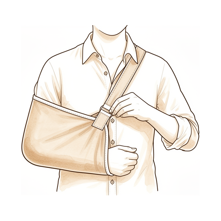
Surgical Approach / Technique: Clear indications for single- versus double-row repair have not been established, as evidence confirms neither is clinically more efficacious than the other; however, biceps tenodesis has proven more successful than tenotomy in non-isolated cuff tears [22]. In comparisons of side-to-side sutures versus traditional tendon-to-bone fixation for supraspinatus tears, the side-to-side group was immobilized in a sling for the first 6 weeks, while the traditional group wore a sling for 4 weeks and initiated formal therapy with passive range of motion at 10 days postoperatively [128]. For isolated subscapularis reconstruction, the arm is placed in a shoulder sling for 6 weeks, with passive mobilization limited to 0° external rotation and flexion/abduction limited to 60° during the first 3 weeks [139].
Biological Augmentation and Adjuvants: Platelet-rich plasma (PRP) may be a safe adjuvant to rotator cuff repair, but it has not been shown to improve healing or function [126]. Platelet-rich fibrin (PRF) has demonstrated no benefit in improving tendon healing rates or functional outcomes in arthroscopic rotator cuff repair [125]. While acceleration of muscle and tendon healing with PRP appears promising, there is currently little clinical evidence, and clinical use should be approached cautiously until high-level evidence supporting efficacy is available [25].
Rehabilitation and Mechanical Loading: A traditional 4-week immobilization followed by a 2-week rehabilitation protocol after arthroscopic rotator cuff repair resulted in excellent outcomes at 1 year without evidence of tendon un-healing or re-tearing [29]. A delayed postoperative physical therapy protocol was associated with improvements in outcome measures and shoulder function compared to the preoperative state, with rotator cuff healing demonstrated by MRI [11]. Frequency of controlled passive motion rehabilitation is a significant factor in accelerating the healing response following tendon repair, and higher frequency-controlled passive motion has a beneficial effect [129]. Contrary to expectations, shoulders that were immobilized demonstrated superior structural, compositional, and quasilinear viscoelastic properties compared to those that were exercised [33]. Strong clinical evidence for a specific rehabilitation protocol over another does not exist, but early mobilization in a controlled fashion does not appear to be harmful [141]. Standardized rehabilitation protocols and technological advances are required to continue to optimize recovery following musculoskeletal trauma [58].
Superior Capsular Reconstruction (SCR) Specifics: There is currently no standardized rehabilitation protocol for superior capsular reconstruction (SCR) [119, 153]. Generally, six weeks of immobilization is recommended for SCR, with passive range of motion work initiated after six weeks and no strengthening allowed for the first six months postoperative [119]. If a patient undergoing SCR is not progressing as expected, MRI assessment is recommended because non-healing of the graft to the glenoid or avulsion of the origin of the long head of biceps is difficult to view on ultrasound images [119]. A specific institutional protocol for SCR advocates for no active movement of the shoulder joint until 6 weeks postoperatively, with Phase 1 (day 9 to 2 weeks) involving continuous sling use and passive/active range of motion of the elbow, wrist, and neck [106]. In this protocol, Phase 2 (weeks 2 to 5) involves weaning the sling and restoring passive range of motion, while Phase 3 begins around 6 weeks with no active abduction initiated during the first 8 weeks [106]. Phase 4 is initiated at 12 to 14 weeks postoperatively with the goal of attaining full rotator cuff strength and endurance at 90° of abduction [106].
Other Considerations: In the early postoperative period (1-3 months) after rotator cuff repair with the suture-bridge technique, worse clinical outcomes correlated with higher signal intensity on the proximal cuff tendon [4]. Healing of the tendon took place without evidence of vascular contribution from the sheath in an experimental dog model [15]. The repair site was revascularized by day 7 in regions with close tendon stump approximation and by 10 to 14 days in regions with larger gaps between the tendon ends [6]. COX inhibitors should be avoided in the early repair phase after tendon injury until further data on humans are available [148].
Complications
Healing Failure and Retear
Repair results in the formation of fibrovascular scar that never attains the gross, histological, or mechanical characteristics of normal tendon [30]. Most retears after rotator cuff repair occur in the first 3 months postoperatively [143]. In one series, a total of 76 (78.4%) rotator cuff reconstructions were healed 1 year postoperatively [143]. There were 17 (17.5%) MRI-confirmed retears after 3 months and 21 (21.7%) after 12 months [143]. Healing failure after rotator cuff repair in small- to medium-sized tears is associated with poorer outcomes [54]. Increased age and longer duration of follow-up were associated with lower healing rates after double-row rotator cuff repair [52]. Older age and higher body mass index are independent risk factors for tendon healing in small- to medium-sized rotator cuff tears [54]. Patients with a large critical shoulder angle had poor tendon integrity after rotator cuff repair [56]. Age is associated with diminished capacity for healing of torn tendons and higher failure rates for attempted repairs [150].
Regarding distal biceps rupture, at more than two weeks, there is a risk of greater loss of force following repair [32]. MRI found poorer cicatrization with late reinsertion of distal biceps rupture [32]. The sole iterative rupture in a distal biceps repair series was associated with a three-month interval to reinsertion [32].
Biological and Pharmacological Factors
Systemic ibuprofen delivery has detrimental effects on tendon healing that are time-dependent [53]. NSAIDs may inhibit tendon cell migration and proliferation, which are critical during the initial response to tendon injury [53]. Additionally, NSAIDs inhibit tendon-to-bone healing in a rat rotator cuff model via inhibition of the formation and maturation of collagen at the tendon insertion site [53]. Growth factor expression shows an initial increase followed by a return to control or undetectable levels by 16 weeks [28]. After arthroscopic supraspinatus tendon repair, image-guided PRP treatment on two occasions does not improve early tendon-bone healing or functional recovery [7, 9]. However, the long-term results of subgroups of grade 1 and 2 tears suggest that PRP positively affected rotator cuff healing [40]. Acceleration of muscle and tendon healing with PRP appears to be promising, but there is currently little clinical evidence [25].
Rehabilitation and Mechanical Factors
There is no evidence that immobilization after arthroscopic rotator cuff repair was superior to early-motion rehabilitation in terms of tendon healing or clinical outcome [18]. A delayed postoperative physical therapy protocol was associated with improvements in outcome measures and shoulder function compared to the preoperative state [11]. A traditional 4-week immobilization followed by a 2-week rehabilitation protocol showed no evidence of tendon un-healing or re-tearing at 1 year [29]. Healing of the tendon took place without evidence of vascular contribution from the sheath [15].
Recovery
Light activity (weeks): The evidence does not specify a distinct week range for light activity such as desk work or driving. However, a traditional protocol involving 4 weeks of immobilization followed by 2 weeks of rehabilitation has been associated with excellent outcomes at 1 year [29].
Full activity (months): Functional recovery after arthroscopic rotator cuff repair can occur as early as 3 months [2]. Further functional improvement is observed over the first year following arthroscopic rotator cuff repair [2].
Complete recovery / outcome plateau (months): Functional outcomes stabilize after the first year following arthroscopic rotator cuff repair [2].
Rehabilitation protocol: Early controlled passive motion stimulates an intrinsic healing response in flexor tendons and restores the gliding surface at an early stage [16]. Repairs subjected to early controlled passive motion are mechanically stronger and structurally more similar to the normal tendon-sheath interface compared to immobilization [16]. A traditional 4-week immobilization followed by a 2-week rehabilitation protocol resulted in excellent outcomes at 1 year after rotator cuff repair, with no evidence of tendon un-healing or re-tearing at that time point [29]. Tendon healing was demonstrated by MRI in patients following a delayed postoperative physical therapy protocol [11].
Functional milestones: Arthroscopic repair of rotator cuff lesions yields very good results in terms of functional recovery [2]. Improved clinical outcomes and an overall high rate of tendon healing were seen at midterm follow-up after arthroscopic isolated subscapularis tendon repair [8]. Improvement in functional outcome after arthroscopic repair of a subscapularis tendon tear is maintained long-term [51]. Short-term results measured by the modified UCLA score were not significantly different between anatomic and single-row repairs in patients over 65 [5].
Other Considerations: Healing of flexor tendons occurs without evidence of vascular contribution from the sheath [15]. The repair site in flexor tendons was revascularized by day 7 in regions with close tendon stump approximation and by 10 to 14 days in regions with larger gaps between the tendon ends [6]. In a murine supraspinatus model, gait, tensile mechanical properties, and bone properties were significantly reduced after injury and repair [17].
Worse clinical outcomes in the early postoperative period (1-3 months) correlated with higher signal intensity on the proximal cuff tendon [4]. The speed at which tendon healing occurs was not significantly different between anatomic and single-row repairs in patients over 65 [5]. Increased age was associated with lower healing rates after double-row rotator cuff repair [52]. Longer duration of follow-up was associated with lower healing rates after double-row rotator cuff repair [52]. Older age and higher body mass index are independent risk factors for tendon healing in small- to medium-sized rotator cuff tears [54]. A large critical shoulder angle is associated with poor tendon integrity after rotator cuff repair, although it did not appear to influence postoperative functional outcomes [56].
Delayed repair of tendon to bone injuries leads to decreased biomechanical properties and bone loss [26]. However, delay of supraspinatus repair by up to 12 weeks does not impair enthesis formation in a rabbit model, supporting a trial of conservative treatment and positive outcomes even if delayed by up to 12 weeks [12]. Image-guided platelet-rich plasma (PRP) treatment on two occasions does not improve functional recovery after arthroscopic supraspinatus tendon repair [7]. Long-term results for grade 1 and 2 tears suggest that PRP positively affected rotator cuff healing [40]. Growth factor expression in the supraspinatus tendon shows an initial increase followed by a return to control or undetectable levels by 16 weeks [28].
Key Evidence
- [L5] Primary repair remains the standard of care, but repaired tendon tissue rarely achieves functionality equal to that of the preinjured state. [1] (10.5435/00124635-201103000-00002)
- [L4] Arthroscopic repair of rotator cuff lesions gives very good results in terms of functional recovery, with recovery as early as 3 months, further improvement over the first year, and subsequent stabilization. [2] (10.1016/j.arthro.2007.07.023)
- [L2] With regard to tendon healing during the first follow-up year, the T2 values of the healing site decreased with the improvement of clinical outcomes over time. [3] (10.1007/s00167-019-05811-w)
- [L4] Early after surgery (1-3 months), worse clinical outcomes correlated with higher signal intensity on the proximal cuff tendon. [4] (10.1177/0363546519899357)
- [L3] However, the speed at which tendon healing occurs and short term results as measured by the modified UCLA score were not significantly different. [5] (10.1016/j.arthro.2012.04.078)
- [Paper] The repair site was revascularized by day 7 in regions with close tendon stump approximation and by 10 to 14 days in regions with larger gaps between the tendon ends. [6] (10.1016/s0363-5023(85)80151-9)
- [L1] After arthroscopic supraspinatus tendon repair, image-guided PRP treatment on two occasions does not improve early tendon-bone healing or functional recovery. [7] (10.1016/j.arthro.2017.08.032)
- [L4] Improved clinical outcomes and an overall high rate of tendon healing were seen at the midterm follow-up after AISR. [8] (10.1177/23259671241229429)
- [L1] After arthroscopic supraspinatus tendon repair, image-guided PRP treatment on 2 occasions does not improve early tendon-bone healing or functional recovery. [9] (10.1177/0363546515572602)
- [L4] Arthroscopic repair in patients aged 65 years or older can yield tendon healing resulting in significant functional improvement. [10] (10.1016/j.arthro.2009.08.027)
- [L4] The delayed postoperative physical therapy protocol was associated with improvements in the outcome measures and shoulder function compared to the preoperatory state and rotator cuff healing demonstrated by MRI. [11] (10.3390/osteology1010003)
- [L3] These results support both a trial of conservative treatment after a rotator cuff tear and a positive outcome of rotator cuff repair even if delayed by up to 12 weeks. [12] (10.1002/jor.20031)
- [Paper] Sheath closure did not improve flexor tendon function in a delayed primary repair. [13] (10.1016/0363-5023(94)90273-9)
- [L4] This technique allows the tendon to be harvested with minimal scarring. [14] (10.1016/0266-7681(93)90008-4)
- [Paper] Healing of the tendon took place without evidence of vascular contribution from the sheath. [15] (10.1016/s0363-5023(77)80096-8)
- [L5] Early controlled passive motion stimulates an intrinsic healing response, restores the gliding surface at an early stage, and results in a repair that is mechanically stronger and structurally more similar to the normal tendon-sheath interface compared to immobilization. [16] (10.2106/00004623-198365010-00010)
- [Paper] Gait, tensile mechanical properties, and bone properties were significantly reduced after injury and repair. [17] (10.1002/jor.24769)
- [L1] We found no evidence that immobilization after arthroscopic rotator cuff repair was superior to early-motion rehabilitation in terms of tendon healing or clinical outcome. [18] (10.1007/s00402-014-2028-2)
- [L4] Arthroscopic repair of higher-grade isolated SSC lesions provides reliable tendon healing accompanied by excellent functional results 1 year after surgery. [19] (10.1016/j.arthro.2014.02.030)
- [L5] Healing tendon may also be responsive to long-term delivery of platelet-derived growth factor. [20] (10.1097/01.blo.0000205875.97468.e4)
- [L2] Pain, range of motion, muscle strength, and function all significantly improved after arthroscopic rotator cuff repair, regardless of early postoperative rehabilitation protocols. [21] (10.1016/j.arthro.2011.07.012)
- [L5] Clear indications have yet to be established for the use of single- versus double-row repair because evidence confirms neither is clinically efficacious than the other, though biceps tenodesis has proven more successful than tenotomy in non-isolated cuff tears. [22] (10.1016/j.arthro.2013.07.265)
- [L5] These findings suggest that rotator cuff tears should be repaired early in the clinical setting. [23] (10.1097/01.blo.0000136831.17696.80)
- [Paper] These findings suggest that future tendon treatment approaches consider modulation of the inflammatory phase of healing. [24] (10.1002/jor.22575)
- [L5] Acceleration of muscle and tendon healing with PRP appears to be promising, but there is currently little clinical evidence; clinical use should be approached cautiously until high-level clinical evidence supporting efficacy is available. [25] (10.5435/00124635-200910000-00002)
- [Paper] Inferior rotator cuff healing was demonstrated when there was a delay between injury and repair. [26] (10.1016/j.orthres.2005.05.005)
- [L5] [27] (10.1002/jor.22869)
- [L5] Growth factors exhibit unique temporal profiles that correlate with specific stages in the injury and repair process of the supraspinatus tendon, with an initial increase in expression followed by a return to control or undetectable levels by 16 weeks. [28] (10.1016/j.jse.2007.04.003)
- [L4] The study confirmed an excellent outcome at 1 year after rotator cuff repair using a traditional 4-week immobilization followed by a 2-week rehabilitation protocol without evidence of tendon un-healing or re-tearing. [29] (10.1055/s-0038-1673701)
- [L5] [30] (10.1002/jor.22853)
- [L3] [31] (10.1016/j.otsr.2019.01.003)
- [L4] [32] (10.1016/j.otsr.2010.11.010)
- [L5] Shoulders that were immobilized demonstrated superior structural (significantly higher collagen orientation), compositional (expression of extracellular matrix genes similar to the uninjured insertion), and quasilinear viscoelastic properties (A 5 0.30 6 0.10 MPa vs. 0.16 6 0.08 MPa, B 5 17.4 6 2.9 vs. 15.1 6 0.9, and t 2 5 344 6 161 s vs. 233 6 40 s) compared to those that were exercised, contrary to expectations. [33] (10.1115/1.1536660)
- [Paper] Inferior rotator cuff healing was demonstrated when there was a delay between injury and repair. [34] (10.1016/j.orthres.2005.05.005.1100230629)
- [L3] [35] (10.1016/j.arthro.2013.12.018)
- [L5] Optimising tendon healing requires understanding the biological processes of inflammation, repair, and remodelling. [36] (10.1136/bjsm.36.5.315)
- [L2] PRP application during arthroscopic rotator cuff repair did not clearly demonstrate accelerated recovery clinically or anatomically except for an improvement in internal rotation. [38] (10.1177/0363546511413454)
- [L5] [39] (10.1016/j.jse.2022.05.030)
- [L1] The long-term results of subgroups of grade 1 and 2 tears suggest that PRP positively affected cuff rotator healing. [40] (10.1016/j.jse.2011.02.008)
- [L4] [44] (10.1016/j.asmr.2024.101009)
- [L5] This study identifies scapular dyskinesis as one underlying mechanism leading to compromise of supraspinatus healing after repair. [45] (10.1016/j.jse.2014.12.029)
- [L3] The etiology of the rotator cuff tear did not significantly affect postoperative outcomes or healing rates. [46] (10.1186/s12891-023-06174-7)
- [L3] Female gender and Sugaya grade III tendon healing were identified as independent predictive factors for poor outcome. [47] (10.1007/s00167-023-07508-7)
- [L3] MR imaging–derived RC muscle PDFF is associated with isometric strength independent of muscle atrophy and tendon rupture in shoulders with early and advanced degenerative changes. [48] (10.1177/0363546517703086)
- [L5] [49] (10.1186/s13018-025-06436-1)
- [Paper] Differences between types of repair for all biomechanical outcome measures (load to failure, energy to failure, and stiffness) at 6- and 12-wk were not significant (p > 0.50). [50] (10.1016/s1058-2746(96)80093-0)
- [L4] This study shows that improvement in functional outcome after arthroscopic repair of a subscapularis tendon tear is maintained long-term. [51] (10.1016/j.arthro.2012.02.031)
- [L4] Increased age and longer duration of follow-up were associated with lower healing rates after double-row rotator cuff repair. [52] (10.1177/0363546510382835)
- [L5] [53] (10.1007/s11999-013-3258-2)
- [L3] Healing failure after rotator cuff repair in small- to medium-sized tears is associated with poorer outcomes. [54] (10.1007/s00167-022-07234-6)
- [L3] CSA did not appear to influence postoperative functional outcomes, while those in the large CSA group had poor tendon integrity after rotator cuff repair. [56] (10.1177/0363546518767634)
- [L4] Data are lacking to allow definitive conclusions on the use of these techniques for routine management of tendon ailments. [57] (10.1093/bmb/ldq030)
- [L5] Standardized rehabilitation protocols and technological advances are required to continue to optimize recovery. [58] (10.1016/j.injury.2020.03.047)
- [L3] [87] (10.1177/0363546512456519)
- [L4] [89] (10.2106/jbjs.m.00583)
- [L4] [90] (10.1016/j.jse.2023.10.016)
- [L3] [91] (10.1007/s00402-016-2429-5)
- [L5] Simulated isolated supraspinatus cord and strap tears significantly reduced shoulder abduction force, with cord tears causing a larger decline than strap tears. [93] (10.1016/j.jse.2023.07.003)
- [L3] Diagnostic performance of the modified Patte classification system was excellent for reparability and acceptable for rotator cuff healing, with high measurement reliability. [94] (10.1002/ksa.12162)
- [L5] With superiorly directed loads, complete supraspinatus tendon tears created superior translations at all abduction angles, and posterior instability in the middle ranges of rotation for 20 and 40 degrees of abduction. [97] (10.1016/j.jse.2021.04.012)
- [L3] Shoulder stiffness at 3 months post-ARCR predicts 12-month shoulder stiffness but indicates better tendon integrity, with limited long-term clinical impact. [99] (10.1016/j.arthro.2024.01.038)
- [L5] The repair of tendon injuries still presents a major clinical challenge to orthopedic medicine. [100] (10.1159/000347059)
- [L5] The microstructure of the tendon to bone insertion serves to simultaneously reduce stress concentrations and material mass. [103] (10.1016/j.jbiomech.2005.05.021)
- [L5] [106] (10.1016/j.asmr.2021.01.001)
- [L4] [111] (10.3389/fbioe.2023.1187974)
- [L5] [117] (10.5435/jaaos-d-25-00069)
- [L5] Apoptotic changes of tenocytes are most marked in the surface region and in the junction region of the healing tendon in the middle and late healing stages. [118] (10.1016/j.jhsa.2011.10.049)
- [L4] [119] (10.1016/j.otsr.2023.103758)
- [Paper] The tendon to bony insertion site varies dramatically along its length in terms of its viscoelastic properties, collagen structure, and extracellular matrix composition. [120] (10.1016/s0736-0266(03)0057-3)
- [L2] [121] (10.1016/j.jse.2025.06.004)
- [L5] [122] (10.2106/jbjs.d.01850)
- [L1] In contrast, PRF has been shown to have no benefit in improving tendon healing rates or functional outcomes. [125] (10.1177/0363546517751397)
- [L5] Platelet-rich plasma may be a safe adjuvant to rotator cuff repair, but it has not been shown to improve healing or function. [126] (10.5435/00124635-201110000-00002)
- [L5] The biomechanics are clear: one should not decorticate the greater tuberosity to cancellous bone. [127] (10.1016/j.arthro.2016.01.009)
- [L3] [128] (10.1177/23259671251321470)
- [L5] Frequency of controlled passive motion rehabilitation is a significant factor in accelerating the healing response following tendon repair, and higher frequency-controlled passive motion has a beneficial effect. [129] (10.1002/jor.1100090510)
- [L5] [130] (10.3390/ijms242015183)
- [L3] Postoperative shoulder stiffness was associated with better healing of the rotator cuff after arthroscopic rotator cuff repair. [132] (10.2106/jbjs.15.00923)
- [L5] [133] (10.1002/jor.24667)
- [L3] This progression may be a source of persistent anterior shoulder pain. [134] (10.1016/j.jse.2016.09.052)
- [L5] Postoperative shoulder stiffness is associated with a lower risk of rotator cuff retear, likely due to an increased healing response involving elevated cytokine levels, though it is also associated with increased pain and restricted function in the short term. [137] (10.1016/j.xrrt.2025.07.012)
- [L3] Male patients with subacromial impingement have more histologic and ultrastructural degenerative changes in their shoulder compared with patients with posttraumatic recurrent shoulder instability. [138] (10.1007/s00167-017-4442-9)
- [L3] [139] (10.1007/s00402-015-2334-3)
- [L5] [141] (10.1016/j.jse.2011.11.002)
- [L3] Both suture-bridge and single-row techniques resulted in significant improvements in clinical scores and tendon healing at 12 months, with no significant differences in healing patterns between the two groups. [142] (10.1177/23259671221137835)
- [L2] [143] (10.1177/23259671231196875)
- [L5] Our findings indicate that tendon healing is impaired in aged rats and is characterized by a significantly lower histological score, decreased collagen synthesis and more adipocyte accumulation in patellar tendon after repair. [144] (10.1186/s12891-021-04838-w)
- [L1] Despite low-quality evidence, nonoperative treatment has been shown to be efficacious for patients with chronic, massive, irreparable rotator cuff tears. [146] (10.1016/j.jse.2020.11.002)
- [L5] COX inhibitors should be avoided in the early repair phase after tendon injury until further data on humans are available. [148] (10.1007/s00167-009-0763-7)
- [L5] [150] (10.2106/jbjs.16.00817)
- [L3] Clinical results were better compared to preoperative clinical assessment even in patients with non watertight healing, supporting arthroscopic repair in patients older than 65 years. [151] (10.1016/j.arthro.2008.04.037)
- [L4] After successful arthroscopic rotator cuff repair, there was a slight (11.3%-13.9%) increase in muscle volume from preoperatively to final follow-up, as seen on serial MRI. [152] (10.1177/0363546515625211)
- [L4] The majority of published protocols were descriptive with little agreement on rehabilitation guidelines after superior capsular reconstruction, precluding specific clinical best practice suggestions. [153] (10.1177/23259671221120052)
- [L2] This study indicated a healing-related relationship between clinical outcomes and quantitative UTE-T2 values, which highlights the potential of using UTE-T2 mapping to track the tendon-healing process noninvasively. [154] (10.1177/0363546520946772)
- [L4] Arthroscopic repair of medium to large rotator cuff tears using triple-loaded medially based single-row repair augmented with marrow vents resulted in a 92% healing rate by MRI and excellent patient-reported outcomes. [155] (10.1016/j.arthro.2020.08.003)
- [L3] Preoperative MRI scans of the shoulder interpreted by orthopaedic surgeons with the described systematic approach resulted in improved accuracy in diagnosing subscapularis tendon tears compared with previous studies. [157] (10.1016/j.arthro.2012.04.142)
See Also
- Rotator Cuff Repair
- Rotator Cuff
- Biceps Tenodesis
- Degenerative Conditions
- Shoulder Instability
- Calcific Tendinitis
References
[1] Tissue Engineering Solutions for Tendon Repair. American Academy of Orthopaedic Surgeon. 2011. DOI: 10.5435/00124635-201103000-00002
[2] The Time for Functional Recovery After Arthroscopic Rotator Cuff Repair: Correlation With Tendon Healing Controlled by Computed Tomography Arthrography. Arthroscopy. 2007. DOI: 10.1016/j.arthro.2007.07.023
[3] Quantitative T2 mapping‐based tendon healing is related to the clinical outcomes during the first year after arthroscopic rotator cuff repair. Knee Surgery, Sports Traumatology, Arthroscopy. 2019. DOI: 10.1007/s00167-019-05811-w
[4] Tendon Healing Progression Evaluated With Magnetic Resonance Imaging Signal Intensity and Its Correlation With Clinical Outcomes Within 1 Year After Rotator Cuff Repair With the Suture-Bridge Technique. The American Journal of Sports Medicine. 2020. DOI: 10.1177/0363546519899357
[5] Serial Ultrasound Evaluation of Tendon Healing of Anatomic Rotator Cuff Repair Compared to Single Row Repair in Patients Over 65 (SS‐18). Arthroscopy. 2012. DOI: 10.1016/j.arthro.2012.04.078
[6] The early stages of flexor tendon healing: A morphologic study of the first fourteen days. The Journal of Hand Surgery. 1985. DOI: 10.1016/s0363-5023(85)80151-9
[7] Paper #42: Do Postoperative Platelet‐Rich Plasma Injections Accelerate Early Tendon Healing and Functional Recovery After Arthroscopic Supraspinatus Repair? A Randomized Controlled Trial. Arthroscopy. 2017. DOI: 10.1016/j.arthro.2017.08.032
[8] Clinical Outcomes and Tendon Healing After Arthroscopic Isolated Subscapularis Tendon Repair: Results at Midterm Follow-up. Orthopaedic Journal of Sports Medicine. 2024. DOI: 10.1177/23259671241229429
[9] Do Postoperative Platelet-Rich Plasma Injections Accelerate Early Tendon Healing and Functional Recovery After Arthroscopic Supraspinatus Repair?. The American Journal of Sports Medicine. 2015. DOI: 10.1177/0363546515572602
[10] Arthroscopic Repair of Full‐Thickness Rotator Cuff Tears: Is There Tendon Healing in Patients Aged 65 Years or Older?. Arthroscopy. 2010. DOI: 10.1016/j.arthro.2009.08.027
[11] Delayed Rehabilitation Protocol after Rotator Cuff Repair. Osteology. 2020. DOI: 10.3390/osteology1010003
[12] Delay of supraspinatus repair by up to 12 weeks does not impair enthesis formation: A quantitative histologic study in rabbits. Journal of Orthopaedic Research. 2005. DOI: 10.1002/jor.20031
[13] Flexor sheath closure during delayed primary tendon repair. The Journal of Hand Surgery. 1994. DOI: 10.1016/0363-5023(94)90273-9
[14] Harvest of Palmaris Longus Tendon: Technique. Journal of Hand Surgery. 1993. DOI: 10.1016/0266-7681(93)90008-4
[15] Tendon healing: An experimental model in the dog. The Journal of Hand Surgery. 1977. DOI: 10.1016/s0363-5023(77)80096-8
[16] Flexor tendon healing and restoration of the gliding surface. An ultrastructural study in dogs.. The Journal of Bone & Joint Surgery. 1983. DOI: 10.2106/00004623-198365010-00010
[17] Cell lineage tracing and functional assessment of supraspinatus tendon healing in an acute repair murine model. Journal of Orthopaedic Research. 2020. DOI: 10.1002/jor.24769
[18] Does immobilization after arthroscopic rotator cuff repair increase tendon healing? A systematic review and meta-analysis. Archives of Orthopaedic and Trauma Surgery. 2014. DOI: 10.1007/s00402-014-2028-2
[19] Arthroscopic Repair of Traumatic Isolated Subscapularis Tendon Lesions (Lafosse Type III or IV): A Prospective Magnetic Resonance Imaging–Controlled Case Series With 1 Year of Follow‐Up. Arthroscopy. 2014. DOI: 10.1016/j.arthro.2014.02.030
[20] Supplementation-time Dependence of Growth Factors in Promoting Tendon Healing. Clinical Orthopaedics and Related Research. 2006. DOI: 10.1097/01.blo.0000205875.97468.e4
[21] Effect of Two Rehabilitation Protocols on Range of Motion and Healing Rates After Arthroscopic Rotator Cuff Repair: Aggressive Versus Limited Early Passive Exercises. Arthroscopy. 2011. DOI: 10.1016/j.arthro.2011.07.012
[22] Management of Disorders of the Rotator Cuff: Proceedings of the ISAKOS Upper Extremity Committee Consensus Meeting. Arthroscopy. 2013. DOI: 10.1016/j.arthro.2013.07.265
[23] The Tension Required at Repair to Reappose the Supraspinatus Tendon to Bone Rapidly Increases after Injury. Clinical Orthopaedics and Related Research. 2004. DOI: 10.1097/01.blo.0000136831.17696.80
[24] The early inflammatory response after flexor tendon healing: A gene expression and histological analysis. Journal of Orthopaedic Research. 2014. DOI: 10.1002/jor.22575
[25] Platelet-rich Plasma: Current Concepts and Application in Sports Medicine. Journal of the American Academy of Orthopaedic Surgeons. 2009. DOI: 10.5435/00124635-200910000-00002
[26] Delayed repair of tendon to bone injuries leads to decreased biomechanical properties and bone loss. Journal of Orthopaedic Research. 2005. DOI: 10.1016/j.orthres.2005.05.005
[27] Tendon basic science: Development, repair, regeneration, and healing. Journal of Orthopaedic Research. 2015. DOI: 10.1002/jor.22869
[28] Temporal expression of 8 growth factors in tendon-to-bone healing in a rat supraspinatus model. Journal of Shoulder and Elbow Surgery. 2007. DOI: 10.1016/j.jse.2007.04.003
[29] The Effects of a Standard Postoperative Rehabilitation Protocol for Arthroscopic Rotator Cuff Repair on Pain, Function, and Health Perception. Joints. 2018. DOI: 10.1055/s-0038-1673701
[30] Tendon regeneration and scar formation: The concept of scarless healing. Journal of Orthopaedic Research. 2015. DOI: 10.1002/jor.22853
[31] Autologous Conditioned Plasma for tendon healing following arthroscopic rotator cuff repair. Prospective comparative assessment with magnetic resonance arthrography at 6 months’ follow-up. Orthopaedics & Traumatology: Surgery & Research. 2019. DOI: 10.1016/j.otsr.2019.01.003
[32] Suture anchor reinsertion of distal biceps rupture: Clinical results and radiological assessment of tendon healing. Orthopaedics & Traumatology: Surgery & Research. 2011. DOI: 10.1016/j.otsr.2010.11.010
[33] Tendon to Bone Healing: Differences in Biomechanical, Structural, and Compositional Properties Due to a Range of Activity Levels. Journal of Biomechanical Engineering. 2003. DOI: 10.1115/1.1536660
[34] Delayed repair of tendon to bone injuries leads to decreased biomechanical properties and bone loss. Journal of Orthopaedic Research. 2005. DOI: 10.1016/j.orthres.2005.05.005.1100230629
[35] Does Autologous Leukocyte‐Platelet–Rich Plasma Improve Tendon Healing in Arthroscopic Repair of Large or Massive Rotator Cuff Tears?. Arthroscopy. 2014. DOI: 10.1016/j.arthro.2013.12.018
[36] Tendon healing: can it be optimised?. British Journal of Sports Medicine. 2002. DOI: 10.1136/bjsm.36.5.315
[38] Does Platelet-Rich Plasma Accelerate Recovery After Rotator Cuff Repair? A Prospective Cohort Study. The American Journal of Sports Medicine. 2011. DOI: 10.1177/0363546511413454
[39] Review of human supraspinatus tendon mechanics. Part II: tendon healing response and characterization of tendon health. Journal of Shoulder and Elbow Surgery. 2022. DOI: 10.1016/j.jse.2022.05.030
[40] Platelet rich plasma in arthroscopic rotator cuff repair: a prospective RCT study, 2-year follow-up. Journal of Shoulder and Elbow Surgery. 2011. DOI: 10.1016/j.jse.2011.02.008
[43] Rockwood And Matsen S The Shoulder. Arthroscopic Management of Prearthritic and Arthritic Conditions of the Shoulder and the Postarthroplasty Shoulder > Radiographic Evaluation.
[44] The Use of a Suture Bridge Technique for Arthroscopic Rotator Cuff Repair in Patients Under 40 Years of Age Resulted in Successful Tendon Healing, Pain Relief, Improved Shoulder Function, and High Patient Satisfaction at a Minimum of 5‐Year Follow‐Up. Arthroscopy, Sports Medicine, and Rehabilitation. 2024. DOI: 10.1016/j.asmr.2024.101009
[45] Effect of scapular dyskinesis on supraspinatus repair healing in a rat model. Journal of Shoulder and Elbow Surgery. 2015. DOI: 10.1016/j.jse.2014.12.029
[46] “Functional outcomes and MRI-based tendon healing after (antero-) superior rotator cuff repair among patients under 50 years: retrospective analysis of traumatic versus non-traumatic rotator cuff tears”. BMC Musculoskeletal Disorders. 2023. DOI: 10.1186/s12891-023-06174-7
[47] Gender and degree of tendon healing are independent predictive factors for clinical outcome in successfully healed rotator cuff tears. Knee Surgery, Sports Traumatology, Arthroscopy. 2023. DOI: 10.1007/s00167-023-07508-7
[48] Proton Density Fat-Fraction of Rotator Cuff Muscles Is Associated With Isometric Strength 10 Years After Rotator Cuff Repair: A Quantitative Magnetic Resonance Imaging Study of the Shoulder. The American Journal of Sports Medicine. 2017. DOI: 10.1177/0363546517703086
[49] Anti-adhesive agents in tendon repair: mechanisms, preclinical evidence, clinical challenges, and future perspectives—a narrative review. Journal of Orthopaedic Surgery and Research. 2025. DOI: 10.1186/s13018-025-06436-1
[50] Tendon healing to cortical bone versus a cancellous trough. Journal of Shoulder and Elbow Surgery. 1996. DOI: 10.1016/s1058-2746(96)80093-0
[51] Long‐Term Outcome of a Consecutive Series of Subscapularis Tendon Tears Repaired Arthroscopically. Arthroscopy. 2012. DOI: 10.1016/j.arthro.2012.02.031
[52] Factors Affecting Healing Rates after Arthroscopic Double-Row Rotator Cuff Repair. The American Journal of Sports Medicine. 2010. DOI: 10.1177/0363546510382835
[53] The Detrimental Effects of Systemic Ibuprofen Delivery on Tendon Healing Are Time-Dependent. Clinical Orthopaedics & Related Research. 2014. DOI: 10.1007/s11999-013-3258-2
[54] Older age and higher body mass index are independent risk factors for tendon healing in small‐ to medium‐sized rotator cuff tears. Knee Surgery, Sports Traumatology, Arthroscopy. 2022. DOI: 10.1007/s00167-022-07234-6
[56] Large Critical Shoulder Angle Has Higher Risk of Tendon Retear After Arthroscopic Rotator Cuff Repair. The American Journal of Sports Medicine. 2018. DOI: 10.1177/0363546518767634
[57] Tissue engineered biological augmentation for tendon healing: a systematic review. British Medical Bulletin. 2010. DOI: 10.1093/bmb/ldq030
[58] Current Concepts in Rehabilitation Protocols to Optimize Patient Function Following Musculoskeletal Trauma. Injury. 2020. DOI: 10.1016/j.injury.2020.03.047
[67] Apley And Solomon S Concise System Of Orthopaedics And Trauma. DISORDERS OF THE ROTATOR CUFF.
[71] Campbell S Operative Orthopaedics 4 Volume Set. RECONSTRUCTIVE PROCEDURES OF THE SHOULDER AND ELBOW IN ADULTS > ANATOMY AND BIOMECHANICS.
[72] Campbell S Operative Orthopaedics 4 Volume Set. ANTERIOR CRUCIATE LIGAMENT RECONSTRUCTION WITH BONE-PATELLAR TENDON-BONE GRAFT > SHOULDER INJURIES > ANATOMY AND BIOMECHANICS.
[74] Orthopaedic Knowledge Update Sports Medicine 6. Diagnostic Ultrasonography and Ultrasonography-Guided Procedures > Annotated References.
[76] Apley And Solomon S Concise System Of Orthopaedics And Trauma. INVESTIGATION.
[79] Rockwood And Matsen S The Shoulder. Developmental Anatomy of the Shoulder and Anatomy of the Glenohumeral Joint > SENIOR EDITOR COMMENTARY.
[81] Rockwood And Green S Fractures In Adults. 29: Principles of Nonunion and Bone Defect Treatment > Magnetic Resonance Imaging and Arthrography.
[83] Orthopaedic Knowledge Update 13 Ebook Without Multimedia. Shoulder Anatomy and Biomechanics, Clinical Evaluation, Imaging > Clinical Evaluation > Imaging.
[86] Rockwood And Matsen S The Shoulder. Fractures, Dislocations, and Acquired Problems of the Shoulder in Children > Indications for Rotator Cuff Repair.
[87] Failed Healing of Rotator Cuff Repair Correlates With Altered Collagenase and Gelatinase in Supraspinatus and Subscapularis Tendons. The American Journal of Sports Medicine. 2012. DOI: 10.1177/0363546512456519
[89] Factors Affecting Rotator Cuff Healing. Journal of Bone and Joint Surgery. 2014. DOI: 10.2106/jbjs.m.00583
[90] Arthroscopic repair for isolated subscapularis tear: successful functional outcomes and high tendon healing rate can be expected nine years after surgery. Journal of Shoulder and Elbow Surgery. 2024. DOI: 10.1016/j.jse.2023.10.016
[91] Serial structural MRI evaluation of arthroscopy rotator cuff repair: does Sugaya’s classification correlate with the postoperative clinical outcomes?. Archives of Orthopaedic and Trauma Surgery. 2016. DOI: 10.1007/s00402-016-2429-5
[93] Relative contributions of the supraspinatus cord and strap tendons to shoulder abduction and translation. Journal of Shoulder and Elbow Surgery. 2024. DOI: 10.1016/j.jse.2023.07.003
[94] A modified Patte classification system for rotator cuff tendon retraction to predict reparability and tendon healing in arthroscopic rotator cuff repair. Knee Surgery, Sports Traumatology, Arthroscopy. 2024. DOI: 10.1002/ksa.12162
[97] Biomechanical analysis of progressive rotator cuff tendon tears on superior stability of the shoulder. Journal of Shoulder and Elbow Surgery. 2021. DOI: 10.1016/j.jse.2021.04.012
[99] Early Postoperative Stiffness After Arthroscopic Rotator Cuff Repair Correlates With Improved Tendon Healing. Arthroscopy. 2024. DOI: 10.1016/j.arthro.2024.01.038
[100] Therapeutic Strategies for Tendon Healing Based on Novel Biomaterials, Factors and Cells. Pathobiology. 2013. DOI: 10.1159/000347059
[103] Collagen fiber orientation at the tendon to bone insertion and its influence on stress concentrations. Journal of Biomechanics. 2006. DOI: 10.1016/j.jbiomech.2005.05.021
[106] Rehabilitation Protocols for Superior Capsular Reconstruction Are Variable: A Systematic Review. Arthroscopy, Sports Medicine, and Rehabilitation. 2021. DOI: 10.1016/j.asmr.2021.01.001
[111] Platelet-rich plasma in the pathologic processes of tendinopathy: a review of basic science studies. Frontiers in Bioengineering and Biotechnology. 2023. DOI: 10.3389/fbioe.2023.1187974
[117] Orthobiologic Augmentation to Improve Rotator Cuff Repair Outcomes: Current and Future Strategies. Journal of the American Academy of Orthopaedic Surgeons. 2025. DOI: 10.5435/jaaos-d-25-00069
[118] Cellular Apoptosis and Proliferation in the Middle and Late Intrasynovial Tendon Healing Periods. The Journal of Hand Surgery. 2012. DOI: 10.1016/j.jhsa.2011.10.049
[119] Superior capsular reconstruction for irreparable posterosuperior rotator cuff tears. Orthopaedics & Traumatology: Surgery & Research. 2024. DOI: 10.1016/j.otsr.2023.103758
[120] Variation of biomechanical, structural, and compositional properties along the tendon to bone insertion site. Journal of Orthopaedic Research. 2003. DOI: 10.1016/s0736-0266(03)0057-3
[121] From tear to repair: transcriptomic signatures related to rotator cuff tendon healing outcomes: a prospective clinical and laboratory study. Journal of Shoulder and Elbow Surgery. 2026. DOI: 10.1016/j.jse.2025.06.004
[122] Tendon Injury and Tendinopathy. The Journal of Bone & Joint Surgery. 2005. DOI: 10.2106/jbjs.d.01850
[125] The Efficacy of Platelet-Rich Plasma and Platelet-Rich Fibrin in Arthroscopic Rotator Cuff Repair: A Meta-analysis of Randomized Controlled Trials. The American Journal of Sports Medicine. 2018. DOI: 10.1177/0363546517751397
[126] Biologic and Pharmacologic Augmentation of Rotator Cuff Repairs. American Academy of Orthopaedic Surgeon. 2011. DOI: 10.5435/00124635-201110000-00002
[127] Editorial Commentary: Save the Subchondral Bone in Rotator Cuff Repair Greater Tuberosity Preparation. Arthroscopy. 2016. DOI: 10.1016/j.arthro.2016.01.009
[128] Arthroscopic Repair With Side-to-Side Sutures for Full-Thickness Transtendinous Supraspinatus Tears Versus Traditional Tendon to Bone Fixation: Outcomes and Retear Rates at 4-Year Follow-up. Orthopaedic Journal of Sports Medicine. 2025. DOI: 10.1177/23259671251321470
[129] The effects of frequency and duration of controlled passive mobilization on tendon healing. Journal of Orthopaedic Research. 1991. DOI: 10.1002/jor.1100090510
[130] Effect of Aging on Tendon Biology, Biomechanics and Implications for Treatment Approaches. International Journal of Molecular Sciences. 2023. DOI: 10.3390/ijms242015183
[132] The Relationship Between Shoulder Stiffness and Rotator Cuff Healing. Journal of Bone and Joint Surgery. 2016. DOI: 10.2106/jbjs.15.00923
[133] The role of the macrophage in tendinopathy and tendon healing. Journal of Orthopaedic Research. 2020. DOI: 10.1002/jor.24667
[134] Progression of degenerative changes of the biceps tendon after successful rotator cuff repair. Journal of Shoulder and Elbow Surgery. 2017. DOI: 10.1016/j.jse.2016.09.052
[137] Postoperative stiffness and rotator cuff tendon healing: a narrative review. JSES Reviews, Reports, and Techniques. 2025. DOI: 10.1016/j.xrrt.2025.07.012
[138] More histologic and ultrastructural degenerative signs in the subscapularis tendon and the joint capsule in male patients with shoulder impingement. Knee Surgery, Sports Traumatology, Arthroscopy. 2017. DOI: 10.1007/s00167-017-4442-9
[139] Recovery of subscapularis and shoulder function following arthroscopic treatment of isolated anterior and combined anterosuperior rotator cuff lesions. Archives of Orthopaedic and Trauma Surgery. 2015. DOI: 10.1007/s00402-015-2334-3
[141] The role of mechanobiology in tendon healing. Journal of Shoulder and Elbow Surgery. 2012. DOI: 10.1016/j.jse.2011.11.002
[142] Quantitative Magnetic Resonance Imaging–Based Tendon Healing of Different Regions of the Shoulder: Comparison Between the Suture-Bridge and Single-Row Techniques. Orthopaedic Journal of Sports Medicine. 2023. DOI: 10.1177/23259671221137835
[143] Structural Musculotendinous Parameters That Predict Failed Tendon Healing After Rotator Cuff Repair. Orthopaedic Journal of Sports Medicine. 2023. DOI: 10.1177/23259671231196875
[144] Effects of aging on the histology and biochemistry of rat tendon healing. BMC Musculoskeletal Disorders. 2021. DOI: 10.1186/s12891-021-04838-w
[146] Nonoperative treatment of chronic, massive irreparable rotator cuff tears: a systematic review with synthesis of a standardized rehabilitation protocol. Journal of Shoulder and Elbow Surgery. 2021. DOI: 10.1016/j.jse.2020.11.002
[148] Negative effects of parecoxib and indomethacin on tendon healing: an experimental study in rats. Knee Surgery, Sports Traumatology, Arthroscopy. 2009. DOI: 10.1007/s00167-009-0763-7
[150] Stiffness, Tears, Repairs, and Healing: Terminology Matters. Journal of Bone and Joint Surgery. 2016. DOI: 10.2106/jbjs.16.00817
[151] Arthroscopic Repair of Full‐Thickness Rotator Cuff Tears: Is There a Tendon Healing in Patients Older Than 65 Years? (SS‐37). Arthroscopy. 2008. DOI: 10.1016/j.arthro.2008.04.037
[152] Reversibility of Supraspinatus Muscle Atrophy in Tendon-Bone Healing After Arthroscopic Rotator Cuff Repair. The American Journal of Sports Medicine. 2016. DOI: 10.1177/0363546515625211
[153] A Scoping Review of Postoperative Rehabilitation Protocols After Superior Capsular Reconstruction for Irreparable Rotator Cuff Tears. Orthopaedic Journal of Sports Medicine. 2022. DOI: 10.1177/23259671221120052
[154] Quantitative Magnetic Resonance Imaging UTE-T2 Mapping of Tendon Healing After Arthroscopic Rotator Cuff Repair: A Longitudinal Study. The American Journal of Sports Medicine*. 2020. DOI: 10.1177/0363546520946772
[155] Arthroscopic Repair of Medium to Large Rotator Cuff Tears With a Triple‐Loaded Medially Based Single‐Row Technique Augmented With Marrow Vents. Arthroscopy. 2020. DOI: 10.1016/j.arthro.2020.08.003
[157] A Systematic Approach for Diagnosing Subscapularis Tendon Tears With Preoperative Magnetic Resonance Imaging Scans. Arthroscopy. 2012. DOI: 10.1016/j.arthro.2012.04.142