Clinicians › Hand
Mucous Cyst
Mucous (myxoid) cysts at the DIP joint — retinacular ganglia driven by underlying OA.
For patients: a plain-language version of this topic is available. See the patient guide.
Overview
Digital mucous cysts are definitively treated through surgical excision, which must include complete removal of the marginal osteophyte to eradicate the lesion and prevent recurrence [1, 4]. While the scientific literature consists almost entirely of retrospective studies and recommendations often rely on expert opinion [5], surgical intervention remains the standard of care. In-office excision is a safe and effective option for these lesions [8], and surgery provides definitive treatment with no major long-term problems [12]. Although some literature reports glowing results, surgical treatment is not a totally benign procedure, and complications do occur [7]. However, complications are rare and occur only in cysts associated with fistula, justifying their early surgical treatment [9].
Operative techniques vary, but the goal is to minimize damage to the friable skin, which is not the culprit of the cyst [17]. A total dorsal capsulectomy alone is a simple treatment that did not lead to any recurrence [10]. For larger cysts, surgical excision with a local advancement skin flap is a reliable treatment, demonstrating a low recurrence rate of 1.4% and high patient satisfaction regarding the scar and willingness to undergo the procedure again [15]. A simple modification of the rotation flap originally described by Kleinert et al is also presented for excision of large mucous cysts [19]. In one series, a 2% recurrence rate (2 of 113) illustrated the advantage of the procedure [18], while another cohort reported 2 cyst recurrences (3%) and only 1 secondary procedure [3]. Osteophyte excision without cyst excision may be a good treatment choice, providing a less invasive method with complete resolution in most cases [21]. This report provides additional basis for prophylactic extirpation of mucous cyst of the finger [2].
Anatomy & Pathophysiology
Clinical Presentation and Location
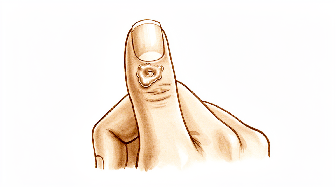
Mucous cysts of the finger are common lesions frequently associated with degenerative changes at the distal interphalangeal joints [26]. These cysts typically present in middle-aged or elderly patients who exhibit other signs of degenerative arthritis [41]. The lesion is usually small and firm, located to one side as it emerges from the joint beside the extensor mechanism [26], specifically lying dorso-lateral to the distal interphalangeal joint [41]. Occasionally, the cyst may spread across the entire dorsum of the digit between the distal interphalangeal joint and the nail fold [26]. The overlying skin is frequently thin, meaning minimal trauma may result in rupture [26]. Rupture of the cyst results in the discharge of clear gelatinous fluid [41].
Histology and Ultrastructure
Digital mucous cysts and wrist ganglia show no anatomic difference at the light and scanning electron microscopic levels [22]. The wall of digital mucous cysts consists of multidirectional strata of collagen fibers with rare fibroblast-like cells on the inner surface [22]. There are no signs of myxomatous degeneration in the wall of digital mucous cysts [22]. The surface of the cyst wall is generally flat, with some areas showing localized elevations that may represent degenerating cells or multifunctional mesenchymal cells [22]. Multifunctional mesenchymal cells beneath the lining of the cyst are postulated to produce collagen fibers, elastic fibers, and the interfibrillary mucopolysaccharide matrix [22]. The ganglion fluid may be produced by these multifunctional cells and reaches the cyst cavity by flowing through the porous collagen matrix of the cyst wall [22]. The porous nature of the cyst wall is demonstrated by a porous network of collagen fibers in the wall on cross section [22]. Histological descriptions by Gross and King suggested there was no connection between the cyst and the underlying joint and that there was no endothelial lining [41]. Gross and King concluded that the cysts resulted from degeneration of connective tissue within the dermis [41]. Nishimura et al. supported the view of dermal degeneration based on the chemical composition of the contents of mucous cysts differing from that of "ordinary" ganglia [41].
Pathogenesis Theories
Theories on the pathogenesis of mucous cysts include myxoid degeneration, repeated local trauma, vascular insufficiency, and synovial herniation [26]. Studies suggest that mucous cysts arise due to excessive hyaluronic acid production from a degenerate distal interphalangeal joint [26]. Other theories proposed for the pathogenesis of mucous cysts include embryological abnormalities resulting in extra-articular synovial tissue [41].
Classification
Clinical Presentation: Mucous cysts are common lesions found in association with osteoarthritis of the distal interphalangeal joint [41]. The cyst is small, firm, and lies dorso-lateral to the joint [41]. Periungual myxoid cysts have a characteristic clinical presentation [14].
Histopathology: The wall of digital mucous cysts consists of a porous network of collagen fibers [22]. The fluid within the digital mucous cyst is produced by underlying mesenchymal cells [22].
Other Considerations: The skin over the cyst is frequently thin, and minimal trauma may result in rupture and discharge of clear gelatinous fluid [41]. The presence of a definite pedicle connecting the cyst to the joint capsule indicates the lesion is analogous to a ganglion [24].
Clinical Presentation
Mucous cysts typically present as a painless subungual mass accompanied by nail deformity [4]. On inspection, the overlying skin may appear thinned [39], and a fistula can be present [9]. The fluid within these digital mucous cysts is produced by underlying mesenchymal cells [22]. Clinically, these lesions are associated with marginal osteophytes [1].
Investigations
Physical Examination: A careful physical examination is essential to direct care and future testing if indicated [31]. A high index of suspicion is necessary to establish the diagnosis, as the disease often presents with non-specific clinical features and delayed diagnosis [35].
Laboratory: Diagnostic tests such as serum laboratory studies are useful in the determination of pathology but can be expensive, time consuming, and often nonspecific [31].
MRI: High-resolution magnetic resonance imaging findings raise important questions about the role of ligaments in hand osteoarthritis [36].
Ultrasound: Ultra-sonography provides a new ability to study the complex anatomical structures of the hand, with the technique being quick and simple to perform [34].
Treatment
Non-Operative
Aspiration with injection of steroid serves as a convenient conservative option that can be performed in an outpatient setting; however, this technique is associated with a recurrence rate of 50% [26].
Operative
Indications: Surgery provides definitive treatment with no major long-term problems, as demonstrated in a review of 134 cases with a minimum 2-year follow-up evaluation [12]. Radical treatment involving complete excision of the stalk and underlying osteophytes appears to reduce recurrence rates [26]. In a consecutive series, 80% of mucous cysts show osteophytes radiologically, while 20% exhibit minimal changes [98].
Surgical Approach / Technique: Both biquadrangular and Hueston flaps yield comparable aesthetic satisfaction and clinical outcomes following surgical excision [88]. An osteophyte-sparing technique is unlikely to lead to a clinically significant loss of motion [28]. When performing osteophyte excision without cyst excision, the principle is to avoid causing further damage to the friable skin, as the skin is not the culprit of the cyst [17]. A critical step in this approach is draining the cyst at the point where it communicates with the distal interphalangeal joint capsule [98]. If the cyst is small and can be easily excised with primary closure, leaving it in position during osteophyte excision is considered inappropriate [98].
Outcomes and Complications: Surgical treatment demonstrates low recurrence rates, with one series reporting a 2% recurrence rate (2 of 113) [18]. Another study noted 2 cyst recurrences (3%) and only 1 secondary procedure following mucous cystectomy and osteophytectomy using a random nonadvancement flap technique [3]. Complications associated with surgical treatments include infection, recurrence, nail deformity, swelling, stiffness, and pain [26].
Complications
Infection (PJI): Complications are rare and occur only in cysts associated with fistula [9].
Other Considerations: Recurrence rates vary significantly by treatment modality. Aspiration with injection of steroid is followed by a recurrence rate of 50% [26]. Surgical excision with a local advancement skin flap demonstrated a low recurrence rate of 1.4% [15]. A total dorsal capsulectomy alone did not lead to any recurrence [10]. In a series of surgically treated mucous cysts, the recurrence rate was 2% (2 of 113) [18].
Recovery
Other Considerations: In a series of 12 consecutive cases involving free composite grafts for claw nail deformity, there was no complaint regarding the donor sites [30].
Key Evidence
- [L4] Excision of the cyst and complete removal of the marginal osteophyte eradicates mucous cysts with extremely rare recurrence. [1] (10.2106/00004623-197355030-00013)
- [L4] This report provides additional basis for prophylactic extirpation of mucous cyst of the finger. [2] (10.1016/s0363-5023(84)80152-5)
- [L4] There were 2 cyst recurrences in the study cohort (3%) and only 1 secondary procedure. [3] (10.1177/15589447241288257)
- [L5] Surgical excision is the definitive treatment for mucous cysts and should include osteophyte debridement. [4] (10.1016/j.jhsa.2006.01.013)
- [L4] The scientific data regarding mucous cysts consist almost entirely of retrospective studies, and much of what is done or recommended is based on expert opinion. [5] (10.1016/j.jhsa.2010.01.029)
- [L4] Surgical treatment of mucous cysts is not a totally benign procedure, and complications do occur contrary to literature reporting glowing results. [7] (10.1016/s0266-7681(97)80067-8)
- [Paper] In-office excision is a safe and effective option for treating digital mucous cysts. [8] (10.1177/15589447251350168)
- [L4] Complications are rare and occur only in cysts associated with fistula, justifying their early surgical treatment. [9] (10.1016/j.main.2015.06.001)
- [L4] A total dorsal capsulectomy alone was a simple treatment for mucous cysts and did not lead to any recurrence. [10] (10.1016/j.jhsa.2014.03.004)
- [L4] Surgery provided definitive treatment with no major long-term problems. [12] (10.1053/jhsu.2003.50088)
- [L4] Periungual myxoid cysts have a characteristic clinical presentation. [14] (10.21276/aimdr.2017.3.5.dt1)
- [L4] Surgical excision with a local advancement skin flap is a reliable treatment for digital mucous cysts, demonstrating a low recurrence rate of 1.4% and high patient satisfaction regarding the scar and willingness to undergo the procedure again. [15] (10.1177/1753193413508540)
- [L5] The authors state that their principle is not to do more damage to the friable skin because the skin is not the culprit of the cyst. [17] (10.1177/1753193414546990)
- [L4] The 2% recurrence rate (2 of 113) obtained in this series illustrates the advantage of this procedure. [18] (10.1016/0363-5023(94)90071-x)
- [L4] The authors present a simple modification of the rotation flap originally described by Kleinert et al after excision of large mucous cysts. [19] (10.1097/bth.0b013e31803c4e19)
- [L4] Osteophyte excision without cyst excision may be a good treatment choice for mucous cyst of the finger, providing a less invasive method with complete resolution in most cases. [21] (10.1177/1753193413478549)
- [L5] [22] (10.1016/s0363-5023(88)80143-6)
- [L4] The presence of a definite pedicle connecting the cyst to the joint capsule indicates the lesion is analogous to a ganglion. [24] (10.2106/00004623-197254070-00008)
- [L4] [26] (10.1177/1753193408103498)
- [L4] The described technique is unlikely to lead to a clinically significant loss of motion. [28] (10.5435/jaaosglobal-d-21-00164)
- [L4] In the series of 12 consecutive cases, the largest size of the grafted tissue was 9 mm in width, but there was no complaint regarding the donor sites. [30] (10.1097/00130911-199803000-00006)
- [L4] Ultra-sonography provides a new ability to study the complex anatomical structures of the hand, with the technique being quick and simple to perform. [34] (10.1016/0266-7681(90)90133-o)
- [L5] A high index of suspicion is necessary to establish the diagnosis, as the disease often presents with non-specific clinical features and delayed diagnosis. [35] (10.1007/s00402-012-1527-2)
- [L4] The findings also raise important questions about the role of ligaments in hand OA. [36] (10.1002/art.21210)
- [L4] It allows excision of the cyst and thinned skin with no added risk to the nail matrix. [39] (10.1016/j.jhsa.2017.03.013)
- [L4] [41] (10.1016/0266-7681_90_90064-b)
- [L4] Both biquadrangular and Hueston flaps yield comparable aesthetic satisfaction and clinical outcomes after surgical excision of digital mucous cysts. [88] (10.1177/17531934261433822)
- [Commentary] [98] (10.1177/1753193413510663)
References
[1] Marginal Osteophyte Excision in Treatment of Mucous Cysts. The Journal of Bone & Joint Surgery. 1973. DOI: 10.2106/00004623-197355030-00013
[2] Infected mucous cyst of the finger. The Journal of Hand Surgery. 1984. DOI: 10.1016/s0363-5023(84)80152-5
[3] Outcomes of Mucous Cystectomy and Osteophytectomy Using a Random Nonadvancement Flap Technique. HAND. 2024. DOI: 10.1177/15589447241288257
[4] Painless Subungual Mass With Nail Deformity. The Journal of Hand Surgery. 2006. DOI: 10.1016/j.jhsa.2006.01.013
[5] Mucous Cysts. The Journal of Hand Surgery. 2010. DOI: 10.1016/j.jhsa.2010.01.029
[7] Complications Following Mucous Cyst Excision. Journal of Hand Surgery. 1997. DOI: 10.1016/s0266-7681(97)80067-8
[8] Safety and Cost-Effectiveness of In-Office Digital Mucous Cyst Excisions. HAND. 2025. DOI: 10.1177/15589447251350168
[9] Surgical treatment of mucous cysts by subcutaneous excision and osteophyte resection: Results in 68 cases at a mean 6.63 years’ follow-up. Chirurgie de la Main. 2015. DOI: 10.1016/j.main.2015.06.001
[10] Total Dorsal Capsulectomy for the Treatment of Mucous Cysts. The Journal of Hand Surgery. 2014. DOI: 10.1016/j.jhsa.2014.03.004
[12] Treatment of mucous cysts of the fingers: Review of 134 cases with minimum 2-year follow-up evaluation. The Journal of Hand Surgery. 2003. DOI: 10.1053/jhsu.2003.50088
[14] Excision of Periungual Myxoid Cyst Using Proximal Nail Fold Flap. Annals of International medical and Dental Research. 2017. DOI: 10.21276/aimdr.2017.3.5.dt1
[15] A reliable surgical treatment for digital mucous cysts. Journal of Hand Surgery (European Volume). 2013. DOI: 10.1177/1753193413508540
[17] Re: Lee, H.-J., Kim, P.-T., Jeon, I.-H., Kyung, H.-S., Ra, I.-H. and Kim, T.-K. Osteophyte excision without cyst excision for a mucous cyst of the finger. J Hand Surg Eur. 2014, 39: 258–61. Journal of Hand Surgery (European Volume). 2014. DOI: 10.1177/1753193414546990
[18] Outcome of surgically treated mucous cysts of the hand. The Journal of Hand Surgery. 1994. DOI: 10.1016/0363-5023(94)90071-x
[19] Flap Advancement Coverage After Excision of Large Mucous Cysts. Techniques in Hand & Upper Extremity Surgery. 2007. DOI: 10.1097/bth.0b013e31803c4e19
[21] Osteophyte excision without cyst excision for a mucous cyst of the finger. Journal of Hand Surgery (European Volume). 2013. DOI: 10.1177/1753193413478549
[22] A surface ultrastructure study of ganglia and digital mucous cysts. The Journal of Hand Surgery. 1988. DOI: 10.1016/s0363-5023(88)80143-6
[24] Etiology and Treatment of the So-Called Mucous Cyst of the Finger. The Journal of Bone & Joint Surgery. 1972. DOI: 10.2106/00004623-197254070-00008
[26] Use of Wolfe Graft for the Treatment of Mucous Cysts. Journal of Hand Surgery (European Volume). 2009. DOI: 10.1177/1753193408103498
[28] Osteophyte-Sparing Treatment of Mucous Cysts: Case Analysis and Surgical Technique. JAAOS: Global Research and Reviews. 2021. DOI: 10.5435/jaaosglobal-d-21-00164
[30] Free Composite Graft to Claw Nail Deformity Using the Ice Water Cooling Method. Techniques in Hand & Upper Extremity Surgery. 1998. DOI: 10.1097/00130911-199803000-00006
[31] Orthopaedic Knowledge Update 13 Ebook Without Multimedia. Anatomy, Evaluation, Clinical Examination, and Imaging > Evaluation and Clinical Examination: Current Concepts.
[34] Diagnostic medical ultrasound in the management of hand injuries. The Journal of Hand Surgery: Journal of the British Society for Surgery of the Hand. 1990. DOI: 10.1016/0266-7681(90)90133-o
[35] Tuberculous extensor tenosynovitis of the hand. Archives of Orthopaedic and Trauma Surgery. 2012. DOI: 10.1007/s00402-012-1527-2
[36] High‐resolution magnetic resonance imaging for the assessment of hand osteoarthritis. Arthritis & Rheumatism. 2005. DOI: 10.1002/art.21210
[39] The Zitelli Bilobed Flap on Skin Coverage After Mucous Cyst Excision: A Retrospective Cohort of 33 Cases. The Journal of Hand Surgery. 2017. DOI: 10.1016/j.jhsa.2017.03.013
[41] Mucous Cyst of the Distal Interphalangeal Joint: Treatment by Simple Excision or Excision and Rotation Flap. Journal of Hand Surgery. 1990. DOI: 10.1016/0266-7681_90_90064-b
[88] Aesthetic outcome of biquadrangular and Hueston dorsal flaps in digital mucous cyst surgery. Journal of Hand Surgery (European Volume). 2026. DOI: 10.1177/17531934261433822
[98] Commentary on Lee et al. Osteophyte excision without cyst excision for a mucous cyst of the finger. Journal of Hand Surgery (European Volume). 2014. DOI: 10.1177/1753193413510663