Clinicians › Elbow
Distal biceps repair
Surgeon-side topic for distal biceps repair. Backed by 437 articles from the corpus, retrieved via combined MeSH + title-text matching.
For patients: a plain-language version of this topic is available. See the patient guide.
Overview¶
Distal biceps tendon repair is indicated for acute and chronic ruptures, as well as partial tears that have failed nonsurgical management [11, 18]. Surgical intervention reliably restores supination and flexion strength in appropriate patients [21]. Clinical and functional outcomes at more than one year post-repair are excellent, with no measurable differences in biceps strength between patients who develop radioulnar heterotopic ossification and those who do not [1, 4]. Patients treated with a delay of more than 21 days can expect functional outcomes similar to those treated acutely [7]. While most morbidity is attributed primarily to delayed repair and secondarily to extensive anterior exposure, the overall rate of serious complications remains low regardless of the surgical approach or technique employed [8, 10].
The procedure is associated with a 7.5% major complication rate and a 4.5% reoperation rate [5]. Complication rates are generally low, though the overall frequency of reported complications is higher for single-incision repair than for double-incision repair [3, 15]. Minimally invasive techniques and specific fixation methods, including cortical button fixation and tensionable repairs with intramedullary knotless all-suture anchors, demonstrate safety and effectiveness with low major complication rates [16, 20, 27, 28]. Both single-incision Endobutton and two-incision modified Boyd–Anderson techniques are effective and similarly safe [22]. A modified two-incision repair allows for a safe immediate active range of motion protocol, facilitating early return of nearly full range of motion and strength without clinically significant disability [37].
Long-term patient-reported outcomes are excellent, with high degrees of patient satisfaction observed in large cohorts of two-incision repairs and single-incision knotless cortical button devices [9, 42, 44]. Although transient minor complications are common with single-incision approaches, major complication rates remain low [44]. Patients undergoing distal biceps reconstruction with a graft exhibit similar failure rates, reoperation rates, and final range of motion compared to primary repair [6]. Surgical repair consistently provides good patient-scored outcomes [14, 2].
Anatomy & Pathophysiology¶
Bony Anatomy¶
The elbow is a trocho-ginglymoid joint comprising medial and lateral articulations that provide bony stability [73]. The ulnohumeral joint forms where the trochlea articulates with the ulna within the greater sigmoid notch, while the radiocapitellar joint involves the capitellum and radial head [73]. The proximal radioulnar joint maintains radial approximation to the ulna via the annular ligament [73]. The radial head is a concave elliptical structure covered by articular cartilage along the radiocapitellar joint and approximately 270° of the articular margin [73].
The distal humeral articulation is angled 30° from the longitudinal axis [73] and 30 degrees anterior to the humeral shaft axis [79]. The axis of rotation is angulated 5° to 7° in the coronal plane relative to the epicondylar axis, with the medial side positioned more distally than the lateral side [73]. The distal humerus articular surface aligns in 4 degrees to 8 degrees of valgus relative to the shaft [84] and is angulated 35 to 40 degrees anteriorly in the sagittal plane [84]. The normal valgus carrying angle is 5 to 10 degrees for men and 10 to 15 degrees for women [79]. The distal humerus consists of medial and lateral columns, with the medial column diverging from the humeral shaft at a 45-degree angle and the lateral column diverging at a 20-degree angle [79, 81].
The trochlea features a 300-degree arc of cartilage [81] and is covered by articular cartilage anteriorly, inferiorly, and posteriorly, creating an arc of almost 270 degrees [84]. The olecranon fossa matches the olecranon to accept it during extension, while the coronoid fossa matches the coronoid to accept it during flexion [84]. In full extension, 60% of axial load transmits through the radiocapitellar joint [79].
Normal elbow range of motion spans 0° to 140° from extension to flexion, with 75° and 85° in pronation and supination respectively [46]. Alternatively, normal flexion/extension is described as 0 to 150 degrees, and normal forearm pronosupination as 80 to 85 degrees in each direction [79]. The functional arc for the elbow is 100° for flexion and extension and forearm rotation [46], or specifically 30 to 130 degrees flexion/extension and 50 degrees pronosupination [79].
Soft Tissue Anatomy¶
The biceps brachii inserts at the ulnar margin of the radial tuberosity, with the long head positioned proximal and the short head distal [74]. The brachialis, the strongest elbow flexor, attaches to the coronoid 11 mm distal to the tip [74]. The triceps, the primary elbow extensor, inserts on the olecranon process [74].
The medial collateral ligament (MCL) serves as the primary valgus stabilizer of the elbow [74, 79]. It originates on the posterior medial epicondyle and inserts on the sublime tubercle of the medial coronoid process [79]. The anterior bundle of the MCL is the most important component for stability [74] and acts as the primary restraint to valgus stress within functional elbow ROM [79]. The posterior bundle becomes taut at flexion beyond 120 degrees [74] and serves as the primary restraint to valgus stress with the elbow in maximal flexion [79]. Stability in full elbow extension is provided by the MCL, joint capsule, and ulnohumeral articulation [79].
The lateral collateral ligament complex functions as an important restraint to varus and posterolateral rotatory instability [84]. It consists of the radial collateral ligament, the lateral ulnar collateral ligament (LUCL), and the annular ligament [84]. The radial collateral ligament originates from an isometric point on the lateral epicondyle and fans out to attach to the annular ligament [84]. The LUCL arises from the isometric point on the lateral epicondyle and attaches to the crista supinatoris of the proximal ulna [84]. The annular ligament attaches to the anterior and posterior margins of the lesser sigmoid notch [84]. The lateral ulnar collateral ligament is the posterolateral stabilizer of the elbow [74].
Osborne’s ligament stabilizes the ulnar nerve in the cubital tunnel [74]. The ligament of Struthers is a variant anatomy arising from the supracondylar process to attach to the medial epicondyle and represents a potential site of median nerve compression [74].
Vascular & Neural Anatomy¶
The median nerve and brachial artery are located just superficial to the anteromedial joint capsule and brachialis muscle [85]. The posterior interosseous nerve is just superficial to the anterior joint capsule at the level of the radiocapitellar joint and courses longitudinally along the medial side of the capitellum [85]. At the level of the radial neck, the posterior interosseous nerve may come in direct contact with the joint capsule [85]. The ulnar nerve is located just superficial to the posteromedial joint capsule [85].
Biomechanics & Pathophysiology¶
The biceps muscle is the strongest supinator of the forearm and assists the brachialis in elbow flexion [41]. It is more active in flexion of the supinated forearm than in flexion of the pronated forearm [41]. There is little, if any, electrical activity in the biceps muscle when flexion of the pronated forearm is attempted, unless a substantial pronating force resists supination [41]. The contribution of the biceps to forearm supination increases with elbow flexion, reaching a maximum at about 90 degrees of flexion [41]. Beyond 90 degrees of elbow flexion, there is no substantial increase in biceps activity as the muscle-tendon unit shortens [41]. Biceps tendon rotation does not play a role in supination moment arm or flexion force [132].
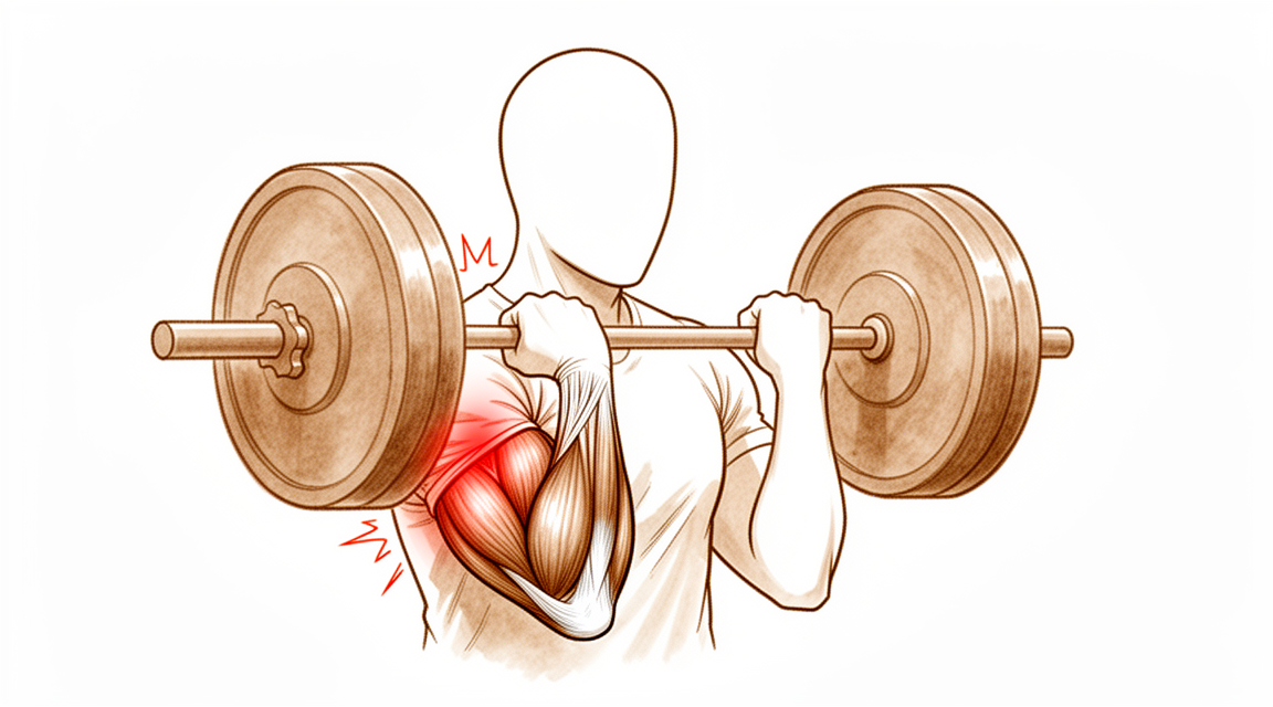
Rupture of the distal biceps tendon is most likely to occur in the dominant extremity of men between the fourth and sixth decades of life [41]. The average age at rupture is approximately 50 years, with a range of 18 to 72 years [41]. All reported cases of complete distal biceps tendon rupture have occurred in men, while partial rupture has been reported in women [41]. Distal biceps injuries frequently occur in middle-aged males after an eccentric load to the elbow in flexion [123]. The mechanism is usually a single traumatic event involving an unexpected extension force applied to an arm flexed to 90 degrees [41]. Ruptures were most commonly observed in weightlifting with a slightly flexed or isometrically extended elbow and forearm supination [147].
The distal biceps tendon typically avulses from the radial tuberosity, although ruptures within the tendon substance and at the musculotendinous junction have been reported [41]. An intact lacertus fibrosus contributes to elbow strength and should be preserved in distal biceps tendon repair [138]. The trough technique for distal biceps repair resulted in a significant decrease in the moment arm of a 60° supinated forearm and a significant reduction in radial tuberosity height [124]. Two tested techniques for distal biceps repair had the same effect on flexion force [151]. Distal biceps tendon repairs using cortical button fixation have the potential of creating important biomechanical consequences in the proximal radius but may not be clinically relevant during routine physiologic conditions [109].
Classification¶
Complete vs. Partial: Distal biceps tendon ruptures are classified as either complete or partial [67]. Partial ruptures represent a spectrum of patterns with varying involvement of the long head and short head tendons [160]. These injuries are exceedingly rare, having been reported only in small series and case reports [67]. There is no formal classification system for partial distal biceps tendon ruptures [67].
Acute vs. Chronic: Complete distal biceps tendon ruptures are subdivided into acute and chronic based on the duration of time from injury [67]. Injuries occurring within 4 weeks of injury are classified as acute [67]. Injuries presenting after 4 weeks are classified as chronic [67].
Lacertus Fibrosus Integrity: Chronic distal biceps tendon ruptures are further separated based on the integrity of the lacertus fibrosus [67]. A torn lacertus fibrosus is associated with proximal retraction of the biceps tendon, scarring of the tendon to the brachialis muscle, and myostatic contraction [67]. An intact lacertus fibrosus usually restricts the tendon from retracting proximally [67]. An intact lacertus fibrosus may facilitate the repair of chronic distal biceps tendon ruptures [67].
Other Considerations: The distal biceps tendon has a complex fiber arrangement that spirals in a predictable manner [155]. This complex fiber arrangement is not currently accounted for in surgical repair techniques [155]. The distal biceps tendon has a bifid insertion [31]. Isolated rupture of the short head component of a bifurcated distal biceps tendon is a recognized entity [31]. All reported cases of isolated single head rupture of the distal biceps involved the short head tendon [31].
Clinical Presentation¶
Demographics and Epidemiology¶
Distal biceps tendon rupture predominantly affects men between the fourth and sixth decades of life, with an average age of approximately 50 years and a reported range of 18 to 72 years [41]. The injury most frequently involves the dominant extremity [41]. In contrast, distal biceps tendon tears in women present differently than in men [32]. Women account for only 3.2% of patients sustaining a distal biceps tendon tear [40]. Within this female population, partial tears are statistically more common than complete ruptures [40]. Bilateral distal biceps ruptures occurred in 7.3% of patients in a cohort of 190 repairs [40].
Mechanism and Pathology¶
The mechanism of injury is usually a single traumatic event involving an unexpected extension force applied to an arm flexed to 90 degrees [41]. The tendon typically avulses from the radial tuberosity, although ruptures within the tendon substance and at the musculotendinous junction have been reported [41]. Some authors describe the rupture as occurring in stages, with initial disruption of the insertion to the radial tuberosity [41]. The bicipital aponeurosis may or may not rupture acutely during distal biceps tendon injury [41]. Isolated single head rupture of the distal biceps involves the short head tendon in all reported cases [31]. Patients with isolated short head tears typically present with a clear history of acute injury and no history of prodromal symptoms [31]. The mean age for isolated short head distal biceps tears is 42 years, with a range of 33 to 58 years [31]. Bruising was documented in 5 of 9 patients with isolated short head distal biceps tears [31].
Diagnostic Findings¶
MRI was performed in all patients with isolated short head distal biceps tears, confirming the diagnosis in all cases [31]. In one case of isolated short head tear, the MRI was reported as indicating a complete tear, but the long head tendon was found to be intact during surgery [31]. The overall sensitivity and specificity of MRI were 92.4% and 100%, respectively, in detecting distal biceps tendon ruptures [110]. For complete distal biceps tears, the sensitivity and specificity of MRI were 100% and 82.8%, respectively [110]. For partial distal biceps tears, the sensitivity and specificity of MRI were 59.1% and 100%, respectively [110]. MRI is extremely sensitive in diagnosing complete distal biceps tears but is substantially less sensitive in diagnosing partial tears [110].
Investigations¶
Imaging¶
Plain radiography: Plain radiographs remain the hallmark and best screening test for elbow evaluation [46].
MRI: MRI has a negative predictive value of 99% for distal biceps tendon pathology, indicating that patients without signal changes on MRI may be assumed to have no distal biceps tendon pathology [171]. Both the flexion-abduction-supination (FABS) view and standard elbow MRI demonstrate high sensitivity and specificity for the diagnosis of partial distal biceps tendon injuries, with no significant differences in sensitivity and specificity found between the two techniques [180]. MRI was performed in all patients with isolated short head tears of the distal biceps, confirming the diagnosis in all cases [31]. In one patient with an isolated short head tear, MRI was reported as indicating a complete tear, but the long head tendon was found to be intact during surgery [31]. MRI can be used to evaluate ligaments and tendons of the elbow, but it is rarely indicated for stiffness evaluation [87].
CT: CT is helpful when assessing for malunion architecture and the location and pattern of osteophytes and/or loose bodies in the elbow [87]. Three-dimensional CT is used to check for heterotopic ossification in the elbow [87].
Other Considerations: Patients with a smaller radioulnar window have a higher risk of rupturing the distal biceps tendon [183].
Physical Examination¶
The physical exam for the elbow is directed by history and the location of the patient's pain in the anterior, posterior, medial, or lateral aspect of the elbow [46]. Active and passive flexion, extension, supination, and pronation should be evaluated during physical examination of the elbow [87]. A goniometer should be used for accurate measurement of elbow range of motion [87]. The contralateral elbow should be examined for comparison during physical examination [87].
Treatment¶
Non-Operative¶
Nonoperative treatment for partial distal biceps tendon tears yields moderate success rates of 47%, with no significant differences between strategies, though injection therapy offers the quickest relief [119]. Surgical management of distal biceps tears was a predictor of improved patient-reported outcomes only for patients with complete tears [50].
Operative¶
Indications: Operative management of complete distal biceps and triceps tendon ruptures is required in most cases, especially for active patients, laborers, and athletes wishing to return to competition [162]. The indications for a small anterior incision for repairing a distal biceps rupture are a partial tear that has been diagnosed with certainty or a complete tear that is less than approximately 3 weeks old [54].
Surgical Approach / Technique: The clinical evidence reported in the literature to date does not support a clear single approach that should be used as the standard of care for distal biceps tendon ruptures [17]. Both surgical techniques for distal biceps tendon repair are effective and are similarly safe methods of treatment [22]. Distal biceps tendon repair surgery can be safely and successfully executed with either a single- or double-incision approach [48]. Until further information is available, both one- and two-incision methods are acceptable for the repair of a ruptured distal biceps tendon [101]. The authors prefer reattachment of the distal biceps tendon rupture to the radial tuberosity through a single anterior exposure and the use of suture anchors for fixation [156]. Anatomic reattachment of the distal biceps tendon is a successful operative treatment option [25]. When diagnosed appropriately, selective disruption of the short head of the biceps distal tendon may be effectively treated with anatomic repair [11].
Fixation Methods: Cortical Button and Suture Anchor Fixation for Distal Biceps Tendon Repair Provide Similar Clinical Outcomes and Complication Profiles [24]. Intramedullary Unicortical Button and All‐Suture Anchors Provide Similar Maximum Strength for Onlay Distal Biceps Tendon Repair [163]. Using suture anchors to repair the biceps tendon may predispose the repaired tendon to impingement when compared with other fixation techniques [169]. Distal biceps repair with cortical button fixation places the PIN at risk of injury regardless of the approach used [30].
Timing and Chronic Injuries: Reinsertion of a distal biceps through a 2-incision approach should be performed within 2 weeks of the injury, when possible [57]. Despite a high rate of initial complications, patients treated with distal biceps tendon repair after a delay (>21 days) can expect similar functional outcomes to those treated acutely [7]. Late reconstruction of the distal biceps tendon results in improved but not full strength in flexion and supination, with deficits of 13% to 20% in flexion and 14% to 15% in supination compared to the uninvolved side [45]. Chronic distal biceps tendon ruptures can be repaired successfully with a single incision using a suture button technique without the use of a graft [51]. Reconstruction of a chronic distal biceps tendon rupture with a single incision, suture anchors and tibialis anterior allograft achieves good to excellent clinical results with minimal complications [174]. Such an incision is contraindicated in chronic tears because the tendon may be adherent to the surrounding tissues proximally and the tunnel to the bicipital tuberosity closed [54]. Patients who have delayed repair frequently require a more significant anterior dissection and the use of tendon graft to restore length [54].
Rehabilitation and Post-operative Care: Early motion after distal biceps tendon repair with cortical button fixation is well tolerated and does not appear to be associated with adverse outcomes [69]. This suggests that patients undergoing open subpectoral biceps tenodesis may be managed using either early or delayed active motion protocols without compromising functional outcome [64]. Based on our findings and the risk of gastrointestinal complications, we do not recommend indomethacin prophylaxis for two-incision distal biceps repairs [184].
Other Considerations: Surgical repair of distal biceps tendon ruptures has an overall low rate of serious complications, regardless of approach or technique [10]. Minor complications were common after distal biceps tendon repair; however, most were sensory nerve injuries that resolved with time [33]. Repair of distal biceps ruptures using an Endobutton fixation results in nearly normal return of strength and function, which is significantly better than in those managed nonoperatively [68]. WC patients who underwent distal biceps tendon repair took longer to return to work and had worse DASH scores than non-WC patients [65]. Despite the cited approach-related morbidity, the authors report excellent long-term patient-reported outcomes for the double-incision distal biceps repair technique [9]. A 2-incision distal biceps repair via nonabsorbable suture allows for immediate protected range of motion, reliably achieving nearly full range of motion with excellent return of strength and function [62]. A double incision technique for distal biceps tendon repair is a minimally invasive procedure with reliable results [88]. The mini-open 2-incision technique is a safe and effective method for repairing distal biceps tendon ruptures, resulting in pleasing cosmesis with minimal surgical dissection [97]. A 2-incision approach for distal biceps tendon repair with suture anchor fixation can restore elbow function and satisfy patients [56]. In this large cohort of 2-incision distal biceps repairs, we found a high degree of patient satisfaction and a low complication rate [42]. Acute distal biceps tendon repair using cortical button fixation was found to result in excellent patient-reported outcomes and health-related quality of life [165]. The intramedullary fixation button technique to repair the distal biceps tendon has excellent functional outcomes at 6 months [166]. In this series, minimally invasive distal biceps repair was safe and effective with a low rate of major complications [28]. The data support the use of this technique and implant combination for acute and chronic distal biceps tendon rupture, demonstrating a low major complication rate and high patient satisfaction despite common transient minor complications [44]. Current biomechanical and clinical literature supports this as producing a functionally equivalent construct compared with double-incision techniques using bone tunnels without increasing, and likely minimizing, the associated complications [53]. The method may improve treatment outcome by preserving anatomic features key to distal biceps tendon function, enhancing repair integrity, and improving patient safety [16]. This will help surgeons determine the best method of fixation and approach with the goal of minimizing postoperative complications in patients with distal biceps ruptures [12]. Incorporating this into a distal biceps rupture procedure gives the surgeon a new alternative for fixation [43]. This technique is simple and cost effective, minimizes additional morbidity in revision distal biceps surgery, and can potentially be used in revision surgery in other anatomic locations where cortical buttons were used [49]. The novel technique for distal biceps reconstruction demonstrated similar patient-reported outcomes scores as prior studies with high patient satisfaction while implementing techniques that significantly decrease the gap between the bone-tendon junction and safely utilize distal fixation methods with the highest load to failure [158]. In a single surgery center, single-incision distal biceps repairs utilizing an implant were performed more expeditiously than double-incision repairs with a transosseous technique but incurred greater surgical costs [159]. Biceps strength can be maintained with no measurable differences in clinical outcome when compared to individuals who do not suffer this complication following distal biceps repair [4]. Surgeons treating distal biceps tendon injuries should be aware of this entity and the treatment options available [26]. Endoscopy is a useful and minimally invasive diagnostic and treatment approach for distal biceps tendon pathology [23]. The authors have performed this technique in over 50 patients with a partial distal biceps tendon tear [13]. Endoscopic distal biceps tendon repair was technically feasible with both fixation techniques without a major rise in compartment pressures [102].
Complications¶
Overall Complication Rates: Distal biceps repair carries a major complication rate of 7.5% and a reoperation rate of 4.5% [5]. The largest analysis of complications after distal biceps repair indicates a major complication rate of 4.6% [36]. Complication rates did not differ significantly between one and two-incision distal biceps repairs [189]. Complication rates after distal biceps tendon repair performed by newly trained surgeons were similar to those previously reported in large cohort studies [143]. The early complication rate following partial distal biceps tendon repair was 20.5% [34]. Data support the use of single incision with knotless cortical button device for acute and chronic distal biceps tendon rupture, demonstrating a low major complication rate and high patient satisfaction despite common transient minor complications [44]. This study supports the use of SPOC distal biceps repair for both partial and complete biceps ruptures, with a low rerupture rate, similar complication rate to other commonly used techniques, and low return to OR rate [190]. In this large cohort of 2-incision distal biceps repairs, a high degree of patient satisfaction and a low complication rate were found [42].
Nerve Palsy: Nerve injury is the most common complication after distal biceps tendon repair [143]. The incidence of posterior interosseous nerve palsy after a single-incision distal biceps repair was 3.2% in the reported series [191]. The most common complication in a comparative study of three fixation techniques was neurapraxia, with 6 cases [149].
Stiffness / Arthrofibrosis: Two patients in the Mitek anchors group required an open arthrolysis due to stiffness of the elbow [149].
Other Considerations: Repair of the distal biceps tendon can lead to complications such as nerve injury, heterotopic ossification, stiffness, brachial artery injury, weakness, complex regional pain syndrome, infection, and rerupture [117]. Despite the cited approach-related morbidity, excellent long-term patient-reported outcomes were reported for the double-incision distal biceps repair technique [9]. The re-rupture rate after primary repair of the distal biceps tendon is low at 1.5% and occurs within 3 weeks of index repair [40]. Rerupture of the distal biceps after primary repair can occur due to technical factors, timing after injury, and patient compliance [63]. All 3 reruptures in a comparative study of three fixation techniques were registered in the Mitek anchors group [149]. Primary repair of chronic distal biceps tendon tears greater than 6 weeks from injury demonstrated excellent patient-reported outcomes and elbow range of motion [39].
Recovery¶
Functional Outcomes and Strength: Distal biceps repair and reconstruction can restore near-normal flexion and supination strength [185]. In cases of chronic biceps ruptures undergoing reconstruction, patients are highly functional and satisfied, although the procedure does not restore peak supination strength [186].
Range of Motion and Rehabilitation Protocols: Early active motion with a 0.9-kg weight restriction may be possible for patients undergoing distal biceps tendon repair using a single-incision EndoButton technique with FiberWire [113]. Repair of acute distal biceps tendon ruptures using a soft tissue button and interference screw technique through a limited anterior incision can allow for accelerated rehabilitation and early return to function [122].
Return to Work: After surgical repair of distal biceps rupture, the majority of patients are able to return to work and sport [35]. Specifically, 1128 (89%) patients were able to fully return to work without any modification of duties [60]. The pooled mean time to return to work after distal biceps repair was 14.37±0.52 weeks [60]. Workers' compensation patients who underwent distal biceps tendon repair took longer to return to work and had worse DASH scores than non-workers' compensation patients [65].
Return to Sport: Patients can expect high levels of return to sport following distal biceps tendon repair with some residual impairment compared with baseline [38]. Following distal biceps repair, 95% of athletes return to sports and 82% to the same preinjury level of competition [66]. The time to return to sports following distal biceps repair is about 40 weeks, depending on the type of sport [66]. Athletes sustaining distal biceps tendon rupture have a high postoperative return to sport rate, independently of selected surgical technique or rehabilitation program [107]. There was a high rate of return to sport after distal biceps tendon repair at 6 months postoperatively [125]. Distal biceps tendon repair in competitive strength athletes resulted in a high return-to-sport rate and excellent recovery [172].
Complications and Re-rupture: Despite the cited approach-related morbidity, the double-incision distal biceps repair technique reports excellent long-term patient-reported outcomes [9].
Key Evidence¶
- [L3] Clinical and functional outcome at more than 1 year after distal biceps tendon repair was excellent in both groups. [1] (10.1016/j.jse.2015.12.007)
- [L3] Overall, patients have excellent outcome after distal biceps tendon rupture repair. [2] (10.1007/s00402-018-3018-6)
- [L1] The overall frequency of reported complications is higher for single-incision distal biceps repair than for double-incision repair. [3] (10.1177/2325967116668137)
- [L4] Biceps strength can be maintained with no measurable differences in clinical outcome when compared to individuals who do not suffer this complication following distal biceps repair. [4] (10.1016/j.jhsa.2007.06.018)
- [L3] Distal biceps repair is associated with a 7.5% major complication rate and 4.5% reoperation rate. [5] (10.1016/j.jse.2018.06.028)
- [L3] Patients that underwent distal biceps reconstruction with a graft had similar failure rates, reoperation rates, and final range of motion compared to primary repair. [6] (10.1016/j.jse.2020.01.062)
- [L3] Despite a high rate of initial complications, patients treated with distal biceps tendon repair after a delay (>21 days) can expect similar functional outcomes to those treated acutely. [7] (10.1016/j.jse.2017.02.025)
- [L4] Most morbidity from repair of the distal biceps tendon can be attributed primarily to a delay in the timing of the repair and secondarily to an extensive anterior exposure. [8] (10.2106/00004623-200011000-00010)
- [L4] Despite the cited approach-related morbidity, the authors report excellent long-term patient-reported outcomes for the double-incision distal biceps repair technique. [9] (10.1177/2325967120944812)
- [L3] The surgical repair of distal biceps tendon ruptures has an overall low rate of serious complications, regardless of approach or technique. [10] (10.1177/0363546517720200)
- [L4] When diagnosed appropriately, selective disruption of the short head of the biceps distal tendon may be effectively treated with anatomic repair. [11] (10.1016/j.jse.2016.09.050)
- [L4] This will help surgeons determine the best method of fixation and approach with the goal of minimizing postoperative complications in patients with distal biceps ruptures. [12] (10.1177/17585732241312212)
- [L4] The authors have performed this technique in over 50 patients with a partial distal biceps tendon tear. [13] (10.1097/bte.0000000000000136)
- [L4] Surgical repair of distal biceps ruptures provides consistently good results in terms of patient-scored outcomes. [14] (10.1016/j.injury.2012.10.029)
- [L3] Complication rates after distal biceps repair are low. [15] (10.5397/cise.2021.00472)
- [L5] The method may improve treatment outcome by preserving anatomic features key to distal biceps tendon function, enhancing repair integrity, and improving patient safety. [16] (10.1016/j.eats.2022.08.019)
- [L5] The clinical evidence reported in the literature to date does not support a clear single approach that should be used as the standard of care for distal biceps tendon ruptures. [17] (10.1097/00132589-200603000-00008)
- [L4] Surgical treatment of partial distal biceps tendon tears is a viable option after failed nonsurgical treatment. [18] (10.1016/j.jhsa.2010.04.024)
- [L5] Distal biceps repair using cortical button fixation has been associated with excellent patient-reported outcomes and satisfaction with minimal complications. [20] (10.1016/j.eats.2023.07.028)
- [L4] Surgical repair of the distal biceps insertion reliably regains both supination and flexion strength in appropriate patients. [21] (10.5435/00124635-201003000-00003)
- [L4] Both surgical techniques for distal biceps tendon repair are effective and are similarly safe methods of treatment. [22] (10.1080/00913847.2016.1129260)
- [L5] Endoscopy is a useful and minimally invasive diagnostic and treatment approach for distal biceps tendon pathology. [23] (10.1016/j.eats.2022.10.003)
- [L4] [24] (10.1016/j.jse.2026.07.033)
- [Case_report] Anatomic reattachment of the distal biceps tendon is a successful operative treatment option. [25] (10.1186/s12891-020-03304-3)
- [L5] Surgeons treating distal biceps tendon injuries should be aware of this entity and the treatment options available. [26] (10.1016/j.tcr.2022.100636)
- [L4] Distal biceps repair using single incision, unicortical button fixation yields a low rate of complications with good clinical results compared to other techniques reported in the literature. [27] (10.1177/2325967119s00365)
- [L4] In this series, minimally invasive distal biceps repair was safe and effective with a low rate of major complications. [28] (10.5397/cise.2023.00227)
- [L5] Distal biceps repair with cortical button fixation places the PIN at risk of injury regardless of the approach used. [30] (10.1016/j.jhsa.2018.09.002)
- [L4] [31] (10.1016/j.jse.2020.04.038)
- [L4] Distal biceps tendon tears in women present differently than in men. [32] (10.1016/j.jse.2010.01.015)
- [L4] Minor complications were common after distal biceps tendon repair; however, most were sensory nerve injuries that resolved with time. [33] (10.1016/j.jhsa.2012.06.022)
- [L4] The early complication rate following partial distal biceps tendon repair was 20.5%. [34] (10.1016/j.jhsa.2025.04.012)
- [L3] After surgical repair of distal biceps rupture, the majority of patients are able to return to work and sport. [35] (10.1177/2325967119s00367)
- [L2] This is the largest analysis of complications after distal biceps repair, indicating a major complication rate of 4.6%. [36] (10.1177/0363546519899933)
- [L4] A modified 2-incision distal biceps repair allows a safe immediate active range of motion protocol with early return of nearly full range of motion and strength, without any clinically significant disability. [37] (10.1177/0363546508323749)
- [L4] Distal biceps tendon rupture is a significant injury; however, patients can expect high levels of return to sport following DBR with some residual impairment compared with baseline. [38] (10.1016/j.jse.2021.01.034)
- [L4] Primary repair of chronic distal biceps tendon tears greater than 6 weeks from injury demonstrated excellent PROMs and elbow ROM. [39] (10.1177/15589447221107691)
- [L4] [40] (10.1016/j.jse.2014.02.006)
- [L5] [41] (10.5435/00124635-199905000-00006)
- [L3] In this large cohort of 2-incision distal biceps repairs, we found a high degree of patient satisfaction and a low complication rate. [42] (10.1016/j.jse.2014.12.032)
- [L4] Incorporating this into a distal biceps rupture procedure gives the surgeon a new alternative for fixation. [43] (10.1097/01.bte.0000160547.61893.79)
- [L4] The data support the use of this technique and implant combination for acute and chronic distal biceps tendon rupture, demonstrating a low major complication rate and high patient satisfaction despite common transient minor complications. [44] (10.1177/17585732211060356)
- [Paper] Late reconstruction of the distal biceps tendon results in improved but not full strength in flexion and supination, with deficits of 13% to 20% in flexion and 14% to 15% in supination compared to the uninvolved side. [45] (10.1097/00132589-200001020-00002)
- [L4] Distal biceps tendon repair surgery can be safely and successfully executed with either a single- or double-incision approach. [48] (10.1016/j.jse.2010.11.009)
- [L5] This technique is simple and cost effective, minimizes additional morbidity in revision distal biceps surgery, and can potentially be used in revision surgery in other anatomic locations where cortical buttons were used. [49] (10.1016/j.eats.2022.11.006)
- [L2] Surgical management of distal biceps tears was a predictor of improved patient-reported outcomes only for patients with complete tears. [50] (10.1016/j.jhsa.2025.12.027)
- [L4] Chronic distal biceps tendon ruptures can be repaired successfully with a single incision using a suture button technique without the use of a graft. [51] (10.1016/j.jse.2020.01.103)
- [L5] Current biomechanical and clinical literature supports this as producing a functionally equivalent construct compared with double-incision techniques using bone tunnels without increasing, and likely minimizing, the associated complications. [53] (10.1097/00130911-200006000-00004)
- [L4] [54] (10.1097/00132589-200203000-00011)
- [L4] A 2-incision approach for distal biceps tendon repair with suture anchor fixation can restore elbow function and satisfy patients. [56] (10.1016/j.jse.2007.07.006)
- [L4] Reinsertion of a distal biceps through a 2-incision approach should be performed within 2 weeks of the injury, when possible. [57] (10.1016/j.jse.2007.04.008)
- [L4] [60] (10.1016/j.jse.2019.12.006)
- [L4] A 2-incision distal biceps repair via nonabsorbable suture allows for immediate protected range of motion, reliably achieving nearly full range of motion with excellent return of strength and function. [62] (10.1016/j.jse.2004.12.003)
- [Case_report] Rerupture of the distal biceps after primary repair can occur due to technical factors, timing after injury, and most importantly, patient compliance. [63] (10.1016/j.jse.2006.09.012)
- [L3] This suggests that patients undergoing open subpectoral biceps tenodesis may be managed using either early or delayed active motion protocols without compromising functional outcome. [64] (10.1177/23259671211026619)
- [L3] WC patients who underwent distal biceps tendon repair took longer to return to work and had worse DASH scores than non-WC patients. [65] (10.1016/j.jse.2012.11.011)
- [L5] [66] (10.1016/j.jisako.2023.02.004)
- [L5] [67] (10.1016/j.csm.2004.06.001)
- [L3] Repair of distal biceps ruptures using an Endobutton fixation results in nearly normal return of strength and function, which is significantly better than in those managed nonoperatively. [68] (10.1016/j.jse.2015.10.008)
- [L1] Early motion after distal biceps tendon repair with cortical button fixation is well tolerated and does not appear to be associated with adverse outcomes. [69] (10.2106/jbjs.20.02047)
- [L4] A double incision technique for distal biceps tendon repair is a minimally invasive procedure with reliable results. [88] (10.1016/j.otsr.2018.10.012)
- [L4] The mini-open 2-incision technique is a safe and effective method for repairing distal biceps tendon ruptures, resulting in pleasing cosmesis with minimal surgical dissection. [97] (10.1016/j.jse.2006.10.021)
- [Paper] Until further information is available, both one- and two-incision methods are acceptable for the repair of a ruptured distal biceps tendon. [101] (10.1097/00132589-200206000-00004)
- [L5] Endoscopic distal biceps tendon repair was technically feasible with both fixation techniques without a major rise in compartment pressures. [102] (10.1016/j.jse.2018.04.020)
- [L4] Athletes sustaining distal biceps tendon rupture have a high postoperative return to sport rate, independently of selected surgical technique or rehabilitation program. [107] (10.1016/j.jse.2022.02.027)
- [L5] Distal biceps tendon repairs using cortical button fixation have the potential of creating important biomechanical consequences in the proximal radius but may not be clinically relevant during routine physiologic conditions. [109] (10.1177/1558944717701236)
- [L3] [110] (10.1016/j.jhsa.2009.08.016)
- [L5] Early active motion with a 0.9-kg weight restriction may be possible in those patients undergoing distal biceps tendon repair using this technique. [113] (10.1007/s00167-010-1348-1)
- [L5] [117] (10.1016/j.ocl.2015.10.003)
- [L3] Nonoperative treatment for partial distal biceps tendon tears yields moderate success rates (47%) with no significant differences between strategies, though injection therapy offers the quickest relief. [119] (10.1016/j.jse.2025.04.017)
- [L4] Repair of acute distal biceps tendon ruptures using a soft tissue button and interference screw technique through a limited anterior incision can allow for accelerated rehabilitation and early return to function. [122] (10.1177/0363546508330130)
- [Paper] Distal biceps injuries frequently occur in middle-aged males after an eccentric load to the elbow in flexion. [123] (10.1016/j.injury.2025.112556)
- [L5] The trough technique resulted in a significant decrease in the moment arm of a 60° supinated forearm and a significant reduction in radial tuberosity height. [124] (10.2106/jbjs.n.01221)
- [L4] There was a high rate of return to sport after distal biceps tendon repair at 6 months postoperatively. [125] (10.1177/03635465241295618)
- [L5] The study showed that biceps tendon rotation does not play a role in supination moment arm or flexion force. [132] (10.1016/j.jses.2019.06.001)
- [L3] An intact lacertus fibrosus contributes to elbow strength and should be preserved in distal biceps tendon repair. [138] (10.1007/s00167-019-05673-2)
- [L4] Complication rates after distal biceps tendon repair performed by newly trained surgeons were similar to those previously reported in large cohort studies, with nerve injury as the most common complication. [143] (10.1016/j.jse.2022.09.014)
- [L4] Distal biceps tendon ruptures were most commonly observed in weightlifting with a slightly flexed or isometrically extended elbow and forearm supination. [147] (10.1186/s12891-022-05546-9)
- [L3] [149] (10.1007/s00167-011-1591-0)
- [L5] The two tested techniques had the same effect on flexion force. [151] (10.1016/j.jse.2014.11.028)
- [L5] The distal biceps tendon spirals in a predictable manner with a complex fiber arrangement that is not currently accounted for in surgical repair techniques. [155] (10.1097/blo.0b013e31802f78aa)
- [L4] The authors prefer reattachment of the distal biceps tendon rupture to the radial tuberosity through a single anterior exposure and the use of suture anchors for fixation. [156] (10.1097/00130911-199812000-00005)
- [L4] The novel technique for distal biceps reconstruction demonstrated similar patient-reported outcomes scores as prior studies with high patient satisfaction while implementing techniques that significantly decrease the gap between the bone-tendon junction and safely utilize distal fixation methods with the highest load to failure. [158] (10.1016/j.xrrt.2026.100713)
- [L4] In a single surgery center, single-incision distal biceps repairs utilizing an implant were performed more expeditiously than double-incision repairs with a transosseous technique but incurred greater surgical costs. [159] (10.1016/j.xrrt.2021.09.006)
- [L4] Partial ruptures of the distal biceps brachii tendon represent a spectrum of patterns with varying involvement of the LH and SH tendons. [160] (10.1016/j.jse.2020.04.021)
- [L5] Operative management of complete distal biceps and triceps tendon ruptures is required in most cases, especially for active patients, laborers, and athletes wishing to return to competition. [162] (10.1016/j.hcl.2016.08.019)
- [L5] This study demonstrates that onlay distal biceps repair with 2 all-suture anchors has similar maximum strength to repair with an intramedullary button and that both are viable options for fixation. [163] (10.1016/j.arthro.2021.06.036)
- [L3] Acute distal biceps tendon repair using cortical button fixation was found to result in excellent patient-reported outcomes and health-related quality of life. [165] (10.1302/0301-620x.103b7.bjj-2020-2246.r1)
- [L4] The intramedullary fixation button technique to repair the distal biceps tendon has excellent functional outcomes at 6 months. [166] (10.1016/j.jse.2021.06.006)
- [L5] Using suture anchors to repair the biceps tendon may predispose the repaired tendon to impingement when compared with other fixation techniques. [169] (10.1016/j.jse.2014.02.023)
- [L3] The negative predictive value of 99% shows that patients without signal changes on MRI may be assumed to have no distal biceps tendon pathology. [171] (10.1016/j.jhsa.2022.01.020)
- [L4] Distal biceps tendon repair in competitive strength athletes resulted in a high return-to-sport rate and excellent recovery. [172] (10.1177/23259671251322700)
- [Paper] Reconstruction of a chronic distal biceps tendon rupture with a single incision, suture anchors and tibialis anterior allograft achieves good to excellent clinical results with minimal complications. [174] (10.1007/s00264-013-2182-0)
- [L4] No significant differences in sensitivity and specificity were found between the FABS view and standard elbow MRI in the diagnosis of partial distal biceps tendon injuries, with high sensitivity and specificity for both views. [180] (10.1016/j.jse.2020.05.014)
- [L3] Therefore, patients with a smaller radioulnar window have a higher risk of rupturing the distal biceps tendon. [183] (10.1016/j.jse.2023.09.020)
- [L3] Based on our findings and the risk of gastrointestinal complications, we do not recommend indomethacin prophylaxis for two-incision distal biceps repairs. [184] (10.1177/2325967118s00152)
- [L5] Distal biceps repair/reconstruction can restore near-normal flexion and supination strength. [185] (10.1016/j.jhsa.2019.09.014)
- [L4] Chronic biceps ruptures undergoing reconstruction are highly functional and patients are satisfied, but the procedure does not restore peak supination strength. [186] (10.1016/j.jse.2019.12.016)
- [L4] The complication rate did not differ significantly between one and two-incision distal biceps repairs; however, the bone tunnel and cortical button methods had significantly lower complication rates compared with suture anchors and intraosseous screws. [189] (10.2106/jbjs.m.00481)
- [L4] This study supports the use of SPOC distal biceps repair for both partial and complete biceps ruptures, with a low rerupture rate, similar complication rate to other commonly used techniques, and low return to OR rate. [190] (10.1016/j.jhsg.2026.101067)
- [L4] The incidence of PIN palsy after a single-incision distal biceps repair was 3.2% in our series. [191] (10.1016/j.jse.2012.08.001)
See Also¶
- Distal Biceps Rupture
References¶
[1] Distal biceps tendon repair: comparison of clinical and radiological outcome between bioabsorbable and nonabsorbable screws. Journal of Shoulder and Elbow Surgery. 2016. DOI: 10.1016/j.jse.2015.12.007
[2] Functional outcome in patients who underwent distal biceps tendon repair. Archives of Orthopaedic and Trauma Surgery. 2018. DOI: 10.1007/s00402-018-3018-6
[3] Complications of Distal Biceps Tendon Repair. Orthopaedic Journal of Sports Medicine. 2016. DOI: 10.1177/2325967116668137
[4] Radioulnar Heterotopic Ossification After Distal Biceps Tendon Repair: Results Following Surgical Resection. The Journal of Hand Surgery. 2007. DOI: 10.1016/j.jhsa.2007.06.018
[5] Major complications after distal biceps tendon repairs: retrospective cohort analysis of 970 cases. Journal of Shoulder and Elbow Surgery. 2018. DOI: 10.1016/j.jse.2018.06.028
[6] Chronic Distal Biceps Repair with Tendon Grafting: A Matched Comparison to Primary Repair. Journal of Shoulder and Elbow Surgery. 2020. DOI: 10.1016/j.jse.2020.01.062
[7] Delayed repair of distal biceps tendon ruptures is successful: a case-control study. Journal of Shoulder and Elbow Surgery. 2017. DOI: 10.1016/j.jse.2017.02.025
[8] Complications of Repair of the Distal Biceps Tendon with the Modified Two-Incision Technique†. The Journal of Bone and Joint Surgery-American Volume*. 2000. DOI: 10.2106/00004623-200011000-00010
[9] Distal Biceps Tendon Repair Using a Modified Double-Incision Technique: Patient-Reported Outcomes With 10-Year Follow-up. Orthopaedic Journal of Sports Medicine. 2020. DOI: 10.1177/2325967120944812
[10] Surgical Treatment of Distal Biceps Tendon Ruptures: An Analysis of Complications in 784 Surgical Repairs. The American Journal of Sports Medicine. 2017. DOI: 10.1177/0363546517720200
[11] Rupture of the short head component of a bifurcated distal biceps tendon. Journal of Shoulder and Elbow Surgery. 2017. DOI: 10.1016/j.jse.2016.09.050
[12] Outcomes of utilizing double-incision technique with combination of cortical button and interference screw fixation for distal biceps rupture: A case series. Shoulder & Elbow. 2025. DOI: 10.1177/17585732241312212
[13] Distal Biceps Endoscopy at the Elbow. Techniques in Shoulder & Elbow Surgery. 2018. DOI: 10.1097/bte.0000000000000136
[14] Distal biceps tendon rupture: Current concepts. Injury. 2013. DOI: 10.1016/j.injury.2012.10.029
[15] Determining the incidence and risk factors for short-term complications following distal biceps tendon repair. Clinics in Shoulder and Elbow. 2022. DOI: 10.5397/cise.2021.00472
[16] Tensionable Distal Biceps Tendon Repair With Intramedullary Knotless All‐Suture Anchors and FiberLoop w/FiberTag Suture. Arthroscopy Techniques. 2022. DOI: 10.1016/j.eats.2022.08.019
[17] Distal Biceps Tendon Repair. Techniques in Shoulder and Elbow Surgery. 2006. DOI: 10.1097/00132589-200603000-00008
[18] Surgical Treatment of Partial Distal Biceps Tendon Ruptures. The Journal of Hand Surgery. 2010. DOI: 10.1016/j.jhsa.2010.04.024
[20] Single‐Incision Distal Biceps Tendon Repair With Bicortical Tensionable Locking Button Fixation. Arthroscopy Techniques. 2023. DOI: 10.1016/j.eats.2023.07.028
[21] Surgical Treatment of Distal Biceps Rupture. American Academy of Orthopaedic Surgeon. 2010. DOI: 10.5435/00124635-201003000-00003
[22] A functional analysis of distal biceps tendon repair: single-incision Endobutton technique vs. two-incision modified Boyd–Anderson technique. The Physician and Sportsmedicine. 2015. DOI: 10.1080/00913847.2016.1129260
[23] Nanoscopic Distal Biceps Repair With Cortical Button and Interference Screw. Arthroscopy Techniques. 2023. DOI: 10.1016/j.eats.2022.10.003
[24] Cortical Button and Suture Anchor Fixation for Distal Biceps Tendon Repair Provide Similar Clinical Outcomes and Complication Profiles: A Systematic Review. Journal of Shoulder and Elbow Surgery. 2026. DOI: 10.1016/j.jse.2026.07.033
[25] No functional differences in anatomic reconstruction with one vs. two suture anchors after non-simultaneous bilateral distal biceps brachii tendon rupture: a case report and review of the literature. BMC Musculoskeletal Disorders. 2020. DOI: 10.1186/s12891-020-03304-3
[26] Elbow heterotopic ossification after distal biceps tendon repair presenting as median nerve neuropathy: A case report. Trauma Case Reports. 2022. DOI: 10.1016/j.tcr.2022.100636
[27] Distal Biceps Tendon Repair Using A Single-incision Approach With Unicortical Intramedullary Button Fixation: Report Early Post-operative Outcomes And Complications. Orthopaedic Journal of Sports Medicine. 2019. DOI: 10.1177/2325967119s00365
[28] Minimally invasive distal biceps tendon repair: a case series. Clinics in Shoulder and Elbow. 2023. DOI: 10.5397/cise.2023.00227
[30] Posterior Interosseous Nerve Proximity to Cortical Button Implant for Distal Biceps Repair With Single and 2-Incision Approaches. The Journal of Hand Surgery. 2019. DOI: 10.1016/j.jhsa.2018.09.002
[31] Distal biceps short head tears: repair, reconstruction, and systematic review. Journal of Shoulder and Elbow Surgery. 2020. DOI: 10.1016/j.jse.2020.04.038
[32] Distal biceps tendon tears in women. Journal of Shoulder and Elbow Surgery. 2010. DOI: 10.1016/j.jse.2010.01.015
[33] Complications Following Distal Biceps Repair. The Journal of Hand Surgery. 2012. DOI: 10.1016/j.jhsa.2012.06.022
[34] Early Postoperative Complications Following Partial Distal Biceps Tendon Surgical Repair. The Journal of Hand Surgery. 2026. DOI: 10.1016/j.jhsa.2025.04.012
[35] Return to Work and Sport After Distal Biceps Repair. Orthopaedic Journal of Sports Medicine. 2019. DOI: 10.1177/2325967119s00367
[36] Complications After Distal Biceps Tendon Repair: A Systematic Review. The American Journal of Sports Medicine. 2020. DOI: 10.1177/0363546519899933
[37] Immediate Active Range of Motion after Modified 2-Incision Repair in Acute Distal Biceps Tendon Rupture. The American Journal of Sports Medicine. 2008. DOI: 10.1177/0363546508323749
[38] Return to sport and weightlifting analysis following distal biceps tendon repair. Journal of Shoulder and Elbow Surgery. 2021. DOI: 10.1016/j.jse.2021.01.034
[39] Primary Repair of Chronic Distal Biceps Tendon Tears. HAND. 2022. DOI: 10.1177/15589447221107691
[40] Re-rupture rate of primarily repaired distal biceps tendon injuries. Journal of Shoulder and Elbow Surgery. 2014. DOI: 10.1016/j.jse.2014.02.006
[41] Distal Biceps Tendon Injuries: Diagnosis and Management. Journal of the American Academy of Orthopaedic Surgeons. 1999. DOI: 10.5435/00124635-199905000-00006
[42] Endobutton versus transosseous suture repair of distal biceps rupture using the two-incision technique: a comparison series. Journal of Shoulder and Elbow Surgery. 2015. DOI: 10.1016/j.jse.2014.12.032
[43] Repair of Distal Biceps Tendon Ruptures Using a Combined Anatomic Interference Screw and Cortical Button. Techniques in Shoulder & Elbow Surgery. 2005. DOI: 10.1097/01.bte.0000160547.61893.79
[44] Distal biceps repair through a single incision with the use of a knotless cortical button device: Mid-term results. Shoulder & Elbow. 2021. DOI: 10.1177/17585732211060356
[45] Late Distal Biceps Tendon Reconstruction. Techniques in Shoulder and Elbow Surgery. 2000. DOI: 10.1097/00132589-200001020-00002
[46] Orthopaedic Knowledge Update 13 Ebook Without Multimedia. Anatomy, Biomechanics, Physical Examination, and Imaging of the Elbow > Summary and Conclusions.
[48] Controversies in the surgical treatment of distal biceps tendon ruptures: single versus double-incision repairs. Journal of Shoulder and Elbow Surgery. 2011. DOI: 10.1016/j.jse.2010.11.009
[49] Revision Distal Biceps Tendon Repair Using Original Intramedullary Buttons. Arthroscopy Techniques. 2023. DOI: 10.1016/j.eats.2022.11.006
[50] Thirty-Six Percent Conversion to Surgery Rate for Nonsurgical Treatment of Complete and Partial Distal Biceps Tears. The Journal of Hand Surgery. 2026. DOI: 10.1016/j.jhsa.2025.12.027
[51] Chronic distal biceps avulsion treated with suture button. Journal of Shoulder and Elbow Surgery. 2020. DOI: 10.1016/j.jse.2020.01.103
[53] Limited Single-Incision Repair of Distal Tendon Ruptures Using Suture Anchors. Techniques in Hand & Upper Extremity Surgery. 2000. DOI: 10.1097/00130911-200006000-00004
[54] Mini-Incision Technique for Acute Distal Biceps Tendon Repair. Techniques in Shoulder & Elbow Surgery. 2002. DOI: 10.1097/00132589-200203000-00011
[56] Elbow function after repair of the distal biceps brachii tendon with a two-incision approach. Journal of Shoulder and Elbow Surgery. 2008. DOI: 10.1016/j.jse.2007.07.006
[57] Complications associated with repair of a distal biceps rupture using the modified two-incision technique. Journal of Shoulder and Elbow Surgery. 2008. DOI: 10.1016/j.jse.2007.04.008
[60] Return to work following a distal biceps repair: a systematic review of the literature. Journal of Shoulder and Elbow Surgery. 2020. DOI: 10.1016/j.jse.2019.12.006
[62] Immediate range of motion after distal biceps tendon repair. Journal of Shoulder and Elbow Surgery. 2005. DOI: 10.1016/j.jse.2004.12.003
[63] Acute failure of distal biceps reconstruction: A case report. Journal of Shoulder and Elbow Surgery. 2007. DOI: 10.1016/j.jse.2006.09.012
[64] Early Versus Delayed Active Range of Motion After Open Subpectoral Biceps Tenodesis. Orthopaedic Journal of Sports Medicine. 2021. DOI: 10.1177/23259671211026619
[65] Outcomes after distal biceps repair in patients with workers' compensation claims. Journal of Shoulder and Elbow Surgery. 2013. DOI: 10.1016/j.jse.2012.11.011
[66] Return to sports following distal biceps tendon repair: A current concepts review. Journal of ISAKOS. 2023. DOI: 10.1016/j.jisako.2023.02.004
[67] Biceps tendon and triceps tendon injuries. Clinics in Sports Medicine. 2004. DOI: 10.1016/j.csm.2004.06.001
[68] A comparison of nonoperative vs. Endobutton repair of distal biceps ruptures. Journal of Shoulder and Elbow Surgery. 2016. DOI: 10.1016/j.jse.2015.10.008
[69] Is Immobilization Necessary for Early Return to Work Following Distal Biceps Repair Using a Cortical Button Technique?. Journal of Bone and Joint Surgery. 2021. DOI: 10.2106/jbjs.20.02047
[73] Orthopaedic Knowledge Update 13 Ebook Without Multimedia. Anatomy, Biomechanics, Physical Examination, and Imaging of the Elbow > Anatomy > Bony Anatomy.
[74] Miller S Review Of Orthopaedics. SECTION 16 PATELLAR TRACKING IN TOTAL KNEE ARTHROPLASTY > TABLE 2.3 Shoulder Spaces.
[79] Miller S Review Of Orthopaedics. ELBOW.
[81] A Lange Medical Book Current Diagnosis Treatment In Orthopedics Fifth Edition. 2Musculoskeletal Trauma Surgery > INJURIES AROUND THE ELBOW.
[84] Rockwood And Green S Fractures In Adults. 29: Principles of Nonunion and Bone Defect Treatment > Pathoanatomy and Applied Anatomy Related to Distal Humerus Fractures.
[85] Green S Operative Hand Surgery. PERTINENT ANATOMY AND PORTALS.
[87] Aaos Comprehensive Orthopaedic Review 3. Elbow Stiffness* > IV. Evaluation.
[88] Double incision repair technique with immediate mobilization for acute distal biceps tendon ruptures provides good results after 2 years in active patients. Orthopaedics & Traumatology: Surgery & Research. 2019. DOI: 10.1016/j.otsr.2018.10.012
[97] Mini-open 2-incision technique for repair of distal biceps tendon ruptures. Journal of Shoulder and Elbow Surgery. 2007. DOI: 10.1016/j.jse.2006.10.021
[101] Distal Biceps Tendon Repair: One- and Two-Incision Techniques. Techniques in Shoulder and Elbow Surgery. 2002. DOI: 10.1097/00132589-200206000-00004
[102] Analysis of technical feasibility and neurovascular safety of endoscopic distal biceps repair: a cadaveric study. Journal of Shoulder and Elbow Surgery. 2018. DOI: 10.1016/j.jse.2018.04.020
[107] Systematic review of distal biceps tendon rupture in athletes: treatment and rehabilitation. Journal of Shoulder and Elbow Surgery. 2022. DOI: 10.1016/j.jse.2022.02.027
[109] Biomechanics of the Proximal Radius Following Drilling of the Bicipital Tuberosity to Mimic Cortical Button Distal Biceps Repair Technique. HAND. 2017. DOI: 10.1177/1558944717701236
[110] Effectiveness of Magnetic Resonance Imaging in Detecting Partial and Complete Distal Biceps Tendon Rupture. The Journal of Hand Surgery. 2010. DOI: 10.1016/j.jhsa.2009.08.016
[113] Biomechanical analysis suggests early rehabilitation is possible after single‐incision EndoButton distal biceps repair with FiberWire. Knee Surgery, Sports Traumatology, Arthroscopy. 2010. DOI: 10.1007/s00167-010-1348-1
[117] Complications of Distal Biceps Repair. Orthopedic Clinics of North America. 2016. DOI: 10.1016/j.ocl.2015.10.003
[119] Efficacy of conservative treatment strategies for partial distal biceps tendon ruptures: a case-control study. Journal of Shoulder and Elbow Surgery. 2026. DOI: 10.1016/j.jse.2025.04.017
[122] A Combined Technique for Distal Biceps Repair Using a Soft Tissue Button and Biotenodesis Interference Screw. The American Journal of Sports Medicine. 2009. DOI: 10.1177/0363546508330130
[123] Distal biceps injuries: an overview. Injury. 2025. DOI: 10.1016/j.injury.2025.112556
[124] The Importance of Preserving the Radial Tuberosity During Distal Biceps Repair. The Journal of Bone and Joint Surgery-American Volume. 2015. DOI: 10.2106/jbjs.n.01221
[125] Return to Sport After Distal Biceps Tendon Repair: A Systematic Review. The American Journal of Sports Medicine. 2025. DOI: 10.1177/03635465241295618
[132] The effect of tendon rotation on distal biceps repair. JSES Open Access. 2019. DOI: 10.1016/j.jses.2019.06.001
[138] An intact lacertus fibrosus improves strength after reinsertion of the distal biceps tendon. Knee Surgery, Sports Traumatology, Arthroscopy. 2019. DOI: 10.1007/s00167-019-05673-2
[143] Trends and complications of distal biceps tendon repair among American Board of Orthopaedic Surgery part II oral examination candidates. Journal of Shoulder and Elbow Surgery. 2023. DOI: 10.1016/j.jse.2022.09.014
[147] Distal biceps tendon ruptures occur with the almost extended elbow and supinated forearm – an online video analytic study. BMC Musculoskeletal Disorders. 2022. DOI: 10.1186/s12891-022-05546-9
[149] Surgical repair of the distal biceps brachii tendon: a comparative study of three surgical fixation techniques. Knee Surgery, Sports Traumatology, Arthroscopy. 2011. DOI: 10.1007/s00167-011-1591-0
[151] Effect of Radial Tuberosity Preservation on Supination and Flexion Strength Following a Distal Biceps Repair. Journal of Shoulder and Elbow Surgery. 2015. DOI: 10.1016/j.jse.2014.11.028
[155] Anatomy of the Distal Biceps Brachii Tendon and Its Clinical Relevance. Clinical Orthopaedics & Related Research. 2007. DOI: 10.1097/blo.0b013e31802f78aa
[156] Single Incision Repair of Distal Biceps Tendon Rupture. Techniques in Hand & Upper Extremity Surgery. 1998. DOI: 10.1097/00130911-199812000-00005
[158] A novel tension-slide double intramedullary cortical button technique for distal biceps tendon reconstruction: a case series. JSES Reviews, Reports, and Techniques. 2026. DOI: 10.1016/j.xrrt.2026.100713
[159] Distal biceps tendon repair: cost analysis of single- versus double-incision techniques in an ambulatory surgery center. JSES Reviews, Reports, and Techniques. 2022. DOI: 10.1016/j.xrrt.2021.09.006
[160] Partial rupture of the distal biceps brachii tendon: a magnetic resonance imaging analysis. Journal of Shoulder and Elbow Surgery. 2020. DOI: 10.1016/j.jse.2020.04.021
[162] Biceps and Triceps Ruptures in Athletes. Hand Clinics. 2017. DOI: 10.1016/j.hcl.2016.08.019
[163] Intramedullary Unicortical Button and All‐Suture Anchors Provide Similar Maximum Strength for Onlay Distal Biceps Tendon Repair. Arthroscopy. 2021. DOI: 10.1016/j.arthro.2021.06.036
[165] Acute distal biceps tendon repair using cortical button fixation results in excellent short- and long-term outcomes. The Bone & Joint Journal. 2021. DOI: 10.1302/0301-620x.103b7.bjj-2020-2246.r1
[166] In vivo evaluation of a new intramedullary distal biceps tendon fixation device. Journal of Shoulder and Elbow Surgery. 2021. DOI: 10.1016/j.jse.2021.06.006
[169] Radioulnar space available at the level of the biceps tuberosity for repaired biceps tendon: a comparison of 4 techniques. Journal of Shoulder and Elbow Surgery. 2014. DOI: 10.1016/j.jse.2014.02.023
[171] Evaluation of MRI Signal Changes of the Distal Biceps Tendon in Asymptomatic Patients. The Journal of Hand Surgery. 2022. DOI: 10.1016/j.jhsa.2022.01.020
[172] Distal Biceps Tendon Repair in Competitive Strength Athletes: A Retrospective Series of 183 Athletes. Orthopaedic Journal of Sports Medicine. 2025. DOI: 10.1177/23259671251322700
[174] Single-incision chronic distal biceps tendon repair with tibialis anterior allograft. International Orthopaedics. 2013. DOI: 10.1007/s00264-013-2182-0
[180] Is the flexion-abduction-supination magnetic resonance imaging view more accurate than standard magnetic resonance imaging in detecting distal biceps pathology?. Journal of Shoulder and Elbow Surgery. 2020. DOI: 10.1016/j.jse.2020.05.014
[183] Smaller radioulnar window is associated with a distal biceps tendon rupture in patients with limited forearm rotation: a 3-dimensional computed tomography comparison study of proximal impingement caused by radial tuberosity hypertrophy—a single-center case series. Journal of Shoulder and Elbow Surgery. 2024. DOI: 10.1016/j.jse.2023.09.020
[184] Indomethacin Does Not Reduce Heterotopic Ossification In Two-incision Distal Biceps Repairs. Orthopaedic Journal of Sports Medicine. 2018. DOI: 10.1177/2325967118s00152
[185] Distal Biceps Tendon Repair and Reconstruction. The Journal of Hand Surgery. 2020. DOI: 10.1016/j.jhsa.2019.09.014
[186] Restoration of peak strength and endurance following distal biceps reconstruction with allograft for chronic ruptures. Journal of Shoulder and Elbow Surgery. 2020. DOI: 10.1016/j.jse.2019.12.016
[189] Repair Techniques for Acute Distal Biceps Tendon Ruptures. The Journal of Bone and Joint Surgery-American Volume. 2014. DOI: 10.2106/jbjs.m.00481
[190] Rerupture and Total Complication Rate After Single-Incision Power Optimizing Cost-Effective Distal Biceps Repair. Journal of Hand Surgery Global Online. 2026. DOI: 10.1016/j.jhsg.2026.101067
[191] Prognosis for recovery of posterior interosseous nerve palsy after distal biceps repair. Journal of Shoulder and Elbow Surgery. 2013. DOI: 10.1016/j.jse.2012.08.001