Clinicians › Hand
Swan-Neck Deformity of the Finger
Swan-neck deformity: PIP/DIP imbalance and causes, Nalebuff staging, splinting vs FDS tenodesis, volar plate advancement, lateral-band relocation and salvage.
For patients: a plain-language version of this topic is available. See the patient guide.
Overview
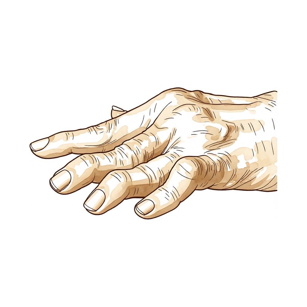
Swan-neck deformity is characterized by hyperextension of the proximal interphalangeal (PIP) joint, often accompanied by concomitant flexion of the distal interphalangeal (DIP) and metacarpophalangeal (MCP) joints [21]. This condition can affect the DIP, PIP, and MCP joints due to multiple etiologies [21]. The inability to flex the digit constitutes a significant functional impairment, as patients find it difficult to grasp objects [6]. In rheumatoid arthritis, carpal collapse and proximal migration of the carpal-hand unit create an imbalance resulting in an "extrinsic-minus" phenomenon [3]. Other causes include synovitis stretching the volar plate and flexor digitorum superficialis (FDS) [21], weakening of radial sagittal bands [21], lack of FDS function [11], IP joint flexion contracture following zone T1 flexor tendon repair [14], and proximal phalangeal osteochondroma [8]. The deformity can progress significantly over time due to increasing DIP joint flexion contracture [2]. Patients with rheumatoid arthritis experience impairments in function and limitations in daily activities [9].
Treatment strategies depend on the flexibility of the deformity and the presence of articular wear. Splinting and soft tissue reconstructions are suitable for flexible swan neck deformities [21]. A fixed deformity in a joint free of articular wear can be converted into a flexible deformity prior to soft tissue reconstruction [21]. Conversely, a fixed swan neck deformity caused by articular wear should be treated with fusion or implant arthroplasty [21]. The classic teaching in rheumatoid hand surgery is that swan neck deformities should be corrected in a proximal-to-distal direction [21]. Distal deformities may improve with conservative treatment once the MCP joint deformity is corrected, potentially circumventing the need for further reconstruction [21].
Headline outcomes indicate that lateral band translocation is a reliable procedure that prevents PIP joint hyperextension and is applicable to all causes of swan-neck deformity [10]. In cases associated with intrinsic tightness, lengthening of the lateral bands is possible using a percutaneous needle technique [5]. A new lateral extensor band technique provides stable correction in rheumatoid arthritis [4]. Palmar arthroplasty of the PIP joint in rheumatoid stiff swan-neck deformity results in increased range of motion and adjustment to a more functional arc [15]. Surface replacement arthroplasty and transfer of the FDS tendon to the radial lateral band is a successful technique for treating traumatic osteoarthritic changes leading to swan-neck deformity and ulnar deviation at the PIP joint [16]. A soft-tissue arthrodesis using FDS hemitenodesis is a safe and effective treatment option for patients with swan neck deformities following unconstrained PIP arthroplasty [28]. The Mitek mini anchor offers a practical, reliable and functional reconstruction of the volar plate in the management of congenital swan neck deformities [25]. Dynamic tenodesis of the PIP joint is a satisfactory surgical procedure for congenital swan neck deformity that produces a better cosmetic result than Swanson tenodesis [41]. An experienced hand therapist is essential to achieve optimal outcomes by emphasizing MCP joint extension, PIP joint flexion, and DIP joint extension [30]. A PIP joint extension block in 10 to 30 degrees for 4 to 6 weeks is essential to avoid recurrence of the deformity [30].
Anatomy & Pathophysiology
General Mechanisms and Etiology
Swan-neck deformity is defined by hyperextension at the proximal interphalangeal (PIP) joint and flexion of the distal interphalangeal (DIP) joint [20]. This deformity may present as flexible or fixed and is associated with varying degrees of PIP joint destruction [20]. While swan-neck deformities can occur at the DIP, PIP, and metacarpophalangeal (MCP) joints due to multiple etiologies [21], the specific etiology involves the onset of a collapsing linked joint appearance where joint disruption causes a zigzag deformity [6]. Displacement can begin at various joints and progresses in either a proximal-to-distal or distal-to-proximal direction [6].
The biomechanical result of many forces acting on the PIP joint is characterized by a common factor: dorsal migration of the extensor lateral bands [24]. This dorsal migration determines a loss of PIP joint excursion and a loss of power for DIP joint extension [24]. Additionally, the deep flexor tendon contributes to the mechanism because a pronounced curve at the PIP joint forces the tendon to enhance its flexor action over the DIP joint while losing force over the PIP joint [24]. Understanding the structural and dynamic factors of normal muscle and tendon balance in the digits is key to understanding pathogenesis [27]. The most important factor in the development of finger deformities is the changes occurring in the tendons and related structures, especially in early stages [72].
Rheumatoid Arthritis Pathophysiology
Synovitis of the PIP joint with a variable degree of tenosynovitis within the finger flexor sheath is the most common cause of swan-neck deformity [20]. Attenuation of the volar plate, collateral ligaments, and flexor digitorum superficialis (FDS) insertion allows the PIP joint to hyperextend due to extensor mechanism forces [20]. These extensor mechanism forces are accentuated by intrinsic muscle contractures and intrinsic tightness due to MCP joint flexion and volar subluxation [20].
Synovitis in the DIP joint with attenuation or rupture of the terminal tendon of the extensor mechanism concentrates extensor forces at the PIP joint, which can initiate or accentuate a swan-neck deformity [20]. Specifically, synovitis in the PIP joint causes attenuation of the volar plate, which acts as a checkrein to PIP joint hyperextension [20]. Furthermore, synovitis produces attenuation of the transverse retinacular ligaments, which are responsible for maintaining the normal position of the lateral bands [20]. When these ligaments become attenuated, the lateral bands can subluxate dorsal to the axis of PIP joint rotation and produce a constant and accentuated PIP joint extension force [20].
The extensor mechanism over the PIP joint, especially the triangular ligament, and the dorsal skin become contracted, resulting in a fixed hyperextension contracture of the joint [20]. Articular destruction secondary to persistent synovitis, as well as altered joint-contact mechanics, contributes to a fixed and occasionally painful deformity [20]. With carpal collapse and proximal migration of the carpal-hand unit, long or extrinsic motor systems are no longer working at physiological length or optimal efficiency, creating an imbalance resulting in an "extrinsic-minus" phenomena and swan-neck finger deformity [3].
Synovitis at the PIP joint level causes stretching of the volar plate and the FDS that inserts at the base of the volar middle phalanx [21]. This stretching causes an unopposed pull of the dorsal central tendon and induces an extension deformity in the PIP joint [21]. Synovitis can cause a flexion deformity at the DIP joint, and a zigzag deformity propagates proximally because there is stretching of the terminal tendon [21]. Additionally, synovitis can stretch out both the extrinsic tendons over the MCP joint and the sagittal bands that keep the central tendon in the midline position [21].
Posttraumatic and Secondary Mechanisms
Posttraumatic boutonnière and swan neck deformities should not be confused with those associated with inflammatory arthritis because the treatment options are often very different [19]. Swan neck deformity may result from chronic mallet injury provided that the PIP deformity is capable of hyperextension [35]. PIP hyperextension results from innate or acquired laxity of the volar plate combined with increased tension in the central slip and lateral bands owing to elongation of the terminal tendon [35].
Swan neck deformity may arise from overactivity of the extrinsic or intrinsic extensor mechanism due to spasticity or intrinsic contracture and can result from insufficiency of the volar stabilizers of the PIP joint, such as volar plate incompetence or flexor digitorum superficialis injury [35]. The deformity may become symptomatic due to locking of the PIP joint in hyperextension (pseudotriggering) upon initiating PIP flexion from the hyperextended position [35]. The swan neck deformity can progress significantly with time because of increasing DIPJ flexion contracture [2].
Functional Implications
Patients with swan neck deformity suffer from a considerable functional loss and a tight grip of the finger around an object is impaired [24].
Classification
Swan-neck deformity is characterized by hyperextension at the proximal interphalangeal (PIP) joint and flexion of the distal interphalangeal (DIP) joint [20]. Several classification systems exist to categorize these deformities based on etiology, severity, and mechanical behavior.
Zancolli: This system classifies swan-neck deformity into three fundamental types based on the cause of the deformity: extrinsic, intrinsic, and articular [24].
Feldon: This classification system categorizes severity based on the extent of joint stiffness and articular wear [21]. Type III swan neck deformity is characterized by a stiff PIP joint in all positions of the MCP joint with a good radiograph [21].
Souter: This classification defines Type I swan neck deformity as having a PIP joint that is flexible in all positions, with PIP joint flexion limited only when the MCP joint is extended or radially deviated [69]. Type IIIa is defined as a preliminary stage "snapping swan neck," while Type IIIb represents a fully established deformity where PIP joint flexion is limited in all positions [69]. Type IV swan neck deformity is defined as a stiff PIP joint with X-ray evidence of advanced intra-articular changes [69].
Other Considerations: The concept of flexible versus fixed deformities applies to swan neck treatment and is relevant for determining treatment options [21].
Clinical Presentation
Swan neck deformities typically present with hyperextension of the proximal interphalangeal (PIP) joint [6]. This configuration is often accompanied by concomitant flexion of the distal interphalangeal (DIP) and metacarpophalangeal (MCP) joints [21]. The inability to flex the digit constitutes a significant functional impairment, as patients find it difficult to grasp objects [6]. In patients with rheumatoid arthritis (RA), these deformities result in a variety of problems, including impairments in hand functions and limitations in daily activities [9]. The severity of the deformity depends on the extent of joint stiffness and articular wear [21].
Etiology and Pathomechanics
The etiology of swan neck and boutonnière deformities relates to the onset of a collapsing linked joint appearance [6]. Digit configuration assumes a formation resulting from a train accident; disruption of the joints, or railcars, causes a zigzag deformity [6]. Displacement can begin at various joints and progresses in either a proximal-to-distal or distal-to-proximal direction [6]. Neither swan neck nor boutonnière deformity is unique to rheumatoid arthritis (RA) [6]. Understanding the etiology of a deformity is necessary to devise an appropriate treatment plan [6].
Synovitis in the DIP joint with attenuation or rupture of the terminal tendon of the extensor mechanism concentrates extensor forces at the PIP joint [20]. This concentration can initiate or accentuate a swan-neck deformity [20]. When transverse retinacular ligaments become attenuated, the lateral bands can subluxate dorsal to the axis of PIP joint rotation [20]. Dorsal subluxation of lateral bands produces a constant and accentuated PIP joint extension force [20]. Articular destruction secondary to persistent synovitis, along with altered joint-contact mechanics, contributes to a fixed and occasionally painful deformity [20].
The dorsal migration of the extensor lateral bands determines a loss of excursion of the PIP joint [24]. This migration results in a loss of power of extension of the DIP joint [24]. Consequently, all extensor forces are transmitted to the base of the middle phalanx where the central slip is inserted, resulting in hyperextension of the PIP joint [24]. The deep flexor tendon also contributes to the mechanism due to a pronounced curve at the PIP joint [24]. This curve forces the deep flexor tendon to enhance its flexor action over the DIP joint and lose force over the PIP joint [24]. Additionally, swan neck deformity is caused by excessive tension on the middle band with relative relaxation of the lateral bands of the extensor mechanism [33].
With carpal collapse and proximal migration of the carpal-hand unit, long or extrinsic motor systems are no longer working at physiological length or optimal efficiency [3]. This carpal collapse creates an imbalance resulting in an "extrinsic-minus" phenomena and swan-neck finger deformity [3]. As the deformity progresses, the extensor tendon over the MCP joint descends volar to the joint axis [21]. This descent causes flexion of the MCP joint and hyperextension of the PIP joint [21]. A zigzag deformity propagates proximally because there is stretching of the terminal tendon [21].
Diagnostic Evaluation
Each type of swan neck deformity can be identified quite readily by reviewing the hand posture [21]. A proper evaluation is necessary to make a definite diagnosis and establish treatment targets [21]. Physical examination, including Elson and intrinsic-plus tests, and plain radiography are important tools for diagnosis of posttraumatic swan neck deformities [19]. An accurate clinical assessment is essential for selecting the appropriate treatment method for posttraumatic swan neck deformities [19]. Posttraumatic swan neck deformities should not be confused with those associated with inflammatory arthritis because the treatment options are often very different [19]. Correct diagnosis of the etiology of deformity is imperative for successful treatment [38]. Understanding the structural and dynamic factors of the normal muscle and tendon balance of the digits is key to understanding the pathogenesis [27]. Treating swan neck and boutonniere deformities of the PIP joint is a difficult challenge that requires understanding the cause, biomechanical changes, and articular status [31].
Classification and Special Considerations
Feldon and colleagues developed a classification system for swan neck deformities [21]. Type III swan neck deformity is characterized by a stiff PIP in all positions of the MCP joint and a good radiograph [21]. The concept of flexible versus fixed deformities that applies to boutonnière treatment is also relevant for swan neck treatment [21]. Splinting and soft tissue reconstructions are suitable treatment options for flexible deformities [21]. If the deformity is fixed, the condition of the joint should be assessed [21]. A joint that is free of articular wear can be converted into a flexible deformity prior to soft tissue reconstruction [21]. A joint that is fixed because of articular wear should be treated with fusion or implant arthroplasty [21]. A deformity at the MCP joint can be corrected with soft tissue reconstruction or joint replacement before distal procedures are performed [21]. Distal deformities may improve with conservative treatment once the MCP joint deformity is corrected, circumventing the need for further reconstruction [21].
Functional and esthetic considerations dictate whether surgical intervention is necessary and indicate the expected treatment outcomes [6]. Boutonnière deformities are characterized by flexion of the PIP joint [6]. Flexion, particularly in the ulnar digits, facilitates gripping [6]. A boutonnière deformity is often considered an esthetic rather than a functional concern [6].
Rare etiologies and complications include a 4-year-old child with a proximal phalangeal osteochondroma of the index finger presenting with a rigid swan-neck deformity [8]. Contraction of a dorsal Dupuytren's cord caused a rigid swan-neck deformity [40]. In a patient with Ehlers-Danlos Syndrome, the deformity in the fingers of both hands originated from PIP joint hyperextension, and DIP joint flexion was secondary [49]. The swan neck deformity in an individual progressed significantly with time because of increasing DIPJ flexion contracture [2]. Swan-neck deformity can complicate the Agee technique, though it may be transient [13]. Strain in the volar plate increased progressively with the creation of mallet and swan neck deformity conditions [26].
Investigations
Other Considerations: Understanding the etiology of the deformity is essential to devise an appropriate treatment plan [6]. Identifying the cause is critical for successful treatment [92]. Treating swan neck and boutonniere deformities of the PIP joint requires understanding the cause, biomechanical changes, and articular status [31]. A careful physical examination is essential to direct care and future testing if indicated [50]. Diagnostic tests such as imaging and serum laboratory studies are useful in determining pathologic processes but can be expensive, time consuming, and often nonspecific [50].
Treatment
Non-Operative
Splinting is a suitable treatment option for flexible swan-neck deformities [21]. For type I swan-neck deformity, a silver ring splint can help correct PIP hyperextension while allowing motion [30]. When the deformity is caused primarily by PIP laxity, restricting PIP hyperextension may be accomplished through figure-of-eight splinting [35].
Operative
Indications: Swan-neck deformities are amenable to surgical treatment with good results if based on an adequate study of the mechanism [1]. Lateral band mobilization is a treatment option for type II and III swan-neck deformities [21]. Arthroplasty and fusion are treatment options for type III swan-neck deformities with severe arthritic changes [21].
Surgical Approach / Technique: The Zancolli-Tonkin procedure (lateral band translocation) is a simple and reliable technique that provides lasting correction of an incapacitating deformity associated with impaired overall hand function [42]. FDS tenodesis is indicated for flexible swan-neck deformities or fixed deformities that can be made flexible, creating a static volar restraint against hyperextension [80]. FDS hemitenodesis significantly reduced volar plate strain compared to the swan-neck deformity condition, suggesting it is a biomechanically effective treatment [81]. A soft-tissue arthrodesis using FDS hemitenodesis is a safe and effective treatment option for patients with swan-neck deformities following unconstrained PIP arthroplasty, offering low revision rates, retained motion, and favorable pain and satisfaction scores [28]. The FDS volar tendon sling for reconstruction of PIPJ volar instability is a simple and safe procedure that effectively corrects the swan-neck deformity and improves the prehension function in the hand [29]. Inverted king post-truss artificial ligament reconstruction is effective for treating severe swan-neck deformity with snapping finger, though it is not expected to correct an associated mallet finger deformity [22]. Dynamic tenodesis of the proximal interphalangeal joint is a satisfactory surgical procedure for congenital swan-neck deformity that does not produce as great a fixed flexion deformity as the Swanson tenodesis and produces a better cosmetic result [41]. Central slip tenotomy is a reliable treatment for dynamic swan-neck deformity in cerebral palsy in patients without dynamic metacarpophalangeal flexion deformity [32]. By addressing directly the underlying pathophysiology of swan-neck deformities in cerebral palsy, effective surgical correction can be seen via lateral band rerouting [23]. Correction of the swan-neck deformity can be accomplished via the Fowler tenotomy, which reduces central slip tone while increasing extensor force at the DIP joint [35]. Surgical reconstruction of an ORL, either by using one of the existing lateral bands or by tendon graft procedures, can correct swan-neck deformity when PIP hyperextension restriction alone does not correct DIP extensor lag [35]. Volar plate imbrication, lateral band translocation, and FDS tenodesis are surgical means to restrict PIP hyperextension for swan-neck deformity caused primarily by PIP laxity [35].
Implant Selection: DIP arthrodesis is preferred particularly when the mallet deformity is primary [30]. Screw fixation, Kirschner wires, and tension band wiring are the most commonly used techniques for DIP arthrodesis [30]. The screw technique has the advantage of immediate strong fixation with a low complication rate [30]. Screw fixation is most often performed with a 2.0 mm screw, although a 1.5 mm screw may be necessary for the small finger [30]. Kirschner wires may be needed in difficult bone situations where screw fixation is not possible or in cases with a significant mismatch between the screw size and the phalanx dimensions [30]. Newer implants such as endomedullary staples provide intramedullary fixing only and have the advantage of neither requiring any secondary hardware removal nor causing any irritation of the fingertip [30].
Other Considerations: The fusion of the DIP joint is generally well tolerated, especially if there is a mobile PIP and MCP joint [30]. Even with a successful DIP fusion, the fine motor skills of a finger may be compromised [30]. Stern and Fulton reported a 20% complication rate for DIP arthrodesis [30]. The majority of problems related to DIP arthrodesis complications were related to the fixation method of the arthrodesis and hardware used [30]. Kirschner wires may provoke pin track infections, fall out, or migrate in such a way that they have to be taken out earlier, which can lead to delayed or nonunion [30]. In rheumatoid stiff swan-neck deformity, increased function is gained by palmar arthroplasty of the PIP joint, resulting not only in increased range of motion, but also adjustment of this range to a more functional arc [15]. Surface replacement arthroplasty and transfer of the flexor digitorum superficialis tendon to the radial lateral band is a successful technique for treating traumatic osteoarthritic changes that lead to swan-neck deformity and ulnar deviation at the proximal interphalangeal joint [16]. Proximal interphalangeal motion deteriorated over time in the long-term group, with a mean loss of 17° of early postoperative flexion following dorsal capsulotomy and lateral band mobilization [12]. We prefer to leave half of the superficialis to prevent development of 'swan-neck' deformity which is caused or accelerated by lack of function of this tendon [11].
Complications
Hardware and Fixation: DIP joint arthrodesis for swan-neck deformity carries a 20% complication rate, with the majority of problems related to the fixation method and hardware used [30]. Kirschner wire fixation may provoke pin track infections, fall out, or migrate, potentially leading to delayed or nonunion [30].
Nail Bed Injury: Violation of the nail's germinal matrix during DIP arthrodesis can lead to severe permanent nail deformity [30].
Soft Tissue and Ligamentous Complications: Release of the central slip during soft tissue procedures for rheumatoid swan-neck deformity has resulted in boutonnière deformities [37]. Soft tissue reconstruction for rheumatoid swan-neck deformity showed a mean loss of 17° of early postoperative flexion in the long-term group [12].
Other Considerations: Swan-neck deformity can occur as a complication of the Agee technique, though it may be transient [13]. Inverted king post-truss artificial ligament reconstruction for severe swan-neck deformity is not expected to correct an associated mallet finger deformity [22]. Strain in the volar plate increases progressively with the creation of mallet and swan neck deformity conditions [26].
Recovery
Rehabilitation protocol: Postoperative splinting must be implemented immediately to prevent recurrence of flexion deformity, which can occur within days if immobilisation is delayed [95]. For central slip repair, early removal of a pin risks stretching of the repaired slip and subsequent recurrence of the deformity [93]. In cases of Kirner's deformity, continuous splinting for 18 months has resulted in excellent cosmetic and functional outcomes [83].
Other Considerations: Long-term functional stability varies by procedure and pathology. PIP joint flexion motion following dorsal capsulotomy and lateral band mobilization for rheumatoid swan-neck deformity deteriorates over time, with a mean loss of 17° of early postoperative flexion observed in the long-term group [12]. Conversely, the Zancolli-Tonkin procedure provides lasting correction of swan-neck deformity associated with impaired overall hand function [42].
Key Evidence
- [L5] There are several forms of swan-neck deformity which are amenable to surgical treatment with good results, if based on an adequate study of the mechanism. [1] (10.1016/0072-968x(76)90004-8)
- [L5] The swan neck deformity in this individual progressed significantly with time because of increasing DIPJ flexion contracture. [2] (10.1016/j.jht.2009.11.005)
- [L4] With carpal collapse and proximal migration of the carpal-hand unit, long or extrinsic motor systems are no longer working at physiological length or optimal efficiency, creating an imbalance resulting in an "extrinsic-minus" phenomena and swan-neck finger deformity. [3] (10.1016/s0363-5023(82)80045-2)
- [L4] This new technique improves some unappealing aspects of previous techniques and provides a stable and reliable correction of swan neck deformity. [4] (10.1177/1753193408092787)
- [L5] In cases of swan neck deformity associated with intrinsic tightness, lengthening of the lateral bands is possible using a percutaneous needle technique. [5] (10.1016/j.hansur.2021.03.021)
- [L5] A 4-year-old child with a proximal phalangeal osteochondroma of the index finger presented with a rigid swan-neck deformity. [8] (10.1016/s0363-5023(86)80106-x)
- [L4] RA patients with swan neck deformities experience a variety of problems, including impairments in functions and limitations in daily activities. [9] (10.1002/msc.180)
- [L4] Lateral band translocation is a reliable and technically straightforward procedure that prevents PIP joint hyperextension and is applicable to all causes of swan-neck deformity. [10] (10.1016/0363-5023(92)90402-b)
- [L4] We prefer to leave half of the superficialis to prevent development of 'swan-neck' deformity which is caused or accelerated by lack of function of this tendon. [11] (10.1016/s0363-5023(78)80039-2)
- [L4] Ninety-two fingers with rheumatoid swan-neck deformity treated with dorsal capsulotomy and lateral band mobilization showed an initial increase of 55° of motion into flexion, but this proximal interphalangeal motion deteriorated over time, with a mean loss of 17° of early postoperative flexion in the long-term group. [12] (10.1016/0363-5023(93)90387-i)
- [L5] This is the first report of a swan-neck deformity complicating the Agee technique which fortunately was transient. [13] (10.1016/s0266-7681(05)80065-8)
- [L5] This in-depth biomechanical analysis explains how IP joint flexion contracture following zone T1 flexor tendon repair can result in secondary swan neck deformity of the thumb. [14] (10.1016/j.jht.2011.07.026)
- [L4] In rheumatoid stiff swan-neck deformity, increased function is gained by palmar arthroplasty of the PIP joint, resulting not only in increased range of motion, but also adjustment of this range to a more functional arc. [15] (10.1016/s0363-5023(83)80156-7)
- [L4] The experience with surface replacement arthroplasty and transfer of the flexor digitorum superficialis tendon to the radial lateral band is a successful technique for treating traumatic osteoarthritic changes that lead to swan-neck deformity and ulnar deviation at the proximal interphalangeal joint. [16] (10.1177/17531934251408609)
- [L5] [19] (10.5435/jaaos-d-14-00272)
- [L5] [20] (10.5435/00124635-199903000-00002)
- [L4] Inverted king post-truss artificial ligament reconstruction is effective for treating severe swan neck deformity with snapping finger, but is not expected to correct an associated mallet finger deformity. [22] (10.1016/j.hansur.2016.11.005)
- [L4] By addressing directly the underlying pathophysiology of swan neck deformities in cerebral palsy, effective surgical correction can be seen. [23] (10.1097/00130911-199709000-00006)
- [L5] [24] (10.1142/s0218810417720236)
- [L5] The Mitek mini anchor offers a practical, reliable and functional reconstruction of the volar plate in the management of congenital swan neck deformities. [25] (10.1016/j.bjps.2005.01.017)
- [L5] Strain in the volar plate increased progressively with the creation of mallet and swan neck deformity conditions. [26] (10.1177/1558944720966736)
- [Paper] Understanding the structural and dynamic factors of the normal muscle and tendon balance of the digits is key to understanding the pathogenesis. [27] (10.1016/0049-0172(83)90007-0)
- [L4] A soft-tissue arthrodesis using FDS hemitenodesis is a safe and effective treatment option for patients with swan neck deformities following unconstrained PIP arthroplasty, offering low revision rates, retained motion, and favorable pain and satisfaction scores. [28] (10.1007/s11552-013-9571-0)
- [L5] The FDS volar tendon sling for reconstruction of PIPJ volar instability is a simple and safe procedure that effectively corrects the swan-neck deformity and improves the prehension function in the hand. [29] (10.1142/s0218810417720182)
- [L4] Treating swan neck and boutonniere deformities of the PIP joint is a difficult challenge that requires understanding the cause, biomechanical changes, and articular status. [31] (10.1016/j.hcl.2017.12.006)
- [L4] Central slip tenotomy is a reliable treatment for dynamic swan-neck deformity in cerebral palsy in patients without dynamic metacarpophalangeal flexion deformity. [32] (10.1016/j.jhsa.2007.07.002)
- [L4] The swan-neck deformity is caused by excessive tension on the middle band with relative relaxation of the lateral bands of the extensor mechanism. [33] (10.2106/00004623-196042060-00004)
- [L5] [37] (10.1097/00130911-200003000-00004)
- [L4] Correct diagnosis of the etiology of deformity is imperative for successful treatment. [38] (10.1016/j.jhsa.2014.01.039)
- [L5] Contraction of this cord caused a rigid swan-neck deformity. [40] (10.1016/j.jhsb.2004.06.003)
- [L4] The new surgical procedure is satisfactory for congenital swan neck deformity, does not produce as great a fixed flexion deformity as the Swanson tenodesis, and produces a better cosmetic result. [41] (10.1054/jhsb.2000.0498)
- [Paper] The Zancolli-Tonkin procedure is a simple and reliable technique that provides lasting correction of an incapacitating deformity associated with impaired overall hand function. [42] (10.1016/j.otsr.2016.03.008)
- [L5] The deformity in the fingers of both hands originated from PIP joint hyperextension, and DIP joint flexion was secondary. [49] (10.1016/s0266-7681(97)80039-3)
- [L4] [69] (10.1177/175899830000500401)
- [L4] The most important factor in the development of finger deformities is the changes occurring in the tendons and related structures, especially in early stages. [72] (10.2106/00004623-195739030-00006)
- [L5] FDS tenodesis is indicated for flexible swan neck deformities or fixed deformities that can be made flexible, creating a static volar restraint against hyperextension. [80] (10.1016/j.jhsa.2015.07.018)
- [L5] FDS hemitenodesis significantly reduced volar plate strain compared to the swan neck deformity condition, suggesting it is a biomechanically effective treatment. [81] (10.1177/15589447211040877)
- [L5] After 18 months of continuous splinting, the fingers have an excellent cosmetic and functional result. [83] (10.1016/s0363-5023(86)80095-8)
- [L5] Identifying the cause of the deformity is critical for successful treatment. [92] (10.1016/j.hcl.2018.06.011)
- [L4] The one poor result was probably because the pin was removed too early, so the repaired central slip stretched out and the deformity recurred. [93] (10.1016/0266-7681(90)90081-e)
- [L5] Hand therapy is essential to prolong the interoperative intervals, and the author has noted recurrence of flexion deformity within days when postoperative splinting has not been implemented immediately. [95] (10.1016/s0894-1130(98)80022-7)
See Also
- Flexor tendon repair
- Boutonnière Deformity
- Mallet Finger
References
[1] Swan-neck deformity: Mechanism and surgical treatment. Hand. 1976. DOI: 10.1016/0072-968x(76)90004-8
[2] Swan Neck Deformity after Distal Interphalangeal Joint Flexion Contractures: A Biomechanical Analysis. Journal of Hand Therapy. 2010. DOI: 10.1016/j.jht.2009.11.005
[3] Wrist involvement in rheumatoid swan-neck deformity. The Journal of Hand Surgery. 1982. DOI: 10.1016/s0363-5023(82)80045-2
[4] Correction of Swan Neck Deformity in Rheumatoid Arthritis Using a New Lateral Extensor Band Technique. Journal of Hand Surgery (European Volume). 2008. DOI: 10.1177/1753193408092787
[5] Percutaneous management of swan neck deformity using WALANT: case report featuring a new surgical technique. Hand Surgery and Rehabilitation. 2021. DOI: 10.1016/j.hansur.2021.03.021
[6] Green S Operative Hand Surgery. Swan Neck and Boutonnière Deformities.
[8] Phalangeal osteochondroma: An unusual cause of swan-neck deformity. The Journal of Hand Surgery. 1986. DOI: 10.1016/s0363-5023(86)80106-x
[9] Swan neck deformities in rheumatoid arthritis: a qualitative study on the patients' perspectives on hand function problems and finger splints. Musculoskeletal Care. 2010. DOI: 10.1002/msc.180
[10] Lateral band translocation for swan-neck deformity. The Journal of Hand Surgery. 1992. DOI: 10.1016/0363-5023(92)90402-b
[11] Flexor tenosynovectomy in the rheumatoid finger. The Journal of Hand Surgery. 1978. DOI: 10.1016/s0363-5023(78)80039-2
[12] Soft tissue reconstruction for rheumatoid swan-neck and boutonniere deformities: Long-term results. The Journal of Hand Surgery. 1993. DOI: 10.1016/0363-5023(93)90387-i
[13] Swan-Neck Deformity as a Complication of the Agee Technique. Journal of Hand Surgery. 1995. DOI: 10.1016/s0266-7681(05)80065-8
[14] Biomechanical Analysis of Secondary Swan Neck Deformity Following IP Joint Flexion Contracture of the Thumb. Journal of Hand Therapy. 2011. DOI: 10.1016/j.jht.2011.07.026
[15] Palmar arthroplasty for the treatment of the stiff swan-neck deformity. The Journal of Hand Surgery. 1983. DOI: 10.1016/s0363-5023(83)80156-7
[16] Surface replacement arthroplasty for a proximal interphalangeal joint with swan neck deformity and ulnar deviation. Journal of Hand Surgery (European Volume). 2025. DOI: 10.1177/17531934251408609
[19] Posttraumatic Boutonnière and Swan Neck Deformities. Journal of the American Academy of Orthopaedic Surgeons. 2015. DOI: 10.5435/jaaos-d-14-00272
[20] Operative Correction of Swan-Neck and Boutonniere Deformities in the Rheumatoid Hand. Journal of the American Academy of Orthopaedic Surgeons. 1999. DOI: 10.5435/00124635-199903000-00002
[21] Green S Operative Hand Surgery. Swan Neck Deformities.
[22] Extra-articular subcutaneous “inverted king post-truss” ligament reconstruction for severe swan neck deformity (snapping finger). Hand Surgery and Rehabilitation. 2017. DOI: 10.1016/j.hansur.2016.11.005
[23] Lateral Band Rerouting in the Treatment of Swan Neck Deformities Due to Cerebral Palsy. Techniques in Hand & Upper Extremity Surgery. 1997. DOI: 10.1097/00130911-199709000-00006
[24] Acute Traumatic Swan Neck Deformity: A Case Report of the Oblique Retinacular Ligament Lesion. The Journal of Hand Surgery (Asian-Pacific Volume). 2017. DOI: 10.1142/s0218810417720236
[25] Correction of bilateral congenital swan-neck deformity by use of Mitek mini anchor: a new technique. British Journal of Plastic Surgery. 2005. DOI: 10.1016/j.bjps.2005.01.017
[26] Development of an In Vitro Swan Neck Deformity Biomechanical Model. HAND. 2020. DOI: 10.1177/1558944720966736
[27] Pathogenesis and differential diagnosis of the swan-neck deformity. Seminars in Arthritis and Rheumatism. 1983. DOI: 10.1016/0049-0172(83)90007-0
[28] Reconstruction of Swan Neck Deformities after Proximal Interphalangeal Joint Arthroplasty. HAND. 2013. DOI: 10.1007/s11552-013-9571-0
[29] Flexor Digitorum Superficialis Tendon Sling Volar Plate Reconstruction for Swan-Neck Deformity in the Spastic Finger. The Journal of Hand Surgery (Asian-Pacific Volume). 2017. DOI: 10.1142/s0218810417720182
[30] Green S Operative Hand Surgery. Swan Neck Deformities > Postoperative Care.
[31] Treating the Proximal Interphalangeal Joint in Swan Neck and Boutonniere Deformities. Hand Clinics. 2018. DOI: 10.1016/j.hcl.2017.12.006
[32] Surgical Treatment of Swan-Neck Deformity in Hemiplegic Cerebral Palsy. The Journal of Hand Surgery. 2007. DOI: 10.1016/j.jhsa.2007.07.002
[33] Surgery of the Hand in Cerebral Palsy and the Swan-Neck Deformity. The Journal of Bone & Joint Surgery. 1960. DOI: 10.2106/00004623-196042060-00004
[35] Green S Operative Hand Surgery. Treatment of Swan Neck Deformity Associated With Chronic Mallet Injury.
[37] Soft Tissue Procedures for the Rheumatoid Swan Neck Finger Deformity. Techniques in Hand & Upper Extremity Surgery. 2000. DOI: 10.1097/00130911-200003000-00004
[38] Treatment of Swan Neck Deformity in Cerebral Palsy. The Journal of Hand Surgery. 2014. DOI: 10.1016/j.jhsa.2014.01.039
[40] Dorsal Dupuytren’s Disease Causing A Swan-Neck Deformity. Journal of Hand Surgery. 2004. DOI: 10.1016/j.jhsb.2004.06.003
[41] TREATMENT OF CONGENITAL SWAN NECK DEFORMITY WITH DYNAMIC TENODESIS OF PROXIMAL INTERPHALANGEAL JOINT. Journal of Hand Surgery. 2001. DOI: 10.1054/jhsb.2000.0498
[42] Lateral band translocation for swan-neck deformity: Outcomes of 41 digits after a mean follow-up of eight years. Orthopaedics & Traumatology: Surgery & Research. 2016. DOI: 10.1016/j.otsr.2016.03.008
[49] Dynamic Swan Neck Deformity in a Patient with Ehlers-Danlos Syndrome. Journal of Hand Surgery. 1997. DOI: 10.1016/s0266-7681(97)80039-3
[50] Orthopaedic Knowledge Update 13 Ebook Without Multimedia. Anatomy, Evaluation, Clinical Examination, and Imaging > Evaluation and Clinical Examination: Current Concepts.
[69] A Study Comparing Use and Effects of Custom-Made versus Prefabricated Splints for Swan Neck Deformity in Patients with Rheumatoid Arthritis. The British Journal of Hand Therapy. 2000. DOI: 10.1177/175899830000500401
[72] Finger Deformities Caused by Rheumatoid Arthritis. The Journal of Bone & Joint Surgery. 1957. DOI: 10.2106/00004623-195739030-00006
[80] Superficialis Sling (Flexor Digitorum Superficialis Tenodesis) for Swan Neck Reconstruction. The Journal of Hand Surgery. 2015. DOI: 10.1016/j.jhsa.2015.07.018
[81] Effect of a Flexor Digitorum Superficialis Hemitenodesis on Reducing Volar Plate Strains for Swan Neck Deformities. HAND. 2021. DOI: 10.1177/15589447211040877
[83] Kirner's deformity: A review of the literature and case presentation. The Journal of Hand Surgery. 1986. DOI: 10.1016/s0363-5023(86)80095-8
[92] Management of Joint Contractures in the Spastic Upper Extremity. Hand Clinics. 2018. DOI: 10.1016/j.hcl.2018.06.011
[93] Operative Treatment of the Post-Traumatic Boutonnière Deformity. Journal of Hand Surgery. 1990. DOI: 10.1016/0266-7681(90)90081-e
[95] A review of the management of the hand in dystrophic epidermolysis bullosa. Journal of Hand Therapy. 1998. DOI: 10.1016/s0894-1130(98)80022-7