Clinicians › Shoulder
Shoulder Arthroplasty
Reverse total shoulder arthroplasty for cuff tear arthropathy, complex fractures, and arthritis — comparing stemmed vs stemless humeral components.
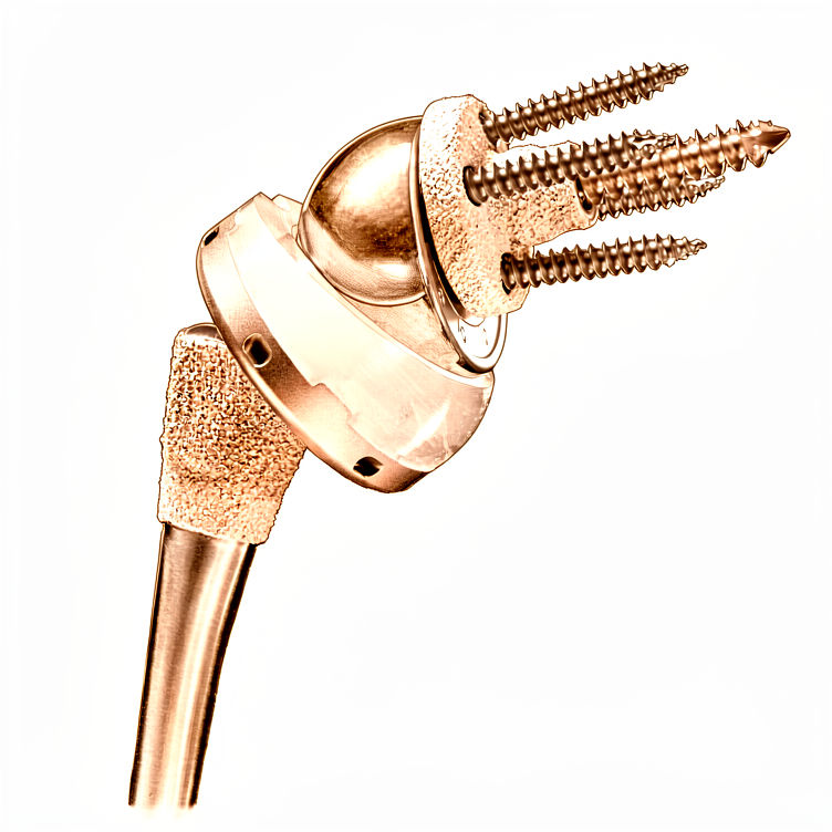
Overview
Shoulder arthroplasty is an effective and safe treatment option, considered as safe as more commonly performed major joint arthroplasties such as hip and knee arthroplasties [6, 23]. It provides substantial and durable improvement in shoulder function [52], with outcomes following anatomic total shoulder arthroplasty proven to be durable and reproducible [208]. The procedure is a safe and reliable option for the management of symptomatic glenoid dysplasia, offering improved clinical outcomes and favorable satisfaction [31]. While age 70 years or older does not appear to be a contraindication to stemless anatomic total shoulder arthroplasty, patients who have undergone prior ipsilateral shoulder surgery derive benefit from the procedure, though their magnitude of improvement and final scores are significantly lower than patients without prior surgery [213, 214, 38].
Indications continue to broaden, with reverse shoulder arthroplasty highly successful in cuff-deficient shoulders [47]. Simultaneous bilateral shoulder arthroplasty is feasible under optimal circumstances [41], and outpatient total shoulder arthroplasty is a safe and cost-effective alternative to inpatient care for appropriately selected patients [43, 207]. Knowledge of the array of shoulder prostheses available and their indications, along with the use of treatment algorithms, can lead to optimized patient outcomes [48]. The American Academy of Orthopaedic Surgeons has developed appropriate use criteria to determine the appropriateness of various humeral component designs during primary anatomic total shoulder arthroplasty [8].
Reported 30-day, 90-day, and 1-year mortality rates for shoulder arthroplasty are 0%, 0.16%, and 2%, respectively [212]. Consensus recommendations for long-term follow-up and surveillance of elective primary shoulder arthroplasty have been developed using a real-time Delphi technique [1]. Surgical implant type, indication, patient comorbidities, and hospital factors contribute to differential surgical cost for total shoulder arthroplasty [49]. Clinical results of revision shoulder arthroplasty are generally inferior to primary shoulder arthroplasty, but careful patient selection and adherence to surgical principles can lead to satisfactory results [53]. The outcome of revision shoulder arthroplasty can be predicted on the basis of the indication for the procedure [179], and patients undergoing glenoid component revision for symptomatic loosening tend to achieve good to excellent results [209].
Anatomy & Pathophysiology
Bony Anatomy
The glenoid is a convex structure of shallow depth shaped like an inverted pear [75]. The subchondral bone of the glenoid is relatively flat, with the articular concavity augmented by cartilage and a circumferential labrum [77]. The glenoid cavity is a shallow socket, approximately one third the size of the humeral head [76]. The glenoid averages 5° of retroversion in relation to the axis of the scapular body [77], with the normal position of the glenoid surface ranging from 2 degrees of anteversion to 7 degrees of retroversion relative to the scapular body [84]. The glenoid is retroverted approximately 5 degrees relative to the scapular body [87]. The glenoid articular surface radius of curvature is 2 to 3 mm larger than that of the humeral head [84].
The humeral head is spherical with a diameter of 37 to 57 mm [75]. The articular surface of the humeral head is essentially spherical, with an arc of approximately 160 degrees covered by articular cartilage [84]. The radius of curvature of the humeral head is approximately 25 mm and is slightly larger in men than in women [84]. The most superior portion of the articular surface of the humeral head averages 8 mm above the greater tuberosity [75], and the superior margin of the humeral head articular surface is normally superior to the top of the greater tuberosity by 8 to 10 mm [84]. The humeral head height is approximately 5.6 cm above the superior border of the pectoralis major tendon [87].
Humeral version averages 29.8 degrees, with a range of 10 to 55 degrees [75]. Proximal humeral retroversion is highly variable, ranging from 0 to 55 degrees depending on the method used for measurement [84]. The humeral head is retroverted an average of 30 degrees [76], and is retroverted 30 degrees relative to the transepicondylar axis of the humerus [87]. The humeral head averages 19° of retroversion [77]. The humeral head is inclined approximately 130 degrees with respect to the humeral shaft [75], and averages 41° of inclination (neck-shaft angle) [77]. The average neck-shaft angle is 45 degrees (±5 degrees), with a range of 30 to 50 degrees [84]. Arthritic shoulders have a flatter neck-shaft angle close to 50 degrees [84]. The neck-shaft angle measures an average of 135 degrees [76].
The distance from the lateral base of the coracoid process to the lateral margin of the greater tuberosity is called the lateral humeral offset [84]. The anatomic neck of the proximal humerus is located at the junction of the articular surface and the tuberosities [75]. The surgical neck represents an indistinct region (metadiaphyseal junction) below the tuberosities but above the humeral shaft [75]. The greater tuberosity serves as the attachment site for the supraspinatus, infraspinatus, and teres minor tendons of the rotator cuff [75]. The lesser tuberosity serves as the attachment site for the subscapularis tendon [75]. The bicipital groove lies between the greater and lesser tuberosities and serves as a pathway for the long head of the biceps [75]. The distal aspect of the bicipital groove is internally rotated with respect to the proximal portion [75].
The acromion, the coracoacromial ligament, and the coracoid process form the coracoacromial arch [75]. The rotator cuff, subacromial bursa, and subdeltoid bursa pass underneath the coracoacromial arch [75]. The scapula spans second through seventh ribs and serves as an attachment for 17 muscles [87]. The scapula is anteverted on the chest wall approximately 30 degrees relative to the body [87]. The scapula has only one true diarthrodial articulation, the acromioclavicular (AC) joint [77]. Normal shoulder motion is approximately two-thirds glenohumeral and one-third scapulothoracic [77].
The proximal humerus has three centers of ossification: the humeral head (4 to 6 months), the greater tuberosity (1 to 3 years), and the lesser tuberosity (3 to 5 years) [77]. The ossification center for the humeral head is usually present at birth [81]. The greater tuberosity ossification center appears by 1 to 3 years of age [81]. The lesser tuberosity ossification center appears by 5 years of age [81]. The proximal humeral ossification centers fuse to the shaft at age 17 to 20 years [77]. The proximal humeral physis closes by 14 to 17 years of age in girls and by 16 to 18 years in boys [81]. Humeral retroversion averages 65 degrees in infants and young children and gradually decreases, approaching adult values by 11 years of age [81]. Eighty percent of subsequent humeral growth comes from the proximal humeral physis [81]. The proximal humeral physis is irregularly shaped, with its apex located on the posteromedial portion of the proximal humerus [81]. The periosteum is thicker and stronger in the posteromedial portion of the proximal humerus as opposed to the anterolateral portion [81].
The clavicle is the first bone to ossify (fifth week of gestation) and is the only long bone to ossify by intramembranous ossification [77]. The medial (sternal) epiphysis of the clavicle is the last ossification center to fuse, at age 20 to 25 years [77]. The primary blood supply of the clavicle is periosteal, with no nutrient artery present [77]. Ossification of the scapular body begins at the eighth week of gestation [77]. The acromion has three ossification centers: the metacromion (base), the mesoacromion (middle), and the preacromion (tip) [77]. Failure of fusion of acromial ossification centers results in os acromiale [77]. The coracobrachialis muscle and the short head of the biceps tendon originate from the coracoid process [77]. The pectoralis minor muscle inserts onto the medial coracoid process [77].
The superior shoulder suspensory complex (SSSC) is composed of the glenoid, the coracoid process, the coracoclavicular ligaments, the distal clavicle, the AC joint, and the acromion [77]. The superior strut of the SSSC comprises the middle clavicle [77]. The inferior strut of the SSSC comprises the lateral scapular border/spine of the scapula [77].
Vascular Supply
The proximal humerus receives its blood supply from the anterior and posterior humeral circumflex branches from the third division of the axillary artery [75]. The anterior humeral circumflex artery (AHCA) arises from the axillary artery at the inferior border of the subscapularis [75]. The AHCA provides vascular inflow to the humeral head by way of its terminal anterolateral branch known as the artery of Laing (also known as the arcuate artery) [75]. The ascending branch of the AHCA courses parallel to the lateral aspect of the long head biceps tendon and enters the humeral head at the interface of the bicipital groove and greater tuberosity [75]. The anterolateral ascending branch of the anterior humeral circumflex artery provides the primary blood supply to the humeral head [77]. The anterolateral ascending branch travels proximally in the lateral aspect of the intertubercular groove [77]. The terminal intraosseous portion of the artery enters at the proximal aspect of the intertubercular groove as the arcuate artery [77]. The major blood supply to the humeral head is through the ascending branch of the anterior humeral circumflex artery, which penetrates the head at the bicipital groove and becomes the arcuate artery [76]. Injury to the arcuate artery may result in osteonecrosis of the humeral head [75]. Additional extraosseous collateral branches can permit humeral head perfusion despite complete ligation of the arcuate artery [75].
The posterior humeral circumflex artery travels with the axillary nerve, enters the quadrilateral space posteriorly, and anastomoses with a branch of the anterior circumflex to supply the posterior cuff [75]. Quantitative assessment has shown that 64% of the humeral head blood supply arises from the posterior humeral circumflex artery [81]. Fractures of the anatomic neck have a poor prognosis because of complete disruption of the blood supply to the head [76]. Surgical neck fractures are common, and with these, the blood supply to the head is preserved [76].
Nerve Anatomy
The brachial plexus and axillary artery are anterior to the coracoid process of the scapula and humeral head [76]. Nerves innervating muscles around the shoulder include the axillary, suprascapular, subscapular, and musculocutaneous nerves [76]. The radial nerve is commonly injured in humeral shaft fractures, particularly at the junction of the middle and distal third (Holstein-Lewis fracture) [76]. An axillary nerve injury from proximal humeral fracture or fracture-dislocation would result in paralysis of the deltoid muscle and anesthesia over the “badge” region at the lateral proximal arm [76].
The axillary nerve is a terminal branch coming off the posterior cord of the brachial plexus just proximal to the coracoid process [80]. The axillary nerve passes beneath the conjoined tendon anterior to the subscapularis 3 to 5 mm medial to the musculotendinous junction [80]. The axillary nerve is adjacent to the inferior capsule before entering the quadrilateral space posteriorly [80]. The axillary nerve splits into the anterior and posterior branches within the quadrangular space [80]. The anterior and middle deltoid muscle receives sole innervation from the anterior branch of the axillary nerve [80]. Posterior deltoid muscle innervation varies, with supply only from the anterior branch in 2.3% of cases, from the posterior branch in 8.5%, and from both branches in 89.1% [80]. The posterior branch of the axillary nerve branches to supply the teres minor muscle and then terminates as the superior lateral brachial cutaneous nerve [80]. The axillary nerve circles the humeral neck just inferior to the glenohumeral joint as it courses posteriorly [81].
In the anterior deltopectoral approach, the axillary nerve can be palpated by sweeping a finger inferiorly across the subscapularis muscle tendon interface [80]. In the anterolateral deltoid splitting approach, the axillary nerve crosses approximately 5 cm inferior to the anterolateral acromial corner [80]. Shoulder abduction brings the axillary nerve closer to the acromion landmark [80]. No accessory motor branches to the anterior deltoid cross the tendinous raphe between the anterior and lateral deltoid [80]. In the posterior deltoid splitting approach, the axillary nerve is approximately 7 cm from the posterior acromial corner [80].
The superior transverse scapular ligament arises from the medial base of the coracoid overlying the suprascapular notch [77]. The suprascapular artery runs superior to the superior transverse scapular ligament [77]. The suprascapular nerve runs deep to the superior transverse scapular ligament [77]. Entrapment of the suprascapular nerve at the superior transverse scapular ligament causes denervation of both the supraspinatus and the infraspinatus [77]. The spinoglenoid ligament overlies the suprascapular nerve at the spinoglenoid notch [77]. Entrapment, traction, or compression of the suprascapular nerve at the spinoglenoid notch causes denervation of the infraspinatus [77]. The suprascapular artery passes superior to the superior transverse scapular ligament and the suprascapular nerve passes inferior to the ligament through the notch [87]. At the spinoglenoid notch, both the artery and nerve are inferior to the inferior transverse scapular ligament [87].
Soft Tissue Anatomy
The rotator cuff consists of four muscles: the subscapularis, supraspinatus, infraspinatus, and teres minor [76]. The teres major is not a rotator cuff muscle [76]. The cuff muscles serve as depressors of the humeral head to allow the deltoid to efficiently abduct the humerus [76]. The infraspinatus and teres minor are external rotators, while the subscapularis is an internal rotator of the humerus [76]. The deltoid and pectoralis major muscles, along with the rotator cuff, cause predictable displacement of fractures around the proximal humerus [76].
The subscapularis is the largest and strongest of the rotator cuff tendons [249]. The subscapularis is responsible for active internal rotation of the humerus and contributes to the stability of the shoulder [249]. The subscapularis forms the anterior portion of the transverse plane “force couple” of the rotator cuff [249]. The subscapularis serves to balance the forces generated across the joint to maintain glenohumeral congruency throughout movement [249]. The subscapularis originates from the anterior scapula and inserts anteriorly onto the lesser tuberosity [81].
The deltoid has anatomic subdivisions (acromial, clavicular, and scapular) [79]. The deltoid is a well-defined muscle that is easily resected but extremely difficult to reconstruct functionally after proximal humeral resections for bone tumors [79]. The deltoid forward flexes and abducts the shoulder and courses from the clavicle and acromion superiorly, coalescing into a common tendinous insertion onto the lateral upper third of the humeral shaft [81]. The pectoralis major powers adduction and internal rotation due to its tendinous insertion anteriorly onto the lateral wall of the bicipital groove [81]. The pectoralis major forms the roof of the distal continuation of the bicipital tunnel, which is a closed space that extends proximally to the glenohumeral joint [81].
The greater tuberosity provides attachment superiorly and posteriorly for the supraspinatus, infraspinatus, and teres minor [81]. The tendinous insertions of the rotator cuff muscles, the articular capsule, the coracohumeral ligament, and the glenohumeral ligament complex blend into a confluent sheet before insertion into the humeral tuberosities [85]. The tendons of the infraspinatus and supraspinatus muscles join approximately 15 mm proximal to their insertion and cannot be readily separated by blunt dissection [85]. The infraspinatus and teres minor fuse near their musculotendinous junctions [85]. The supraspinatus and subscapularis tendons join as a sheath that surrounds the biceps tendon at the entrance of the bicipital groove [85]. The roof of the biceps sheath consists of a portion of the supraspinatus tendon, and a sheet.
The glenoid labrum provides concavity and up to 50% of marginal glenoid socket depth [77]. The glenoid articular surface and the labrum combine to create a socket that is approximately 9 mm deep in the superoinferior direction and 5 mm deep in the anteroposterior direction [85]. Adding the glenoid labrum increases the glenoid surface to 75% of the humeral head vertically and 57% horizontally [85]. The fibrocartilaginous glenoid labrum deepens the socket by 50% and provides a bumper to translation [87].
The coracoacromial ligament contributes to anterosuperior stability in rotator cuff deficiency and should be preserved with irreparable cuff tears to prevent anterosuperior escape [87]. The acromial branch of the thoracoacromial artery runs on the medial aspect of the coracoacromial ligament [87]. The coracoacromial ligament is the arthroscopic landmark for a complete release of the rotator interval for adhesive capsulitis [87]. The transverse humeral ligament is an important stabilizer of the biceps tendon [87].
Classification
Glenoid Morphology and Bone Loss
Walch: This morphologic classification describes the glenoid in primary glenohumeral osteoarthritis [353]. It demonstrated fair to substantial agreement in a study of anatomic total shoulder arthroplasty with keeled glenoids in patients younger than 60 years [296].
Alternative Systems: Alternative glenoid classification systems or predictive models should be considered to provide more precise prognoses for patients before and after shoulder arthroplasty performed for osteoarthritis with an intact rotator cuff [231]. A proposed classification system addresses the surgical management of the glenoid during prosthetic replacement and allows direct follow-up comparison of similarly treated glenoid replacements [99]. Another proposed system serves as a helpful guide to the degree of glenoid bone loss when embarking on revision shoulder arthroplasty [102].
Revision Defects: Current classifications exhibit poor reliability in categorizing glenoid defects post-reverse shoulder arthroplasty removal [169]. The formation of clusters based on glenoid morphology indicates that patterns exist in the types of glenoid defects, highlighting a need to further investigate a three-dimensional classification system and potentially new standardized revision implant component designs [186]. Patient-specific planning brings all surgeons into closer agreement with the recommendations of experts for glenoid classification and surgical planning [204].
Scapular Notching
Nerot-Sirveaux: This is the most commonly used grading system to define the extent of scapular notching in reverse shoulder arthroplasty [9]. Scapular notching is identified as the osseous defect of the lateral scapular pillar and neck that is created by impingement of the humeral component when the arm is adducted [9]. This osseous defect typically occurs inferior to the glenosphere, but can also be found anterior or posterior depending on the implant design and positioning of the glenosphere and humeral component [9].
The classification tiers are defined as follows: * Grade 1: Notching concerns only the scapular pillar [9]. * Grade 2: Erosion to the inferior screw of the baseplate [9]. * Grade 3: Erosion beyond the inferior screw [9]. * Grade 4: Progression of the defect to the central baseplate peg [9].
Some authors prefer to simplify the Nerot-Sirveaux classification by regrouping grades 1 and 2 into a single group and grades 3 and 4 into another group [9].
Humeral Bone Loss
PHAROS: The Proximal Humeral Arthroplasty Revision Osseous inSufficiency (PHAROS) classification system may be useful to anticipate the complexity of humeral reconstruction [132].
Fracture Sequelae
Pathophysiologic Classification: This system distinguishes 4 basic types of lesions that dominate the clinical picture and allow fracture sequelae of the proximal humerus to be grouped [71]. The inconsistent results reported with the use of shoulder prostheses to treat fracture sequelae were partly due to enormous variability in the anatomic lesions and comparisons between very heterogeneous groups of sequelae [71].
Arthritis and Rotator Cuff Integrity
Samilson-Prietro: This is the most widely adopted classification to determine the extent of osteoarthritis of the glenohumeral joint [70]. The grades are defined by osteophyte size and articular congruity: * Grade 0: Normal [70]. * Grade 1: Mild with osteophytes less than 3 mm on the humeral head [70]. * Grade 2: Moderate with osteophytes between 3 and 7 mm on the humeral head or glenoid rim [70]. * Grade 3: Severe with osteophytes of more than 7 mm with or without articular incongruity [70].
Hamada-Fukuda: This radiographic morphological description assesses the height of the acromiohumeral space in the natural course of massive rotator cuff tear [70]. * Type 1: Represents normal joint morphology and acromiohumeral distance of more than 6 mm [70].
Rheumatoid Arthritis
Larsen: This classification grades shoulder disease in rheumatoid arthritis [348]. * Grade I: Medium destructive abnormality with erosions and joint space narrowing [348]. * Grade IV: Severe destructive abnormality with bone deformation [348]. * Grade V: Mutilating abnormality where the original articular surface has disappeared [348].
Other Considerations
Complications and Outcomes: A system of three complication categories was introduced to serve as a standardised routine for the presentation of complications in shoulder arthroplasty [20]. However, a clear standardised set of shoulder arthroplasty complication definitions is lacking [193]. The development of a consensus definition of a periprosthetic shoulder infection is critical to future investigations of these devastating complications [39]. The 2018 ICM shoulder infection criteria provided a new scoring system to diagnose periprosthetic joint infection, with C acnes identified as the most common infectious organism [223]. The majority of revision shoulder arthroplasties are performed for patients who are unlikely to have a periprosthetic joint infection, with less than 10% meeting ICM criteria for definite PJI [227].
Complications of shoulder arthroplasty depend on the prosthesis type used [45]. Arthroplasty for glenohumeral arthropathies have specific complications and the final results are sometimes more dependent upon the type of shoulder arthroplasty than the initial etiology [34]. The same minimal clinically important difference value may not be appropriate for different types of shoulder arthroplasty [224]. Consensus on validated and clinically-meaningful tools from multiple categories is necessary to increase the generalizability and applicability of published studies in shoulder arthroplasty literature [221]. The conversion equations generated for the Normalized Constant Score and Patient-reported Outcomes may provide utility in the evaluation of shoulder arthroplasty outcomes across studies in systematic reviews and meta-analyses [58].
Recovery and Durability: Six distinct early recovery trajectories were identified after total shoulder arthroplasty, with 83.7% of patients experiencing very low pain scores after only 2 weeks [59]. The functional and radiologic results of the stemless shoulder arthroplasty are comparable to the third and fourth generation of standard stem arthroplasty [63]. The use of a convertible prosthetic system to revise a failed anatomical shoulder arthroplasty reduces morbidity and minimises the rate of complications [211]. Over 80% of shoulder replacements last more than 10 years, and 75% last more than 20 years [225]. The incidence of shoulder replacement and revision is expected to increase in the following years, constituting a burden for the healthcare systems [217]. There are considerable differences between Nordic countries regarding incidence of shoulder arthroplasty, age, diagnoses, and choice of arthroplasty type and brand [192].
Guidelines and Data: The AAOS developed appropriate use criteria to determine the appropriateness of various humeral component designs during primary anatomic total shoulder arthroplasty based on evidence-based information and clinical expertise [8]. This is the first study to use a national data set to examine risk factors for intraoperative complications during all types of primary shoulder arthroplasty, and identifies several previously unrecognized risk factors, such as surgical approach [55]. Successful postoperative radiologic evaluation of shoulder reconstructions requires an understanding of their fundamental hardware design, physiologic objective, preoperative imaging assessment, normal postoperative radiologic appearance, and findings and types of complication [54]. An algorithm represents the first step to automatically classify and organize shoulder radiographs on a large scale in very little time, which will profoundly enrich shoulder arthroplasty registries [51]. The majority of the fifty most cited publications in shoulder arthroplasty research focused on clinical outcomes, with five articles introducing novel classification systems developed between 2000 and 2006 [301].
Clinical Presentation
General Evaluation and History
A thorough and systematic approach incorporating history, physical examination, and appropriate diagnostic studies is necessary to evaluate failed shoulder arthroplasty, as the cause of pain and disability is often multifactorial [112]. Patients with an unsatisfactory outcome after shoulder arthroplasty present with poor shoulder function and pain [7]. The diagnosis of a stiff shoulder depends on awareness of the problem, with history and physical examination being paramount [4]. Preoperative clinical examination and radiological assessment is fundamental to anticipating specific difficulties in glenoid exposure [32]. Recognition and management of altered glenoid morphology and diminished bone stock are important for successful shoulder arthroplasty [26]. Shoulder surgeons should be familiar with the clinical manifestations of fibrous dysplasia and the unique surgical challenges they present as the incidence of shoulder arthroplasty continues to increase [172].
Specific patient factors influence outcomes and perioperative risk. A preoperative diagnosis of a stroke in patients undergoing primary shoulder arthroplasty is associated with higher rates of perioperative complications and mortality when compared to a matched cohort [137]. Patients with a diagnosis of depression should be counseled that they will experience a significant clinical improvement from baseline after total shoulder arthroplasty [144]. Prosthetic shoulder arthroplasty in young patients provides substantial improvement in active range of motion, irrespective of diagnosis and glenoid management, though residual shoulder pain is common [116]. The diagnosis, shoulder pathology, and prosthesis specifics were significant predictors of outcomes [103].
Painful Shoulder Arthroplasty
Common causes of pain after shoulder arthroplasty include instability, periprosthetic joint infection (PPJI), component loosening or malposition, acromioclavicular joint osteoarthritis, subacromial impingement, rotator cuff tear, adhesive capsulitis, and synovitis [135]. Radiographs, computed tomography (CT), and magnetic resonance imaging are all imperfect methods of evaluating the painful shoulder arthroplasty patient population [135]. An organized approach to diagnose and manage stiff or unstable total shoulder arthroplasty is needed to improve patient satisfaction and long-term survival [148].
Diagnostic shoulder arthroscopy provided clinically meaningful diagnostic information in patients with painful shoulder arthroplasty and nondiagnostic preoperative evaluations [17]. When used alongside other investigations in a comprehensive assessment protocol, arthroscopy can play an important role in the diagnosis and treatment of the problematic shoulder arthroplasty [18]. Shoulder arthroscopy in patients after arthroplasty is most frequently used as a diagnostic tool; however, it has utility in treating a number of predetermined pathologies [19]. Arthroscopy after shoulder arthroplasty is useful for the diagnosis and treatment of pain and loss of motion in selected patients but can be technically challenging [145]. Diagnostic arthroscopy is a useful adjunct in identifying causes of failure in patients with painful reverse total shoulder arthroplasty, especially when the cause of failure is unclear [29]. Diagnostic arthroscopy supports considering it in the evaluation of painful anatomic shoulder arthroplasties when there is a high likelihood of infection but cultures of aspirated fluid are negative [155].
Periprosthetic Joint Infection (PJI)
The clinical presentation of periprosthetic joint infection (PJI) of the shoulder can vary widely, from obvious infection with gross purulence and sinus tract formation to insidious presentations of stiffness and pain without overt signs of infection [136]. The clinical findings, symptoms, and signs of any infection include erythema, pain, swelling, and warmth, and the patient can present with signs of infection such as fever and skin fluctuance [46]. Early infections and acute hematogenous infections are usually associated with local and systemic signs of inflammation, while local and systemic signs of inflammation are absent in cases of late periprosthetic infections, making accurate diagnosis much more difficult [141]. Patients with indolent PJI due to C. acnes or other similar organisms do not typically have acute symptoms and instead present later after surgery with more subtle symptoms [147].
Infection is classified into early (within the first 4 weeks after an intervention) and late (anything after this period), with infections occurring within the first year potentially associated with the index surgery and those developing after 1 year considered to have followed the hematogenous pathway [46]. Specific clinical symptoms including pain at rest, systemic symptoms including fevers, chills, or sweats, and WBC bone scan are poorly associated with the presence of infection in revision shoulder arthroplasty [118]. Around one in 25 cases develop a fever following shoulder arthroplasty; most have no infective aetiology [164]. About 45% of patients with no clinical signs of infection and a history of prior ipsilateral shoulder surgery undergoing primary shoulder arthroplasty grew positive intraoperative cultures [24]. Determining the presence of infection in revision shoulder arthroplasty can be difficult, and a standardized approach is needed to determine the best course of treatment in this particular clinical setting [105]. This systematic review demonstrates that the evaluation workup and criteria used to diagnose shoulder PJI remain inconsistent [25].
Diagnosis of periprosthetic shoulder infection should be based on clinical suspicion, patient factors, and intraoperative findings when perioperative testing is equivocal, particularly due to the indolent nature of organisms like P. acnes [124]. The article reviews current approaches to diagnosing and treating prosthetic joint infection of the shoulder, noting that while the 2018 International Consensus Meeting criteria have strengthened validation, many studies have not adopted them, affecting reported sensitivities and specificities [114]. Infection in the shoulder is clearly different from the hip and knee periprosthetic infection, where preoperative blood work and aspiration is thought to be of significant value in determining whether a hip or knee prosthesis is infected, but these tests have been shown to be less reliable in the shoulder [11]. P. acnes can be slow growing and the average time to a positive culture can be greater than 5 days, making it imperative that a laboratory keep cultures for a minimum of 10 to 14 days to detect this organism [11].
A rapid diagnosis for early or acute hematogenous infections can be achieved by determining the level of C-reactive protein in the blood and the leukocyte count in the joint fluid, where the leukocyte count is usually raised to levels much greater than 10,000/μL [141]. In a patient with normal labs, no lucency on radiographs, and a negative or dry aspiration for indolent PJI, clinicians typically rule out other causes of pain such as acromial stress reaction or fracture, and recommend repeat evaluation at an appropriate interval to detect progressive lucency/loosening [147]. The incidence of infection after shoulder surgery has been reported to be between 0% and 2.9%, with an increase in more constrained implants up to 15.4% [46]. Patients undergoing primary reverse shoulder arthroplasty were found to have six times greater risk of infection than patients undergoing primary unconstrained total shoulder arthroplasty [46]. Arthroplasties for trauma were more at risk of infection than other etiologies [46].
P. acnes has been identified as a main pathogen involved in shoulder joint infections and has been shown to be sometimes difficult to detect [46]. The incidence of shoulder infection from P. acnes has been cited as being 16 times that in other joints [46]. In Richard’s study, P. acnes was found in 31% of the cases [46]. Shoulder arthroplasty performed on the male population must be carefully checked after surgery for the possible presence of P. acnes [152]. After the clinical examination, laboratory evaluation is performed including the erythrocyte sedimentation rate, C-reactive protein value, and differential blood count [46]. Synovial fluid analysis in septic arthritis will most likely show a high leukocyte count, a low glucose level, and more than 75% polymorphonuclear cells [46]. Sensitivity in diagnosing P. acnes via intraoperative frozen section is improved by utilizing a new histologic threshold value of 10 polymorphonuclear neutrophils (PMNs) per 5 hpf, while maintaining 100% specificity [46].
Joint aspiration and culture is still the gold standard for identifying the causing agent, and any antibiotic therapy must be stopped for at least 2 weeks to not compromise the results negatively [46]. The culture must be followed for 15 to 21 days because of the slow-growing nature of some bacteria, such as P. acnes [46]. Standard radiographs are taken to demonstrate any local erosions or secondary changes in an advanced infection [46]. CT will help in demonstrating the amount of bone loss or loosening of the prosthesis, while MRI will show the soft tissue involvement at the same time [46]. Specific scans, such as an indium-labeled leukocyte scan, will also help in revealing activity at the local area [46].
Osteolysis and Component Issues
Radiographic osteolysis after total shoulder arthroplasty (TSA) remains a challenging clinical entity, as it may not initially manifest clinically apparent symptoms but can lead to clinically important complications, such as aseptic loosening [3]. A thorough consideration of medical history and physical examination is essential to rule out other causes of symptomatic TSA—namely, periprosthetic joint infection—as symptoms often progress to vague pain or discomfort due to subtle component loosening [3]. Early results of total shoulder arthroplasty in young patients with shoulder chondrolysis show an opportunity for improvements in pain and function; however, progressive glenoid radiolucencies may develop in these patients [10].
Subscapularis tear after total shoulder arthroplasty is a common finding that cannot be diagnosed reliably by physical examination or radiographs [15]. Clinically symptomatic subscapularis failure after anatomic total shoulder arthroplasty is rare (1.4%) [37]. The utility of ultrasound examination of the subscapularis tendon following shoulder arthroplasty is limited by timing and may be most useful when used by the physician within clinical context [151].
Scapular notching in reverse shoulder arthroplasty is identified as the osseous defect of the lateral scapular pillar and neck that is created by impingement of the humeral component when the arm is adducted [9]. The scapular defect and resultant osteolysis is potentially compounded by the biological response to polyethylene wear debris [9]. The classification of scapular notching includes: * Grade 2: Demarcated by erosion to the inferior screw of the baseplate [9]. * Grade 3: Describes erosion beyond the inferior screw [9]. * Grade 4: Describes progression of the defect to the central baseplate peg [9].
Some authors prefer to simplify this classification by regrouping Sirveaux grades 1 and 2 into a single group and grades 3 and 4 into another group [9]. Radiographic evaluation of scapular notching includes true anteroposterior (AP) and axillary roentgenograms [9]. The Grashey view or true AP of the shoulder in the scapular plane allows direct examination of the scapular neck, without overlap of the humeral prosthesis [9].
Periprosthetic Fractures
Diagnosis of periprosthetic fractures is often subtle and requires a high index of suspicion [292]. Workup for periprosthetic fractures should begin with a complete history and examination, elucidating the underlying diagnosis for the index surgery and subsequent surgeries [292]. Any complications including infection should be accounted for in the history of periprosthetic fractures [292]. The examiner should understand the patient's shoulder function and level of disability before surgery, after surgery, and at present, as well as the time course of these changes [292]. Past medical history must identify risk factors including a history of steroid use, osteoporosis, subacromial decompression, or rotator cuff tear arthropathy [292].
Typically, patients will present around their 8th decade of life after a sudden increase in pain or loss of function in an otherwise smooth postoperative course [292]. This presentation is generally within 1 year but up to 2 years from surgery [292]. Patients who go on to have periprosthetic scapular fractures initially outperform those who don't [292]. In the case of a stress reaction, new pain at the base of the acromion may be the only finding, and should raise suspicion and spark further imaging or a period of rest [292]. Often these stress fractures can actually be more painful than after it propagates into a displaced fracture [292].
The physical examination starts with inspection, where deformity is concerning for dislocation, hematoma, or displaced fracture [292]. Erythema or incisional dehiscence is concerning for infection [292]. Tenderness along the acromion or scapular spine raises suspicion for fracture which should be confirmed with imaging [292]. A complete neurovascular examination is performed as well as assessment of active and passive motion [292]. Fracture can result in motion limited by pain, new weakness, or loss of function [292]. Infection should be investigated with laboratory tests [292]. A sudden loss of function or increase in pain is consistent with both scapular fracture and infection and should trigger further workup [292]. Unlike periprosthetic humeral fractures, scapular fractures are universally associated with stable glenoid implants [292]. This injury has been noted, rarely, to result in new glenohumeral instability due to the change of the orientation of the glenosphere and loss of deltoid tension [292].
Workup for periprosthetic humeral fractures should begin with a thorough history including review of the operative note, and underlying diagnosis [238]. Any red flags concern for periprosthetic infection should be ruled out and a clear understanding of preinjury shoulder function should be elicited [238]. If preinjury function was impaired, this should be further characterized and correlated with the clinical situation [238]. History should identify risk factors for nonunion such as smoking that has not only been correlated with nonunion but also with periprosthetic infection, higher overall complications, and periprosthetic fracture [238]. Bone fragility workup and treatment is mandatory in all low-energy fractures and females over age 60 [238]. This includes laboratory work (complete blood count [CBC], parathyroid hormone, thyroid stimulating hormone, calcium, vitamin D, phosphate) initiation of calcium and vitamin D supplement as well as training on fall prevention [238]. Underlying causes for future falls should be identified and treated including cardiac disease, syncope, visual impairment, gait imbalance, and living independence [238].
Following a traditional trauma survey, the focused physical examination starts with inspection, noting open fractures, pallor, and deformity, as well as muscle wasting (supraspinatus fossa, infraspinatus fossa, and deltoid flattening) [238]. Previous incisions are documented [238]. Anterosuperior escape of a previous TSA or hemiarthroplasty can often be identified clinically in thin individuals [238]. Neurovascular examination includes assessment of the peripheral nerves and radial pulse, with radial nerve status well documented [238]. Specific attention should be paid to the axillary nerve, since many of these patients may require conversion to an RSA which is dependent on a functional deltoid neuromuscular unit [238]. This can be confirmed with symmetric sensation over the lateral shoulder and the ability to set the deltoid [238]. In the acutely injured patient, the lateral muscle belly can be tested by placing one hand over the lateral epicondyle of the elbow and another hand over the lateral deltoid, asking the patient to push the elbow gently into the examiners hand while the examiner notes contractility of the deltoid [238]. This can be repeated for the posterior belly by asking the patient to gently push the elbow back into the bed which is often more tolerable [238]. Most acute traumatic axillary nerve palsies are neuropraxias and have a good natural history [238]. When injury is suspected and the surgeon is considering revision to an RSA, an electromyogram (EMG) may better elicit the status of the nerve and prognosticate recovery prior to proceeding [238].
Imaging and Monitoring
Our work presents a monitoring tool developed with international consensus for the assessment of asymptomatic patients after shoulder arthroplasty and including a structured core set of radiographic parameters [27]. Incidental findings are relatively common in preoperative CTs obtained for shoulder arthroplasty, occurring in nearly one-quarter of patients [36]. Standard radiographs in anteroposterior (AP) (with internal and external rotation) and axial views were obtained for radiographic and clinical evaluation [125].
Investigations
Plain radiography: Standardized plain films are almost always sufficient to garner the information needed for shoulder arthroplasty evaluation [5]. Radiographs are the first imaging modality used to evaluate the presence and degree of arthritis of the glenohumeral joint [70]. The anteroposterior view in the plane of the scapula shows the superoinferior position of the humeral head relative to the glenoid, presence of osteophytes, joint space narrowing, and degree of medial displacement of the humerus [5]. The axillary view taken with the arm in the functional position of elevation is referred to as the "truth view" because it demonstrates glenohumeral relationships in the functional position [5]. This standardized axillary view enables measurement of posterior subluxation or "functional decentering" that is not evident in images taken with the arm at the side [5]. The degree of posterior subluxation can be measured by the point of contact of the humeral articular surface on the glenoid articular surface, which reflects the degree of centering of the net humeral joint reaction force on the glenoid [5]. Measurement of soft tissue thickness about the shoulder prior to anatomic total shoulder arthroplasty using plain radiographs is reliable and reproducible [361]. However, radiographs may be unreliable for detecting acromial fractures after reverse total shoulder arthroplasty [381]. In 68 of 100 total shoulder arthroplasties evaluated, clinically useful information was identified on fluoroscopically positioned localized views that was not visualized on standard radiographic examination [349]. Preoperative radiographic evaluation of glenoid component loosening may often differ from intraoperative findings [371].
CT: CT scans may offer a few degrees of increased precision in the measurement of glenoid version, but this precision does not necessarily improve the quality of the surgery or the clinical outcome [5]. CT scans have the disadvantage of being taken with the arm in the adducted position, unlike the axillary truth view [5]. The use of preoperative three-dimensional imaging for anatomic total shoulder arthroplasty for a diagnosis of osteoarthritis has increased dramatically, with the use of computed tomography increasing the most [218]. CT scans are often needed to identify acromial fractures after reverse total shoulder arthroplasty [381]. No signs of loosening were found in 88% of patients with an uncemented all-polyethylene glenoid on computed tomography scans [379].
MRI: MRI provides information not otherwise afforded by other imaging modalities, aiding in the complex decision-making process for managing a painful shoulder arthroplasty [201]. The presence of a partial cuff tear on preoperative MRI does not significantly affect function after anatomic total shoulder replacement in the medium term [341].
Arthroscopy: Shoulder arthroscopy in patients after arthroplasty is most frequently used as a diagnostic tool [19]. Arthroscopy is a valuable tool for identifying loosening missed by computed tomography arthrography in painful total shoulder arthroplasty [365].
Ultrasound: Sonography is a useful tool for examining soft tissues after shoulder arthroplasty in order to demonstrate pathological changes and is not negatively affected by the implant [368].
Monitoring and Assessment: A structured core set of radiographic parameters has been developed with international consensus for the assessment of asymptomatic patients after shoulder arthroplasty [27]. The evaluation workup and criteria used to diagnose shoulder periprosthetic joint infection remain inconsistent [25]. Radiographic changes at the periprosthetic interface are significantly more common in total shoulder arthroplasties compared to hemiarthroplasties [330]. Fifty-four (95%) of the 57 shoulders had no periprosthetic radiolucent lines at most recent follow-up after primary total shoulder arthroplasty using a cementless metal-backed pegged polyethylene glenoid [346]. Radiolucencies were noted around the glenoid component and/or screws in 45% of shoulders with an uncemented glenoid component [356]. The radiographic changes seen at the implant-bone interfaces are much less frequent than those seen following cementing of the glenoid component [362]. Patients with mild radiographic signs of arthritis have about sevenfold higher odds of failing to achieve the minimum clinically important difference (MCID) after anatomic total shoulder replacement compared to patients with severe arthritis [383]. Immediate postoperative radiographs after shoulder arthroplasty are often poor quality and do not alter care [367]. Elimination of immediate postoperative radiographs and radiographic interpretation after shoulder arthroplasty may reduce charges without changing clinical care [367].
Treatment
Non-Operative
Nonoperative management is a prerequisite for surgical intervention in patients with cuff tear arthropathy, where reverse total shoulder arthroplasty is indicated only when nonoperative management has failed and no repair option exists [312].
Operative
Indications: Shoulder arthroplasty is an effective and safe treatment option, with safety profiles comparable to other major joint arthroplasties [23, 6]. Total shoulder arthroplasty (TSA) generally provides superior patient satisfaction, function, and strength compared to hemiarthroplasty, particularly at longer-term follow-up [357]. While a Cochrane review found TSA associated with better shoulder function than hemiarthroplasty without other significant clinical benefits [357], economic models indicate TSA is more cost-effective for improving quality of life in elderly patients (≥64 years) with osteoarthritis and in young patients with shoulder arthritis [357]. Elective shoulder arthroplasty is feasible in patients aged 90 years and older, providing excellent pain relief and functional improvement [307]. However, nonagenarians face increased risks of medical complications, longer hospital stays, periprosthetic fractures, and death [324]. Reverse total shoulder arthroplasty (RTSA) is the standard of care for cuff tear arthropathy without a repair option [312], offering a solution for the unstable, cuff-deficient shoulder [13].
Surgical Approach / Technique: The long deltopectoral approach is the standard exposure for shoulder arthroplasty in osteoarthritis and rheumatoid arthritis [347]. The anteromedial approach improves exposure in difficult cases [91], while the superior approach provides good exposure with a low complication rate [123]. Glenoid exposure is critical, requiring frontal access to allow ancillary tools and facilitate implant positioning [163]. This involves circumferential tissue release through a deltopectoral approach without iatrogenic damage to the rotator cuff or neurovascular structures [338]. In revision TSA, creating an anterior cortical window is an excellent technique [166]. The posterior approach is not recommended for shoulder arthroplasty outside of prospective research trials after sufficient training [150]. Technical refinements allow exposure of the proximal humerus and glenoid through the rotator interval in shoulders with an intact rotator cuff [170]. A combined minimally invasive approach preserving most of the subscapularis decreases the risk of devastating complications while allowing full access [171]. Subscapularis-sparing TSA has been used to reduce postoperative subscapularis failure, though patient selection is crucial to avoid jeopardizing intact tendons [359]. If optimal visualization is not obtained in a subscapularis-sparing approach, the procedure can be converted to the standard technique by releasing the subscapularis tendon via tenotomy, peel, or lesser-tuberosity osteotomy [359]. Specific stepwise approaches exist for glenoid exposure in the context of a prior Latarjet procedure [160] and for general dissection and soft tissue management in anatomic TSA [133].
Implant Selection: The ideal humeral component allows secure, durable placement of an ample articular surface to optimize glenohumeral motion and stability [12]. Humeral implants are modular, allowing independent selection of head and body components [12]. Surgeons prioritize optimization of glenohumeral mechanics over restoring normal anatomy [12]. Partial resurfacing prostheses create a major discontinuity in joint surface deformation under load at the prosthesis margin due to a 200,000-fold difference in modulus of elasticity between metal and cartilage [12]. Head replacement prostheses offer versatility in humeral head selection and excellent access to glenoid bone [12]. In hemiarthroplasty without glenoid arthroplasty, the humeral component’s articular surface should match the diameter of curvature of the resected humeral head [12]. Both cemented and press-fit humeral fixation yield durable functional improvements with similar 10-year survival rates [310]. Stemless humeral components for TSA have been investigated and approved by the FDA [246]. For unstable intraoperative humeral fractures, a longer stem prosthesis extending at least two humeral cortical diameters beyond the fracture is advantageous over dynamic compression plating or cerclage wiring alone [69]. Alternative techniques for cerclage fixation may use materials other than metallic or polymer [302].
The major unsolved challenge in shoulder arthroplasty lies on the glenoid side [282]. Most current glenoid implants are polyethylene, fixed with cement in pegged or keeled configurations [337]. Anchor-pegged devices to improve fixation and encourage bony ingrowth are also popular [337]. Glenoid components initially designed for cementless fixation using screws and porous coating have been largely abandoned due to high complication rates [337]. To ensure secure fixation and reduce loosening risk, the glenoid trial must sit securely against subchondral bone without rocking after preparation [246]. Cement cannot adjust for poor seating [246]. Peg holes or keels must be thoroughly dried before cementation, and tuberculin syringes should be used to pressurize cement into these spaces [246]. Some systems use polyethylene or metal ingrowth posts for press-fit stability, preventing reliance on digital pressure during cement curing [246]. Although totally uncemented, metal-backed glenoid components exist, they are associated with higher revision and failure rates [246]. All-polyethylene, pegged glenoid components utilizing hybrid fixation demonstrate excellent long-term clinical and radiological outcomes [280] and durable medium-term results [295]. Deficient glenoid bone is not always a contraindication to unconstrained total shoulder prostheses [159]. Specific approaches exist for evaluating and treating marked glenoid deficiency or deformity [128]. Nonconstrained total shoulder replacements yield satisfactory results, with 90% of patients experiencing significant pain relief regardless of preoperative diagnosis [304]. Biologic resurfacing of the glenoid may have a minimal and undefined role in young active patients compared to hemiarthroplasty or TSA [305]. Younger patients are likely to experience implant failure in their lifetime; thus, alternative treatments focus on avoiding prosthetic glenoid implants, preserving glenoid bone stock, and using humeral implants that facilitate revision [306]. TSA implant materials are carefully chosen to minimize hardware-related complications [290].
Alignment / Balancing Strategy: In the Grammont reverse prosthesis, the center of rotation is medial to the glenoid component–bone interface to decrease shear stress and provide compressive stress, reducing glenoid loosening [319]. The humeral component is inset, and the polyethylene opening angle is relatively horizontal (155 degrees) compared with conventional arthroplasty [319]. Once articulated, the humerus is more medial and distal than preoperatively, providing a mechanical advantage to the deltoid for active elevation in the absence of a rotator cuff [319]. The humeral component is recommended to be implanted in more anteversion (0 to 10 degrees of retroversion) than conventional arthroplasty [319]. Subsequent designs have modified these features, including reverse designs with a larger portion of a sphere and a more lateral center of rotation [319].
Pain Management: Appropriate patient selection, multimodal pain management strategies, minimizing blood loss, and efficient operative times are paramount to successful outpatient shoulder arthroplasty [278].
Adjuncts: Standardized plain films are almost always sufficient for planning, with proper radiographic technique being as important as surgical technique [5]. The first key view is the anteroposterior (AP) in the plane of the scapula, showing superoinferior humeral head position, osteophytes, joint space narrowing, and medial humeral displacement [5]. The second key view is the axillary view taken with the arm in functional elevation in the plane of the scapula, referred to as the "truth view" because it demonstrates glenohumeral relationships in functional elevation [5]. This standardized axillary "truth view" enables measurement of posterior subluxation or "functional decentering" not evident with the arm at the side [5]. CT scans may offer increased precision in measuring glenoid version, but this precision does not necessarily improve surgical quality or clinical outcome [5]. Preoperative CT scans with or without planning software can aid in understanding glenoid orientation and morphology [246]. Good preoperative axillary radiographs or CT scans help determine if posterior glenoid wear is present [69]. Knowledge of available shoulder prostheses, their indications, and treatment algorithms leads to optimized outcomes [48]. Surgeons should consider patient and implant factors and patient goals when determining the appropriate implant [235]. A collaborative and comprehensive approach to preoperative medical evaluation of patients with diabetes is critical [143].
Setting of Care: Under optimal circumstances, simultaneous shoulder arthroplasty is feasible [41]. Prolonged length of stay after shoulder arthroplasty is multifactorial, involving non-modifiable demographic factors compounded by modifiable social and structural elements [334].
Revision: Complication rates are higher and functional improvement more modest when RTSA is performed as a revision of a prior arthroplasty [13]. Infection is a common complication following RTSA, likely related to multiple prior surgeries and dead space created by distal humeral displacement [13]. Instability following RTSA can result from falls, suboptimal component selection, malposition, bulky posterior tissues, leverage of the humeral component against the glenoid, or insufficient deltoid compressive effect [13]. Early closed reduction can be successful for instability [13]. Recurrent or chronic instability may require surgical revision [13]. Stability may be restored by changing to a larger diameter of curvature and increasing the thickness of the polyethylene humeral cup [13]. The risk of humeral fracture is increased by revision surgery, falls, and abrupt transitions between cemented or press-fitted diaphyseal stem tips and osteopenic bone distal to the prosthesis [13]. In the presence of previous non-arthroplasty operative interventions, there is a significantly higher risk for infection in shoulders undergoing RTSA [298].
Other Considerations: Previous lower extremity periprosthetic joint infection is not a relative contraindication to shoulder arthroplasty [33]. Axillary lymph node dissection is not a contraindication to shoulder arthroplasty [154]. Scapular notching in RTSA has been well-documented since the Grammont-style design in 1985 [9]. Early reports indicated an incidence of scapular notching ranging from 44 to 96% [9]. A classification system defines grade 1 notching as concerning only the scapular pillar, grade 2 as erosion to the inferior screw of the baseplate, grade 3 as erosion beyond the inferior screw, and grade 4 as progression to the central baseplate peg [9]. The semiconstrained nature of the reverse prosthesis provides a stable fulcrum allowing the deltoid to elevate the shoulder even without a functional rotator cuff [319]. Unique radiographic outcomes and complications exist for both surgical approaches and should factor into decision-making for RTSA [35]. The durability of total shoulder replacement is similar to hip and knee replacements, with 85% component retention at 20 years and revision rates for all causes averaging less than 10% [357]. With advances in implant design and techniques, the complication profile after anatomic shoulder arthroplasty is changing and outcomes are improving [158]. These findings highlight the need for further research and consensus to establish evidence-based perioperative protocols in elective shoulder replacement surgery [263]. Recommendations, along with limited published evidence, can contribute to guidelines on long-term follow-up and surveillance of elective primary shoulder arthroplasty [1].
Complications
The overall complication rate for total shoulder arthroplasty (TSA) is approximately 15% [56], with specific studies reporting rates of 11.6% [20] and 14% for nonconstrained or semiconstrained TSA [74]. Anatomic TSA carries a 10% complication rate, whereas reverse TSA (rTSA) carries a 16% rate [273]. These rates have declined significantly over the past decade, with a notable reduction in component loosening [404, 273]. Primary TSA is associated with low 90-day reoperation and complication rates [61], and short-term reoperation is infrequent [104]. Readmission within 90 days occurs in 5.9% of patients, with a 1% incidence of never events [392]. The 1-year mortality rate is approximately 1% [273]. Male patients face significantly higher odds of adverse events, death, myocardial infarction, and sepsis [415], and long-term corticosteroid users are at heightened risk for 90-day medical complications, including pneumonia and venous thromboembolism [398].
Revision TSA complications are similar to those in the primary setting but are more frequently encountered and difficult to manage [22]. Although clinical results for revision TSA are generally inferior to primary TSA, careful patient selection and adherence to surgical principles can yield satisfactory outcomes [53]. Complication and reoperation rates for revision TSA are higher than for primary glenohumeral joint arthritis, though this difference is not statistically significant [424].
Infection (PJI): Prosthetic joint infection (PJI) incidence ranges from 0.4% to 3% for primary anatomic TSA, 1% to 10% for primary rTSA, and up to 15.4% for revision TSA [384]. Reported rates are 1.2% to 3.0% overall, with anatomic TSA at 0.5% to 3.9% and rTSA at 5.1% to 10.0% [273]. PJI is a rare but devastating complication where eradication is paramount to clinical success [431, 336]. It commonly presents as a painful arthroplasty with minimal clinical and laboratory findings [273]. Indolent organisms, including Cutibacterium acnes (formerly Propionibacterium acnes) and coagulase-negative staphylococci, predominate [273]. Propionibacterium species is frequently cultured from failed rTSA and can present with loosening without typical infection signs [13]. Risk factors include prior nonarthroplasty-related surgery [380], one or more nonarthroplasty operative procedures for rTSA [430], and younger male gender [382]. Incomplete vancomycin administration (infusion to incision time under 30 min) adversely increases infectious complications, whereas complete administration does not increase rates compared to cefazolin [340, 403]. Preoperative tranexamic acid use is not associated with a decrease in 5-year revision for deep infection [420]. PJI incidence remained stable at 0.98% from 2002 to 2011 [423], though overall PJI numbers are increasing due to higher procedure volumes and early-onset infections [385].
Management of shoulder PJI includes two-stage revision with aggressive débridement, antibiotic spacer placement, prolonged intravenous antibiotics, and second-stage reimplantation, showing 63% to 100% success in eradicating infection [273]. Two-stage revision for complex or late-presenting PJI is associated with an 80% infection eradication rate [413]. One-stage revision is advocated for indolent infections, with similar eradication rates and comparable complication rates to two-stage revision [273, 343]. A systematic review found average Constant scores of 51 after one-stage revision and 41 after two-stage revision [273]. Numerous independent risk factors exist for repeat stage 1, no re-implantation, or death within one year following index stage 1 [406]. Shoulder arthroplasty for primary or secondary native shoulder infections has a high complication rate and low functional outcome but low re-infection rates at short-term follow-up [370].
Instability: Instability is the second leading cause of complications, with a reported prevalence of 4% and accounting for 30% of all complications [351]. In a meta-analysis of 838 patients, postoperative dislocation incidence was 1.2% over 20 to 54 months [351]. Instability was the most common complication in a systematic review of rTSA studies from 2006 to 2015, accounting for 31.3% of all complications and 5.0% of all shoulders [273]. Approximately 80% of instability complications involve anterior or superior instability, usually resulting from soft-tissue deficiency [351]. Anterior instability is associated with subscapularis failure, glenoid component malposition, or anterior deltoid dysfunction [351]. Subscapularis rupture is often a consequence of operative technique, tissue quality, inappropriate physical therapy, or oversized components [351]. A torn subscapularis tendon is likely underreported and must be repaired to regain stability; however, it is often irreparable, necessitating conversion to reverse arthroplasty [351]. Platform systems allowing conversion from anatomic to reverse replacement without humeral stem removal have simplified this process with lower complication rates [351].
Progressive superior migration of the humeral head is associated with dynamic muscle dysfunction, supraspinatus attenuation, failed rotator cuff repairs, or frank rupture [351]. Long-term superior migration can result in glenoid component loosening [351]. Posterior instability is attributed to component malposition, posterior glenoid erosion with excessive retroversion, and soft-tissue imbalance [351]. Patients with posterior glenohumeral subluxation or chronic posterior instability are at increased risk [351]. Inferior instability is related to loss of normal humeral height, most common after hemiarthroplasty for proximal humeral fractures, and can result from excessive proximal humeral resection [351]. Instability can also result from falls, suboptimal component selection, malposition, bulky posterior tissues, leverage of the humeral component against the glenoid, or lack of deltoid compressive effect in rTSA [13]. Stability in rTSA may be restored by increasing the diameter of curvature and polyethylene humeral cup thickness [13]. The primary causes of failure for cuff tear arthropathy prostheses are weakness and instability, and maintaining subscapularis integrity is important for outcomes [13]. In a systematic review of return to work, the most common complications were postoperative stiffness/pain (n = 47), anterior dislocation or instability (n = 16), and progressive arthritis requiring revision (n = 7) [44].
Periprosthetic Fracture: The overall rate of periprosthetic humeral fracture is approximately 0.5% to 3%, representing the most common complication and accounting for 11% to 20% of TSA complications [65]. In a series of 2,588 TSAs, the humeral fracture rate was 2.1% compared with a 0.4% glenoid fracture rate [65]. Periprosthetic humeral fractures occur more commonly intraoperatively than postoperatively [65]. Intraoperative rates are approximately 0.9% to 1.5% [65], with one large series reporting 1.2% intraoperative versus 0.9% postoperative [65]. According to the Swedish registry, intraoperative fractures are three times more common than postoperative fractures [273]. Intraoperative fractures are likely underreported as they can be undetected or managed conservatively with good outcomes [65]. Risk factors include female gender, older age, TSA (compared with hemiarthroplasty), revision arthroplasty, press-fit humeral stem, proximal stress shielding, osteopenia, rheumatoid arthritis (RA), posttraumatic osteoarthritis, severe joint stiffness, manipulation under anesthesia, and malunion or deformity [65]. RA is a significant risk factor, with a fracture incidence of 3.4% in RA patients compared with 1.1% in others [65]. Osteopenia or severe cortical thinning is a risk factor, especially in revision stems [65]. The incidence of periprosthetic humeral fractures is reported as high as 4.6% and is associated with revision, osteoporosis, RA, prolonged steroid use, and frequent falls [273].
Glenoid fractures are rare and typically occur intraoperatively due to retraction, poor bone stock, or implantation errors [68, 65]. Most glenoid fractures occur in osteopenic bone [69]. Intraoperative humeral fractures are usually caused by surgical technique errors such as inadvertent reaming, overzealous impaction, or manipulation during glenoid exposure [69]. Spiral fractures result from excessive external rotation during exposure attempts [69]. Complete anterior and inferior capsular releases and use of a bone hook to deliver the proximal humerus may minimize torsion forces and reduce fracture risk [69]. Humeral shaft fractures are most likely during reaming when resistance is met with firm assistant hold, or during reduction/dislocation maneuvers to test implant stability [69].
Management depends on implant stability. If the humeral implant is well-fixed, open reduction and internal fixation (ORIF) using a locking plate and screws or strut graft with cerclage wires is preferred [273]. Available biomechanical and clinical data support surgical fixation using cables, plates, and/or screws for periprosthetic fractures around a stable humeral implant [68]. A plate should span the fracture and provide adequate overlap with the implant to avoid stress risers, which decreases strain on the stem and increases construct stiffness [68]. Adding proximal screws increases stiffness but not significantly [68]. Simple cerclage wiring is sufficient for fractures proximal to the tip of the humeral prosthesis [69]. For loose implants, revision with a longer humeral stem augmented with a locking plate or strut graft is recommended [273]. A longer stem extending at least two humeral cortical diameters beyond the fracture obviates the need for secure screw purchase in poor-quality bone [69]. Converting to a longer-stem prosthesis for unstable intraoperative fractures has advantages over dynamic compression plating or cerclage wiring alone [69]. For severe proximal bone loss precluding rotational stability, proximal humeral allograft or tumor prosthesis should be considered [273]. Nonsurgical management is an option for all fracture types, especially for patients with comorbidities, minimal function, or minimal displacement [68]. Type A fractures with minimal displacement can be managed with a protocol similar to nonsurgical rotator cuff management [68]. Nondisplaced type B and C fractures can undergo a trial of nonsurgical management with a fracture brace, but due to higher incidence of delayed union and nonunion, a low threshold for surgical intervention at 3-month follow-up is recommended [68]. For type A, B, and even C fractures with a loose implant, exchange for a longer implant bypassing the fracture by at least two cortical diameters is recommended [68]. Cerclage cables alone can be used for very proximal type A fractures, especially if intraoperative [68]. In postoperative fractures, careful circumferential dissection is required to avoid neurovascular injury [68]. A large 2013 series of 36 patients reported a 97% union rate and a 39% complication rate [68].
Other Considerations: The most common intraoperative complications in shoulder arthroplasty are fractures, usually of the humeral shaft in the mid to distal diaphysis [69].
Recovery
Light activity (weeks): Driving performance returns to preoperative levels at 6 weeks after shoulder arthroplasty [126]. By 12 weeks postoperatively, driving performance improves beyond preoperative levels, with a significant decrease in collisions during simulated driving courses compared to preoperative and 2-week trials [219].
Full activity (months): Most patients return to preoperative sports activities within 6 months postoperatively [268]. Patients who were active in sports prior to surgery successfully resume these activities, whereas those who were not active before surgery are unlikely to resume sports [412]. Activities entailing greater shoulder demands may hinder a patient's ability to return after arthroplasty [220].
Complete recovery / outcome plateau (months): Patient-reported outcomes and range of motion plateau at one year postoperatively without additional complications [411]. Patients achieve maximum medical improvement at 1 postoperative year following reverse total shoulder arthroplasty [429]. Sleep quality improves meaningfully between 6 weeks and 6 months postoperatively, with improvements plateauing thereafter [432].
Rehabilitation protocol: A well-designed, progressed, and executed rehabilitation program is vital to successful functional outcomes [196]. Protocols should be tailored to the biomechanical principles and soft-tissue considerations unique to each form of total shoulder arthroplasty [269]. Early, active rehabilitation after reverse total shoulder arthroplasty is safe and effective, and may offer early clinical benefits over conservative, delayed mobilisation [242]. Symptomatic subscapularis rupture necessitates additional surgery and a period of protected or delayed rehabilitation [258].
Functional milestones: Six distinct early pain recovery trajectories exist after total shoulder arthroplasty, with 83.7% of patients in the "Faster group" experiencing very low pain scores after only 2 weeks [59]. Acute and chronic recovery can be assessed via maximum elevation and time above 90 degrees, respectively [440]. Preoperative baseline scores serve as strong predictors of success in primary reverse shoulder arthroplasty, aiding in patient counseling and protocol tailoring [435].
Other Considerations: Sixty-seven percent of patients successfully return to work after shoulder arthroplasty [199]. Workers' compensation patients experience higher reoperation rates, inferior patient-reported outcomes, and higher rates of persistent pain compared to non-workers' compensation patients, despite significant clinical improvements in both groups [216]. Smoking increases the risk for revision, reoperation, and complications in patients aged 65 years or younger undergoing primary reverse shoulder arthroplasty [67]. Patients residing in distressed communities face significantly increased risks of unplanned readmission and increased health care utilization postoperatively [255].
The incidence of complications is approximately 14% in nonconstrained or semiconstrained total shoulder arthroplasty, with instability representing 38% of all complications [74]. The most common postoperative complications include postoperative stiffness/pain (n = 47), anterior dislocation or shoulder instability (n = 16), and progressive arthritis requiring revision (n = 7) [44]. Periprosthetic fracture (n = 3) and infection (n = 1) are also reported [44]. In total, 14 patients were reported to have returned to the operating room for revision in a systematic review of return to work after shoulder arthroplasty [44].
Most patients are able to return to one or more sports following shoulder arthroplasty, with anatomic total shoulder arthroplasty having the highest rate of return [240]. A high return to sport can be expected after total shoulder arthroplasty [259]. In patients aged 65 years and younger, anatomic total shoulder arthroplasty on the nondominant shoulder was associated with a significantly higher rate of return to sport [417].
Although initially exhibiting slower recovery, patients with proximal humerus fractures can expect similar functional recovery and satisfaction at 1-year compared to those who received reverse shoulder arthroplasty for degenerative indications [434]. In elderly patients who have undergone a reverse shoulder arthroplasty for acute proximal humeral fractures, anatomic tuberosity healing improves objective and subjective outcomes [436]. Primary shoulder arthroplasty is an effective treatment modality for the improvement of pain, motion, and strength in patients with a history of prior external beam radiation therapy [264]. At mid-term follow-up, patients with a history of anterior shoulder instability undergoing total shoulder arthroplasty can expect continued improvement in function compared with preoperative values [396].
Total shoulder arthroplasty demonstrates excellent long-term survivorship with significant improvements in pain and function [439]. Reverse total shoulder arthroplasty restores function in the shoulder with significant improvements in function and moderate complications [433]. Primary reverse shoulder arthroplasty in patients aged 65 years or younger yields good short-term to medium-term outcomes with high implant survival [67]. The study establishes a benchmark for early clinical value of new glenoid components by demonstrating significant improvement in patient-reported outcomes at 1 and 2 years post-surgery across a large multicenter cohort [421]. Within a group of patients with primary glenohumeral degenerative joint disease, shoulder arthroplasty was effective within a relatively short time frame in improving the patients' assessment of both shoulder function and overall health status [261].
Consensus recommendations using a real-time Delphi technique can contribute to guidelines on long-term follow-up and surveillance of elective primary shoulder arthroplasty [1]. Rankabilities were low for revision rates following primary shoulder arthroplasty, meaning that much of the other (smaller) variation in performance could not be detected, rendering revisions unsuitable to rank hospital performances [409]. In a cohort of 12 shoulders with late glenoid component failure, the mean SST score dropped by 6.7 ± 2.7 to a mean of 4.6 ± 3.1 at a mean of 7.4 years after the index arthroplasty [21]. All revision surgeries for late glenoid component failure took place more than 2 years after the index arthroplasty [21]. Prevalent deficits in patients with late glenoid component failure included the inability to sleep comfortably, the inability to reach the back of the opposite shoulder, and difficulty lifting loads to shoulder level [21]. None of the shoulders in a cohort of late glenoid component failure had infections, and none of the humeral components were loose [21].
Patients reported a more passive role in the decision-making process with an overall preference for a surgeon-led approach in primary total shoulder arthroplasty [252]. Patients traveling after total shoulder replacement are often delayed and subjected to more rigorous screening when traveling, especially in the post-9/11 environment [239]. Improvements in function, pain, and motion were not markedly different between the first and second shoulder arthroplasty surgeries [236]. Patients with good outcomes after first reverse total shoulder arthroplasty can be counseled on contralateral TSA as early as 3 months postoperatively with confidence of a similar result on the contralateral side [438]. There is a paucity of high-quality evidence to guide rehabilitation protocols and practice following shoulder arthroplasty [250]. Although significant strength improvement from baseline was observed at 2 years after shoulder arthroplasty, subscapularis strength returned to normal in only a minority of patients [437].
Key Evidence
- [L4] We anticipate that these recommendations, in addition to the limited applicable published evidence available, can contribute to guidelines on long-term follow-up and surveillance of elective primary shoulder arthroplasty. [1] (10.1177/17585732251356963)
- [L5] [3] (10.5397/cise.2021.00738)
- [L2] The authors believe shoulder arthroplasty is as safe as the more commonly performed major joint arthroplasties. [6] (10.1097/01.blo.0000238839.26423.8d)
- [L4] Patients with an unsatisfactory outcome after shoulder arthroplasty present with poor shoulder function and pain. [7] (10.1016/j.jse.2006.11.004)
- [L5] The AAOS developed appropriate use criteria to determine the appropriateness of various humeral component designs during primary anatomic total shoulder arthroplasty based on evidence-based information and clinical expertise. [8] (10.5435/jaaos-d-23-00758)
- [L4] Early results of total shoulder arthroplasty show an opportunity for improvements in pain and function; however, progressive glenoid radiolucencies may develop in these patients. [10] (10.1016/j.jse.2007.11.004)
- [L4] Subscapularis tear after total shoulder arthroplasty is a common finding that cannot be diagnosed reliably by physical examination or radiographs. [15] (10.1016/j.jse.2010.04.001)
- [L4] Diagnostic shoulder arthroscopy provided clinically meaningful diagnostic information in patients with painful shoulder arthroplasty and nondiagnostic preoperative evaluations. [17] (10.1016/j.jse.2026.07.016)
- [L4] When used alongside other investigations in a comprehensive assessment protocol, arthroscopy can play an important role in the diagnosis and treatment of the problematic shoulder arthroplasty. [18] (10.1007/s12178-016-9318-y)
- [L4] Shoulder arthroscopy in patients after arthroplasty is most frequently used as a diagnostic tool; however, it has utility in treating a number of predetermined pathologies. [19] (10.1016/j.jse.2015.09.013)
- [Paper] [20] (10.1007/s00264-009-0780-7)
- [L4] [21] (10.1016/j.jse.2004.10.008)
- [L5] Complications after revision shoulder arthroplasty are similar to those in the primary setting but are more frequently encountered and difficult to manage. [22] (10.1007/s12178-014-9249-4)
- [L5] Shoulder arthroplasty is now an effective and safe treatment option. [23] (10.1016/j.jbspin.2010.09.004)
- [L3] About 45% of patients with no clinical signs of infection and a history of prior ipsilateral shoulder surgery undergoing primary shoulder arthroplasty grew positive intraoperative cultures. [24] (10.1016/j.jseint.2019.12.011)
- [L4] This systematic review demonstrates that the evaluation workup and criteria used to diagnose shoulder PJI remain inconsistent. [25] (10.1016/j.jseint.2024.09.022)
- [L5] Recognition and management of altered glenoid morphology and diminished bone stock are important for successful shoulder arthroplasty. [26] (10.5435/jaaos-20-09-604)
- [L4] Our work presents a monitoring tool developed with international consensus for the assessment of asymptomatic patients after shoulder arthroplasty and including a structured core set of radiographic parameters. [27] (10.2106/jbjs.oa.19.00025)
- [L4] Diagnostic arthroscopy is a useful adjunct in identifying causes of failure in patients with painful reverse total shoulder arthroplasty, especially when the cause of failure is unclear. [29] (10.1016/j.jse.2007.02.131)
- [L4] Shoulder arthroplasty represents a safe and reliable option for the management of symptomatic GD, offering improved clinical outcomes and favorable satisfaction following surgery. [31] (10.1016/j.xrrt.2025.03.001)
- [Paper] [32] (10.1016/j.otsr.2017.10.008)
- [L4] Thus, previous lower extremity PJI should not be considered a relative contraindication to shoulder arthroplasty. [33] (10.1016/j.jse.2016.05.024)
- [L3] Arthroplasty for glenohumeral arthropathies have specific complications and the final results are sometimes more dependent upon the type of shoulder arthroplasty than the initial etiology. [34] (10.1016/j.otsr.2012.04.003)
- [L5] Unique radiographic outcomes and complications exist for both surgical approaches and should factor into the decision-making process regarding the appropriate surgical approach for reverse total shoulder arthroplasty. [35] (10.1016/j.ocl.2014.09.015)
- [L3] Incidental findings are relatively common in preoperative CTs obtained for shoulder arthroplasty, occurring in nearly one-quarter of patients. [36] (10.5397/cise.2023.00836)
- [L4] Clinically symptomatic subscapularis failure after anatomic total shoulder arthroplasty is rare (1.4%). [37] (10.1177/17585732261441853)
- [L3] Shoulder arthroplasty after undergoing prior shoulder surgery results in overall clinically improved outcomes, however these results are inferior compared to patients without a history of prior shoulder surgery. [38] (10.1177/2325967115s00168)
- [L5] The development of a consensus definition of a periprosthetic shoulder infection is critical to future investigations of these devastating complications. [39] (10.2106/jbjs.m.00402)
- [L5] Under optimal circumstances simultaneous shoulder arthroplasty is feasible. [41] (10.1016/s1058-2746(96)80400-9)
- [Abstract] At long-term follow-up, indications for initial shoulder replacement do not influence the clinical outcome after conversion to reverse total shoulder arthroplasty. [42] (10.1016/j.jse.2022.01.004)
- [L4] Outpatient shoulder arthroplasty is a safe option for appropriately selected patients. [43] (10.1016/j.jse.2019.04.006)
- [L1] [44] (10.1016/j.jse.2018.12.011)
- [L5] Complications of shoulder arthroplasty depend on the prosthesis type used. [45] (10.1007/s00330-008-1093-8)
- [L4] The reverse shoulder arthroplasty has been highly successful in cuff deficient shoulders, with indications continuing to broaden. [47] (10.1007/s12178-011-9097-4)
- [L5] Knowledge of the array of shoulder prostheses currently available and the indications for each, as well as the use of treatment algorithms, can lead to optimized patient outcomes. [48] (10.5435/00124635-200907000-00002)
- [L3] Surgical implant type, indication, patient comorbidities, and hospital factors contribute to differential surgical cost for total shoulder arthroplasty. [49] (10.1016/j.jse.2025.02.055)
- [L5] This algorithm represents the first step to automatically classify and organize shoulder radiographs on a large scale in very little time, which will profoundly enrich shoulder arthroplasty registries. [51] (10.1016/j.jse.2023.09.021)
- [L3] Total shoulder arthroplasty can provide substantial and durable improvement in shoulder function. [52] (10.1067/mse.2001.117122)
- [L5] Although clinical results of revision shoulder arthroplasty are, in general, inferior to primary shoulder arthroplasty, careful patient selection and adherence to surgical principles can lead to satisfactory results. [53] (10.1016/s1048-6666(03)00084-3)
- [L5] Successful postoperative radiologic evaluation of shoulder reconstructions requires an understanding of their fundamental hardware design, physiologic objective, preoperative imaging assessment, normal postoperative radiologic appearance, and findings and types of complication. [54] (10.1055/s-0034-1384833)
- [L3] This is the first study to use a national data set to examine risk factors for intraoperative complications during all types of primary shoulder arthroplasty, and identifies several previously unrecognized risk factors, such as surgical approach. [55] (10.1080/17453674.2017.1362155)
- [L3] In addition, the conversion equations generated here may provide utility in the evaluation of shoulder arthroplasty outcomes across studies in systematic reviews and meta-analyses. [58] (10.5435/jaaosglobal-d-22-00247)
- [L2] Six distinct early recovery trajectories were identified after total shoulder arthroplasty, with 83.7% of patients (the 'Faster group') experiencing very low pain scores after only 2 weeks. [59] (10.1016/j.jse.2025.06.016)
- [L4] Primary shoulder arthroplasty was associated with low 90-day reoperation and complication rates. [61] (10.1016/j.jse.2019.12.008)
- [L4] The functional and radiologic results of the stemless shoulder arthroplasty are comparable to the third and fourth generation of standard stem arthroplasty. [63] (10.1016/j.jse.2015.02.023)
- [L3] Primary reverse shoulder arthroplasty in patients aged 65 years or younger yields good short-term to medium-term outcomes with high implant survival, though smoking increases the risk for revision, reoperation, and complications. [67] (10.1016/j.jse.2016.05.026)
- [L5] [70] (10.1007/s00256-019-03183-3)
- [L4] [71] (10.1067/mse.2001.115985)
- [L5] The incidence of complications is approximately 14% in nonconstrained or semiconstrained total shoulder arthroplasty, with instability representing 38% of all complications. [74] (10.1016/s0030-5898(05)70025-0)
- [L4] The anteromedial approach is a reliable technique to improve surgical exposure in difficult shoulder arthroplasty cases. [91] (10.1016/j.jse.2009.10.016)
- [L5] The proposed classification system addresses the surgical management of the glenoid during prosthetic replacement and allows direct follow-up comparison of similarly treated glenoid replacements. [99] (10.1016/j.jse.2011.01.035)
- [L4] The proposed classification system is a helpful guide to the degree of glenoid bone loss when embarking on revision shoulder arthroplasty. [102] (10.1302/0301-620x.98b3.36664)
- [L2] The diagnosis, shoulder pathology, and prosthesis specifics were significant predictors of outcomes. [103] (10.1080/03009740600759720)
- [L4] Short-term reoperation after shoulder arthroplasty was infrequent. [104] (10.2106/jbjs.m.00127)
- [L5] Determining the presence of infection in revision shoulder arthroplasty can be difficult, and a standardized approach is needed to determine the best course of treatment in this particular clinical setting. [105] (10.1016/j.jse.2014.10.005)
- [L5] A thorough and systematic approach including history, physical examination, and appropriate diagnostic studies is necessary to evaluate failed shoulder arthroplasty, as the cause of pain and disability is often multifactorial. [112] (10.1016/j.jse.2013.12.003)
- [L4] The article reviews current approaches to diagnosing and treating prosthetic joint infection of the shoulder, noting that while the 2018 International Consensus Meeting criteria have strengthened validation, many studies have not adopted them, affecting reported sensitivities and specificities. [114] (10.5435/jaaos-d-24-00720)
- [L4] Prosthetic shoulder arthroplasty in young patients provides substantial improvement in active range of motion, irrespective of diagnosis and glenoid management, though residual shoulder pain is common. [116] (10.1016/j.arthro.2017.08.204)
- [L4] Specific clinical symptoms including pain at rest, systemic symptoms including fevers, chills, or sweats, and WBC bone scan are poorly associated with the presence of infection in revision shoulder arthroplasty. [118] (10.1016/j.jseint.2025.02.005)
- [L4] The superior approach is reliable for shoulder surgery and provides good exposure with a low rate of complication. [123] (10.1016/s1058-2746(95)80088-3)
- [L4] Diagnosis of periprosthetic shoulder infection should be based on clinical suspicion, patient factors, and intraoperative findings when perioperative testing is equivocal, particularly due to the indolent nature of organisms like P. acnes. [124] (10.2106/jbjs.rvw.m.00055)
- [L3] [125] (10.1007/s00264-016-3310-4)
- [L4] Driving performance returned to preoperative levels at 6 weeks after shoulder arthroplasty, with improved performance compared with preoperative levels by 12 weeks postoperatively. [126] (10.2106/jbjs.15.00162)
- [L5] The article reviews the literature and provides a cogent approach to the evaluation and treatment of surgical dilemmas associated with marked glenoid deficiency or deformity in total shoulder arthroplasty. [128] (10.1067/mse.2000.103656)
- [L3] This classification system may be useful to anticipate the complexity of humeral reconstruction. [132] (10.1097/corr.0000000000000590)
- [L5] In Part 2 of this technique series for the management of glenohumeral osteoarthritis, we describe our technical approach for dissection, exposure, and management of soft tissues in anatomic total shoulder arthroplasty, including pearls and pitfalls, as well as a discussion of the benefits and risks of the most common approaches. [133] (10.1016/j.eats.2023.07.007)
- [L4] [135] (10.1016/j.arthro.2020.01.045)
- [L3] [136] (10.1016/j.jse.2026.04.036)
- [L3] A preoperative diagnosis of a stroke in patients undergoing primary shoulder arthroplasty is associated with higher rates of perioperative complications and mortality when compared to a matched cohort. [137] (10.1016/j.jse.2022.10.014)
- [L5] [141] (10.1155/2017/4582756)
- [L4] A collaborative and comprehensive approach to the pre-operative medical evaluation of patients with DM is critical, as is future investigation into alternative methods associated with outcomes after shoulder arthroplasty in patients with DM. [143] (10.1007/s00264-018-3874-2)
- [L3] Patients with a diagnosis of depression should be counseled that they will experience a significant clinical improvement from baseline after total shoulder arthroplasty. [144] (10.2106/jbjs.16.00541)
- [L4] Arthroscopy after shoulder arthroplasty is useful for the diagnosis and treatment of pain and loss of motion in selected patients but can be technically challenging. [145] (10.1067/mse.2002.122257)
- [L4] [147] (10.1007/s12178-020-09670-8)
- [L5] An organized approach to diagnose and manage stiff or unstable total shoulder arthroplasty is needed to improve patient satisfaction and long-term survival. [148] (10.5435/jaaos-21-01-23)
- [L5] The authors do not recommend the posterior approach for shoulder arthroplasty at this time and suggest it should only be performed in prospective research trials after sufficient training. [150] (10.1097/corr.0000000000001779)
- [L4] The utility of ultrasound examination of the subscapularis tendon following shoulder arthroplasty is limited by timing and may be most useful when used by the physician within clinical context. [151] (10.1177/2471549219832442)
- [L2] Shoulder arthroplasty performed on the male population must be carefully checked after surgery for the possible presence of P. [152] (10.1186/s12891-020-03332-z)
- [L4] Axillary lymph node dissection is not a contraindication to shoulder arthroplasty. [154] (10.1177/1758573218780519)
- [L4] Diagnostic arthroscopy supports considering it in the evaluation of painful anatomic shoulder arthroplasties when there is a high likelihood of infection but cultures of aspirated fluid are negative. [155] (10.2106/jbjs.n.00961)
- [L4] With advances in implant design and techniques, the complication profile after anatomic shoulder arthroplasty is changing and outcomes are improving. [158] (10.1016/j.ocl.2021.03.002)
- [L4] Deficient glenoid bone need not always be a contraindication to the use of an unconstrained total shoulder prosthesis. [159] (10.2106/00004623-198870080-00006)
- [L5] This article describes a stepwise approach to glenoid exposure and preparation for anatomic total shoulder arthroplasty in the context of a prior Latarjet procedure. [160] (10.1016/j.eats.2025.103942)
- [L5] Glenoid exposure is a key step in total shoulder arthroplasty that should offer frontal access to the glenoid to allow ancillary tools to be used freely and facilitate good positioning of the glenoid implant. [163] (10.1302/2058-5241.4.180057)
- [L3] Around one in 25 cases develop a fever following shoulder arthroplasty; most have no infective aetiology. [164] (10.1302/0301-620x.99b11.bjj-2017-0469.r1)
- [L4] The creation of the anterior cortical window is an excellent technique to implement in selecting cases of revision total shoulder arthroplasty. [166] (10.1097/00132589-200606000-00008)
- [Paper] Current classifications exhibit poor reliability in categorizing glenoid defects post-reverse shoulder arthroplasty removal. [169] (10.1016/j.jseint.2024.08.170)
- [L5] These technical refinements allow exposure of both the proximal humerus and the glenoid through the rotator interval in shoulders with an intact rotator cuff undergoing shoulder arthroplasty. [170] (10.1016/j.xrrt.2026.100738)
- [L4] A combined minimally invasive approach to the glenohumeral joint that preserves most of the subscapularis will decrease the risk of a potentially devastating complication while allowing full access to the glenohumeral joint for replacement. [171] (10.1016/j.csm.2018.05.006)
- [L4] Shoulder surgeons should be familiar with the clinical manifestations of fibrous dysplasia and the unique surgical challenges they present as the incidence of shoulder arthroplasty continues to increase. [172] (10.1016/j.xrrt.2020.100002)
- [L4] The formation of clusters based on glenoid morphology indicates that patterns exist in the types of glenoid defects, highlighting a need to further investigate a three-dimensional classification system and potentially new standardized revision implant component designs. [186] (10.1016/j.jse.2026.04.002)
- [L3] We found considerable differences between the 3 countries regarding incidence of shoulder arthroplasty, age, diagnoses, and choice of arthroplasty type and brand. [192] (10.1016/j.jse.2016.02.034)
- [L1] A clear standardised set of shoulder arthroplasty complication definitions is lacking. [193] (10.1007/s00402-017-2635-9)
- [L5] A well-designed, progressed, and executed rehabilitation program is vital to successful functional outcomes after shoulder arthroplasty. [196] (10.1016/j.csm.2018.05.007)
- [L4] Sixty-seven percent of patients successfully returned to work after shoulder arthroplasty. [199] (10.1016/j.jsea.2026.100058)
- [L4] MRI provides information not otherwise afforded by other imaging modalities, aiding in the complex decision-making process for managing a painful shoulder arthroplasty. [201] (10.1007/s11420-014-9399-3)
- [L5] A postoperative total shoulder arthroplasty rehabilitation protocol and algorithm, founded on basic science principles and tailored toward the specific clinical condition, are presented. [203] (10.2519/jospt.2005.35.12.821)
- [L4] The information provided by PSP has the greatest impact on the surgical decision-making of low volume surgeons (those who perform fewer than ten shoulder arthroplasties annually), and PSP brings all surgeons in to closer agreement with the recommendations of experts for glenoid classification and surgical planning. [204] (10.1302/0301-620x.102b3.bjj-2019-1153.r1)
- [L3] There was no difference in final outcomes between patients with shoulder periprosthetic joint infection and those revised for noninfectious indications. [206] (10.1016/j.jse.2018.07.014)
- [L4] Outpatient total shoulder arthroplasty in appropriately selected patients is a safe and cost-effective alternative to inpatient total shoulder arthroplasty. [207] (10.5435/jaaos-d-21-00562)
- [L5] Outcomes following anatomic total shoulder arthroplasty have proven to be durable and reproducible. [208] (10.1097/bte.0000000000000075)
- [Abstract] When the glenoid component revision is the indication for revision total shoulder replacement, these patients tend to achieve good to excellent results. [209] (10.1016/j.jse.2007.02.098)
- [L4] The use of a convertible prosthetic system to revise a failed anatomical shoulder arthroplasty reduces morbidity and minimises the rate of complications. [211] (10.1302/0301-620x.97b12.35176)
- [L4] This study reports 30-day, 90-day and 1-year mortality of 0%, 0.16% and 2% in 640 patients undergoing shoulder arthroplasty for all indications. [212] (10.1016/j.jor.2020.04.005)
- [L3] Age 70 years or older does not appear to be a contraindication to stemless anatomic total shoulder arthroplasty, as postoperative improvements in patient-determined outcome scores and range of motion were similar between patients aged <70 years and those aged 70 years or older. [213] (10.1016/j.jse.2022.08.003)
- [L3] While patients who have undergone prior ipsilateral shoulder surgery derive benefit from shoulder arthroplasty, these patients are significantly younger, and their magnitude of improvement and final scores are significantly lower than patients without prior surgery. [214] (10.1016/j.arthro.2017.04.086)
- [L3] Although both workers' compensation and non-workers' compensation patients experienced significant clinical improvements after shoulder arthroplasty, workers' compensation patients had a higher reoperation rate, inferior patient-reported outcomes, and a higher rate of persistent pain. [216] (10.1016/j.jse.2018.10.007)
- [L3] The incidence of shoulder replacement and revision is expected to increase in the following years, constituting a burden for the healthcare systems. [217] (10.1186/s12891-022-05849-x)
- [L3] The use of preoperative three-dimensional imaging for anatomic total shoulder arthroplasty for a diagnosis of osteoarthritis has increased dramatically, with the use of computed tomography increasing the most. [218] (10.1177/1758573220908865)
- [Abstract] At 12 weeks status post anatomic or reverse total shoulder arthroplasty, patients showed improved driving performance, with a significant decrease in the number of collisions in the simulated driving course compared to preoperative and 2-week post-operative trials. [219] (10.1016/j.jse.2014.06.005)
- [L4] Activities entailing greater shoulder demands may hinder a patient's ability to return after arthroplasty. [220] (10.1177/23259671251326076)
- [L1] Consensus on validated and clinically-meaningful tools from multiple categories is necessary to increase the generalizability and applicability of published studies in shoulder arthroplasty literature. [221] (10.1177/1758573220935865)
- [L4] The 2018 ICM shoulder infection criteria provided a new scoring system to diagnose PJI, with C acnes identified as the most common infectious organism. [223] (10.1016/j.jse.2021.04.009)
- [L3] The same MCID value may not be appropriate for different types of shoulder arthroplasty. [224] (10.1016/j.jse.2022.02.010)
- [L1] Over 80% of shoulder replacements last more than 10 years, and 75% last more than 20 years. [225] (10.1177/17585732221075037)
- [L3] The majority of revision shoulder arthroplasties are performed for patients who are unlikely to have a PJI, with less than 10% meeting ICM criteria for definite PJI. [227] (10.1016/j.jse.2025.01.040)
- [L3] Alternative glenoid classification systems or predictive models should be considered to provide more precise prognoses. [231] (10.1016/j.jse.2023.08.029)
- [L5] The shoulder arthroplasty surgeon should consider patient and implant factors and patient goals when determining the appropriate implant for each individual. [235] (10.5435/jaaos-d-23-00257)
- [L4] Improvements in function, pain, and motion were not markedly different between the first and second shoulder arthroplasty surgeries. [236] (10.5435/jaaosglobal-d-17-00073)
- [L4] Patients traveling after total shoulder replacement are often delayed and subjected to more rigorous screening when traveling, especially in the post-9/11 environment. [239] (10.1016/j.jse.2006.10.016)
- [L1] Most patients are able to return to one or more sports following shoulder arthroplasty, with anatomic total shoulder arthroplasty having the highest rate of return. [240] (10.1007/s00167-017-4547-1)
- [L1] Early, active rehabilitation after reverse total shoulder arthroplasty is safe and effective, and may have early clinical benefits over a conservative, delayed mobilisation programme. [242] (10.1177/1758573220937394)
- [L4] There is a paucity of high-quality evidence to guide rehabilitation protocols and practice following shoulder arthroplasty. [250] (10.2106/jbjs.rvw.19.00129)
- [L4] Patients reported a more passive role in the decision-making process with an overall preference for a surgeon-led approach in primary total shoulder arthroplasty. [252] (10.1016/j.jse.2022.09.016)
- [L3] Following primary total shoulder arthroplasty, patients who reside in distressed communities are at significantly increased risk of experiencing an unplanned readmission and increased health care utilization postoperatively. [255] (10.1016/j.jse.2023.03.035)
- [L4] Symptomatic subscapularis rupture after shoulder arthroplasty introduces the need for additional surgery and a period of protected or delayed rehabilitation. [258] (10.1016/j.jse.2005.02.013)
- [L1] A high return to sport can be expected after total shoulder arthroplasty. [259] (10.1016/j.jseint.2025.05.028)
- [L4] The data presented indicate that within a group of patients with primary glenohumeral degenerative joint disease, shoulder arthroplasty was effective within a relatively short time frame in improving the patients' assessment of both shoulder function and overall health status. [261] (10.1016/s1058-2746(95)80203-7)
- [L5] These findings highlight the need for further research and consensus to establish evidence-based perioperative protocols in elective shoulder replacement surgery. [263] (10.1016/j.jseint.2026.101758)
- [L3] Primary shoulder arthroplasty is an effective treatment modality for the improvement of pain, motion, and strength in patients with a history of prior external beam radiation therapy. [264] (10.1016/j.jse.2022.08.014)
- [L4] Most patients return to preoperative sports activities following shoulder arthroplasty, usually within 6 months postoperatively. [268] (10.1016/j.csm.2018.06.002)
- [L5] Rehabilitation after total shoulder arthroplasty should be tailored to the biomechanical principles and soft-tissue considerations unique to each form of total shoulder arthroplasty. [269] (10.1002/arj.70509)
- [L4] Appropriate patient selection, multimodal pain management strategies, minimizing blood loss, and efficient operative times are paramount to successful outpatient shoulder arthroplasty. [278] (10.1016/j.ocl.2017.08.011)
- [L3] Total Shoulder Arthroplasty with an all-polyethylene, pegged glenoid component, utilising hybrid fixation, demonstrates excellent clinical and radiological outcomes at long term follow-up. [280] (10.1016/j.jse.2021.03.003)
- [L5] Although much attention has been directed to the development of the humeral components used in shoulder arthroplasty, the major unsolved challenge lies on the glenoid side of the articulation. [282] (10.1016/j.jse.2007.02.112)
- [L4] The materials that are used in total shoulder arthroplasty (TSA) implants have been carefully chosen in an attempt to minimize hardware-related complications. [290] (10.2106/jbjs.rvw.19.00212)
- [L4] Shoulder arthroplasty with an all-polyethylene, hybrid-fixation (bone ingrowth and cement) pegged glenoid component has durable clinical and radiographic outcomes at medium-term follow-up. [295] (10.2106/jbjs.20.00084)
- [L4] [296] (10.1016/j.jse.2024.11.039)
- [L3] In the presence of previous non-arthroplasty operative interventions there is a significantly higher risk for infection in shoulders undergoing rTSA. [298] (10.1016/j.jse.2014.11.008)
- [L5] The purpose of this report is to review the current literature on press-fit fixation of the humeral component during total shoulder arthroplasty and propose minimum requirements for radiographic descriptions of stress shielding. [299] (10.1016/j.jse.2017.12.020)
- [L4] [301] (10.1177/1758573221989669)
- [L4] This technique may be used as an alternative to metallic or polymer materials for cerclage fixation of the humerus during shoulder arthroplasty. [302] (10.1016/j.jseint.2020.03.002)
- [L4] The overall results of nonconstrained total shoulder replacements have been very satisfactory, with an average of 90% of patients having significant relief of pain regardless of their preoperative diagnosis. [304] (10.1016/s1058-2746(05)80013-8)
- [L4] Our results suggest that biologic resurfacing of the glenoid may have a minimal and as yet undefined role in the management of glenohumeral arthritis in the young active patient over more traditional methods of hemiarthroplasty or total shoulder arthroplasty. [305] (10.1016/j.jse.2013.06.001)
- [L5] Younger patients with shoulder arthroplasty are likely to experience implant failure in their lifetime; therefore, the primary focus of alternative treatment has been to avoid the use of prosthetic glenoid implants, to preserve glenoid bone stock, and to use humeral implants that facilitate revision surgery. [306] (10.1016/j.jse.2014.09.029)
- [L4] Elective shoulder arthroplasty can be performed in patients 90 years of age and older, providing excellent pain relief, improved functional outcome, and enhanced general health status. [307] (10.1016/j.jse.2007.09.005)
- [L3] Both cemented and press-fit humeral fixation techniques yield durable and significant improvements in shoulder function with similar rates of survival at 10 years of follow-up. [310] (10.1016/j.jse.2023.11.029)
- [L5] Reverse total shoulder arthroplasty has become the standard of care for patients with cuff tear arthropathy who do not have a repair option and when nonoperative management has failed. [312] (10.1016/j.jse.2018.07.020)
- [L3] Nonagenarians are at an increased risk of medical complications, longer hospital stays, periprosthetic fractures, and death following total shoulder arthroplasty. [324] (10.1177/17585732241269174)
- [L4] Radiographic changes at the periprosthetic interface are significantly more common in total shoulder arthroplasties compared to hemiarthroplasties. [330] (10.1067/mse.2001.118482)
- [L3] Prolonged LOS after shoulder arthroplasty is multifactorial, with non-modifiable demographic factors compounded by modifiable social and structural elements. [334] (10.1016/j.xrrt.2026.100856)
- [L4] Infection in the setting of shoulder arthroplasty is a devastating complication, and eradication of the infection is paramount to clinical success in most patients. [336] (10.1097/bte.0b013e318182aa2d)
- [L5] [338] (10.1097/00132589-200112000-00005)
- [L3] When vancomycin is the primary prophylactic agent used at the time of primary shoulder arthroplasty, incomplete administration (infusion to incision time under 30 min) seems to adversely increase the rates of infectious complications and PJI. [340] (10.1016/j.jse.2022.10.012)
- [L4] The presence of a partial cuff tear on preoperative MRI does not significantly affect function after anatomic total shoulder replacement in the medium term. [341] (10.1016/j.jse.2020.07.037)
- [L3] Single-stage revision demonstrated infection control and complication rates comparable with 2-stage revision for shoulder PJI. [343] (10.2106/jbjs.rvw.26.00055)
- [L4] Fifty-four (95%) of the 57 shoulders had no periprosthetic radiolucent lines at most recent follow-up. [346] (10.1177/1758573218789339)
- [L4] [347] (10.1016/j.cuor.2007.11.002)
- [L2] [348] (10.1016/s1058-2746(96)80030-9)
- [L4] In 68 of 100 total shoulder arthroplasties evaluated, clinically useful information was identified on fluoroscopically positioned localized views that was not visualized on standard radiographic examination. [349] (10.1016/s1058-2746(09)80057-8)
- [L2] [353] (10.1177/17585732211032922)
- [L4] Radiolucencies were noted around the glenoid component and/or screws in 45% of shoulders. [356] (10.1097/00132589-200412000-00002)
- [L5] [359] (10.1016/j.ocl.2020.02.003)
- [L3] Measurement of soft tissue thickness about the shoulder prior to anatomic total shoulder arthroplasty using plain radiographs is reliable and reproducible. [361] (10.1016/j.jseint.2026.101648)
- [L4] The radiographic changes seen at the implant-bone interfaces are much less frequent than those seen following cementing of the glenoid component. [362] (10.1016/s1058-2746(98)90096-9)
- [L1] Arthroscopy is a valuable tool for identifying loosening missed by CTA in painful total shoulder arthroplasty. [365] (10.1016/j.jse.2015.06.027)
- [L3] Elimination of these radiographs and radiographic interpretation after shoulder arthroplasty may reduce charges without changing clinical care. [367] (10.1007/s11999-012-2551-9)
- [L4] Sonography is a useful tool for examining soft tissues after shoulder arthroplasty in order to demonstrate pathological changes and is not negatively affected by the implant. [368] (10.1007/s00256-002-0555-3)
- [L2] Shoulder arthroplasty in the management of either primary or secondary native shoulder infections has a high complication rate and low functional outcome but low re-infection rates at short-term follow-up. [370] (10.1177/17585732241231758)
- [L2] Preoperative radiographic evaluation of glenoid component loosening may often differ from intraoperative findings. [371] (10.1016/j.jse.2019.04.005)
- [L4] The clinical and radiologic evaluation of an uncemented all-polyethylene glenoid is promising, with good clinical results and with no signs of loosening in 88% of the patients on computed tomography scans. [379] (10.1016/j.jse.2013.01.036)
- [L2] The risk of infection after primary shoulder arthroplasty is significantly higher in patients with a history of prior nonarthroplasty-related surgery. [380] (10.1016/j.jse.2016.10.020)
- [L3] Radiographs may be unreliable for detecting acromial fractures after reverse total shoulder arthroplasty, and CT scans are often needed to identify the fracture. [381] (10.2106/jbjs.k.01516)
- [L2] Younger, male patients are at greater risk for deep infection after primary shoulder arthroplasty. [382] (10.1007/s11999-014-3696-5)
- [Paper] Patients with mild radiographic signs of arthritis have about sevenfold higher odds of failing to achieve the minimum clinically important difference (MCID) after anatomic total shoulder replacement compared to patients with severe arthritis. [383] (10.1097/corr.0000000000002747)
- [L2] The incidence of prosthetic joint infection of the shoulder ranges from 0.4% to 3% for primary aTSA, 1% to 10% for primary RSA, and up to 15.4% for revision shoulder arthroplasty. [384] (10.5435/jaaos-d-24-00719)
- [L3] The PJIs are increasing, both because of an increase in the numbers of primary arthroplasties and due to an increase in PJIs occurring within 90 days. [385] (10.1016/j.arth.2026.01.042)
- [L3] Readmission within the first 90 days after shoulder arthroplasty occurred in 5.9% of patients, with a 1% incidence of never events. [392] (10.1016/j.jse.2013.08.007)
- [L3] At mid-term follow-up, patients with a history of anterior shoulder instability undergoing total shoulder arthroplasty can expect continued improvement in function compared with preoperative values. [396] (10.1016/j.jse.2023.07.005)
- [L3] Long-term corticosteroid users are at a heightened risk for 90-day medical complications, including pneumonia and venous thromboembolism following total shoulder arthroplasty. [398] (10.1177/17585732261460807)
- [L3] Complete administration of vancomycin as the primary prophylactic agent does not adversely increase the rates of infectious complications and PJI compared to cefazolin. [403] (10.1016/j.jse.2024.03.059)
- [L4] The overall complication rate of total shoulder arthroplasty has decreased dramatically over time, with a striking diminution of component loosening. [404] (10.1016/j.jse.2005.05.005)
- [L3] Numerous independent risk factors exist for repeat stage 1, no re-implantation or death within one year following index stage 1 shoulder arthroplasty. [406] (10.1016/j.jse.2017.06.014)
- [L3] However, rankabilities were low, meaning that much of the other (smaller) variation in performance could not be detected, rendering revisions unsuitable to rank hospital performances following primary shoulder arthroplasty. [409] (10.1016/j.jse.2022.06.006)
- [L3] Patient-reported outcomes and range of motion plateau at one year postoperatively without additional complications. [411] (10.1177/1758573220922845)
- [L4] Patients with active sports participation before total shoulder arthroplasty are successfully able to return to sports activities after surgery, whereas those not participating before surgery are unlikely to resume sports. [412] (10.1177/0363546514557940)
- [L4] Managing complex and late presentation shoulder PJI with two-stage revision is associated with high rates of infection eradication (80%). [413] (10.1177/1758573219841057)
- [L3] Male patients had significantly higher odds of adverse events, death, MI, and sepsis following shoulder arthroplasty. [415] (10.1007/s00264-017-3547-6)
- [L3] In patients aged 65 years and younger, anatomic total shoulder arthroplasty on the nondominant shoulder was associated with a significantly higher rate of return to sport. [417] (10.1177/03635465261423899)
- [L3] In a multicenter cohort of more than 9000 primary shoulder arthroplasty procedures, the use of preoperative TXA was not associated with a decrease in the 5-year probability of revision for deep infection. [420] (10.1016/j.jse.2022.09.002)
- [L4] The study establishes a benchmark for early clinical value of new glenoid components by demonstrating significant improvement in patient-reported outcomes at 1 and 2 years post-surgery across a large multicenter cohort. [421] (10.1007/s00264-018-4213-3)
- [L3] The incidence of periprosthetic shoulder infection remained stable at 0.98% from 2002 to 2011 despite increasing procedure volumes. [423] (10.1016/j.jse.2014.11.044)
- [L1] Complication and reoperation rates are higher compared to shoulder arthroplasty for primary glenohumeral joint arthritis; however, the difference is not statistically significant. [424] (10.1007/s00402-020-03400-y)
- [L2] Patients achieved maximum medical improvement at 1 postoperative year following reverse total shoulder arthroplasty. [429] (10.1016/j.jse.2018.05.029)
- [L3] Infection is more likely to develop in shoulders undergoing primary rTSA that have had one or more nonarthroplasty operative procedures. [430] (10.1016/j.jse.2014.12.036)
- [L4] Infection is a rare but devastating complication of shoulder arthroplasty. [431] (10.1016/s1058-2746(96)80085-1)
- [L4] Shoulder arthroplasty is generally associated with meaningful improvement in sleep quality between 6 weeks and 6 months postoperatively, with improvements plateauing thereafter. [432] (10.1177/17585732261450975)
- [L2] Reverse total shoulder arthroplasty restores function in the shoulder with significant improvements in function and moderate complications. [433] (10.1177/1758573220977184)
- [L3] Although initially exhibiting slower recovery, patients with proximal humerus fractures can expect similar functional recovery and satisfaction at 1-year compared to those who received reverse shoulder arthroplasty for degenerative indications. [434] (10.1177/17585732221097415)
- [L3] Preoperative baseline scores can serve as strong predictors of success in patients undergoing primary reverse shoulder arthroplasty and can be used to both counsel patients on surgery and to tailor postoperative protocols. [435] (10.1016/j.jse.2021.03.033)
- [L3] In elderly patients who have undergone a reverse shoulder arthroplasty for acute proximal humeral fractures, anatomic tuberosity healing improves objective and subjective outcomes. [436] (10.1016/j.jse.2018.05.030)
- [L4] Although significant strength improvement from baseline was observed at 2 years after shoulder arthroplasty, subscapularis strength returned to normal in only a minority of patients. [437] (10.1016/j.jse.2014.06.042)
- [L4] Patients with good outcomes after first reverse total shoulder arthroplasty can be counseled on contralateral TSA as early as 3 months postoperatively with confidence of a similar result on the contralateral side. [438] (10.1016/j.jse.2023.10.007)
- [L3] Total shoulder arthroplasty demonstrates excellent long-term survivorship with significant improvements in pain and function. [439] (10.1016/j.jse.2005.02.009)
- [L4] This study demonstrates that acute and chronic recovery after total shoulder arthroplasty can be assessed via maximum elevation and time above 90 degrees, respectively. [440] (10.1016/j.jse.2019.01.003)
See Also
- Total shoulder arthroplasty
- Reverse Shoulder Arthroplasty
- Rotator Cuff
- Os Acromiale
- Fractures
- Shoulder Arthroscopy
- Subacromial Decompression
- Shoulder Arthritis
- Latarjet Procedure
- Internal Fixation
- Shoulder Instability
References
[1] Consensus on long-term follow-up and surveillance of elective primary shoulder arthroplasty using a real-time Delphi technique. Shoulder & Elbow. 2025. DOI: 10.1177/17585732251356963
[3] Pathogenesis, Evaluation, and Management of Osteolysis After Total Shoulder Arthroplasty. Clinics in Shoulder and Elbow. 2022. DOI: 10.5397/cise.2021.00738
[4] Rockwood And Matsen S The Shoulder. Arthroscopic Management of Prearthritic and Arthritic Conditions of the Shoulder and the Postarthroplasty Shoulder > SUMMARY.
[5] Rockwood And Matsen S The Shoulder. Arthroscopic Management of Prearthritic and Arthritic Conditions of the Shoulder and the Postarthroplasty Shoulder > Radiographic Evaluation.
[6] Shoulder Arthroplasty versus Hip and Knee Arthroplasties. Clinical Orthopaedics and Related Research. 2007. DOI: 10.1097/01.blo.0000238839.26423.8d
[7] The complex characteristics of 282 unsatisfactory shoulder arthroplasties. Journal of Shoulder and Elbow Surgery. 2007. DOI: 10.1016/j.jse.2006.11.004
[8] American Academy of Orthopaedic Surgeons Appropriate Use Criteria for Humeral Component Design During Primary Anatomic Total Shoulder Arthroplasty. Journal of the American Academy of Orthopaedic Surgeons. 2023. DOI: 10.5435/jaaos-d-23-00758
[9] Reverse Shoulder Arthroplasty. 2017.
[10] Young patients with shoulder chondrolysis following arthroscopic shoulder surgery treated with total shoulder arthroplasty. Journal of Shoulder and Elbow Surgery. 2008. DOI: 10.1016/j.jse.2007.11.004
[11] Rockwood And Matsen S The Shoulder. Sepsis of the Shoulder: Molecular Mechanisms and Pathogenesis > EDITOR COMMENTARY.
[12] Rockwood And Matsen S The Shoulder. Arthroscopic Management of Prearthritic and Arthritic Conditions of the Shoulder and the Postarthroplasty Shoulder > Techniques for Anatomic Arthroplasty > Prosthesis Selection for Anatomic Arthroplasty.
[13] Rockwood And Matsen S The Shoulder. Arthroscopic Management of Prearthritic and Arthritic Conditions of the Shoulder and the Postarthroplasty Shoulder > Results.
[15] Integrity and function of the subscapularis after total shoulder arthroplasty. Journal of Shoulder and Elbow Surgery. 2010. DOI: 10.1016/j.jse.2010.04.001
[17] Assessing the Usefulness of Diagnostic Shoulder Arthroscopy in Evaluating Painful Shoulder Replacement: A Retrospective Case Series. Journal of Shoulder and Elbow Surgery. 2026. DOI: 10.1016/j.jse.2026.07.016
[18] Safety and efficacy of arthroscopy in the setting of shoulder arthroplasty. Current Reviews in Musculoskeletal Medicine. 2016. DOI: 10.1007/s12178-016-9318-y
[19] Indications and outcomes of shoulder arthroscopy after shoulder arthroplasty. Journal of Shoulder and Elbow Surgery. 2016. DOI: 10.1016/j.jse.2015.09.013
[20] Complications in shoulder arthroplasty: an analysis of 485 cases. International Orthopaedics. 2009. DOI: 10.1007/s00264-009-0780-7
[21] Patient functional self-assessment in late glenoid component failure at three to eleven years after total shoulder arthroplasty. Journal of Shoulder and Elbow Surgery. 2005. DOI: 10.1016/j.jse.2004.10.008
[22] Management of complications after revision shoulder arthroplasty. Current Reviews in Musculoskeletal Medicine. 2015. DOI: 10.1007/s12178-014-9249-4
[23] Shoulder arthroplasty: Evolving techniques and indications. Joint Bone Spine. 2010. DOI: 10.1016/j.jbspin.2010.09.004
[24] Incidence and predictors of positive intraoperative cultures in primary shoulder arthroplasty following prior ipsilateral shoulder surgery. JSES International. 2020. DOI: 10.1016/j.jseint.2019.12.011
[25] How is infection diagnostic criteria for shoulder periprosthetic joint infection reported in literature: systematic review. JSES International. 2025. DOI: 10.1016/j.jseint.2024.09.022
[26] Glenoid Bone Loss in Primary Total Shoulder Arthroplasty: Evaluation and Management. Journal of the American Academy of Orthopaedic Surgeons. 2012. DOI: 10.5435/jaaos-20-09-604
[27] Core Set of Radiographic Parameters for Shoulder Arthroplasty Monitoring. JBJS Open Access. 2019. DOI: 10.2106/jbjs.oa.19.00025
[29] Polyethylene dissociation after reverse total shoulder arthroplasty: The use of diagnostic arthroscopy. Journal of Shoulder and Elbow Surgery. 2008. DOI: 10.1016/j.jse.2007.02.131
[31] Shoulder arthroplasty for the management of glenoid dysplasia: a systematic review. JSES Reviews, Reports, and Techniques. 2025. DOI: 10.1016/j.xrrt.2025.03.001
[32] Glenoid exposure in total shoulder arthroplasty. Orthopaedics & Traumatology: Surgery & Research. 2018. DOI: 10.1016/j.otsr.2017.10.008
[33] Safety and efficacy of shoulder arthroplasty following lower extremity periprosthetic joint infection. Journal of Shoulder and Elbow Surgery. 2017. DOI: 10.1016/j.jse.2016.05.024
[34] Total shoulder arthroplasty – Arthroplasty for glenohumeral arthropathies: Results and complications after a minimum follow-up of 8 years according to the type of arthroplasty and etiology. Orthopaedics & Traumatology: Surgery & Research. 2012. DOI: 10.1016/j.otsr.2012.04.003
[35] Surgical Exposure for Reverse Total Shoulder Arthroplasty. Orthopedic Clinics of North America. 2015. DOI: 10.1016/j.ocl.2014.09.015
[36] Rate of incidental findings on routine preoperative computed tomography for shoulder arthroplasty. Clinics in Shoulder and Elbow. 2024. DOI: 10.5397/cise.2023.00836
[37] Rate of symptomatic subscapularis failure in anatomic total shoulder arthroplasty in patients greater than 70 years of age. Shoulder & Elbow. 2026. DOI: 10.1177/17585732261441853
[38] Does Prior Shoulder Surgery Negatively Impact Shoulder Arthroplasty Outcomes?. Orthopaedic Journal of Sports Medicine. 2015. DOI: 10.1177/2325967115s00168
[39] Diagnosis and Management of Periprosthetic Shoulder Infections. Journal of Bone and Joint Surgery. 2014. DOI: 10.2106/jbjs.m.00402
[41] Simultaneous bilateral shoulder arthroplasty, a case report. Journal of Shoulder and Elbow Surgery. 1996. DOI: 10.1016/s1058-2746(96)80400-9
[42] Conversions From Anatomic Shoulder Replacements To Reverse Shoulder Arthroplasty: Do The Indications For Initial Surgery Influence The Outcome?. Journal of Shoulder and Elbow Surgery. 2022. DOI: 10.1016/j.jse.2022.01.004
[43] Outpatient shoulder arthroplasty: outcomes, complications, and readmissions in 2 outpatient settings. Journal of Shoulder and Elbow Surgery. 2019. DOI: 10.1016/j.jse.2019.04.006
[44] Return to work after shoulder arthroplasty: a systematic review and meta-analysis. Journal of Shoulder and Elbow Surgery. 2019. DOI: 10.1016/j.jse.2018.12.011
[45] Shoulder arthroplasty. European Radiology. 2008. DOI: 10.1007/s00330-008-1093-8
[46] Rockwood And Matsen S The Shoulder. Sepsis of the Shoulder: Molecular Mechanisms and Pathogenesis > INTERNATIONAL EXPERT COMMENTARY > Pierre Mansat.
[47] Reverse shoulder arthroplasty. Current Reviews in Musculoskeletal Medicine. 2011. DOI: 10.1007/s12178-011-9097-4
[48] Shoulder Arthroplasty: Prosthetic Options and Indications. Journal of the American Academy of Orthopaedic Surgeons. 2009. DOI: 10.5435/00124635-200907000-00002
[49] Same reimbursement, different cost: risk factors for high-cost total shoulder arthroplasty. Journal of Shoulder and Elbow Surgery. 2025. DOI: 10.1016/j.jse.2025.02.055
[51] Deep learning to automatically classify very large sets of preoperative and postoperative shoulder arthroplasty radiographs. Journal of Shoulder and Elbow Surgery. 2024. DOI: 10.1016/j.jse.2023.09.021
[52] The magnitude and durability of functional improvement after total shoulder arthroplasty for degenerative joint disease. Journal of Shoulder and Elbow Surgery. 2001. DOI: 10.1067/mse.2001.117122
[53] Revision shoulder arthroplasty after failed total shoulder arthroplasty. Operative Techniques in Orthopaedics. 2003. DOI: 10.1016/s1048-6666(03)00084-3
[54] Preoperative Planning and Postoperative Imaging in Shoulder Arthroplasty. Seminars in Musculoskeletal Radiology. 2014. DOI: 10.1055/s-0034-1384833
[55] Risk factors associated with intraoperative complications in primary shoulder arthroplasty. Acta Orthopaedica. 2017. DOI: 10.1080/17453674.2017.1362155
[56] Campbell S Operative Orthopaedics 4 Volume Set. RECONSTRUCTIVE PROCEDURES OF THE SHOULDER AND ELBOW IN ADULTS > COMPLICATIONS OF SHOULDER ARTHROPLASTY.
[58] Correlation and Conversion of the Normalized Constant Score and Patient-reported Outcomes in Shoulder Arthroplasty. J Am Acad Orthop Surg Glob Res Rev. 2023. DOI: 10.5435/jaaosglobal-d-22-00247
[59] Trajectories of pain recovery during the first 8 weeks after shoulder arthroplasty: results from the shoulder diary study using latent growth curve modeling. Journal of Shoulder and Elbow Surgery. 2026. DOI: 10.1016/j.jse.2025.06.016
[61] Cost analysis and complication profile of primary shoulder arthroplasty at a high-volume institution. Journal of Shoulder and Elbow Surgery. 2020. DOI: 10.1016/j.jse.2019.12.008
[63] Midterm results of stemless shoulder arthroplasty: a prospective study. Journal of Shoulder and Elbow Surgery. 2015. DOI: 10.1016/j.jse.2015.02.023
[65] Rockwood And Green S Fractures In Adults. 29: Principles of Nonunion and Bone Defect Treatment > Periprosthetic Humeral Fractures About Shoulder Arthroplasty > Introduction to Periprosthetic Humeral Fractures About Shoulder Arthroplasty.
[67] Primary reverse shoulder arthroplasty in patients aged 65 years or younger. Journal of Shoulder and Elbow Surgery. 2017. DOI: 10.1016/j.jse.2016.05.026
[68] Orthopaedic Knowledge Update Trauma. Periprosthetic Fractures > Upper Extremity Periprosthetic Fractures > Periprosthetic Fractures Associated With Shoulder Arthroplasty.
[69] Campbell S Operative Orthopaedics 4 Volume Set. RECONSTRUCTIVE PROCEDURES OF THE SHOULDER AND ELBOW IN ADULTS > INTRAOPERATIVE COMPLICATIONS.
[70] Current peri-operative imaging concepts surrounding shoulder arthroplasty. Skeletal Radiology. 2019. DOI: 10.1007/s00256-019-03183-3
[71] Shoulder arthroplasty for the treatment of the sequelae of fractures of the proximal humerus. Journal of Shoulder and Elbow Surgery. 2001. DOI: 10.1067/mse.2001.115985
[74] AVOIDING PITFALLS AND COMPLICATIONS IN TOTAL SHOULDER ARTHROPLASTY. Orthopedic Clinics of North America. 1998. DOI: 10.1016/s0030-5898(05)70025-0
[75] Rockwood And Matsen S The Shoulder. Shoulder and Elbow Specialty Clinic Workers’ Survey > ANATOMY.
[76] A Lange Medical Book Current Diagnosis Treatment In Orthopedics Fifth Edition. 2Musculoskeletal Trauma Surgery > SHOULDER AND ARM INJURIES.
[77] Aaos Comprehensive Orthopaedic Review 3. Anatomy of the Shoulder, Arm, and Elbow > I. Shoulder.
[79] Rockwood And Matsen S The Shoulder. Risk Factors for Failure of Arthroscopic Stabilization > ANATOMY.
[80] Rockwood And Matsen S The Shoulder. Developmental Anatomy of the Shoulder and Anatomy of the Glenohumeral Joint > EDITOR COMMENTARY.
[81] Rockwood And Matsen S The Shoulder. Fractures, Dislocations, and Acquired Problems of the Shoulder in Children > FRACTURES OF THE PROXIMAL HUMERUS.
[84] Campbell S Operative Orthopaedics 4 Volume Set. RECONSTRUCTIVE PROCEDURES OF THE SHOULDER AND ELBOW IN ADULTS > ANATOMY AND BIOMECHANICS.
[85] Campbell S Operative Orthopaedics 4 Volume Set. ANTERIOR CRUCIATE LIGAMENT RECONSTRUCTION WITH BONE-PATELLAR TENDON-BONE GRAFT > SHOULDER INJURIES > ANATOMY AND BIOMECHANICS.
[87] Miller S Review Of Orthopaedics. Genetics of musculoskeletal conditions and abnormalities are summarized in Table 1.27 > UPPER EXTREMITY > SHOULDER.
[91] Anteromedial approach for shoulder arthroplasty: Current indications, complications, and results. Journal of Shoulder and Elbow Surgery. 2010. DOI: 10.1016/j.jse.2009.10.016
[99] Post-treatment glenoid classification system for total shoulder arthroplasty. Journal of Shoulder and Elbow Surgery. 2012. DOI: 10.1016/j.jse.2011.01.035
[102] A new classification of glenoid bone loss to help plan the implantation of a glenoid component before revision arthroplasty of the shoulder. The Bone & Joint Journal. 2016. DOI: 10.1302/0301-620x.98b3.36664
[103] Indications, complications, and results of shoulder arthroplasty. Scandinavian Journal of Rheumatology. 2006. DOI: 10.1080/03009740600759720
[104] Thirty and Ninety-Day Reoperation Rates After Shoulder Arthroplasty. Journal of Bone and Joint Surgery. 2014. DOI: 10.2106/jbjs.m.00127
[105] Preoperative and intraoperative infection workup in apparently aseptic revision shoulder arthroplasty. Journal of Shoulder and Elbow Surgery. 2015. DOI: 10.1016/j.jse.2014.10.005
[112] The evaluation of the failed shoulder arthroplasty. Journal of Shoulder and Elbow Surgery. 2014. DOI: 10.1016/j.jse.2013.12.003
[114] Shoulder Arthroplasty: Current Evidence and Techniques on Infection Diagnosis and Treatment. Journal of the American Academy of Orthopaedic Surgeons. 2025. DOI: 10.5435/jaaos-d-24-00720
[116] Paper #242: Outcomes of Prosthetic Shoulder Arthroplasty in Patients Forty Years Old or Less. Arthroscopy. 2017. DOI: 10.1016/j.arthro.2017.08.204
[118] Accuracy of clinical symptoms and nuclear imaging in the diagnosis of infection in revision shoulder arthroplasty. JSES International. 2025. DOI: 10.1016/j.jseint.2025.02.005
[123] Analysis of 177 superior approach of the shoulder. Journal of Shoulder and Elbow Surgery. 1995. DOI: 10.1016/s1058-2746(95)80088-3
[124] Diagnosis of Periprosthetic Infection After Shoulder Arthroplasty. JBJS Reviews. 2013. DOI: 10.2106/jbjs.rvw.m.00055
[125] Radiological and functional 24-month outcomes of resurfacing versus stemmed anatomic total shoulder arthroplasty. International Orthopaedics. 2016. DOI: 10.1007/s00264-016-3310-4
[126] Changes in Driving Performance Following Shoulder Arthroplasty. Journal of Bone and Joint Surgery. 2016. DOI: 10.2106/jbjs.15.00162
[128] The management of significant glenoid deficiency in total shoulder arthroplasty. Journal of Shoulder and Elbow Surgery. 2000. DOI: 10.1067/mse.2000.103656
[132] Humeral Bone Loss in Revision Total Shoulder Arthroplasty: the Proximal Humeral Arthroplasty Revision Osseous inSufficiency (PHAROS) Classification System. Clinical Orthopaedics & Related Research. 2018. DOI: 10.1097/corr.0000000000000590
[133] Custom, 3‐Dimensional Patient‐Specific Instrumentation in Anatomic Total Shoulder Arthroplasty: Part 2, Dissection and Soft Tissue Management. Arthroscopy Techniques. 2023. DOI: 10.1016/j.eats.2023.07.007
[135] The Role of Arthroscopy in Painful Shoulder Arthroplasty: Is Revision Always Necessary?. Arthroscopy: The Journal of Arthroscopic & Related Surgery. 2020. DOI: 10.1016/j.arthro.2020.01.045
[136] Can Standard Preoperative Serum Laboratory Tests Predict Bacterial Presence at the Time of Revision Shoulder Arthroplasty?. Journal of Shoulder and Elbow Surgery. 2026. DOI: 10.1016/j.jse.2026.04.036
[137] Higher rates of mortality and perioperative complications in patients undergoing primary shoulder arthroplasty and a history of previous stroke. Journal of Shoulder and Elbow Surgery. 2023. DOI: 10.1016/j.jse.2022.10.014
[141] Periprosthetic Joint Infection of Shoulder Arthroplasties: Diagnostic and Treatment Options. BioMed Research International. 2017. DOI: 10.1155/2017/4582756
[143] Outcomes of shoulder arthroplasty in diabetic patients as assessed by peri-operative A1C. International Orthopaedics. 2018. DOI: 10.1007/s00264-018-3874-2
[144] Depression and Patient-Reported Outcomes Following Total Shoulder Arthroplasty. Journal of Bone and Joint Surgery. 2017. DOI: 10.2106/jbjs.16.00541
[145] The role of arthroscopy for the problem shoulder arthroplasty. Journal of Shoulder and Elbow Surgery. 2002. DOI: 10.1067/mse.2002.122257
[147] Periprosthetic Infection After Reverse Shoulder Arthroplasty: a Review. Current Reviews in Musculoskeletal Medicine. 2020. DOI: 10.1007/s12178-020-09670-8
[148] Soft-tissue Management in Revision Total Shoulder Arthroplasty. Journal of the American Academy of Orthopaedic Surgeons. 2012. DOI: 10.5435/jaaos-21-01-23
[150] What Are Practical Surgical Anatomic Landmarks and Distances from Relevant Neurologic Landmarks in Cadavers for the Posterior Approach in Shoulder Arthroplasty?. Clinical Orthopaedics & Related Research. 2021. DOI: 10.1097/corr.0000000000001779
[151] Blinded Ultrasound Examination of the Subscapularis Following Anatomic Shoulder Arthroplasty. Journal of Shoulder and Elbow Arthroplasty. 2019. DOI: 10.1177/2471549219832442
[152] Antibiotic prophylaxis in primary and revision shoulder replacement: a systematic review. BMC Musculoskeletal Disorders. 2020. DOI: 10.1186/s12891-020-03332-z
[154] Outcomes of shoulder arthroplasty following axillary lymph node dissection. Shoulder & Elbow. 2018. DOI: 10.1177/1758573218780519
[155] Periprosthetic Shoulder Infections Are Less Elusive to Diagnostic Arthroscopy. Journal of Bone and Joint Surgery. 2014. DOI: 10.2106/jbjs.n.00961
[158] Complications After Anatomic Shoulder Arthroplasty. Orthopedic Clinics of North America. 2021. DOI: 10.1016/j.ocl.2021.03.002
[159] Glenoid bone-grafting in total shoulder arthroplasty.. The Journal of Bone & Joint Surgery. 1988. DOI: 10.2106/00004623-198870080-00006
[160] Glenoid Exposure for Anatomic Shoulder Arthroplasty in the Setting of Previous Latarjet Procedure. Arthroscopy Techniques. 2025. DOI: 10.1016/j.eats.2025.103942
[163] Exposing the glenoid in shoulder arthroplasty. EFORT Open Reviews. 2019. DOI: 10.1302/2058-5241.4.180057
[164] Evaluation of fever in the immediate post-operative period following shoulder arthroplasty. The Bone & Joint Journal. 2017. DOI: 10.1302/0301-620x.99b11.bjj-2017-0469.r1
[166] Anterior Humeral Window for Revision Shoulder Arthroplasty. Techniques in Shoulder and Elbow Surgery. 2006. DOI: 10.1097/00132589-200606000-00008
[169] Glenoid Defects In Revision Reverse Shoulder Arthroplasty: Are Current Classification Systems Appropriate?. JSES International. 2024. DOI: 10.1016/j.jseint.2024.08.170
[170] Rotator cuff sparing approach for shoulder arthroplasty. JSES Reviews, Reports, and Techniques. 2026. DOI: 10.1016/j.xrrt.2026.100738
[171] Subscapularis-Sparing Approaches to Total Shoulder Arthroplasty. Clinics in Sports Medicine. 2018. DOI: 10.1016/j.csm.2018.05.006
[172] Shoulder arthroplasty in the setting of polyostotic fibrous dysplasia. JSES Reviews, Reports, and Techniques. 2021. DOI: 10.1016/j.xrrt.2020.100002
[179] Retrospective, single-centre analysis comparing clinical outcomes of reverse total shoulder replacement for fracture, degenerative changes and revision procedure. 2025.
[186] Three-Dimensional Characterization of Glenoid Defects in Failed Shoulder Arthroplasties. Journal of Shoulder and Elbow Surgery. 2026. DOI: 10.1016/j.jse.2026.04.002
[192] Is it feasible to merge data from national shoulder registries? A new collaboration within the Nordic Arthroplasty Register Association. Journal of Shoulder and Elbow Surgery. 2016. DOI: 10.1016/j.jse.2016.02.034
[193] Towards standardised definitions of shoulder arthroplasty complications: a systematic review of terms and definitions. Archives of Orthopaedic and Trauma Surgery. 2017. DOI: 10.1007/s00402-017-2635-9
[196] Rehabilitation Strategies After Shoulder Arthroplasty in Young and Active Patients. Clinics in Sports Medicine. 2018. DOI: 10.1016/j.csm.2018.05.007
[199] Predictive factors for return to work after shoulder arthroplasty. Journal of Shoulder and Elbow Arthroplasty. 2026. DOI: 10.1016/j.jsea.2026.100058
[201] Magnetic Resonance Imaging of Shoulder Arthroplasty. HSS Journal®: The Musculoskeletal Journal of Hospital for Special Surgery. 2014. DOI: 10.1007/s11420-014-9399-3
[203] Rehabilitation Following Total Shoulder Arthroplasty. Journal of Orthopaedic & Sports Physical Therapy. 2005. DOI: 10.2519/jospt.2005.35.12.821
[204] Patient-specific planning in shoulder arthroplasty. The Bone & Joint Journal. 2020. DOI: 10.1302/0301-620x.102b3.bjj-2019-1153.r1
[206] Outcomes of revision arthroplasty for shoulder periprosthetic joint infection: a three-stage revision protocol. Journal of Shoulder and Elbow Surgery. 2019. DOI: 10.1016/j.jse.2018.07.014
[207] Safety and Cost Effectiveness of Outpatient Total Shoulder Arthroplasty: A Systematic Review. Journal of the American Academy of Orthopaedic Surgeons. 2021. DOI: 10.5435/jaaos-d-21-00562
[208] Postoperative Rehabilitation and Outcomes of Primary Anatomic Shoulder Arthroplasty. Techniques in Shoulder & Elbow Surgery. 2016. DOI: 10.1097/bte.0000000000000075
[209] Symptomatic Glenoid Loosening Complicating Total Shoulder Arthroplasty. Journal of Shoulder and Elbow Surgery. 2007. DOI: 10.1016/j.jse.2007.02.098
[211] The use of a modular system to convert an anatomical total shoulder arthroplasty to a reverse shoulder arthroplasty. The Bone & Joint Journal. 2015. DOI: 10.1302/0301-620x.97b12.35176
[212] Predictors of mortality following shoulder arthroplasty. Journal of Orthopaedics. 2020. DOI: 10.1016/j.jor.2020.04.005
[213] Is stemless total shoulder arthroplasty indicated in elderly patients?. Journal of Shoulder and Elbow Surgery. 2023. DOI: 10.1016/j.jse.2022.08.003
[214] Does Prior Shoulder Surgery Negatively Impact Shoulder Arthroplasty Outcomes?. Arthroscopy. 2017. DOI: 10.1016/j.arthro.2017.04.086
[216] Inferior outcomes and higher complication rates after shoulder arthroplasty in workers' compensation patients. Journal of Shoulder and Elbow Surgery. 2019. DOI: 10.1016/j.jse.2018.10.007
[217] Shoulder replacement: an epidemiological nationwide study from 2009 to 2019. BMC Musculoskeletal Disorders. 2022. DOI: 10.1186/s12891-022-05849-x
[218] Trends and impact of three-dimensional preoperative imaging for anatomic total shoulder arthroplasty. Shoulder & Elbow. 2020. DOI: 10.1177/1758573220908865
[219] Driving Performance after Shoulder Arthroplasty. Journal of Shoulder and Elbow Surgery. 2014. DOI: 10.1016/j.jse.2014.06.005
[220] Return to Outdoorsman Sports After Primary Total Shoulder Arthroplasty. Orthopaedic Journal of Sports Medicine. 2025. DOI: 10.1177/23259671251326076
[221] The popularity of outcome measures used in shoulder arthroplasty literature. Shoulder & Elbow. 2020. DOI: 10.1177/1758573220935865
[223] Validation of new shoulder periprosthetic joint infection criteria. Journal of Shoulder and Elbow Surgery. 2021. DOI: 10.1016/j.jse.2021.04.009
[224] The minimal clinically important differences of the Simple Shoulder Test are different for different arthroplasty types. Journal of Shoulder and Elbow Surgery. 2022. DOI: 10.1016/j.jse.2022.02.010
[225] Anatomical shoulder replacements in young patients: A systematic review and meta-analysis. Shoulder & Elbow. 2022. DOI: 10.1177/17585732221075037
[227] Assessment of periprosthetic joint infection in revision shoulder arthroplasty. Journal of Shoulder and Elbow Surgery. 2025. DOI: 10.1016/j.jse.2025.01.040
[231] Prognostic value of the Walch classification for patients before and after shoulder arthroplasty performed for osteoarthritis with an intact rotator cuff. Journal of Shoulder and Elbow Surgery. 2024. DOI: 10.1016/j.jse.2023.08.029
[235] Advances in Anatomic Total Shoulder Arthroplasty Glenoid Implant Design. Journal of the American Academy of Orthopaedic Surgeons. 2024. DOI: 10.5435/jaaos-d-23-00257
[236] Comparative Outcomes Between the First and Second Operated Shoulders in Bilateral Shoulder Arthroplasty. JAAOS: Global Research and Reviews. 2018. DOI: 10.5435/jaaosglobal-d-17-00073
[238] Rockwood And Green S Fractures In Adults. 29: Principles of Nonunion and Bone Defect Treatment > Signs and Symptoms of Periprosthetic Humeral Fractures About Shoulder Arthroplasty.
[239] Effect of total shoulder replacements on airport security screening in the post-9/11 era. Journal of Shoulder and Elbow Surgery. 2007. DOI: 10.1016/j.jse.2006.10.016
[240] Return to sport after shoulder arthroplasty: a systematic review and meta-analysis. Knee Surgery, Sports Traumatology, Arthroscopy. 2017. DOI: 10.1007/s00167-017-4547-1
[242] A randomised trial comparing two rehabilitation approaches following reverse total shoulder arthroplasty. Shoulder & Elbow. 2020. DOI: 10.1177/1758573220937394
[246] Campbell S Operative Orthopaedics 4 Volume Set. RECONSTRUCTIVE PROCEDURES OF THE SHOULDER AND ELBOW IN ADULTS > SURGICAL TECHNIQUE.
[249] Rockwood And Matsen S The Shoulder. Fractures, Dislocations, and Acquired Problems of the Shoulder in Children > Subscapularis Tears.
[250] Rehabilitation After Anatomic and Reverse Total Shoulder Arthroplasty. JBJS Reviews. 2020. DOI: 10.2106/jbjs.rvw.19.00129
[252] Patients who have undergone total shoulder arthroplasty prefer greater surgeon involvement in shared decision making. Journal of Shoulder and Elbow Surgery. 2023. DOI: 10.1016/j.jse.2022.09.016
[255] Distressed communities demonstrate increased readmission and health care utilization following shoulder arthroplasty. Journal of Shoulder and Elbow Surgery. 2023. DOI: 10.1016/j.jse.2023.03.035
[258] Rupture of the subscapularis tendon after shoulder arthroplasty: Diagnosis, treatment, and outcome. Journal of Shoulder and Elbow Surgery. 2005. DOI: 10.1016/j.jse.2005.02.013
[259] Why do patients not return to sports or work after anatomical or reverse total shoulder arthroplasty? A systematic review and meta-analysis. JSES International. 2025. DOI: 10.1016/j.jseint.2025.05.028
[261] Effectiveness of shoulder arthroplasty in improving function & General health status. Journal of Shoulder and Elbow Surgery. 1995. DOI: 10.1016/s1058-2746(95)80203-7
[263] A worldwide cross-sectional survey seeks the current global practice patterns on perioperative management in elective shoulder replacement surgery. JSES International. 2026. DOI: 10.1016/j.jseint.2026.101758
[264] Shoulder arthroplasty after prior external beam radiation therapy: a matched cohort analysis. Journal of Shoulder and Elbow Surgery. 2023. DOI: 10.1016/j.jse.2022.08.014
[268] Return to Play After Shoulder Replacement Surgery. Clinics in Sports Medicine. 2018. DOI: 10.1016/j.csm.2018.06.002
[269] Postoperative Rehabilitation After Shoulder Arthroplasty. Arthroscopy. 2026. DOI: 10.1002/arj.70509
[273] Orthopaedic Knowledge Update 13 Ebook Without Multimedia. Shoulder Arthritis and Arthroplasty > Imaging > Complications of Shoulder Arthroplasty.
[278] Outpatient Shoulder Arthroplasty. Orthopedic Clinics of North America. 2018. DOI: 10.1016/j.ocl.2017.08.011
[280] Long Term Clinical and Radiological Results of an All-Polyethylene, Pegged, Bone Ingrowth Glenoid Component. Journal of Shoulder and Elbow Surgery. 2021. DOI: 10.1016/j.jse.2021.03.003
[282] Shoulder arthroplasty: The socket perspective. Journal of Shoulder and Elbow Surgery. 2007. DOI: 10.1016/j.jse.2007.02.112
[290] The Biomaterials of Total Shoulder Arthroplasty. JBJS Reviews. 2020. DOI: 10.2106/jbjs.rvw.19.00212
[292] Rockwood And Green S Fractures In Adults. 29: Principles of Nonunion and Bone Defect Treatment > Injuries Associated with Periprosthetic Scapular Fractures About Reverse Shoulder Arthroplasty.
[295] Medium-Term Clinical and Radiographic Results of an All-Polyethylene, Pegged, Bone-Ingrowth Glenoid Component. Journal of Bone and Joint Surgery. 2020. DOI: 10.2106/jbjs.20.00084
[296] Anatomic total shoulder arthroplasty with keeled glenoids in patients younger than 60 years at 10 years minimum: which risk factors of failure are still valid at long-term follow-up?. Journal of Shoulder and Elbow Surgery. 2025. DOI: 10.1016/j.jse.2024.11.039
[298] Deep Infection Rates Following Primary Anatomic and Reverse Total Shoulder Arthroplasty. Journal of Shoulder and Elbow Surgery. 2015. DOI: 10.1016/j.jse.2014.11.008
[299] Stress shielding of the humerus in press-fit anatomic shoulder arthroplasty: review and recommendations for evaluation. Journal of Shoulder and Elbow Surgery. 2018. DOI: 10.1016/j.jse.2017.12.020
[301] The fifty most cited publications in shoulder arthroplasty research. Shoulder & Elbow. 2021. DOI: 10.1177/1758573221989669
[302] Suture cerclage for stabilizing the humeral shaft during shoulder arthroplasty. JSES International. 2020. DOI: 10.1016/j.jseint.2020.03.002
[304] Indications for glenoid resurfacing in shoulder arthroplasty. Journal of Shoulder and Elbow Surgery. 1996. DOI: 10.1016/s1058-2746(05)80013-8
[305] The high failure rate of biologic resurfacing of the glenoid in young patients with glenohumeral arthritis. Journal of Shoulder and Elbow Surgery. 2014. DOI: 10.1016/j.jse.2013.06.001
[306] Shoulder arthroplasty options in young (<50 years old) patients: review of current concepts. Journal of Shoulder and Elbow Surgery. 2015. DOI: 10.1016/j.jse.2014.09.029
[307] Elective shoulder arthroplasty in patients older than ninety years of age. Journal of Shoulder and Elbow Surgery. 2008. DOI: 10.1016/j.jse.2007.09.005
[310] Cemented vs. press-fit humeral stems: a matched cohort analysis at a mean follow-up of 10 years. Journal of Shoulder and Elbow Surgery. 2024. DOI: 10.1016/j.jse.2023.11.029
[312] The pathogenesis and management of cuff tear arthropathy. Journal of Shoulder and Elbow Surgery. 2018. DOI: 10.1016/j.jse.2018.07.020
[319] Rockwood And Green S Fractures In Adults. 29: Principles of Nonunion and Bone Defect Treatment > Reverse Shoulder Arthroplasty.
[324] Perioperative complications and readmission rates following total shoulder arthroplasty in patients aged 90 and older. Shoulder & Elbow. 2024. DOI: 10.1177/17585732241269174
[330] Radiographic assessment of cemented humeral components in shoulder arthroplasty. Journal of Shoulder and Elbow Surgery. 2001. DOI: 10.1067/mse.2001.118482
[334] Determinants of Prolonged Hospitalisation Following Shoulder Arthroplasty: A Systematic Review of Patient and Health System Factors. JSES Reviews, Reports, and Techniques. 2026. DOI: 10.1016/j.xrrt.2026.100856
[336] PROSTALAC Implantation for Two-Stage Eradication of Infected Shoulder Arthroplasty. Techniques in Shoulder & Elbow Surgery. 2008. DOI: 10.1097/bte.0b013e318182aa2d
[337] Campbell S Operative Orthopaedics 4 Volume Set. RECONSTRUCTIVE PROCEDURES OF THE SHOULDER AND ELBOW IN ADULTS > HISTORY.
[338] Management of Glenoid Bone Loss in Shoulder Arthroplasty. Techniques in Shoulder & Elbow Surgery. 2001. DOI: 10.1097/00132589-200112000-00005
[340] When intravenous vancomycin prophylaxis is needed in shoulder arthroplasty, incomplete administration is associated with increased infectious complications. Journal of Shoulder and Elbow Surgery. 2023. DOI: 10.1016/j.jse.2022.10.012
[341] Preoperative partial-thickness rotator cuff tears do not compromise anatomic total shoulder replacement outcomes: medium-term follow-up. Journal of Shoulder and Elbow Surgery. 2021. DOI: 10.1016/j.jse.2020.07.037
[343] One-Stage or Two-Stage Revision for Infected Shoulder Arthroplasty. JBJS Reviews. 2026. DOI: 10.2106/jbjs.rvw.26.00055
[346] Short-term rates of radiolucency after primary total shoulder arthroplasty using a cementless metal-backed pegged polyethylene glenoid. Shoulder & Elbow. 2018. DOI: 10.1177/1758573218789339
[347] (i) Shoulder arthroplasty for osteoarthritis and rheumatoid arthritis. Current Orthopaedics. 2007. DOI: 10.1016/j.cuor.2007.11.002
[348] Total shoulder replacement in rheumatoid arthritis: Proximal migration and loosening. Journal of Shoulder and Elbow Surgery. 1996. DOI: 10.1016/s1058-2746(96)80030-9
[349] Fluoroscopically positioned radiographs of total shoulder arthroplasty. Journal of Shoulder and Elbow Surgery. 1992. DOI: 10.1016/s1058-2746(09)80057-8
[351] Campbell S Operative Orthopaedics 4 Volume Set. RECONSTRUCTIVE PROCEDURES OF THE SHOULDER AND ELBOW IN ADULTS > INSTABILITY.
[353] Early outcomes of augmented glenoid components in anatomic total shoulder arthroplasty: a systematic review. Shoulder & Elbow. 2021. DOI: 10.1177/17585732211032922
[356] Total Shoulder Arthroplasty With an Uncemented Glenoid Component. Techniques in Shoulder and Elbow Surgery. 2004. DOI: 10.1097/00132589-200412000-00002
[357] Campbell S Operative Orthopaedics 4 Volume Set. RECONSTRUCTIVE PROCEDURES OF THE SHOULDER AND ELBOW IN ADULTS > OUTCOMES.
[359] Total Shoulder Arthroplasty Utilizing the Subscapularis-Sparing Approach. Orthopedic Clinics of North America. 2020. DOI: 10.1016/j.ocl.2020.02.003
[361] The relationship between plain radiograph soft tissue thickness and perioperative outcomes following anatomic total shoulder arthroplasty: a preliminary study. JSES International. 2026. DOI: 10.1016/j.jseint.2026.101648
[362] 36 Ingrowth total shoulder arthroplasty. Journal of Shoulder and Elbow Surgery. 1998. DOI: 10.1016/s1058-2746(98)90096-9
[365] Assessment of painful total shoulder arthroplasty using computed tomography arthrography. Journal of Shoulder and Elbow Surgery. 2015. DOI: 10.1016/j.jse.2015.06.027
[367] Immediate Postoperative Radiographs After Shoulder Arthroplasty Are Often Poor Quality and Do Not Alter Care. Clinical Orthopaedics & Related Research. 2013. DOI: 10.1007/s11999-012-2551-9
[368] The value of ultrasound after shoulder arthroplasty. Skeletal Radiology. 2002. DOI: 10.1007/s00256-002-0555-3
[370] Shoulder arthroplasty in the management of native shoulder joint infections has a high complication rate and poor functional outcome – a systematic review. Shoulder & Elbow. 2024. DOI: 10.1177/17585732241231758
[371] Improving preoperative planning of revision surgery after previous anatomic total shoulder arthroplasty. Journal of Shoulder and Elbow Surgery. 2019. DOI: 10.1016/j.jse.2019.04.005
[379] Fully uncemented glenoid component in total shoulder arthroplasty. Journal of Shoulder and Elbow Surgery. 2013. DOI: 10.1016/j.jse.2013.01.036
[380] Is previous nonarthroplasty surgery a risk factor for periprosthetic infection in primary shoulder arthroplasty?. Journal of Shoulder and Elbow Surgery. 2017. DOI: 10.1016/j.jse.2016.10.020
[381] Classification of Postoperative Acromial Fractures Following Reverse Shoulder Arthroplasty. The Journal of Bone and Joint Surgery-American Volume. 2013. DOI: 10.2106/jbjs.k.01516
[382] Patient and Procedure-specific Risk Factors for Deep Infection After Primary Shoulder Arthroplasty. Clinical Orthopaedics & Related Research. 2014. DOI: 10.1007/s11999-014-3696-5
[383] Editor’s Spotlight/Take 5: Patients With Mild Osteoarthritis Are Less Likely to Achieve a Clinically Important Improvement in Pain or Function After Anatomic Total Shoulder Arthroplasty. Clinical Orthopaedics & Related Research. 2023. DOI: 10.1097/corr.0000000000002747
[384] Shoulder Arthroplasty: Current Evidence and Techniques on Infection Prevention. Journal of the American Academy of Orthopaedic Surgeons. 2025. DOI: 10.5435/jaaos-d-24-00719
[385] Timing of Periprosthetic Joint Infections Following Primary and Revision Arthroplasty in Ontario: A Population-Based Retrospective Cohort Study Using Administrative Databases From 2003 to 2017. The Journal of Arthroplasty. 2026. DOI: 10.1016/j.arth.2026.01.042
[392] Readmission after shoulder arthroplasty. Journal of Shoulder and Elbow Surgery. 2014. DOI: 10.1016/j.jse.2013.08.007
[396] Outcomes of total shoulder arthroplasty in patients with prior anterior shoulder instability: minimum 5-year follow-up. Journal of Shoulder and Elbow Surgery. 2024. DOI: 10.1016/j.jse.2023.07.005
[398] Long-term corticosteroid use elevates 90-day medical complications but not 5-year surgical complications after shoulder arthroplasty. Shoulder & Elbow. 2026. DOI: 10.1177/17585732261460807
[403] After primary shoulder arthroplasty appropriate vancomycin antibiotic prophylaxis does not lead to increased infectious complications when compared to cefazolin. Journal of Shoulder and Elbow Surgery. 2024. DOI: 10.1016/j.jse.2024.03.059
[404] Complications of total shoulder arthroplasty: Are they fewer or different?. Journal of Shoulder and Elbow Surgery. 2006. DOI: 10.1016/j.jse.2005.05.005
[406] Stage 1 shoulder arthroplasty: risk factors for repeat stage 1, spacer retention and mortality. Journal of Shoulder and Elbow Surgery. 2017. DOI: 10.1016/j.jse.2017.06.014
[409] The reliability of revision rates following primary shoulder arthroplasty as a quality indicator to rank hospital performance: a national registry analysis including 13,104 shoulders and 87 hospitals. Journal of Shoulder and Elbow Surgery. 2023. DOI: 10.1016/j.jse.2022.06.006
[411] Reassessing the minimum two-year follow-up standard after total shoulder arthroplasty—Is one year sufficient?. Shoulder & Elbow. 2020. DOI: 10.1177/1758573220922845
[412] Do Patients Return to Sports and Work After Total Shoulder Replacement Surgery?. The American Journal of Sports Medicine. 2014. DOI: 10.1177/0363546514557940
[413] The management of infected shoulder arthroplasty by two-stage revision. Shoulder & Elbow. 2019. DOI: 10.1177/1758573219841057
[415] The influence of gender on early adverse events, hospital charges and length of stay after shoulder arthroplasty. International Orthopaedics. 2017. DOI: 10.1007/s00264-017-3547-6
[417] Dominant-Sided Surgery Is Associated With Lower Rates of Return to Sport After Anatomic Total Shoulder Arthroplasty in Active Patients Aged 65 Years and Younger. The American Journal of Sports Medicine. 2026. DOI: 10.1177/03635465261423899
[420] Tranexamic acid is not associated with decreased infection risk after primary shoulder arthroplasty: a cohort study of 9276 patients. Journal of Shoulder and Elbow Surgery. 2023. DOI: 10.1016/j.jse.2022.09.002
[421] One and two-year clinical outcomes for a polyethylene glenoid with a fluted peg: one thousand two hundred seventy individual patients from eleven centers. International Orthopaedics. 2018. DOI: 10.1007/s00264-018-4213-3
[423] Periprosthetic shoulder infection in the United States: incidence and economic burden. Journal of Shoulder and Elbow Surgery. 2015. DOI: 10.1016/j.jse.2014.11.044
[424] Shoulder arthroplasty to address the sequelae of anterior instability arthropathy and stabilization procedures: systematic review and meta-analysis. Archives of Orthopaedic and Trauma Surgery. 2020. DOI: 10.1007/s00402-020-03400-y
[429] Establishing maximum medical improvement following reverse total shoulder arthroplasty for rotator cuff deficiency. Journal of Shoulder and Elbow Surgery. 2018. DOI: 10.1016/j.jse.2018.05.029
[430] Infection after primary anatomic versus primary reverse total shoulder arthroplasty. Journal of Shoulder and Elbow Surgery. 2015. DOI: 10.1016/j.jse.2014.12.036
[431] Infected shoulder arthroplasties: Treatment with staged reimplantations vs. resection arthroplasty. Journal of Shoulder and Elbow Surgery. 1996. DOI: 10.1016/s1058-2746(96)80085-1
[432] Sleep quality after shoulder arthroplasty – A systematic review. Shoulder & Elbow. 2026. DOI: 10.1177/17585732261450975
[433] Prospective multicentre mid-term clinical and radiological outcomes of 159 reverse total shoulder replacements and assessment of the influence of post-operative complications. Shoulder & Elbow. 2020. DOI: 10.1177/1758573220977184
[434] Comparable outcomes of reverse shoulder arthroplasty for proximal humerus fractures and for rotator cuff arthropathy in a predominantly asian population. Shoulder & Elbow. 2022. DOI: 10.1177/17585732221097415
[435] Role of Preoperative PROMIS Scores in Predicting Postoperative Outcomes and Likelihood of Achieving MCID Following Reverse Shoulder Arthroplasty. Journal of Shoulder and Elbow Surgery. 2021. DOI: 10.1016/j.jse.2021.03.033
[436] How the greater tuberosity affects clinical outcomes after reverse shoulder arthroplasty for proximal humeral fractures. Journal of Shoulder and Elbow Surgery. 2018. DOI: 10.1016/j.jse.2018.05.030
[437] The return of subscapularis strength after shoulder arthroplasty. Journal of Shoulder and Elbow Surgery. 2015. DOI: 10.1016/j.jse.2014.06.042
[438] Early outcomes after first reverse total shoulder arthroplasty better prognosticate contralateral success compared with early outcomes after anatomic total shoulder arthroplasty. Journal of Shoulder and Elbow Surgery. 2024. DOI: 10.1016/j.jse.2023.10.007
[439] Total shoulder arthroplasty: Long-term survivorship, functional outcome, and quality of life. Journal of Shoulder and Elbow Surgery. 2005. DOI: 10.1016/j.jse.2005.02.009
[440] Continuously monitoring shoulder motion after total shoulder arthroplasty: maximum elevation and time spent above 90° of elevation are critical metrics to monitor. Journal of Shoulder and Elbow Surgery. 2019. DOI: 10.1016/j.jse.2019.01.003