Clinicians › Wrist
Ulnar-Sided Wrist Pain and Ulnar Impaction
Ulnar impaction / ulnocarpal abutment and ulnar-shortening osteotomy (corpus-synthesised).
For patients: a plain-language version of this topic is available. See the patient guide.
Overview
Ulnar impaction syndrome arises primarily from increased wear on a thinner, less durable triangular fibrocartilage complex rather than solely from increased distal ulna loading in ulnar positive variant wrists [5]. The condition can occur in wrists with zero or negative ulnar variance [13]. Ulnar shortening osteotomy is an excellent procedure for treating this syndrome, as it changes the load of the ulnar side of the wrist and appears useful for ulnar-sided wrist pain in the presence of impaction [4, 8]. This approach is successful for both ulnar impaction syndrome and early-stage ulnar carpal instability in patients with neutral or positive ulnar variances [18].
Headline outcomes indicate that ulnar shortening osteotomy yields reliable long-term pain relief and satisfaction in patients with idiopathic and post-traumatic ulnar impaction syndrome [9]. Specifically, 96% of patients consider themselves cured or improved [10], and the procedure results in a high rate of satisfied patients with improved wrist function if there is no arthrosis of the distal radioulnar joint [24]. The 2.7 mm AO Ulna Shortening Osteotomy System yields predictable results, ensuring consistent rates of bony union with statistically significant improvements in patient-reported outcome and function [6]. Ulnar shortening procedures generally result in improvement in patient-reported outcomes in patients with ulnocarpal impaction [27].
Arthroscopic treatment of ulnar impaction yields generally excellent results when the initial diagnosis is correct, with disappearance of pain in most cases and often spectacular improvement in grip strength [2]. Ulnar-shortening osteotomy alone or combined with arthroscopic debridement is superior to debridement alone [12]. While effective treatment increasingly relies on arthroscopic decompression, ulnar shortening osteotomy and open wafer resection remain options [15]. The Wafer Procedure and Ulnar Shortening Osteotomy achieve similar clinical and radiologic outcomes for idiopathic ulnar impaction syndrome [7], with wafer resection potentially offering less morbidity than osteotomy [37]. Patients with persisting or recurrent pain from ulnar impaction syndrome benefited from ulnar shortening osteotomy as a secondary procedure [16].
Anatomy & Pathophysiology
Bony Anatomy
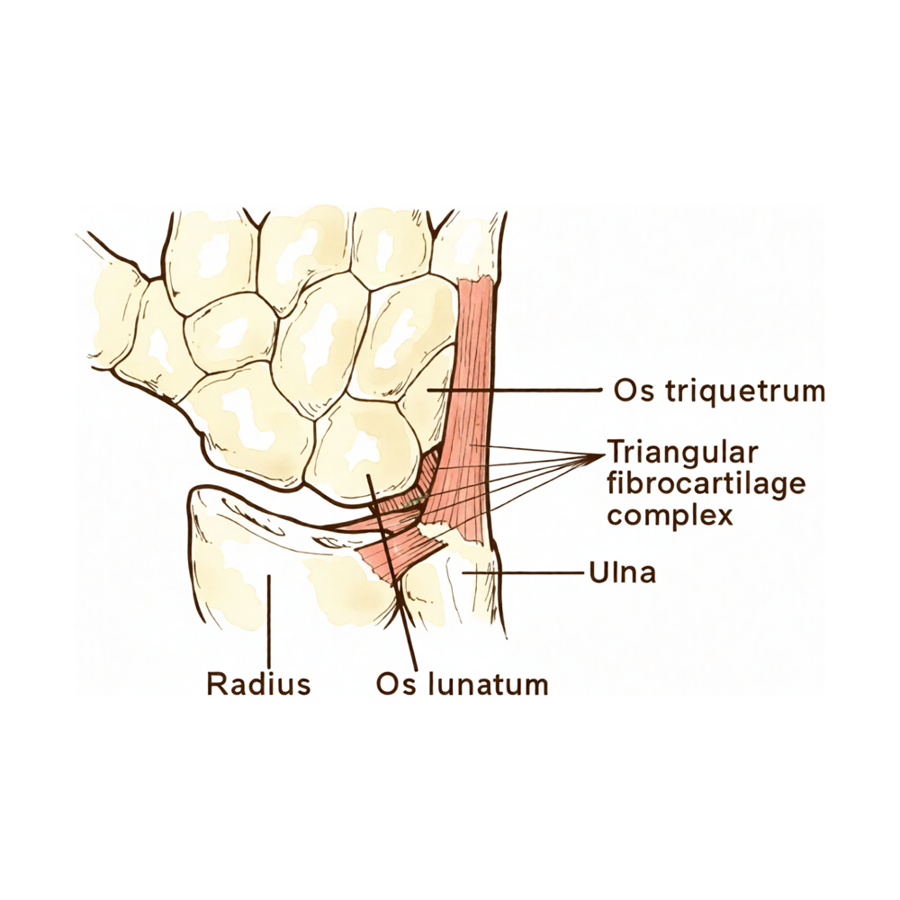
The distal radius articular surface features two concave facets for the scaphoid and lunate, separated by the scapholunate ridge [66]. The sigmoid notch along the ulnar border of the distal radius is a shallow concavity that accommodates the ulnar head at the distal radioulnar joint [66]. The distal radial articular surface has a double obliquity of 12–15 degrees in the lateral view and 15–20 degrees in the anteroposterior view [71]. The distal ulna is covered with hyaline cartilage on its dorsal, lateral, palmar, and distal surfaces [66]. The distal ulnar convexity articulates at the lesser sigmoid notch of the distal radius, with the articular surface accommodating the ulnar head through two-thirds of its arc [64]. The distal ulna has an inclination of approximately 20 degrees at its articulation with the radius [64]. The ulnar styloid projects distally, and its base contains the fovea which serves as the insertion site for the triangular fibrocartilage complex [66]. The ulnar styloid lies dorsal to the ulnar head and extends distally [64]. The triquetrum does not contact the ulnar head directly; a fibrocartilage disc separates the two bones [71].
Ligaments & TFCC
The triangular fibrocartilage complex (TFCC) consists of the ulnar collateral ligament, dorsal and volar radioulnar ligaments, articular disc, meniscal homologue, extensor carpi ulnaris sheath, and ulnolunate and ulnotriquetral ligaments [64]. The TFCC is formed by the central meniscus homolog, dorsal and volar radioulnar ligaments, the floor of the extensor carpi ulnaris tendon sheath, and volar ulnocarpal ligaments [65]. The TFCC attaches to the ulnar margin of the lunate fossa of the radius and separates the hyaline cartilage-covered ulnar head from the styloid [64]. The TFCC arises from the radial border of the distal radius and inserts into the base of the ulnar styloid and distal ulna through the ligamentum subcruentum [65]. The dorsal and volar radioulnar ligaments are the primary stabilizers of the distal radioulnar joint [65]. The ulnotriquetral ligament stabilizes the triquetrum to the fovea of the ulna [65]. The triangular ligament extends the distal radial articular surface to the ulnar styloid and forms the principal link between the radius and ulna [71].
Vascular Supply
Only the peripheral 10% to 40% of the volar, ulnar, and dorsal TFCC has a vascular supply [65].
Biomechanics & Load Transmission
With axial loading through the neutral wrist, approximately 80% of forces are transmitted through the distal radius and 20% through the distal ulna [65]. Axial loading of the wrist increases ulnar variance [44]. Dynamic increases in ulnar variance may accompany forceful grip and pronation [22]. The ulnocarpal ligaments are likely to be stretched tensely in wrist radial extension and wrist extension [114]. The distal radioulnar joint stiffness in dorsal translation decreased significantly with dorsal tilt of 10° and 20° in pronation [111]. Reconstructed animation from four-phase grip MRI demonstrated impairment of the articular disc and longitudinal instability of the distal radioulnar joint simultaneously [30].
Pathophysiology
The ulnar impaction syndrome is a degenerative condition related to excessive load bearing across the ulnar carpus, triangular fibrocartilage complex, and ulnar head [22]. Compressive forces in ulnar impaction syndrome result in ulnar wrist symptoms secondary to a spectrum of pathological changes most commonly resulting from positive ulnar variance [22]. Ulnar impaction syndrome can occur in wrists with ulnar negative or neutral variance, not just positive variance [22]. Pathological staging validates the existence of degenerative changes even before triangular fibrocartilage tears develop [22]. Most degenerative changes in wrists with ulnar plus variance had progressed or changed during a ten-year period [112]. Ulnar styloid impaction syndrome is produced by the tip of an oversized ulnar styloid inappropriately contacting the triquetrum [29]. Ulnar styloid impaction syndrome results in chondromalacia, synovitis, and ulnar-sided wrist pain [29]. Ulnar styloid impaction syndrome is most commonly seen in patients with excessively long or hypertrophic ulnar styloid processes and is not necessarily associated with positive ulnar variance [29]. The ulnar styloid process index (USPI) is calculated as the difference between the ulnar styloid length and the ulnar variance divided by the [29].
Clinical Context
The complex nature of the wrist has hampered the ability to formulate concise, yet thorough, algorithmic approaches to evaluation and treatment [48]. The relative importance of the distal radioulnar joint in coordinated upper extremity motions and function is gaining greater recognition [81]. Ulnar-sided wrist pain in athletes is a common problem often resulting from a combination of overuse and acute injury [101].
Classification
Ulnar Impaction Syndrome (Ulnocarpal Abutment)
Ulnar impaction syndrome is a degenerative condition of the ulnar side of the wrist related to excessive load bearing across the ulnar carpus, triangular fibrocartilage complex, and ulnar head [22]. It is synonymous with ulno-carpal impaction syndrome and ulno-carpal abutment [130]. The condition is characterized by a complex of symptoms resulting from excessive compression of the ulnar head against the triangular fibrocartilage complex [129]. While it occurs most commonly in the ulnar positive wrist, it can also occur in wrists with either ulnar negative or neutral variance [22]. Dynamic increases in ulnar variance may accompany forceful grip and pronation in wrists with neutral or negative variance [22]. An inverse relationship may exist between ulnar variance and the thickness of the articular disk of the triangular fibrocartilage complex [134].
Associated factors of ulno-carpal joint space narrowing include previous fracture of the distal radius, premature physeal arrest of the distal radius, or congenital ulna positive variance [130]. The syndrome may lead to degenerative lesions of the triquetrum, lunate, ulnar head cartilage, or of the triangular fibrocartilage complex [130]. Concomitantly with ulnar impaction syndrome, the triquetrolunate ligaments may be disrupted [130]. In advanced cases, degenerative or cystic changes may be noted on the ulnar aspect of the proximal lunate articular surface and on the "kiss" lesion site [46]. Degenerative lunotriquetral membrane changes were seen in about half of patients with idiopathic ulnar impaction syndrome and were mostly of a mild nature [131]. Once ulnar impaction becomes symptomatic, it remains so and recurs, especially on return to the athletic sport [23].
Ulnar Styloid Impaction Syndrome
Ulnar styloid impaction syndrome is a distinct manifestation from classic ulnocarpal impaction syndrome, described in patients exhibiting neutral or negative ulnar variance with a long ulnar styloid process [53]. It involves repetitive friction between an excessively long ulnar styloid and the carpus [53]. Specifically, ulnar styloid impaction is caused by impaction between the ulnar styloid and the proximal triquetrum [130]. Radiographic evidence displays chondromalacia of the proximal triquetrum and ulnar styloid, as opposed to the classic chondromalacia of the proximal pole of the lunate and ulnar head seen in ulnocarpal impaction [53].
Ulnar Impingement Syndrome
Ulnar impingement syndrome is defined by a shortened ulna impinging on the distal radius [130]. It is a distinct entity from ulnar styloid impaction and ulnar impaction syndrome [130].
Other Causes of Ulnar-Sided Wrist Pain
Triquetrohamate impaction syndrome remains an underreported and often unrecognized cause of ulnar-sided wrist pain [21]. Hamato-lunate and triquetro-hamate impaction syndromes are poorly recognized and underdiagnosed causes of refractory ulnar-sided wrist pain [33]. Ulnar-sided wrist pain is a common cause of upper extremity disability with a complex differential diagnosis [28, 32].
Clinical Presentation
Determining the etiology of ulnar-sided wrist pain is often challenging due to overlapping history and physical examination findings [97]. A systematic approach to evaluating patients with ulnar-sided wrist pain is imperative [31]. A detailed history, systematic physical examination with provocative maneuvers, and appropriate diagnostic imaging are essential for the diagnosis of ulnar-sided wrist pain [97].
History and Examination Findings
The history for ulnar-sided wrist pain should include age, hand dominance, occupation, hobbies, date of injury or onset of symptoms, and correlation of symptoms with activities and modifying factors [122]. For long-standing ulnar-sided wrist problems, it is important to correlate the problem with factors that cause worsening or relief [122]. In the history of traumatic ulnar-sided conditions, it is important to document swelling, bruising, local areas of pain, point tenderness, and sensations of grating, popping, and crunching [122]. Flexion and pronation injuries may contribute more to ligament injuries on the ulnar side of the wrist, especially the lunotriquetral ligament [122].
General Characteristics and Diagnosis
Ulnar impaction syndrome, also termed the ulnocarpal abutment syndrome, results from compressive forces that produce a spectrum of pathological changes most commonly associated with positive ulnar variance [22]. However, ulnar impaction syndrome can occur in wrists with either ulnar negative or neutral variance [22]. The diagnosis of ulnar impaction syndrome can be difficult in wrists with neutral or negative variance [22]. A diagnostic test is required for dynamic ulnar impingement because the condition is frequently missed and to assist in the differential diagnosis of distal forearm pain [56].
Disorders of the distal radioulnar joint are a common source of ulnar-sided wrist pain [57]. The "ulnar fovea sign" represents conditions that are two common sources of ulnar-sided wrist pain [94]. Ulnar extrinsic ligament tears are a source of ulnar-sided wrist pain with associated pathologies and specific clinical presentations [19].
Ulnar Styloid Impaction Syndrome
Ulnar styloid impaction syndrome is most commonly seen in patients with excessively long or hypertrophic ulnar styloid processes [29]. It is not necessarily associated with positive ulnar variance [29]. This condition represents a subset of patients exhibiting neutral or negative ulnar variance with a long ulnar styloid process [53]. Radiographic evidence shows chondromalacia of the proximal triquetrum and ulnar styloid [53]. Ulnar styloid impaction syndrome remains an uncommon cause of ulnar-sided wrist pain and a particularly difficult diagnosis to confirm [29].
Investigations
Clinical Examination: A diagnostic test is required because ulnar-sided pathology is frequently missed and to assist in the differential diagnosis of distal forearm pain [56]. The natural inclination to study radiographs or special imaging studies prior to a thorough history and physical examination should be avoided, as this introduces cognitive bias that affects thinking and decision making [92]. Physical examination must always be preceded by a thorough investigation of the patient’s medical history, with special emphasis on the mechanism of injury and acuity [92]. Palpation for areas of maximal tenderness is one of the most useful tools in the diagnosis of wrist pathology, especially in patients with chronic dysfunctions [92]. A careful assessment of neural and vascular status is imperative, with particular attention being paid to the median and ulnar nerves [92]. A thorough set of provocative maneuvers should be performed, not just to concentrate on the suspected diagnosis but specifically to rule out alternative or even concurrent diagnoses [92]. The TFCC must be carefully examined, and the distal radioulnar joint must be assessed for instability in neutral, supination, and pronation [92]. Ulnar impaction and ulnar styloid impingement must be assessed in pronation and supination [92]. Bilateral grip and pinch strength are useful to uncover underlying pathology in chronic cases [92]. A local injection of anesthetic to a painful joint or selected tendon sheath may help normalize the dynamometer readings and narrow the diagnostic spectrum [92].
MRI: MRI has an expanding role in the evaluation of pathologic conditions of the wrist, requiring high-resolution images best obtained with surface coil technique and high field systems [76]. A dedicated wrist coil provides enhanced resolution of wrist structures [80]. With proper technique, injuries to the triangular fibrocartilage complex (TFCC) can be demonstrated with MRI, where perforations appear as linear defects or gaps filled with hyperintense fluid on coronal gradient-echo or T2-weighted pulse sequences [76]. The addition of arthrographic contrast improves the visualization of carpal ligaments on MR images [76]. MRI is useful in detecting additional marrow abnormalities in osteonecrosis, as seen in the lunate in Kienböck disease or in the scaphoid after fracture [76]. MRI should be added for evaluation of the triangular fibrocartilage, the distal radioulnar joint (DRUJ), and vascularity of carpal bones to confirm clinical suspicion and correlate with physical examination findings [80]. Reconstructed animation from four-phase grip MRI demonstrated impairment of the articular disc and longitudinal instability of the distal radioulnar joint simultaneously and should be of value in investigating dynamic pathophysiology causing ulnar wrist pain [30]. For young subjects, MRI is still valuable, especially in diagnosing ulnar detachment, although the ability to distinguish between proximal and distal laminae remains questionable [141]. Magnetic resonance imaging remains a powerful tool to help diagnose a variety of ulnar wrist conditions, but it is imperative that the surgeon correlates the imaging findings with physical examination [138]. While MRI is a useful adjunct for determining the cause of ulnar wrist pathologies, findings are often discordant when compared with diagnostic arthroscopy [132]. A high rate of false-positive findings on MR images of normal subjects has been reported [80]. MRI is controversial, but newer innovations suggest value in detection and localization of TFCC pathology [84].
Arthroscopy: Arthroscopy is the gold standard for detection of TFCC tears [84]. Arthroscopic assessment of intercarpal ligament injuries and instability is considered by many the “gold standard” for evaluation of these conditions, as well as for examination of patients who have wrist pain of unknown origin [79]. Arthroscopy has been found to be more accurate than arthrography in identifying the location and size of triangular fibrocartilage and interosseous ligament injuries [79]. Arthroscopy is more accurate than triple-injection cinearthrography in detecting tears of the dorsal sensory branch of the ulnar nerve during arthroscopic repair of the triangular fibrocartilage [79].
Other Considerations: Ulnar impaction syndrome is a degenerative condition caused by excessive load across the ulnocarpal joint, often associated with positive ulnar variance [39]. Although the ulnar impaction syndrome occurs most commonly in the ulnar positive wrist, it can also occur in wrists with either ulnar negative or neutral variance [22]. Dynamic increases in ulnar variance may accompany forceful grip and pronation, but diagnosis of the ulnar impaction syndrome can be difficult nonetheless [22]. Hamato-lunate and triquetro-hamate impaction syndromes are potentially treatable with satisfactory results once identified, though they are poorly recognized and underdiagnosed causes of refractory ulnar-sided wrist pain [33].
Treatment
Non-Operative
The provided evidence does not detail specific conservative management protocols such as weight loss, physical therapy, NSAIDs, or injections. Surgical intervention is presented as the primary treatment modality for ulnar impaction syndrome and related conditions.
Operative
Indications: Ulnar shortening osteotomy (USO) is indicated for patients with idiopathic and post-traumatic ulnar impaction syndrome, offering reliable long-term pain relief and satisfaction [9]. Indications have expanded to include luno-triquetral tears and early degenerative arthritis at the distal radioulnar joint [137]. The wafer procedure is suitable for patients with ulnar wrist pain secondary to ulnar impaction who have level or short ulnae on conventional zero rotation radiographs [47]. Arthroscopic treatment is effective when the initial diagnosis is correct [2].
Surgical Approach / Technique: USO yields satisfactory clinical outcomes for more than 5 years, even in the presence of osteoarthritic changes at the distal radioulnar joint [34]. Distal metaphyseal USO is a viable option [17] and offers advantages over other procedures, including more rapid osseous union and buried screw fixation that minimizes the need for hardware removal [139]. An osteochondral shortening osteotomy technique is minimally invasive, preserves the distal ulnar articular joint surface, has an extremely low complication rate, and allows early return of joint motion and function [73]. USO can simultaneously lead to distal radioulnar joint stabilization [86]. The wafer procedure is easier to perform than USO and avoids risks of nonunion and hardware-related complications [47]. However, the wafer procedure should be avoided when a prominent ulnar styloid process is present, as residual stylocarpal impaction may compromise long-term results [47]. Ulnar styloidectomy is a relatively simple procedure that responds well to ulnar styloid impaction syndrome [1]. Arthroscopic treatment of ulnar impaction generally yields excellent results, with disappearance of pain in most cases and often spectacular improvement in grip strength [2].
Implant Selection: Similar postoperative functional scores and complications are observed in patients undergoing USO with a dorsally placed plate compared with other plate placement localizations [3]. Distal metaphyseal USO utilizes buried screw fixation to minimize the potential need for hardware removal [139].
Adjuncts: USO combined with arthroscopic débridement of the TFCC is effective for treating ulnar impaction syndrome [126]. Arthroscopic wafer procedure and USO for idiopathic ulnar impaction syndrome achieve similar clinical and radiologic outcomes [7]. In a randomized controlled trial, both arthroscopic wafer procedure and USO yielded comparable outcomes, with earlier return to work, a lower incidence of complications, and fewer secondary procedures observed in the arthroscopic wafer procedure group [78]. Surgical options for ulnocarpal abutment and degenerative triangular fibrocartilage complex tears include arthroscopic debridement, arthroscopic wafer, open wafer, or ulnar shortening osteotomy [91].
Revision: Patients with persisting or recurrent pain benefited from ulnar shortening osteotomy as a secondary procedure [16]. All patients had resolution of the ulnar forearm pain after hardware removal following ulnar-shortening osteotomy [36]. Symptoms of bilateral ulnar styloid impaction syndrome resolved in both wrists following two separate ulnar styloidectomy procedures [29].
Other Considerations: Ulnar shortening procedures result in improvement in patient-reported outcome scores in patients with ulnocarpal impaction [27].
Complications
Nonunion: Ulnar shortening osteotomy carries a risk of nonunion, with management typically requiring autologous bone grafting or further operation. In a cohort of 30 patients treated with the TriMed dynamic compression system, atrophic nonunion occurred in 2 patients [127]. A separate cohort of 27 patients treated with a dynamic compression plate on the ulnar surface observed one case of nonunion requiring further operation [136]. A trend toward a higher incidence of nonunion was noted in patients treated with the freehand technique, although no statistical difference was demonstrated compared to the Rayhack technique [128].
Hardware Irritation: Plate removal due to irritation is a recognized complication following ulnar shortening osteotomy. Four patients required plate removal due to irritation following treatment with the TriMed dynamic compression system [127]. Similarly, three patients required plate removal following ulnar shortening osteotomy with a dynamic compression plate placed on the ulnar surface of the ulna [136].
Persistent Pain: Persistent ulnar-sided pain may persist post-operatively. Two patients reported persistent ulnar-sided pain following ulnar shortening osteotomy with the TriMed dynamic compression system [127]. Two additional patients reported persistent ulnar-sided wrist pain following ulnar shortening osteotomy with a dynamic compression plate placed on the ulnar surface of the ulna [136].
Infection: No infections were reported in the cohort of 30 patients treated with ulnar shortening osteotomy using the TriMed dynamic compression system [127].
Malalignment: No cases of malalignment of the ulnar shaft or malalignment of the plate against bone were observed in either the freehand or Rayhack technique groups for ulnar shortening osteotomy [128].
Other Considerations: Residual stylocarpal impaction may compromise long-term results if the wafer procedure is performed in the presence of a prominent ulnar styloid process [47]. Ulnar shortening osteotomy in post-traumatic ulnar impaction syndrome achieves improved wrist function only if there is no arthrosis of the distal radioulnar joint [24]. Clinical outcomes remain satisfactory for more than 5 years after ulnar shortening osteotomy for idiopathic ulnar impaction syndrome despite the presence of osteoarthritic changes of the distal radioulnar joint [34].
Recovery
Light activity (weeks): Wrist function recovers after an initial decrease from week 8 onward following ulnar shortening with the UOL [51].
Other Considerations: Ulnar shortening osteotomy achieved excellent long-term results in most cases [62]. At 14 months after open wedge osteotomy with ulnar shortening for Madelung deformity, there has been no loss of correction, instability of the ulnar head, or pain on the ulnar side [14]. Long-term follow-up of forearm shortening and volar radiocarpal capsulotomy for wrist flexion deformity in children with amyoplasia shows that the initial improvement in wrist position is not maintained [52]. Prognosis for return to elite participation is guarded, particularly after distal ulnar resection [142].
Key Evidence
- [L4] Ulnar styloid impaction syndrome responds to the relatively simple procedure of partial ulnar styloidectomy. [1] (10.1016/s0363-5023(97)80131-1)
- [Paper] Results are generally excellent if the initial diagnosis of ulnar impaction was correct, with disappearance of pain in most cases and often spectacular improvement in grip strength. [2] (10.1016/j.main.2006.07.007)
- [L4] Similar postoperative functional scores and complications were seen in patients undergoing an ulnar shortening osteotomy with a dorsally placed plate for ulnar impaction syndrome, compared with other plate placement localizations. [3] (10.1055/s-0037-1608636)
- [L4] This longterm follow-up study revealed that distal ulnar shortening osteotomy is an excellent procedure for the treatment of ulnar impaction syndrome. [4] (10.1016/0363-5023(93)90243-v)
- [L5] The clinically observed incidence of ulnar impaction is more likely the result of increased wear on a thinner and less durable triangular fibrocartilage complex than due to increased distal ulna loading in ulnar positive variant wrists. [5] (10.1016/j.jhsa.2014.10.001)
- [L4] This system yields predictable results for the treatment of ulnar impaction syndrome, and ensures consistent rates of bony union, with statistically significant improvements in patient reported outcome and function. [6] (10.1142/s242483551850011x)
- [L1] The WP and USO for idiopathic ulnar impaction syndrome achieve similar clinical and radiologic outcomes. [7] (10.1016/j.jhsa.2022.08.029)
- [L4] Ulnar shortening osteotomy can change the load of the ulnar side of the wrist and appears useful for ulnar-sided wrist pain in the presence of ulnar impaction. [8] (10.1055/s-0034-1372516)
- [L4] Ulna shortening osteotomy yields reliable long-term pain relief and satisfaction in patients with idiopathic and post-traumatic ulnar impaction syndrome. [9] (10.1016/s0363-5023(12)60047-1)
- [L4] Ulna shortening osteotomy has proven efficacy in ulnar impaction syndrome with 96% of patients considering themselves cured or improved. [10] (10.1016/j.otsr.2021.102970)
- [L4] The study assessed outcomes of 26 patients with ulnar impaction syndrome who underwent ulnar shortening osteotomy over 4 years. [11] (10.1177/1558944716660555it)
- [L4] Ulnar-shortening osteotomy alone or combined with arthroscopic debridement is superior to debridement alone for ulnar impaction syndrome. [12] (10.1016/j.jhsa.2012.07.019)
- [L4] These cases show that ulnocarpal impaction syndrome can occur in wrists with zero or negative ulnar variance, and that ulnar shortening is an effective treatment for such wrists. [13] (10.1016/j.jhsb.2004.09.005)
- [L5] At 14 months after surgery, there has been no loss of correction, instability of the ulnar head, or pain on the ulnar side. [14] (10.1142/s021881041772042x)
- [L5] Effective treatment for ulnar impaction syndrome increasingly relies on arthroscopic decompression, though ulnar shortening osteotomy and open wafer resection remain options. [15] (10.1016/j.hcl.2005.08.011)
- [L3] Patients with persisting or recurrent pain benefited from ulnar shortening osteotomy as a secondary procedure. [16] (10.1055/s-0037-1607073)
- [L4] DMUSO is a viable option for patients with ulnar impaction syndrome. [17] (10.1055/s-0034-1384745)
- [L4] Ulnar shortening osteotomy is a successful treatment both for ulnar impaction syndrome (UIS) and for early stage ulnar carpal instability (UCI) in patients with neutral or positive ulnar variances. [18] (10.1016/s0266-7681(97)80265-3)
- [L4] These findings add information on diagnosis, associated pathologies, surgical management, and long-term outcomes of ulnar extrinsic pathology. [19] (10.1177/15589447251329563)
- [L4] Triquetrohamate impaction syndrome remains an underreported and often unrecognized cause of ulnar-sided wrist pain. [21] (10.1177/1558944716670138)
- [L4] [22] (10.1016/s0266-7681(98)80090-9)
- [L5] Once ulnar impaction becomes symptomatic, it remains so and recurs, especially on return to the athletic sport. [23] (10.1016/j.hcl.2012.05.021)
- [L4] Ulnar shortening osteotomy in post-traumatic ulnar impaction syndrome results in a high rate of satisfied patients, and improved wrist function can be achieved if there is no arthrosis of the distal radioulnar joint. [24] (10.1007/bf00434545)
- [L2] Ulnar shortening procedures result in improvement in PROS in patients with UCI. [27] (10.1055/s-0038-1677534)
- [L5] Ulnar-sided wrist pain is a common cause of upper extremity disability with a complex differential diagnosis. [28] (10.1016/j.jhsa.2012.04.036)
- [L4] [29] (10.1055/s-0038-1673408)
- [L4] Reconstructed animation from four-phase grip MRI demonstrated impairment of the articular disc and longitudinal instability of the distal radioulnar joint simultaneously and should be of value in investigating dynamic pathophysiology causing ulnar wrist pain. [30] (10.1177/1753193413476979)
- [L4] A systematic approach to evaluating patients with ulnar-sided wrist pain is imperative. [31] (10.1016/j.jhsa.2014.07.004)
- [L5] Ulnar-sided wrist pain is a common cause of upper-extremity disability with a complex differential diagnosis. [32] (10.1016/j.jhsa.2008.08.026)
- [L5] Hamato-lunate and triquetro-hamate impaction syndromes are potentially treatable with satisfactory results once identified, though they are poorly recognized and underdiagnosed causes of refractory ulnar-sided wrist pain. [33] (10.1016/j.jhsa.2024.07.016)
- [L4] The clinical outcomes are satisfactory for even more than 5 years after ulnar shortening osteotomy for treating idiopathic ulnar impaction syndrome despite the osteoarthritic changes of the DRUJ. [34] (10.4055/cios.2011.3.4.295)
- [L4] Although patient satisfaction was higher with seven satisfied patients, and postoperative wrist pain scores were good in ten patients, the procedure did not necessarily resolve ulnar-sided wrist pain thought to be caused by ulnar abutment. [35] (10.1016/s1297-3203(01)00055-5)
- [L4] All patients had resolution of the ulnar forearm pain after hardware removal. [36] (10.1016/j.jhsa.2005.06.005)
- [L4] The wafer resection procedure seems to be a viable alternative for the treatment of ulnar impaction syndrome with potentially less morbidity than ulnar shortening osteotomy. [37] (10.1053/jhsu.2000.jhsu025a0055)
- [Paper] Ulnar impaction syndrome is a degenerative condition caused by excessive load across the ulnocarpal joint, often associated with positive ulnar variance. [39] (10.1016/j.hcl.2010.05.011)
- [L2] Axial loading of the wrist increases ulnar variance. [44] (10.1055/s-0038-1627458)
- [L4] [46] (10.1097/00130911-200009000-00007)
- [L4] [47] (10.1054/jhsb.1999.0268)
- [Paper] The complex nature of the wrist has plagued us clinically and hampered our ability to formulate concise, yet thorough, algorithmic approaches to evaluation and treatment. [48] (10.1016/s0894-1130(96)80065-2)
- [L4] In ulnar shortening with the UOL, wrist function recovered after an initial decrease from week 8 onward. [51] (10.1177/1558944717702465)
- [L4] Long-term follow-up of the procedure shows that the initial improvement in wrist position is not maintained. [52] (10.1016/j.jhsa.2011.10.013)
- [L4] [53] (10.1055/s-0035-1570743)
- [L4] A diagnostic test is required because the condition is so frequently missed and also to assist in the differential diagnosis of distal forearm pain in this particular clinical context. [56] (10.1016/s0266-7681(97)80264-1)
- [L5] Disorders of the distal radioulnar joint are a common source of ulnar-sided wrist pain, but increased understanding of anatomy and pathology has facilitated accurate diagnosis and successful treatment in most cases. [57] (10.5435/00124635-199503000-00005)
- [L4] Ulnar shortening osteotomy achieved excellent long-term results in most cases. [62] (10.1016/j.jhsa.2012.09.011)
- [Commentary] The study is just one small piece in the enormous puzzle that constitutes wrist biomechanics, and the information gleaned from this study is not enough at this point to carry over into clinical practice. [65] (10.1016/j.jhsa.2015.08.017)
- [L4] [73] (10.1097/bth.0b013e3180337df9)
- [L2] [78] (10.1016/j.jhsa.2022.04.011)
- [Paper] The relative importance of the distal radioulnar joint in the coordinated upper extremity motions and function is finally gaining greater recognition. [81] (10.1097/bth.0b013e318033d96f)
- [L4] [86] (10.1177/17531934231197942)
- [L5] [91] (10.1016/j.hcl.2012.05.018)
- [L2] The conditions represent 2 common sources of ulnar-sided wrist pain. [94] (10.1016/j.jhsa.2007.01.022)
- [L5] Determining the etiology of ulnar-sided wrist pain is often challenging due to overlapping history and physical examination findings; a detailed history, systematic physical examination with provocative maneuvers, and appropriate diagnostic imaging are essential for diagnosis. [97] (10.5435/jaaos-d-16-00407)
- [L5] Ulnar-sided wrist pain in athletes is a common problem often resulting from a combination of overuse and acute injury, requiring careful understanding of sport-specific injuries and underlying biomechanics for effective diagnosis and treatment. [101] (10.1016/j.csm.2019.12.008)
- [L5] The distal radioulnar joint stiffness in dorsal translation decreased significantly with dorsal tilt of 10° and 20° in pronation. [111] (10.1177/1753193412473036)
- [L4] Most of the degenerative changes in the wrists with ulnar plus variance had progressed or changed during the ten or more years. [112] (10.1016/s0363-5023(10)80011-5)
- [L4] The ulnocarpal ligaments are likely to be stretched tensely in wrist radial extension and wrist extension. [114] (10.1016/j.jhsa.2008.04.033)
- [L4] Ulnar shortening osteotomy combined with arthroscopic débridement of the TFCC is effective for treating ulnar impaction syndrome. [126] (10.1097/blo.0b013e31815a9e21)
- [L4] [127] (10.1016/j.jhsa.2013.04.040)
- [L3] [128] (10.1016/j.jhsa.2005.09.017)
- [L4] [129] (10.1177/1753193420908795)
- [L4] [130] (10.1177/1753193414541749)
- [L4] [131] (10.1016/j.jhsa.2014.03.006)
- [L2] While MRI is a useful adjunct for determining the cause of ulnar wrist pathologies, findings are often discordant when compared with diagnostic arthroscopy. [132] (10.1016/j.jhsa.2024.04.015)
- [L5] [134] (10.1016/j.arthro.2017.11.008)
- [L4] [136] (10.1055/s-0037-1608851)
- [L5] Ulnar shortening is a procedure that has steadily grown in popularity, with indications expanding to include luno-triquetral tears and early degenerative arthritis at the distal radial–ulnar joint. [137] (10.1097/00130911-200306000-00002)
- [L5] Magnetic resonance imaging remains a powerful tool to help diagnose a variety of ulnar wrist conditions, but it is imperative that the surgeon correlates the imaging findings with physical examination. [138] (10.1016/j.jhsa.2024.05.012)
- [L5] This technique offers advantages over other commonly used procedures for treatment of ulnar impaction, including more rapid osseous union and buried screw fixation minimizing the potential need for hardware removal. [139] (10.1016/j.jhsa.2012.02.031)
- [L3] For young subjects, MRI is still valuable, especially in diagnosing ulnar detachment, although the ability to distinguish between proximal and distal laminae remains questionable. [141] (10.1177/17531934221141986)
- [Paper] Prognosis for return to elite participation is guarded, particularly after distal ulnar resection. [142] (10.1016/j.hcl.2012.05.020)
References
[1] Ulnar styloid impaction syndrome. The Journal of Hand Surgery. 1997. DOI: 10.1016/s0363-5023(97)80131-1
[2] Traitement arthroscopique des conflits ulnolunaires. Chirurgie de la Main. 2006. DOI: 10.1016/j.main.2006.07.007
[3] A Dorsal Approach for Ulnar Shortening Osteotomy. Journal of Wrist Surgery. 2017. DOI: 10.1055/s-0037-1608636
[4] The ulnar impaction syndrome: Follow-up of ulnar shortening osteotomy. The Journal of Hand Surgery. 1993. DOI: 10.1016/0363-5023(93)90243-v
[5] Force Variations in the Distal Radius and Ulna: Effect of Ulnar Variance and Forearm Motion. The Journal of Hand Surgery. 2015. DOI: 10.1016/j.jhsa.2014.10.001
[6] Outcomes of Ulnar Shortening for Ulnar Impaction Syndrome Using the 2.7 mm AO Ulna Shortening Osteotomy System. The Journal of Hand Surgery (Asian-Pacific Volume). 2018. DOI: 10.1142/s242483551850011x
[7] Ulnar Shortening Osteotomy Versus the Wafer Procedure in the Treatment of Idiopathic Ulnar Impaction Syndrome: A Systematic Review and Meta-Analysis. The Journal of Hand Surgery. 2024. DOI: 10.1016/j.jhsa.2022.08.029
[8] Ulnar Shortening Osteotomy for Ulnar-Sided Wrist Pain. Journal of Wrist Surgery. 2014. DOI: 10.1055/s-0034-1372516
[9] Long-Term Subjective Satisfaction after Ulna Shortening Osteotomy for Ulnar Impaction Syndrome. The Journal of Hand Surgery. 2012. DOI: 10.1016/s0363-5023(12)60047-1
[10] Ulna shortening osteotomy for ulnar impaction syndrome: Impact of distal radioulnar joint morphology on clinical outcome. Orthopaedics & Traumatology: Surgery & Research. 2021. DOI: 10.1016/j.otsr.2021.102970
[11] Shortening Osteotomy for Ulnar Impaction Syndrome. HAND. 2016. DOI: 10.1177/1558944716660555it
[12] Ulnar Shortening Osteotomy for Ulnar Impaction Syndrome. The Journal of Hand Surgery. 2013. DOI: 10.1016/j.jhsa.2012.07.019
[13] Results of Ulnar Shortening Osteotomy for Ulnocarpal Impaction Syndrome in Wrists with Neutral or Negative Ulnar Variance. Journal of Hand Surgery. 2005. DOI: 10.1016/j.jhsb.2004.09.005
[14] Open Wedge Osteotomy with Ulnar Shortening for Madelung Deformity Using a Computer-Generated Template. The Journal of Hand Surgery (Asian-Pacific Volume). 2017. DOI: 10.1142/s021881041772042x
[15] Ulnar Impaction Syndrome. Hand Clinics. 2005. DOI: 10.1016/j.hcl.2005.08.011
[16] Treatment of Ulnar Impaction Syndrome with and without Central TFC Lesion. Journal of Wrist Surgery. 2017. DOI: 10.1055/s-0037-1607073
[17] Distal Metaphyseal Ulnar Shortening Osteotomy: Technique, Pearls, and Outcomes. Journal of Wrist Surgery. 2014. DOI: 10.1055/s-0034-1384745
[18] Ulnar Shortening Osteotomy for Ulnar Carpal Instability and Ulnar Carpal Impaction. Journal of Hand Surgery. 1997. DOI: 10.1016/s0266-7681(97)80265-3
[19] Ulnar Extrinsic Ligament Tears: Clinical Presentation, Surgical Management, and Outcomes of Surgical Repair Extending Into Long-term Follow-up. HAND. 2025. DOI: 10.1177/15589447251329563
[21] Triquetrohamate Impaction Syndrome: An Unrecognized Cause of Ulnar-Sided Wrist Pain; Its Presentation Further Defined. HAND. 2016. DOI: 10.1177/1558944716670138
[22] Ulnar Impaction Syndrome in the Ulnar Negative and Neutral Wrist. Journal of Hand Surgery. 1998. DOI: 10.1016/s0266-7681(98)80090-9
[23] Central TFC Tear/Ulnar Impaction Injuries in Professional Football Players. Hand Clinics. 2012. DOI: 10.1016/j.hcl.2012.05.021
[24] Mar shortening osteotomy in posttraumatic ulnar impaction syndrome. Archives of Orthopaedic and Trauma Surgery. 1996. DOI: 10.1007/bf00434545
[27] Clinical and Radiographic Correlates for the Treatment of Ulnocarpal Impaction. Journal of Wrist Surgery. 2019. DOI: 10.1055/s-0038-1677534
[28] Ulnar-sided Wrist Pain: Evaluation and Treatment of Triangular Fibrocartilage Complex Tears, Ulnocarpal Impaction Syndrome, and Lunotriquetral Ligament Tears. The Journal of Hand Surgery. 2012. DOI: 10.1016/j.jhsa.2012.04.036
[29] Bilateral Ulnar Styloid Impaction Syndrome Treated with Ulnar Styloidectomy. Journal of Wrist Surgery. 2018. DOI: 10.1055/s-0038-1673408
[30] Reconstructed animation from four-phase grip MRI of the wrist with ulnar-sided pain. Journal of Hand Surgery (European Volume). 2013. DOI: 10.1177/1753193413476979
[31] Examination of the Wrist: Ulnar-Sided Wrist Pain Due to Ligamentous Injury. The Journal of Hand Surgery. 2014. DOI: 10.1016/j.jhsa.2014.07.004
[32] Ulnar-Sided Wrist Pain: Evaluation and Treatment of Triangular Fibrocartilage Complex Tears, Ulnocarpal Impaction Syndrome, and Lunotriquetral Ligament Tears. The Journal of Hand Surgery. 2008. DOI: 10.1016/j.jhsa.2008.08.026
[33] Midcarpal Impaction Syndromes as a Rare Cause of Ulnar-Sided Wrist Pain: A Review. The Journal of Hand Surgery. 2024. DOI: 10.1016/j.jhsa.2024.07.016
[34] Long-term Outcomes of Ulnar Shortening Osteotomy for Idiopathic Ulnar Impaction Syndrome: At Least 5-Years Follow-up. Clinics in Orthopedic Surgery. 2011. DOI: 10.4055/cios.2011.3.4.295
[35] Ulnar shortening after failed arthroscopic treatment of triangular fibrocartilage complex tears. Chirurgie de la Main. 2001. DOI: 10.1016/s1297-3203(01)00055-5
[36] Plate Removal After Ulnar-Shortening Osteotomy. The Journal of Hand Surgery. 2005. DOI: 10.1016/j.jhsa.2005.06.005
[37] Comparison of ulnar shortening osteotomy and the wafer resection procedure as treatment for ulnar impaction syndrome. The Journal of Hand Surgery. 2000. DOI: 10.1053/jhsu.2000.jhsu025a0055
[39] Ulnar Impaction. Hand Clinics. 2010. DOI: 10.1016/j.hcl.2010.05.011
[44] The Effect of Axial Loading on Ulnar Variance. Journal of Wrist Surgery. 2018. DOI: 10.1055/s-0038-1627458
[46] Arthroscopic Wafer for Ulnar Impaction Syndrome. Techniques in Hand & Upper Extremity Surgery. 2000. DOI: 10.1097/00130911-200009000-00007
[47] Results of the Wafer Procedure for Ulnar Impaction Syndrome in the Ulnar Negative and Neutral Wrist. Journal of Hand Surgery. 1999. DOI: 10.1054/jhsb.1999.0268
[48] The Mysterious Wrist. Journal of Hand Therapy. 1996. DOI: 10.1016/s0894-1130(96)80065-2
[51] Time-Dependent Recovery of Outcome Parameters in Ulnar Shortening for Positive Ulnar Variance: A Prospective Case Series. HAND. 2017. DOI: 10.1177/1558944717702465
[52] Long-Term Results of Forearm Shortening and Volar Radiocarpal Capsulotomy for Wrist Flexion Deformity in Children With Amyoplasia. The Journal of Hand Surgery. 2012. DOI: 10.1016/j.jhsa.2011.10.013
[53] Ulnar-Sided Wrist Pain due to Long Ulnar Styloid: A Case Report. Journal of Wrist Surgery. 2016. DOI: 10.1055/s-0035-1570743
[56] The Radiological Demonstration of Dynamic Ulnar Impingement. Journal of Hand Surgery. 1997. DOI: 10.1016/s0266-7681(97)80264-1
[57] The Distal Radioulnar Joint:Problems and Solutions. Journal of the American Academy of Orthopaedic Surgeons. 1995. DOI: 10.5435/00124635-199503000-00005
[62] Clinical, Radiographic, and Arthroscopic Outcomes After Ulnar Shortening Osteotomy: A Long-Term Follow-Up Study. The Journal of Hand Surgery. 2012. DOI: 10.1016/j.jhsa.2012.09.011
[64] Campbell S Operative Orthopaedics 4 Volume Set. NERVE INJURIES AT THE LEVEL OF THE HAND AND WRIST > ANATOMY.
[65] Commentary for “Distal Radioulnar Joint Reaction Force Following Ulnar Shortening: Diaphyseal Osteotomy Versus Wafer Resection”. The Journal of Hand Surgery. 2015. DOI: 10.1016/j.jhsa.2015.08.017
[66] Aaos Comprehensive Orthopaedic Review 3. Anatomy of the Hand and Wrist > VII. The Wrist.
[71] Exam Of The Hand Wrist 2Ed. 1.2 SKELETON OF THE WRIST.
[73] Osteochondral Shortening Osteotomy for the Treatment of Ulnar Impaction Syndrome. Techniques in Hand & Upper Extremity Surgery. 2007. DOI: 10.1097/bth.0b013e3180337df9
[76] Campbell S Operative Orthopaedics 4 Volume Set. WRIST AND ELBOW.
[78] Arthroscopic Wafer Procedure Versus Ulnar Shortening Osteotomy for Treatment of Idiopathic Ulnar Impaction Syndrome: A Randomized Controlled Trial. The Journal of Hand Surgery. 2022. DOI: 10.1016/j.jhsa.2022.04.011
[79] Campbell S Operative Orthopaedics 4 Volume Set. NERVE INJURIES AT THE LEVEL OF THE HAND AND WRIST > ARTHROSCOPY OF THE WRIST.
[80] Campbell S Operative Orthopaedics 4 Volume Set. NERVE INJURIES AT THE LEVEL OF THE HAND AND WRIST > RADIOGRAPHIC TECHNIQUES.
[81] The Distal Radioulnar Joint Contemporary Perspectives. Techniques in Hand & Upper Extremity Surgery. 2007. DOI: 10.1097/bth.0b013e318033d96f
[84] Miller S Review Of Orthopaedics. DISTAL RADIOULNAR JOINT, TRIANGULAR FIBROCARTILAGE COMPLEX, AND WRIST ARTHROSCOPY > 2. TFCC tears.
[86] An alternative treatment for degenerative triangular fibrocartilage complex injuries with distal radioulnar joint instability: first experience with 48 patients. Journal of Hand Surgery (European Volume). 2023. DOI: 10.1177/17531934231197942
[91] The Management of Ulnocarpal Abutment and Degenerative Triangular Fibrocartilage Complex Tears in the Competitive Athlete. Hand Clinics. 2012. DOI: 10.1016/j.hcl.2012.05.018
[92] Green S Operative Hand Surgery. Diagnosis and Treatment > Assessment of the Symptomatic Wrist.
[94] The “Ulnar Fovea Sign” for Defining Ulnar Wrist Pain: An Analysis of Sensitivity and Specificity. The Journal of Hand Surgery. 2007. DOI: 10.1016/j.jhsa.2007.01.022
[97] Evaluation of Ulnar-sided Wrist Pain. Journal of the American Academy of Orthopaedic Surgeons. 2017. DOI: 10.5435/jaaos-d-16-00407
[101] Ulnar-Sided Wrist Pain in the Athlete. Clinics in Sports Medicine. 2020. DOI: 10.1016/j.csm.2019.12.008
[111] The effects of dorsally angulated distal radius fractures on distal radioulnar joint stability: a biomechanical study. Journal of Hand Surgery (European Volume). 2013. DOI: 10.1177/1753193412473036
[112] Radiographic changes in wrists with ulnar plus variance observed over a ten-year period. The Journal of Hand Surgery. 1991. DOI: 10.1016/s0363-5023(10)80011-5
[114] Change in the Length of the Ulnocarpal Ligaments During Radiocarpal Motion: Possible Impact on Triangular Fibrocartilage Complex Foveal Tears. The Journal of Hand Surgery. 2008. DOI: 10.1016/j.jhsa.2008.04.033
[122] Campbell S Operative Orthopaedics 4 Volume Set. NERVE INJURIES AT THE LEVEL OF THE HAND AND WRIST > DIAGNOSIS OF WRIST CONDITIONS.
[126] Factors Affecting Results of Ulnar Shortening for Ulnar Impaction Syndrome. Clinical Orthopaedics & Related Research. 2007. DOI: 10.1097/blo.0b013e31815a9e21
[127] Outcomes of Ulnar Shortening Osteotomy Fixed With a Dynamic Compression System. The Journal of Hand Surgery. 2013. DOI: 10.1016/j.jhsa.2013.04.040
[128] A Comparative Study of Ulnar-Shortening Osteotomy by the Freehand Technique Versus the Rayhack Technique. The Journal of Hand Surgery. 2006. DOI: 10.1016/j.jhsa.2005.09.017
[129] Computed tomography osteoabsorptiometry changes following ulnar shortening osteotomy: observations in nine patients. Journal of Hand Surgery (European Volume). 2020. DOI: 10.1177/1753193420908795
[130] Operative treatment of ulnar impaction syndrome: a systematic review. Journal of Hand Surgery (European Volume). 2014. DOI: 10.1177/1753193414541749
[131] Ulnar Impaction Syndrome: Incidence of Lunotriquetral Ligament Degeneration and Outcome of Ulnar-Shortening Osteotomy. The Journal of Hand Surgery. 2014. DOI: 10.1016/j.jhsa.2014.03.006
[132] Etiology and Diagnostic Challenges of Ulnar Wrist Pain in Pediatric and Adolescent Patients. The Journal of Hand Surgery. 2024. DOI: 10.1016/j.jhsa.2024.04.015
[134] Editorial Commentary: Wrist Ulnar Impaction Syndrome: When I Use the Wafer Procedure and When I Do Not. Arthroscopy. 2018. DOI: 10.1016/j.arthro.2017.11.008
[136] Outcomes of the Ulnar Shortening Osteotomy Using a Dynamic Compression Plate on the Ulnar Surface of the Ulna. Journal of Wrist Surgery. 2017. DOI: 10.1055/s-0037-1608851
[137] Ulnar Shortening. Techniques in Hand and Upper Extremity Surgery. 2003. DOI: 10.1097/00130911-200306000-00002
[138] Magnetic Resonance Imaging Assessment of Ulnar Wrist Pain: A Practical Guide for Surgeons. The Journal of Hand Surgery. 2024. DOI: 10.1016/j.jhsa.2024.05.012
[139] Distal Metaphyseal Ulnar-Shortening Osteotomy: Surgical Technique. The Journal of Hand Surgery. 2012. DOI: 10.1016/j.jhsa.2012.02.031
[141] Abnormal MRI signal intensity of the triangular fibrocartilage complex in asymptomatic wrists. Journal of Hand Surgery (European Volume). 2022. DOI: 10.1177/17531934221141986
[142] Elite Athlete: Chronic DRUJ Instability or Central TFC Tears. Hand Clinics. 2012. DOI: 10.1016/j.hcl.2012.05.020