Clinicians › Hand
Giant Cell Tumour of Tendon Sheath
Giant cell tumour of tendon sheath (a.k.a. tenosynovial giant cell tumour, localised pigmented villonodular synovitis).
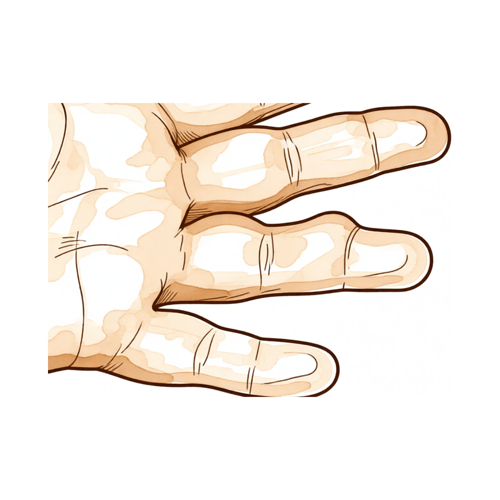
Overview
Giant cell tumour of tendon sheath (GCTTS) is a common benign neoplasm of the hand that is locally aggressive and carries the potential for osseous invasion [6, 10]. While recurrence remains the primary clinical risk, it typically manifests within 36 months of excision [4, 9]. In rare instances, the tumour presents as a lytic osseous lesion through uncharacteristic intraosseous invasion [3]. In a reviewed series of 133 cases, intraosseous expansion occurred in 11.3% of patients [18]. Although reports in the paediatric population are rare [6], the condition shares histological features with pigmented villonodular synovitis, both being benign synovial neoplasms with a capacity for local recurrence [11]. Clinically, GCTTS is frequently confused with fibroma of tendon sheath, as both present as well-circumscribed masses intimately involved with the tendon sheath [5]. Fibromas are sufficiently common to warrant inclusion in the differential diagnosis, comprising 7 of 208 soft tissue hand tumors excised over a 15-year period [2].
The proliferative activity of localized GCTTS, measured by the MIB-1 staining index, is not related to its high recurrence rate or local aggressiveness [14]. Complete surgical resection remains the treatment of choice for most patients with tenosynovial giant cell tumors [13]. Adequate resection should be performed at the initial procedure to mitigate recurrence [51]. For selected cases, curettage, cryosurgery, and cementation performed by experienced surgeons appear to be a safe, effective, and reliable method [44]. In the setting of recurrent giant cell tumour of the hand, en bloc resection of the tumour and reconstruction with an autograft should be considered [36]. Diffuse tenosynovial giant cell tumor disease presents specific challenges due to high recurrence rates [13].
Anatomy & Pathophysiology
Epidemiology and Demographics
Giant cell tumour of tendon sheath is the second most common tumour of the hand [20] and the second most common benign proliferative tumour in the upper extremities after ganglion cysts [27]. It is also identified as the most common benign neoplasm in the hand following the ganglion cyst [32]. These tumours are most frequently found in the fingers and among women in their fourth and fifth decades [27]. The male-to-female ratio for giant cell tumour of tendon sheath of the digits is 1:1.47 [26], with a mean age ranging from 32 to 51 years [26].
Etiology and Pathogenesis
The etiology of giant cell tumors of the tendon sheath is unknown [24]. Pathogenetic theories have included inflammatory process, trauma, immune mechanisms, and neoplasia [24]. It has been suggested that the lesion may be neoplastic, inflammatory, trauma-induced, immune-mediated or associated with abnormal lipid metabolism [20]. More recent reports, including deoxyribonucleic acid (DNA) analysis by flow cytometry, support a truly neoplastic nature for the lesion [20]. Inflammation resulting from reactive or regenerative hyperplasia is the generally accepted theory of pathogenesis [27]. Genetic factors have been observed in previous studies regarding the pathogenesis of these tumours [27]. A definite history of trauma was recorded in 5% of giant cell tumour of tendon sheath lesions [26].
Clinical Presentation and Symptoms
Giant cell tumors usually present as a solitary and firm slow-growing nodular lesion which affects the volar aspect of the hand [24]. These tumours are usually slow-growing, painless, benign and consist of soft tissue [27]. As the tumour grows, some patients may present with swelling, pain and limitation of movement [27]. Pain was reported in 15.7% of cases of giant cell tumour of tendon sheath of the digits [26], while sensory disturbances were reported in 4.57% of cases [26]. The most frequent tumour location was the index finger at 29.7% [26]. A giant cell tumor of tendon sheath can present as a trigger finger [1] or with intraosseous invasion as a lytic osseous lesion [3].
Histology and Classification
The giant cell tumour of tendon sheath has been classified as being of fibrohistiocytic origin [30]. The disease spectrum is described by a number of terms due to varied morphology, including pigmented villonodular synovitis for less well circumscribed tumours that grow more diffusely and incorporate haemosiderin pigment [30]. Different names used to describe these lesions include giant cell tumour of tendon sheath, pigmented villonodular synovitis, fibrous xanthoma, benign synovioma and sclerosing haemangioma [20].
Type I: Defined as single lesions [26], this classification includes a nodular or multinodular lesion surrounded by a capsule [27]. Type II: Defined as two or more distinct tumours that were not joined together [26], this classification describes tumours with no connective tissue membrane and satellite, diffuse or multicentric nodules [27].
Type I tumours (single lesions) were more frequently detected than type II tumours in a systematic review, with 78.7% and 21.3% respectively [26].
Prognostic Markers
nm23 expression was absent in 21% of giant cell tumours and this was associated with a highly significant risk of local recurrence (P<0.0001) [30]. Multivariate analysis showed nm23 expression to be more reliable than other clinicopathological parameters for predicting outcome in giant cell tumours of tendon sheath [30].
Classification
Al-Qattan Classification: The Al-Qattan classification categorizes giant cell tumour of tendon sheath (GCTTS) into two main types based on whether the entire tumour is surrounded by one pseudocapsule as assessed by the surgeon during surgery [20]. Type I consists of nodular or multinodular lesions surrounded by a capsule [27]. Type II describes tumours with no connective tissue membrane and satellite, diffuse, or multicentric nodules [27]. This system categorizes GCTTS according to the risk of recurrence [27].
Recurrence Patterns: In a prospective study of 43 consecutive cases, none of the 30 Type I tumours recurred [20]. Conversely, recurrence occurred in five out of 13 Type II tumours [20]. Second recurrences were observed with Type II B and C tumours but not with Type II A tumours [20]. A systematic review found that Type I tumours (single lesions) were more frequently detected (78.7%) than Type II tumours (two or more distinct tumours that were not joined together) (21.3%) [26]. Type II tumours are associated with a higher risk of recurrence compared to Type I tumours [26].
Other Considerations: GCTTS has been referred to by historic names including pigmented villonodular synovitis, fibrous xanthoma, benign synovioma, and sclerosing haemangioma [20]. The term pigmented villonodular synovitis is largely a descriptive phrase for less well-circumscribed giant cell tumours that grow more diffusely and incorporate haemosiderin pigment [30]. GCTTS are considered to be the extra-articular equivalent of localised pigmented villonodular synovitis [46]. Intra-articular giant cell tumor of tendon sheath and pigmented villonodular synovitis are histologically similar, usually benign tumors characterized by synovial involvement (GCTTS) or overgrowth (PVNS) [28]. The World Health Organization classifies diffuse pigmented villonodular synovitis as 'diffuse-type giant cell tumour' (Dt-GCT) with ICD-O code 9251/0 [46].
Clinical Presentation
Giant cell tumor of tendon sheath (GCTTS) is the second most common tumor of the hand [20] and the second most common benign proliferative tumour in the upper extremities after ganglion cysts [27]. The lesion typically presents as a solitary, firm, slow-growing nodular mass beneath the skin [19, 24]. It is usually not tender to palpation [19] and is often asymptomatic; however, as the tumor grows, patients may present with swelling, pain, and limitation of movement [27]. In a systematic review of 605 patients, pain was reported in 15.7% of cases and sensory disturbances in 4.57% [26]. A definite history of trauma was recorded in 5% of lesions [26].
Demographics and Location: GCTTS most commonly affects the volar aspect of the hand [24] and is found most frequently in the fingers [27]. The index finger is the most frequent tumor location, accounting for 29.7% of cases in a systematic review of 605 patients [26]. The condition predominantly affects women in their fourth and fifth decades [27], with a male-to-female ratio of 1:1.47 and a mean age ranging from 32 to 51 years in the same review [26]. Multifocal lesions are rarely described and commonly involve the same finger or the volar aspect of different fingers [24]. Type I tumours (single lesions) were more frequently detected (78.7%) than type II tumours (two or more distinct tumours that were not joined together) (21.3%) [26].
Atypical Presentations: GCTTS can present as a trigger finger [1], a trigger wrist [21], or with carpal tunnel syndrome [23]. Intraosseous expansion occurs in 11.3% of a reviewed series of 133 cases [18]. When intraosseous invasion is present, the tumor can present as a lytic osseous lesion [3].
Differential Diagnosis: Fibromas of tendon sheath are clinically usually confused with GCTTS, as both are well circumscribed and intimately involved with the tendon sheath [5]. Calcifying aponeurotic fibroma presents as a painless mass primarily on the volar surface of the hands and must be considered in the differential diagnosis [33].
Imaging: Radiographs will demonstrate no specific findings for GCTTS [19], though radiography can be helpful in evaluating cortical destruction but is not helpful in the definitive diagnosis [27]. Ultrasound is useful in distinguishing GCTTS from ganglion cysts [19]. Magnetic resonance imaging (MRI) is the most useful examination for diagnosis and treatment planning [27].
Investigations
MRI: Magnetic resonance imaging is the investigation of choice for diagnosing tumour-like lesions of the infrapatellar fat pad [106] and is essential to diagnose localized pigmented villonodular synovitis, accurately defining its localization and treatment strategy [107]. While MRI provides valuable clues for tenosynovial giant cell tumour, definitive diagnosis relies on histopathological confirmation [109]. Although MRI findings and location might help in the diagnosis of tenosynovial giant cell tumors, careful assessment is mandatory, especially in unusual locations [108]. The absence of disease on MRI should not be used as the sole criterion in determining whether a repeat resection should be performed for unsuspected soft tissue sarcomas of the hand [104].
Other Considerations: A careful physical examination is essential to direct care and future testing if indicated, as diagnostic tests such as imaging and serum laboratory studies can be expensive, time consuming, and often nonspecific [37]. Clinically, fibroma of tendon sheath is usually confused with giant cell tumor of tendon sheath (GCTTS), both being well circumscribed and intimately involved with tendon sheath [5]. Fibromas of tendon sheath are common enough to be considered in the differential diagnosis of a soft tissue tumor in the hand, as they comprised 7 of 208 soft tissue hand tumors excised over a 15-year period [2]. Calcified aponeurotic fibroma is a rare tumor that presents as a painless mass primarily on the volar surface of the hands and plantar aspects of the feet in juveniles and must be considered in the differential diagnosis of GCT of the tendon sheaths or sarcomas, especially in young patients [33].
Giant cell tumour of tendon sheath can present as a trigger finger, with one case report representing the first reported instance of this presentation [1]. A giant cell tumour of the tendon sheath can cause trigger wrist [21]. A diffuse giant cell tumor of the tendon sheath can arise from the carpal tunnel of the wrist, presenting with mild carpal tunnel syndrome and a mid-palmar swelling [23]. Giant cell tumors of the tendon sheaths in the hand are benign lesions where recurrence is the primary risk, typically occurring within 36 months of excision [4]. Pigmented villonoid synovitis and giant-cell tumor of tendon sheath are benign synovial neoplasms with the potential for local recurrence [11]. Giant cell tumor of tendon sheath (GCTTS) is locally aggressive and has the possibility of osseous invasion [10]. Complete surgical resection remains the treatment of choice for most patients with tenosynovial giant cell tumors, though diffuse disease presents challenges due to high recurrence rates [13].
Treatment
Non-Operative
The provided evidence does not support conservative management as a primary treatment modality for giant cell tumor of tendon sheath (GCTTS). Surgical intervention is the standard of care, with non-operative options such as weight loss, physical therapy, NSAIDs, or injections not indicated in the available literature.
Operative
Indications: Marginal excision is the treatment of choice for GCTTS [32]. Surgical excision is the most commonly accepted treatment [27] and is required to address the primary risk of recurrence associated with these tumors [4, 9].
Surgical Approach / Technique: Successful outcomes depend on preoperative planning aided by tissue diagnosis via fine needle aspiration cytology, wide surgical exposure, and meticulous dissection using magnification [31]. The surgeon must ensure complete excision of the tumor and removal of any residual satellite nodules to mitigate factors predisposing to recurrence [32]. A magnifying loupe is utilized to facilitate a careful search for satellite lesions and to respect surrounding structures [32]. Preoperative diagnosis and meticulous surgical technique are identified as the only predictive factors for recurrence in a series of 14 cases [31].
Adjuncts: In cases of infiltrative GCTTS, radiation therapy may provide local tumor control with preservation of hand function [43].
Other Considerations: GCTTS is a locally aggressive benign tumor that may involve osseous invasion [10] and can present with uncharacteristic intraosseous invasion as a lytic osseous lesion [3]. Diffuse disease presents challenges due to high recurrence rates [13]. Recurrence rates tend to be high, with variable rates reported by different authors [19]. The average recurrence rate in the literature is about 15% [32], though only 3 cases (4.7%) of 64 patients reported recurrence in a study comparing literature results with local experience [32]. A recent prospective study of 213 cases showed 27 recurrences (13%) at a minimum 3-year follow-up [19]. Recurrence typically occurs within 36 months of excision [4]. Of recurrent tumors, those involving the extensor tendon, flexor tendon, or joint capsule had the highest risk of recurrence [19]. Close monitoring and/or adjuvant treatment is recommended for tumors involving the extensor tendon, flexor tendon, or joint capsule [19]. In a prospective study of 43 consecutive cases, none of the type I tumours (n=30) recurred [20]. Recurrence occurred in five out of 13 type II tumours [20]. Second recurrences were seen with type II B and C, but not type II A tumours [20].
Complications
Recurrence
Recurrence is the primary risk associated with giant cell tumors of the tendon sheaths in the hand [4]. A systematic review of 605 patients identified an overall tumour recurrence rate of 14.8% [26], while a prospective study of 213 cases reported 27 recurrences (13%) at a minimum 3-year follow-up [19]. In a smaller study of 64 cases, only 3 cases (4.7%) reported recurrence [32]. Type II tumours are associated with higher recurrence rates than Type I tumours [26]. Specifically, in a prospective study of 43 cases, none of the type I tumours recurred, but recurrence occurred in five out of 13 type II tumours [20]. Second recurrences were observed with type II B and C tumours, but not type II A tumours [20]. Tumors involving the extensor tendon, flexor tendon, or joint capsule carry the highest risk of recurrence [19].
Local Invasion and Aggressiveness
GCTTS may present with uncharacteristic intraosseous invasion appearing as a lytic osseous lesion [3]. Intraosseous expansion occurs in 11.3% of a reviewed series of 133 cases [18]. The possibility of osseous invasion is referenced in the context of GCTTS being locally aggressive [10]. Marginal excision is often difficult to perform due to the strict adherence of the tumor to the tendon or neurovascular bundles [32].
Atypical Presentations and Complications
Giant cell tumor of tendon sheath can present as a trigger finger [1]. A diffuse giant cell tumor of the tendon sheath arising from the carpal tunnel can present with mild carpal tunnel syndrome [23]. Multifocal lesions are rarely described and commonly involve the same finger or the volar aspect of different fingers [24]. Multiple giant cell tumors of the tendon sheath can involve both flexor and extensor tendons in a single digit [16]. A rare case of diffuse tenosynovial giant cell tumor of the forearm soft tissues has been reported [17].
Recovery
Other Considerations: Recurrent tumors involving the extensor tendon, flexor tendon, or joint capsule carry the highest risk of recurrence [19]. At 9 years' follow-up, a patient with giant cell tumor of the scaphoid was disease-free and clinical function remained excellent [111].
Key Evidence
- [L5] The authors believe this patient represents the first reported case of a giant cell tumor of tendon sheath presenting as a trigger finger. [1] (10.1016/s0363-5023(85)80172-6)
- [L4] Fibromas of tendon sheath are common enough to be considered in the differential diagnosis of a soft tissue tumor in the hand, as they comprised 7 of 208 soft tissue hand tumors excised over a 15-year period. [2] (10.1016/0363-5023(94)90184-8)
- [L5] The paper describes a giant cell tumor of tendon sheath (GCTTS) with uncharacteristic intraosseous invasion presenting as a lytic osseous lesion. [3] (10.1016/s0363-5023(05)80148-0)
- [L4] Giant cell tumors of the tendon sheaths in the hand are benign lesions where recurrence is the primary risk, typically occurring within 36 months of excision. [4] (10.1016/j.otsr.2013.03.008)
- [L5] Clinically it is usually confused with GCTTS, both being well circumscribed and intimately involved with tendon sheath. [5] (10.1016/s0363-5023(84)80031-3)
- [L4] Giant cell tumour of tendon sheath is a common benign tumour of the hand that can be locally recurrent after excision, and reports in the paediatric population are rare, with this case believed to be the youngest reported. [6] (10.1177/1753193412455792)
- [L4] Giant cell tumors of the synovial sheaths in the hand are benign lesions in which recurrence is the primary risk. [9] (10.1016/j.jhsa.2013.08.051)
- [L5] The authors agree that giant cell tumor of tendon sheath (GCTTS) is locally aggressive and made reference to the possibility of osseous invasion. [10] (10.1016/j.jhsa.2013.04.049)
- [L4] Pigmented villonoid synovitis and giant-cell tumor of tendon sheath are benign synovial neoplasms with the potential for local recurrence. [11] (10.2106/00004623-198466010-00012)
- [L5] Complete surgical resection remains the treatment of choice for most patients with tenosynovial giant cell tumors, though diffuse disease presents challenges due to high recurrence rates. [13] (10.5435/jaaos-d-24-01255)
- [L4] The proliferative activity of localized giant cell tumour of tendon sheath, measured by the MIB-1 staining index, is not related to its high recurrence rate and local aggressiveness. [14] (10.1016/j.jhsb.2004.06.012)
- [L5] [16] (10.1142/s2424835518720189)
- [L5] The study reports a rare case of diffuse tenosynovial giant cell tumor of the forearm soft tissues in a 23-year-old woman. [17] (10.1016/s1297-3203(01)00040-3)
- [L4] Intraosseous expansion of giant cell tumors of the tendon sheath (GCTTS) is not as rare as the literature suggests, occurring in 11.3% of a reviewed series of 133 cases. [18] (10.1016/s0363-5023(98)80062-2)
- [L5] [19] (10.1016/j.jhsa.2010.08.015)
- [L3] [20] (10.1054/jhsb.2000.0522)
- [L5] A case of giant cell tumour of the tendon sheath causing trigger wrist is reported. [21] (10.1016/s0266-7681(85)80038-3)
- [L5] The authors report a case of a diffuse giant cell tumor of the tendon sheath arising from the carpal tunnel of the wrist in a 42-year-old woman, presenting with mild carpal tunnel syndrome and a mid-palmar swelling. [23] (10.1016/j.main.2012.08.006)
- [L5] [24] (10.1007/s12593-015-0185-3)
- [L1] [26] (10.1007/s11552-011-9341-9)
- [L4] [27] (10.1177/17531934231222401)
- [Case_report] [28] (10.1016/j.jhsa.2014.11.010)
- [L3] [30] (10.1016/s0266-7681(98)80084-3)
- [L4] [31] (10.1007/s12593-010-0020-9)
- [L4] [32] (10.11138/gchir/2013.34.5.149)
- [L5] Calcified aponeurotic fibroma is a rare tumor that presents as a painless mass primarily on the volar surface of the hands and plantar aspects of the feet in juveniles and must be considered in the differential diagnosis of GCT of the tendon sheaths or sarcomas, especially in young patients. [33] (10.1016/j.jhsa.2006.04.011)
- [L4] En bloc resection of the tumour and reconstruction with an autograft should be considered in the treatment of recurrent giant cell tumour of the hand. [36] (10.1016/s0266-7681(98)80197-6)
- [L4] In cases of infiltrative GCTTS, radiation therapy may provide local tumor control with preservation of hand function. [43] (10.1016/j.jhsa.2012.01.011)
- [L4] Curettage, cryosurgery, and cementation performed by experienced surgeons appears to be a safe, effective, and reliable method for treating selected giant cell tumors of the hand. [44] (10.1053/jhsu.2001.22525)
- [L4] [46] (10.1302/0301-620x.95b3.30192)
- [L5] Adequate resection of a giant cell tumor should be done at the initial procedure. [51] (10.1016/s0363-5023(77)80123-8)
- [L3] The absence of disease on MRI should not be used as the sole criterion in determining whether a repeat resection should be performed. [104] (10.1016/j.jhsa.2010.05.009)
- [L4] MRI is the investigation of choice for diagnosing these tumour-like lesions. [106] (10.1007/s00167-009-1034-3)
- [L4] Magnetic resonance imaging is essential to diagnose this pathologic condition and to define accurately its localization and treatment strategy. [107] (10.1007/s00167-011-1747-y)
- [L4] Although MRI findings and location might help in the diagnosis of a T-GCT, careful assessment is mandatory, especially in unusual locations. [108] (10.1186/s12891-016-1050-7)
- [L4] MRI provides valuable clues, but definitive diagnosis relies on histopathological confirmation. [109] (10.1186/s12891-026-09563-w)
- [L5] At 9 years' follow-up, the patient was disease-free and clinical function remained excellent. [111] (10.1016/0363-5023(94)90105-8)
See Also
- Tumors
- Trigger Finger
References
[1] An unusual etiology of trigger finger: A case report. The Journal of Hand Surgery. 1985. DOI: 10.1016/s0363-5023(85)80172-6
[2] Fibroma of tendon sheath in the hand. The Journal of Hand Surgery. 1994. DOI: 10.1016/0363-5023(94)90184-8
[3] Giant cell tumor of tendon sheath with intraosseous invasion: A case report. The Journal of Hand Surgery. 1995. DOI: 10.1016/s0363-5023(05)80148-0
[4] Giant cell tumors of the tendon sheaths in the hand: Review of 96 patients with an average follow-up of 12 years. Orthopaedics & Traumatology: Surgery & Research. 2013. DOI: 10.1016/j.otsr.2013.03.008
[5] Fibroma of tendon sheath. The Journal of Hand Surgery. 1984. DOI: 10.1016/s0363-5023(84)80031-3
[6] Giant cell tumour of tendon sheath in a 4-year-old boy. Journal of Hand Surgery (European Volume). 2012. DOI: 10.1177/1753193412455792
[9] Giant Cell Tumors of the Tendon Sheaths in the Hand: Review of 96 Patients With an Average Follow-Up of 12 Years. The Journal of Hand Surgery. 2013. DOI: 10.1016/j.jhsa.2013.08.051
[10] Malignant Giant Cell Tumor of the Tendon Sheath. The Journal of Hand Surgery. 2013. DOI: 10.1016/j.jhsa.2013.04.049
[11] Pigmented villonodular synovitis (giant-cell tumor of the tendon sheath and synovial membrane). A review of eighty-one cases.. The Journal of Bone & Joint Surgery. 1984. DOI: 10.2106/00004623-198466010-00012
[13] Tenosynovial Giant Cell Tumor and Pigmented Villonodular Synovitis. Journal of the American Academy of Orthopaedic Surgeons. 2025. DOI: 10.5435/jaaos-d-24-01255
[14] The Effect of Cellular Proliferative Activity on Recurrence and Local Tumour Extent of Localized Giant Cell Tumour of Tendon Sheath. Journal of Hand Surgery. 2004. DOI: 10.1016/j.jhsb.2004.06.012
[16] Multiple Giant Cell Tumor of Tendon Sheath Involving Both Flexor and Extensor Tendons in a Single Digit: A Case Report and Review of the Literatures. The Journal of Hand Surgery (Asian-Pacific Volume). 2018. DOI: 10.1142/s2424835518720189
[17] Tumeur ténosynoviale à cellules géantes, diffuse des parties molles de l'avant-bras. À propos d'un cas. Chirurgie de la Main. 2001. DOI: 10.1016/s1297-3203(01)00040-3
[18] Intraosseous growth of giant cell tumors of the tendon sheath (localized nodular tenosynovitis) of the digits: Report of 15 cases. The Journal of Hand Surgery. 1998. DOI: 10.1016/s0363-5023(98)80062-2
[19] Benign Bony and Soft Tissue Tumors of the Hand. The Journal of Hand Surgery. 2010. DOI: 10.1016/j.jhsa.2010.08.015
[20] Giant Cell Tumours of Tendon Sheath: Classification and Recurrence Rate. Journal of Hand Surgery. 2001. DOI: 10.1054/jhsb.2000.0522
[21] Trigger Wrist Caused by a Giant Cell Tumour of Tendon Sheath. Journal of Hand Surgery. 1985. DOI: 10.1016/s0266-7681(85)80038-3
[23] Tumeur ténosynoviale à cellules géantes diffuse du poignet révélée par un syndrome du canal carpien : à propos d’un cas. Chirurgie de la Main. 2012. DOI: 10.1016/j.main.2012.08.006
[24] Multiple Giant Cell Tumors of the Tendon Sheath : Separate Volar and Dorsal Lesions Involving Three Digits of the Same Hand Following Repetitive Trauma. Journal of Hand and Microsurgery. 2015. DOI: 10.1007/s12593-015-0185-3
[26] Giant Cell Tumour of Tendon Sheath of the Digits. A Systematic Review. HAND. 2011. DOI: 10.1007/s11552-011-9341-9
[27] The effect of surgical factors on recurrence of tendon sheath giant cell tumours. Journal of Hand Surgery (European Volume). 2024. DOI: 10.1177/17531934231222401
[28] Recurrent Pigmented Villonodular Synovitis and Multifocal Giant Cell Tumor of the Tendon Sheath: Case Report. The Journal of Hand Surgery. 2015. DOI: 10.1016/j.jhsa.2014.11.010
[30] Measurement of Invasive Potential Provides an Accurate Prognostic Marker for Giant Cell Tumour of Tendon Sheath. Journal of Hand Surgery. 1998. DOI: 10.1016/s0266-7681(98)80084-3
[31] Giant Cell Tumor of Tendon Sheath: Case Series and Review of Literature. Journal of Hand and Microsurgery. 2010. DOI: 10.1007/s12593-010-0020-9
[32] Giant cell tumor of tendon sheath: study of 64 cases and review of literature. Giornale di Chirurgia - Journal of Surgery. 2013. DOI: 10.11138/gchir/2013.34.5.149
[33] Calcifying Aponeurotic Fibroma of the Hand: Radiologic Differentiation from Giant Cell Tumors of the Tendon Sheath. The Journal of Hand Surgery. 2006. DOI: 10.1016/j.jhsa.2006.04.011
[36] Recurrent Metacarpal Giant Cell Tumour Treated by En Bloc Resection and Metatarsal Transfer. Journal of Hand Surgery. 1998. DOI: 10.1016/s0266-7681(98)80197-6
[37] Orthopaedic Knowledge Update 13 Ebook Without Multimedia. Anatomy, Evaluation, Clinical Examination, and Imaging > Evaluation and Clinical Examination: Current Concepts.
[43] Radiation Therapy for Infiltrative Giant Cell Tumor of the Tendon Sheath. The Journal of Hand Surgery. 2012. DOI: 10.1016/j.jhsa.2012.01.011
[44] Giant cell tumor of the hand: Superior results with curettage, cryosurgery, and cementation. The Journal of Hand Surgery. 2001. DOI: 10.1053/jhsu.2001.22525
[46] Diffuse pigmented villonodular synovitis (diffuse-type giant cell tumour) of the foot and ankle. The Bone & Joint Journal. 2013. DOI: 10.1302/0301-620x.95b3.30192
[51] Extensive giant cell tumor of the finger: A case history. The Journal of Hand Surgery. 1977. DOI: 10.1016/s0363-5023(77)80123-8
[104] Predictive Value of Magnetic Resonance Imaging in Determining Presence of Residual Disease After Marginal Excision of Unsuspected Soft Tissue Sarcomas of the Hand. The Journal of Hand Surgery. 2010. DOI: 10.1016/j.jhsa.2010.05.009
[106] Tumour‐like lesions of the infrapatellar fat pad. Knee Surgery, Sports Traumatology, Arthroscopy. 2010. DOI: 10.1007/s00167-009-1034-3
[107] Arthroscopic treatment of localized pigmented villonodular synovitis of the knee. Knee Surgery, Sports Traumatology, Arthroscopy. 2011. DOI: 10.1007/s00167-011-1747-y
[108] Tenosynovial giant cell tumors in unusual locations detected by positron emission tomography imaging confused with malignant tumors: report of two cases. BMC Musculoskeletal Disorders. 2016. DOI: 10.1186/s12891-016-1050-7
[109] Two rare intra-articsular knee disorders with overlapping symptoms: separate case reports of tenosynovial giant cell tumour and lipoma arborescens and literature review. BMC Musculoskeletal Disorders. 2026. DOI: 10.1186/s12891-026-09563-w
[111] Giant cell tumor of the scaphoid: A case report and review of the literature. The Journal of Hand Surgery. 1994. DOI: 10.1016/0363-5023(94)90105-8