Clinicians › Shoulder
Clavicle fixation
Surgeon-side topic for clavicle fixation. Backed by 373 articles from the corpus, retrieved via combined MeSH + title-text matching.
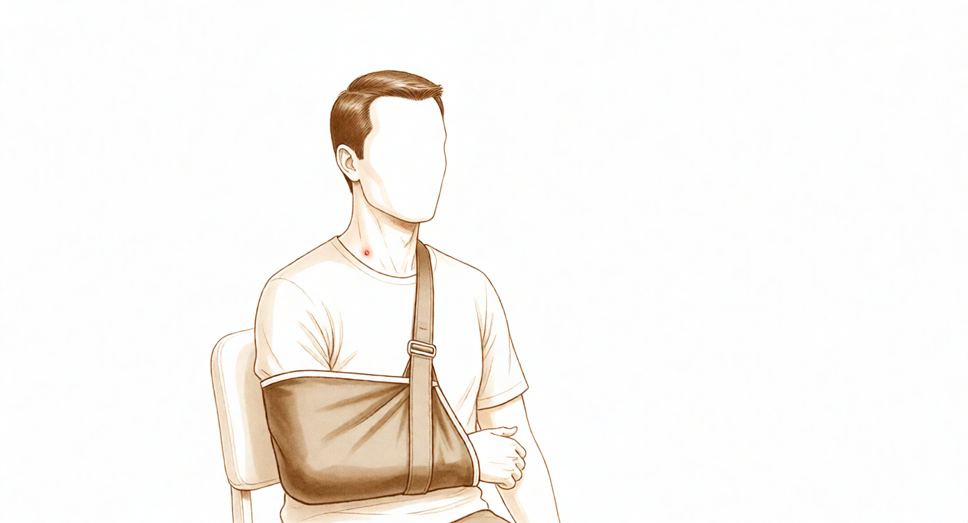
For patients: a plain-language version of this topic is available. See the patient guide.
Overview
Clavicle fracture management is individualized based on fracture characteristics and patient expectations rather than broadly applied protocols [30]. The majority of patients achieve excellent outcomes with conservative management [8]. For displaced mid-third clavicle fractures, surgical treatment increases the likelihood of union at one year [32]. While fixation for delayed or non-union is cost-effective, outcomes are inferior to those of patients who unite non-operatively [5]. In the adolescent population, the majority of clavicular fractures are treated nonoperatively, although operative treatment with plate and screw application yields consistently good outcomes with a low complication rate in selected cases [14]. Although open reduction and internal fixation (ORIF) of displaced midshaft fractures remains controversial in adolescents, specific circumstances beyond absolute indications may warrant initial surgical intervention [86].
Distal clavicle fractures managed nonoperatively exhibit higher nonunion rates, yet shoulder function remains excellent with low risks of complications or delayed surgery [19]. Arthroscopic fixation provides good functional outcomes with union rates comparable to traditional open techniques [74]. Both arthroscopic coracoclavicular button fixation and anatomic locking plate fixation are viable options, presenting similar high union rates and minimal complication risks [22]. Augmented fixation techniques for the distal clavicle are effective with minimal complications [1], and new internal fixation methods for unstable distal fractures show promise, albeit with short-term observations [13]. Medial clavicle fractures managed operatively result in anatomic reconstruction and excellent functional outcomes, even in symptomatic nonunion [16]. If patients survive the initial trauma, good clinical and functional outcomes are expected regardless of the management strategy chosen [23]. Rigid fixation is a safe and effective option for anterior sternoclavicular joint disruption with ipsilateral medial clavicle fracture in adolescents [2].
Surgical intervention for clavicle nonunion and malunion is associated with very good clinical results and a 100% union rate [15]. Open reduction and internal fixation with bone grafting provides good long-term functional and radiological results, allowing most patients to return to a functional level close to the general population [3]. Plate fixation remains a successful method for treating clavicle nonunions [6]. Reliable bony union and improved shoulder function are achievable through thoughtful surgical planning, appropriate implant choice, and meticulous technique [7].
Anatomy & Pathophysiology
Bony Anatomy
The clavicle is the only long bone to ossify by intramembranous ossification [33]. It serves as the primary stabilizer between the axial skeleton via the sternoclavicular joint and the appendicular skeleton via the acromioclavicular joint [33]. The bone forms a unique S-shaped curve on the axial view [33], with the greater radius of curvature at the medial curve, which is anteriorly convex, while the smaller lateral curve is posteriorly convex [106]. The bone is somewhat rounded in its midsection and medially and relatively flat laterally [106], with the distal clavicle flat in the AP plane [33]. The middle third of the clavicle is a junction of two curves where moments of tension, bending, and torsion act [64], forming a strut to hold the glenohumeral joint in the parasaggital plane [64].
Specific anatomical landmarks include a 30% incidence of a rhomboid fossa on the inferior surface of the medial end, where the costoclavicular ligaments insert [106]. The middle portion contains the subclavian groove where the subclavius muscle has a fleshy insertion [106], while the lateral portion has the coracoclavicular process when present [106]. The distance from the lateral edge of the clavicle to the medial edge of the conoid tubercle is approximately 45 mm in male and female specimens [106], and the distance from the lateral edge to the center of the trapezoid tuberosity is approximately 25 mm [106].
Ossification begins from two primary centers by 5 to 6 weeks of gestation [104], with the overall contour and "S" shape assumed by 7 to 8 weeks [104]. Most growth (80%) occurs from the medial physis [104]. The lateral epiphysis forms and fuses at around 18 to 19 years of age [104]. The medial epiphysis is the last in the body to ossify, at the age of 18 to 20 years [104], and is the last to complete ossification, at the age of 23 to 25 years [104]. The medial clavicular epiphysis may persist in patients until 25 to 30 years of age [80].
Ligaments and Soft Tissue
The coracoclavicular ligaments consist of the conoid (medial) and trapezoid (lateral) components [33]. These ligaments are the primary stabilizers to superior (vertical) translation of the distal clavicle [33]. The conoid ligament attaches to the clavicle at the conoid tubercle [106], while the trapezoid ligament attaches at the trapezoid line, which lies in an anteroposterior direction just lateral to the conoid tubercle [106].
The superior shoulder suspensory complex is a bone–soft-tissue ring that provides a stable connection of the glenoid and scapula to the clavicle [33]. It is composed of the distal clavicle, acromion, coracoid process, and glenoid neck, along with the supporting ligamentous complexes of the AC joint and CC ligaments [33]. The primary blood supply to the clavicle is periosteal, with no nutrient blood supply [33].
The muscular envelope includes the platysma, pectoralis major, deltoid, and some of the strap muscles of the neck [33]. The pectoralis major originates from the anterior portion of the medial two-thirds of the clavicle [106] and merges with the origin of the anterior deltoid laterally [109]. The sternocleidomastoid originates superiorly on the clavicle [109], largely on the posterior portion of the middle third [106]. The sternohyoid originates on the clavicle just medial to the origin of the sternocleidomastoid [106]. The deltoid originates on the anterior portion of the inner surface of the lateral curve of the clavicle [106]. The trapezius inserts on the posterosuperior surface of the distal end of the clavicle [106] and blends superiorly with the deltoid origin at the lateral margin [109]. The subclavius muscle inserts on the undersurface of the clavicle and serves as a soft tissue buffer in the subclavicular space superior to the brachial plexus and subclavian vessels [109]. The platysma usually envelopes the anterior and superior aspects of the clavicle and runs in the subcutaneous tissues [109].
Neurovascular Anatomy
The supraclavicular nerves originate from cervical roots C3 and C4 and exit from a common trunk behind the posterior border of the sternocleidomastoid muscle [113]. There are typically three major branches (anterior, middle, and posterior) that cross the clavicle superficially from medial to lateral [113].
The subclavian vein runs directly below the subclavius muscle and above the first rib [113]. The subclavian artery and brachial plexus lie more posteriorly than the subclavian vein, separated from the vein and clavicle by the scalenus anterior muscle medially [113]. The brachial plexus is closest to the clavicle in its midportion [113]. The subclavian vessels are closest to the clavicle at the medial end, with the vein directly apposed to the posterior cortex of the medial clavicle in some cases [113].
In the middle third of the clavicle, the subclavian artery is a mean of 17 mm from the clavicle [113], and the subclavian vein is a mean of 13 mm from the clavicle [113]. The subclavian vessels are located at an approximate angle of 60 degrees to the horizontal in this region [113]. Laterally, the subclavian artery is a mean of 63 mm from the clavicle [113], and the subclavian vein is a mean of 76 mm from the clavicle [113]. The subclavian vein and artery and brachial plexus are located posteriorly to the clavicle [106]. The medial anterior curve of the clavicle is often described as an accommodation for the subclavian vein and artery and brachial plexus [106].
Mechanism of Injury and Deformity
A fall on the shoulder or the outstretched hand may fracture the clavicle [29]. A direct blow on the point of the shoulder is the commonest reported mechanism of injury that produces a midshaft fracture of the clavicle [107]. Most (85%) clavicle fractures occur in the midshaft of the bone where the bone is narrowest and enveloping soft tissue structures are most scarce [107].
The typical deformity of middle-third fractures is caused by a medial fragment pulled superiorly by the sternocleidomastoid muscle, with the weight of gravity pulling downward on the lateral fragment [33]. The medial fragment of a fractured clavicle is held up by the sternomastoid muscle [29] and is elevated by the unopposed pull of the sternocleidomastoid muscle [109]. The lateral fragment of a fractured clavicle is pulled down by the weight of the arm [29] and is held inferiorly by the deltoid and medially by the pectoralis major [109]. The distal fragment of a displaced midshaft clavicle fracture is translated inferiorly, anteriorly, and medially (shortened), and rotated anteriorly [107].
Fractures of the outer third are easily mistaken for acromioclavicular injuries [29]. Vascular and neurological complications of clavicle fractures are rare [29]. Damage to the lung or vessels beneath the clavicle is very rare despite deformity [29].
Epidemiology and Classification
Clavicular fractures account for 3.8% of all fractures and 35.0% to 45.0% of all shoulder girdle injuries [33]. Approximately 15% of clavicular fractures are distal third, 80% are middle third, and 5% are medial third [33]. Medialization of a clavicular fracture more than 20 mm is associated with a measurable decrease in functional outcome [33].
Classification Systems: * Allman: Defines fractures of the proximal (medial), middle (midshaft), and distal (lateral) thirds of the clavicle [33]. * Neer: Classified lateral-third fractures based on the integrity of the CC ligament complex and the involvement of the AC joint [33]. Neer also classified clavicular fractures into midclavicular (80%), distal (15%), and inner third (5%) groups [146]. * Medial Third: Classified according to the displacement and involvement of the SC joint [33]. * Craig: Group I fractures involve the middle third and account for 80% of clavicle fractures [139]. Group II fractures account for 12% to 15% of all clavicle fractures [139]. The Craig classification best predicted nonunion or delayed union of lateral third clavicle fractures [117]. * Robinson: Had the best prognostic potential for middle third clavicle fractures [117].
Distal Clavicle Fracture Types: * Type I: Interligamentous fractures occurring between the conoid and trapezoid or between the coracoclavicular and acromioclavicular ligaments [139]. These present minimal displacement with intact ligaments [146]. * Type II: Involve detachment of the coracoclavicular ligaments from the medial segment [139]. These are displaced with detachment of the ligaments from the proximal fragment [146]. * Type III: Involve the articular surface of the acromioclavicular joint alone [139]. These are fractures of the articular surface [146]. * Type IV: Occur in children with ligaments intact to the periosteum and displacement of the proximal fragment [139]. * Type V: Comminuted fractures with ligaments attached neither proximally nor distally, but to an inferior, comminuted fragment [139].
Pathophysiology of Displacement and Malunion
The more lateral the fracture, the more potential there is for significant angulation in clavicular malunions [56]. The greatest angulation usually occurs at the point at which the clavicle is subtended by the coracoid [56]. Angulation is more acute the closer the break is to the pivot points of clavicoscapular motion [56]. The predominant influence on displacement of lateral clavicle fractures involves the lateral segment being carried forward by its retained acromioclavicular and residual coracoclavicular attachments [56].
In medial-third fractures, the lateral shaft fragment was situated anteriorly to the medial one, usually forming a prominent anterior spike [56]. The anatomy of a medial-third fracture is difficult to appreciate on standard radiographic views and is best understood via 3-dimensional reconstruction [56].
Four forces act on Type II distal clavicle fractures to impair healing: trapezius muscle pull, weight of the arm, trunk muscles attaching the humerus and scapula, and rotary displacement [146]. The trapezius muscle draws the large medial fragment posteriorly within its substance [146]. The weight of the arm pulls the outer fragment downward and forward [146]. Trunk muscles displace the outer fragment medially toward the apex of the thorax [146]. Scapular ligaments may rotate the outer fragment as much as 40° with movement of the arm [146].
Predisposing factors for clavicle nonunion include severe displacement, comminution, additional soft-tissue trauma, soft-tissue interposition, and inadequate initial immobilization [64]. The reported incidence of clavicle nonunion has been less than 2% [64]. Shortening of the clavicle has been discussed as an indication for surgery [64]. The problem of most midshaft nonunions is a deformity with the apex directed upwards [64]. Malunion is inevitable in displaced fractures [29]. In children, the bone is soon remodelled after malunion, but in adults the slight deformity has to be accepted unless there is a very unsightly bump with skin irritation [29]. Non-union sometimes occurs in middle-third fractures and is treated by bone graft and plating [29].
The clavicle is subcutaneous [33]. Tenting of the skin should be evaluated carefully because it can be a sign of impending open fracture [33].
Classification
Robinson: This system defines the lateral one-fifth of the clavicle as Type 3 [194].
Neer: In this classification, Type IIB fractures involve a torn conoid ligament with the trapezoid ligament presumed to remain attached to the distal fragment [43]. Type IV distal clavicle fractures involve disruption of the periosteal sleeve in the pediatric population and are injuries to the growth plate [43]. A modified Neer classification has been proposed to update the system to include an extra-lateral distal clavicle fracture pattern as a type IIC fracture [45].
Edinburgh: This classification has the highest correlation with prognosis for middle-third fractures of the clavicle [41]. Middle-third clavicle fractures are defined as greater than one-third distance from the medial edge of the clavicle [41]. The cutoff for lateral to middle third clavicle fractures is defined as the medial edge of the conoid ligament, described as 31% of the length of the clavicle from the lateral border [41].
CCI: Subtypes within this classification belong to a common group of one entity that are treated with similar surgical procedures or the same approaches [83]. A proposed classification system for monopolar and bipolar combination injuries of the clavicle includes higher-grade, complete, and unstable joint dislocations of the SCJD type Allman III and ACJD at least from type Rockwood III(b) [83].
Other Considerations: A new simple classification system for lateral clavicle fractures showed substantial inter- and intraobserver reliability [112].
Clinical Presentation
Mechanism and Deformity
The lateral fragment of a clavicle fracture is pulled down by the weight of the arm, while the medial fragment is held up by the sternomastoid muscle [29]. Fractures are often displaced, producing a lump along the ‘collar-bone’ [29].
Physical Examination
A distal neurovascular examination is important because of the proximity of the brachial plexus and the subclavian vessels to the zone of injury [33]. Vascular and neurological complications are rare [29].
Imaging
X-rays show that the fracture is usually in the middle third of the bone and the lateral fragment lies below the medial [29]. Outer-third injuries need special views to define any fracture [29]. Upright and supine radiographs, including an AP view of the clavicle and a 15° cephalad tilt view, should be obtained to define displacement when the patient is upright [33]. A bilateral panoramic view of both shoulders should be obtained to measure clavicular shortening [33]. CT is the most accurate modality for determining fracture shortening and morphology but not typically obtained [33]. Lateral clavicle fractures can be well visualized with AP radiographs using a Zanca view (10- to 15-degree cephalic tilt) which helps delineate the fracture by removing the overlap of the thoracic cage [151, 152]. For medial clavicle fractures, CT scanning is the procedure of choice when the anatomy of the fracture is unclear [152]. CT scanning can help distinguish between a medial epiphyseal fracture and true SC dislocations [152].
Investigations
Plain radiography: Simple anteroposterior (AP) radiographs are usually sufficient to establish the diagnosis of a clavicle fracture [63]. To best delineate the fracture, the radiograph should be taken in the upright position, where gravity demonstrates maximal deformity [63]. Ideally, the radiographic beam for the AP view is angled 20 degrees superiorly to eliminate thoracic cage overlap and show the clavicle in profile [63]. If the torso is internally rotated a similar 20 degrees, the scapula and shoulder girdle are placed parallel to the cassette for a true AP film [63]. A single AP chest radiograph evaluates deformity relative to the normal side and identifies associated skeletal injuries, including rib, glenoid, and scapular fractures [63]. Length measurement on the chest radiograph compares the injured to the uninjured side, where shortening of 2 cm or more represents a relative indication for primary fixation [63]. Weighted stress radiographs significantly increase the measured elevation of the clavicle and the coracoclavicular distance compared to non-weighted views [219].
CT: CT scanning of midshaft clavicular fractures is rarely performed in the clinical setting, although this modality demonstrates the complex three-dimensional deformity affecting the shoulder girdle, including significant scapular angulation and protraction [63]. CT is useful for evaluating fractures of the medial third of the clavicle and the remainder of the shoulder girdle, such as the glenoid neck in cases of a “floating shoulder” [63]. CT is the most accurate modality for determining fracture shortening and morphology but is not typically obtained [33].
Other Considerations: Once clavicle fractures are healed, further radiographic imaging does not provide any notable information [38]. Delayed assessment at 6 weeks following displaced midshaft clavicle fracture enables an accurate prediction of patients likely to have union with nonoperative management [21]. The probability of nonunion at various time points following a midshaft clavicle fracture is influenced by a prognostic index that becomes more negative with increasing age, increasing comminution, increasing displacement, and female sex [31].
Treatment
Non-Operative
Conservative treatment of clavicle fractures yields significantly good clinical and radiological outcomes [24]. In adolescents, nonoperative management of mid-shaft clavicle fractures results in excellent functional outcomes at long-term follow-up [25], and comparably excellent outcomes of severe clavicle fractures in adolescent athletes can be achieved with non-operative treatment [141]. A targeted approach is required, with simple fractures treated nonoperatively and complex displaced fractures considered for surgery to prevent non-union [129].
Operative
Indications: Operative treatment of displaced midshaft clavicle fractures in adults is associated with higher union rates and better early patient-reported outcomes than non-operative treatment, though long-term outcomes are similar [51]. In the management of midshaft clavicular fractures, surgery is superior to nonoperative treatment [67]. For healthy, active adults, midshaft clavicular fractures should undergo consideration for surgical stabilization if significantly displaced (2 cm of shortening, 100% displacement or significant comminution) [52]. Fracture-specific indications for operative treatment of midshaft clavicle fractures include displacement >2 cm, shortening >2 cm, increasing comminution (>3 fragments), segmental fractures, open fractures, impending open fractures with soft tissue compromise, and obvious clinical deformity [31]. Associated injuries that serve as indications for operative treatment include vascular injury requiring repair, progressive neurologic deficit, ipsilateral upper extremity injuries/fractures, multiple ipsilateral upper rib fractures, “floating shoulder,” and bilateral clavicle fractures [31]. Patient factors that may indicate operative treatment include polytrauma with requirement for early upper extremity weight-bearing/arm use and patient motivation for rapid return of function (e.g., elite sports or the self-employed professional) [31]. Specific treatment of clavicle fractures should not be broadly applied but rather should be individualized based on fracture characteristics and patient expectations [30]. Patients with displaced clavicle fractures benefit clinically and financially from stabilization, experiencing less chronic pain, deformity, and weakness with better range of motion and earlier return to work [81]. Stabilisation of the clavicle gives better functional scores compared to conservative treatment in the short term in patients with a ‘floating shoulder’ [28]. However, floating shoulder injuries should be carefully assessed on the basis of component injuries individually because surgical management has not been shown to result in improved clinical outcomes [52].
Surgical Approach / Technique: A superior approach and plating is a preferred technique for open reduction and fixation of the fracture with precontoured 3.5-mm clavicular DCP [154]. If a lag screw is placed during midshaft clavicle plating, it is sufficient to insert three bicortical screws (six cortices) proximally and three distally; if a lag screw is not possible, four screws should be placed proximally and four distally [154]. These 2 approaches (oblique and transverse incisions) have equivalent effects on recovery of shoulder joint function in the treatment of fractures of the middle and outer third of the clavicle [46]. The primary technical difficulty with medial clavicle fractures is the fixation in the medial fragment, and it is important to remember that the subclavian vessels are in close proximity to the bone medially [80]. In a cadaveric study by Galley et al., the vascular risk was high in the most medial quarter of the clavicle [201]. On the posterior side of the most medial quarter of the clavicle, the common carotid artery and in particular the internal jugular vein axis is at risk when drilling anterior to posterior [201]. An image study performed by Sinha et al. showed that drilling should be directed superiorly in the most medial quarter and posteriorly [201].
Implant Selection: Finite element analysis indicates that plate fixation significantly stabilizes the fracture gap, reduces the implant stress, and serves as the recommended fixation for the mid-shaft clavicle fracture [159]. The use of a pre-contoured plate facilitates surgical care of clavicular fractures, reducing hardware prominence and secondary surgical procedures, whereas dual plating may be used as an adjuvant in highly comminuted cases [52]. Dual plating with 2.4- or 2.7-mm plates may additionally have a benefit of decreased hardware prominence [52]. A hybrid uni-cortical approach to clavicle plate fixation demonstrates comparable bending strength to current bi-cortical approaches while potentially reducing the risk of intra-operative vascular damage [131]. Fixation of uncomplicated midshaft clavicle fractures with titanium elastic nails (TENs) provides adequate fixation and faster relief of pain and return to normal function of the affected shoulder than fixation with 3.5-mm reconstruction plates [72]. The technique of intramedullary screw fixation is a safe, simple, and reliable method for fixation of displaced mid-shaft clavicle fractures with minimal complications and excellent functional outcomes [123]. Based on evidence from four trials with a high risk of bias, a recent systematic review identified some low-quality evidence of a lack of a clinically important difference in function between intramedullary (IM) fixation and plate fixation [154]. The review indicated that overall adverse events may be less frequent after IM fixation, but the reliability of these findings is uncertain due to limited and low-quality evidence [154]. Three randomized controlled trials comparing plate fixation versus IM nailing for treating displaced midshaft clavicle fractures found no statistical differences between the two methods regarding the functional endpoints and incidence of nonunion at 1-year follow-up [154]. The results of intramedullary fixation appear to be more unpredictable than the results of plate fixation, with anatomy and biomechanical aspects of fixation appearing inferior in resisting displacement compared with plate fixation [154]. For distal clavicle fractures, the described technique of augmented fixation of the distal clavicle with a Fibertape coracoclavicular cerclage is effective, produces good clinical outcomes, and has minimal complications [1]. Although observations are short term, the technique of internal fixation for unstable distal clavicle fractures is promising as an alternative for internal fixation of distal clavicle fractures [13]. Excellent clinical and radiological outcomes can be achieved with a minimally invasive all-suture fixation technique for displaced distal clavicle fractures, which allows for an anatomic reduction and stable fixation [69]. The results demonstrate that arthroscopic stabilization using Tightrope (SS-16) is a safe, simple, cosmetically acceptable and reproducible method of reducing and stabilising the distal clavicle allowing for healing of either the coracoclavicular ligaments or the distal clavicle [121]. A combined surgical strategy of locking plate with titanium cable can be considered an effective method for treating unstable distal clavicle fractures [128]. CC fixation adds stability to type IIB distal clavicle fractures fixed with plate and screws when loaded to failure [85]. A superior locking plate with braided PDS coracoclavicular fixation is a technique used for unstable fractures of the distal clavicle, in which fixation of the distal segment are tenuous, typically involving fractures within 3 cm of the CC ligaments or those involving the CC ligaments [218]. The authors have not observed any complications related to distal clavicle fixation in the skeletally immature, although clavicle and coracoid fractures have been reported with this technique in other populations [27]. For medial clavicle fractures, surgical treatment of displaced medial clavicle fractures using an anatomically precontoured locking plate originally designed for the lateral clavicle led to very good to excellent clinical and functional results [130]. This case adds to the limited literature and supports the role of rigid clavicular fixation as a safe and effective treatment option in presentations of anterior sternoclavicular joint disruption with ipsilateral medial clavicle fracture [2]. Fractures that are significantly displaced may warrant operative repair, especially if there is posterior displacement of the shaft fragment [80]. Fixation of the medial clavicle fracture using smooth wires or pins alone is contraindicated, due to the potential for migration and visceral injury [80].
Pain Management: Ketorolac use following operative clavicle fixation is not associated with increased nonunion or surgical complications and may serve as a useful adjunct for postoperative pain management [192].
Other Considerations: Treatment of middle-third clavicle non-union after initial failure of conservative treatment with stable fixation and bone graft is a reliable, well-suited and effective treatment [20]. Plate fixation with cortical bone grafting of clavicular nonunions is associated with restoration of clavicular length and a high rate of bone union [50]. Superiorly applied plate fixation is an effective treatment for clavicular nonunion [73]. Careful preoperative planning and combining segmental bone graft and stable internal fixation can restore clavicle length, alignment, and shoulder girdle function [144]. Total claviculectomy may be a useful salvage procedure for clinical situations in which the restoration of normal clavicular osseous anatomy is impossible [37]. Both pain and shoulder function were improved with the use of vascularized fibular grafts for salvage reconstruction of clavicle nonunion [40]. This suggests the etiology of midshaft clavicle nonunions may result from a combination of suboptimal mechanical fixation and latent infection [53]. A pain score that exhibits no or minimal change from 2 to 4 weeks after nonoperative treatment of a displaced midshaft fracture of the clavicle is associated with a high risk that symptomatic nonunion will develop [207]. Plate fixation of midshaft clavicle fractures for delayed union and non-union is a cost-effective intervention but functional deficits persist at long-term follow-up [5]. Medial clavicle fractures have favorable functional outcomes and pain relief at minimum 1-year follow-up among those patients who survive the trauma, but a high proportion will die within 3 years of the injury [96]. Functional outcome is excellent following the treatment of both acute and non-united clavicle fractures, but recovery occurs earlier following acute treatment [36]. Women are 4 times more likely than men to have hardware removed, and patients undergoing clavicle hardware removal report worse long-term outcomes than patients with hardware retained [11]. Only 7.7% of patients required hardware removal for symptomatic hardware when using pre-contoured titanium anterior plating of midshaft clavicle fractures, as opposed to the estimated 20%-60% reported in the literature in patients with symptomatic superior clavicle plates [12]. The overall incidence of clavicular hardware removal was 12.6% following operative treatment of middle- and distal-third clavicular fractures [26]. Retaining the clavicular hook-plate indefinitely carries a potential risk of developing osteolysis and fracture around the implant [82]. Ipsilateral os acromiale may be a relative contraindication to the clavicle hook plate [157]. The risk of iatropathic brachial plexus injury can be reduced by thorough release of tissues from the inferior surface of the clavicle and ensuring no shortening occurs during fixation [9]. Minor morbidity includes incisional and proximal chest wall numbness which have been reported to occur in 12% to 29% of clavicle fractures after surgical management, suggesting injury to the supraclavicular nerve branches [212]. Wang et al. reported 83% of sensory disorders after plate fixation in clavicle fractures and recommended using the vertical incision [201]. Leroux et al. reported only five neurologic complications and five vascular complications out of 1,350 clavicle fractures treated with plate fixation [201]. Surgeons should be wary of supraclavicular nerve branches during surgical exposure and consider neuropathy in patients with persistent pain after clavicle fracture [221]. The study supports the need for further development of clavicle fixation devices, specifically variable-shape and gender-specific designs, and suggests that varying plate width and multi-plane pre-contouring would improve anatomic fit [156].
Complications
Non-Operative Management
Symptomatic Nonunion: Symptomatic nonunion occurs in 14% to 24% of patients treated nonoperatively for clavicle fractures [177]. Patients with smoking status or high Disabilities of the Arm Shoulder and Hand scores are at risk for symptomatic nonunion of displaced midshaft fractures, warranting investigation for early surgical fixation to avoid nonunion morbidity [236]. In adolescents, nonunion following nonoperative treatment of displaced midshaft clavicle fractures is rare [31].
Malunion: Malunion is a common complication of nonsurgical management, noted as one of the two most common complications alongside symptomatic nonunion [177]. Five patients in a nonoperative group with a mean shortening of 26 mm developed symptomatic malunion, with four choosing corrective osteotomy [31].
Distal Fracture Outcomes: Nonoperative management of displaced distal clavicle fractures results in higher nonunion rates compared to other fracture locations or treatments, though shoulder function remains excellent [19]. The risk of complications and delayed surgery following nonoperative management of displaced distal clavicle fractures is low [19].
Operative Management: General and Infection
Infection: Rates of infection following clavicle fracture surgery range from 0% to 18% [160]. A cohort study found an infection rate of 2.6% following open reduction and internal fixation (ORIF) of clavicle fractures [177]. Nonunion after clavicle osteosynthesis is associated with a high incidence of Propionibacterium acnes, suggesting etiology may result from a combination of suboptimal mechanical fixation and latent infection [53].
General Complication Rates: The overall complication rate following surgical clavicle fracture care averaged 8.1% [230]. Surgical fixation of midshaft clavicle fractures in a multi-trauma setting shows a 14.5% overall complication rate [241].
Pneumothorax: In a series of 89 patients with isolated clavicular fractures without preoperative pneumothorax, no iatrogenic pneumothoraces occurred after plate fixation [235]. The rate of pneumothorax following clavicle fracture ORIF was reported as 0.01% [177].
Operative Management: Hardware and Implant Issues
Hardware Removal: The overall incidence of clavicular hardware removal was 12.6% [26]. Rates of hardware irritation requiring removal of material range from 50% to 100% according to one expert commentary, though other sources cite lower rates [160]. The rate of implant irritation from any form of surgical fixation ranges from 3.7% to 40%, with the lowest rates reported with dual plating techniques [177]. An overall 24.6% revision surgery rate was found in a cohort of 1,350 patients who had undergone ORIF with at least 2 years of follow-up, with the most common reason being implant irritation [177].
Demographic Risk Factors: Women are 4 times more likely than men to have hardware removed following clavicle fixation [11]. Female patients had the highest risk of secondary surgery for implant removal because of implant irritation at an average time of 12 months from index surgery [177].
Implant-Specific Complications: Only 7.7% of patients required hardware removal for symptomatic hardware when treated with pre-contoured titanium anterior plating, compared to an estimated 20%-60% reported in the literature for symptomatic superior clavicle plates [12]. Clavicular hook plates can cause injury to the rotator cuff, shoulder stiffness, and acromioclavicular joint osteoarthritis, with osteoarthritis reported in 21% of patients in one series [191]. Removal of clavicular hook plates earlier than 6 months can lead to nonunion or refracture [191]. In a study of displaced medial clavicle fractures, 30% of all operative patients experienced plate prominence or irritation, and 27% had additional surgery for implant removal [214].
Construct Failure and Refracture: Overall construct failure after open reduction and plate fixation of displaced midshaft clavicular fractures occurred in 6.9 percent [90]. Major re-intervention and re-fracture after implant removal occurred more frequently after plate fixation of non-comminuted, displaced midshaft clavicle fractures compared to intramedullary fixation [240]. Implant or fixation failure occurred in 3 patients (3%) in a systematic review of displaced medial clavicle fractures treated operatively [214].
Intramedullary Fixation Issues: Reported shortcomings of intramedullary fixation include high rates of implant breakage, temporary brachial plexus palsy, poor rotational control in comminuted fractures, and skin breakdown over entry portals [182]. The majority of surgical complications following endoscopic intramedullary nailing (ESIN) for clavicular mid-shaft fractures were caused by medial nail protrusion [119]. In a study of ESIN for clavicular mid-shaft fractures, non-union was recorded in four patients, with three requiring implant removal and osteosynthesis by open reposition and internal plate fixation [119].
Operative Management: Neurovascular and Soft Tissue
Neurovascular Injury: The rate of neurovascular injury following clavicle fracture ORIF was reported as 0.003% [177]. Transient brachial plexus symptoms are listed as an adverse event associated with surgical interventions for clavicle fractures [160]. Paresthesia of the arm or fingers is defined as transient brachial plexus lesions if it spontaneously recovers within 6 months after surgery [202].
Nerve Injury and Numbness: Supraclavicular numbness from iatrogenic injury to branches of the supraclavicular nerve is a common complication of surgical management [177]. Incisional numbness was reported in 18 of 62 patients (29%) in the operative group compared to 0 in the nonoperative group in a comparative study [55].
Soft Tissue and Hematoma: Hardware irritation or prominence was reported in 11 of 62 patients (17.7%) in the operative group compared to 0 in the nonoperative group [55]. Complications following clavicle fracture surgery are grouped into infection, neurovascular pathology (transient brachial plexus syndrome, hematoma, desensitized skin), implant-related problems, bone-healing problems, and refracture [202].
Operative Management: Union and Malunion
Nonunion Rates: The nonunion rate in the surgical group for midshaft clavicle fractures was 1% in a meta-analysis of six randomized control trials [118]. A low rate of nonunion (2.6%) and malunion (1.1%) was reported in a cohort of 1,350 patients undergoing ORIF [177]. In a study of percutaneous plating for comminuted midshaft fractures, none of the patients developed nonunion or delayed union [47]. All operatively treated displaced medial end clavicle fractures united successfully with no reported nonunions in a systematic review [214].
Functional Outcomes: Although solid union after realignment of symptomatic nonunion or malunion of midshaft clavicle fractures is predictable, patients can remain functionally impaired [91]. Surgical treatment of nonunion and malunions of the clavicle was associated with a 100% union rate in a study comparing computer-assisted planning to conventional surgery [15].
Operative Management: Specific Techniques and Populations
Distal Clavicle Fixation: The described technique of augmented fixation of the distal clavicle with Fibertape coracoclavicular cerclage produces good clinical outcomes and has minimal complications [1]. Both arthroscopic coracoclavicular button fixation and anatomic locking plate fixation for unstable distal clavicular fractures have a minimal risk of complications and present similar, high union rates [22]. Arthroscopically assisted coracoclavicular stabilization of distal clavicle fractures demonstrated high union rates while limiting complications or need for secondary hardware removal [95]. Open reduction and tunneled suspensory device fixation for isolated displaced lateral-end clavicular fractures is associated with good functional outcomes and a low rate of complications in the medium term [97]. Excellent union rates and functional outcomes, as well as low complication rates, can be expected following combined locking plate and tunneled suspensory device fixation of lateral-end clavicle nonunions [242]. In a study of 48 patients with extra-lateral distal clavicle fractures, open reduction and internal fixation yielded good clinical outcomes [45].
Medial and Adolescent Populations: Rigid clavicular fixation is a safe and effective treatment option for anterior sternoclavicular joint disruption with ipsilateral medial clavicle fracture in adolescents [2]. Operative treatment with plate and screw application for clavicular fractures in adolescents has consistently good outcomes with a low complication rate in selected cases [14]. Surgery demonstrated no benefit in patient-reported quality of life, satisfaction, shoulder-specific function, or prevention of complications after completely displaced clavicle shaft fractures in adolescents at 2 years after injury [243].
Technique Comparisons and Oncologic Cases: Clavicular fracture fixation using either locking or hook plates is a safe method of treatment with a very low reoperation rate for either hardware removal or revision [238]. The systematic review of Neer type IIB (IIC) distal clavicle fractures showed similar major complication rates among techniques, but the hook plate technique demonstrated inferior clinical results [244]. Kona et al. reported poor results in 10 of 19 patients treated with operative fixation of a lateral clavicle fracture, including 6 nonunions and 5 infections in the Kirschner wire group [191]. Satisfactory clinical and functional results can be achieved following partial or total claviculectomy without reconstruction for oncologic causes, with a low complication rate [100].
Recovery
Light activity (weeks): Early functional gains are evident at six weeks following plate fixation of midshaft clavicular fractures compared with nonoperative treatment [101]. Fixation with a pre-contoured plate and locking screws facilitates faster functional recovery in the early phase [229]. For adolescent athletes engaged in structure- or kinetic-dependent sports, TEN fixation significantly accelerates return to sport and enhances early functional and psychological recovery [195].
Full activity (months): There is no difference in function between surgical and nonoperative treatment after six months and one year [229]. Surgical treatment of displaced clavicle fractures results in higher union rates and better early patient-reported outcomes, though long-term outcomes are similar to nonsurgical treatment [94]. In a working population under injury compensation, ORIF of displaced middle-third clavicular fractures resulted in shorter time to complete return to work and earlier bone healing [166]. Workers' compensation patients return to work at roughly the same time whether treated surgically or nonoperatively [135].
Complete recovery / outcome plateau (months): Excellent functional outcomes continue to be observed several years after clavicle fracture fixation, with no difference in reoperation rates between plate types or location [10]. Patients report a good quality of life and functional outcome after plating for midshaft clavicular fractures [197]. Late reconstruction of nonunion and malunion restores objective muscle strength similar to immediate fixation, though subtle decreases in endurance strength and outcome persist compared with acute fracture repair [222].
Rehabilitation protocol: A limited incision approach for plating of acute midshaft clavicle fractures achieves good functional and radiographic outcomes with a low complication rate comparable to standard incision techniques [57]. For adolescent athletes, accelerated return to play after ORIF is not associated with a significantly increased risk of refracture, nonunion, or other complications compared with a more traditional timeline [228]. Adolescent patients undergoing anatomic ORIF may return to play more quickly than previously thought [220].
Functional milestones: High-quality evidence supports higher union rates and better early patient-reported outcomes for surgical treatment of displaced clavicle fractures in adults [94]. ORIF of displaced middle-third clavicular fractures yields good and excellent functional results with fewer cases of nonunions [166].
Other Considerations: Non-Operative Management: Teenage patients with completely displaced clavicle fractures treated non-operatively can expect excellent radiographic and clinical outcomes five years post-injury [49]. Adolescent athletes managed following best practice guidelines provide predictable return to athletics, including collision sports [234].
Non-Union and Delayed Union: Open reduction and internal fixation with bone grafting for clavicular non-union provides good long-term functional and radiological results, with most patients returning to a functional level close to the general population [3]. Although solid union after realignment of symptomatic nonunion or malunion is predictable, patients can remain functionally impaired [91].
Hardware and Complications: Only 7.7% of patients required hardware removal for symptomatic hardware, compared with an estimated 20%-60% reported in the literature for symptomatic superior clavicle plates [12]. Dual plating of acute displaced midshaft clavicle fractures may lead to lower rates of reoperation for symptomatic hardware removal without compromising fracture healing [208]. The risk of complications can be reduced by thorough release of tissues from the inferior surface of the clavicle and ensuring no shortening occurs during fixation [9]. Arthroscopically assisted CC stabilization of distal clavicle fractures demonstrates high union rates while limiting complications or need for secondary hardware removal [95]. ORTSD fixation for isolated displaced lateral-end clavicular fractures in medically fit patients is associated with good functional outcomes and a low rate of complications in the medium term [97].
Specific Injury Patterns: Stabilisation of the clavicle gives better functional scores compared to conservative treatment in the short term [28]. Internal plating for bipolar clavicle injury allows early mobilization and results in good joint function [170]. Rigid clavicular fixation is a safe and effective treatment option in similar presentations [2]. Satisfactory clinical and functional results can be achieved following partial or total claviculectomy without reconstruction, with a low complication rate and acceptable mid- to long-term function [100]. Satisfactory recovery of radiographic and functional outcomes was obtained after wide excision with allograft reconstruction for large parosteal osteoma of the clavicle [237].
Key Evidence
- [L4] The described technique of augmented fixation of the distal clavicle is effective, produces good clinical outcomes, and has minimal complications. [1] (10.5397/cise.2022.00913)
- [Case_report] This case adds to the limited literature and supports the role of rigid clavicular fixation as a safe and effective treatment option in similar presentations. [2] (10.1016/j.xrrt.2026.100697)
- [L4] We conclude that open reduction and internal fixation with bone grafting gives good long-term functional and radiological results in clavicular non-union with most patients returning to a functional level close to the general population. [3] (10.1016/s0020-1383(03)00239-0)
- [L3] Clavicle fixation for delayed and non-union is a cost-effective intervention but outcomes are worse compared to patients that unite with non-operative management. [5] (10.1177/1758573221990367)
- [L4] Plate fixation of clavicle nonunions remains a successful method of treatment. [6] (10.1016/j.jse.2020.06.035)
- [L5] Reliable bony union and improved shoulder function can be expected with thoughtful surgical planning, appropriate implant choice, and meticulous surgical technique. [7] (10.1016/j.jse.2013.01.022)
- [L3] Most patients with clavicle fractures have an excellent outcome using conservative management. [8] (10.1016/j.jse.2019.06.022)
- [L4] The risk can be reduced by thorough release of tissues from the inferior surface of the clavicle and ensuring no shortening occurs during fixation. [9] (10.1302/0301-620x.95b1.29625)
- [L4] No difference in reoperation rates between plate types or location could be detected, and excellent functional outcomes continue to be observed several years after clavicle fracture fixation. [10] (10.1016/j.injury.2014.04.032)
- [L3] Women are 4 times more likely than men to have hardware removed, and patients undergoing clavicle hardware removal report worse long-term outcomes than patients with hardware retained. [11] (10.1016/j.jse.2015.09.029)
- [L4] Only 7.7% of patients required hardware removal for symptomatic hardware, as opposed to the estimated 20%-60% reported in the literature in patients with symptomatic superior clavicle plates. [12] (10.1016/j.jse.2021.05.021)
- [L4] Although observations are short term, the technique is promising as an alternative for internal fixation of distal clavicle fractures. [13] (10.1016/j.jse.2007.04.012)
- [L4] Current evidence suggests that the majority of clavicular fractures in adolescents can and should be treated nonoperatively, although operative treatment with plate and screw application has consistently good outcomes with a low complication rate in selected cases. [14] (10.2106/jbjs.22.01036)
- [L3] Surgical treatment of nonunion and malunions of the clavicle was associated with very good clinical results and a 100% union rate. [15] (10.1016/j.jseint.2023.07.005)
- [L4] Operative fixation of displaced medial clavicle fractures results in anatomic reconstruction and excellent functional outcomes, even in the setting of fixation performed for symptomatic nonunion. [16] (10.1016/j.jse.2015.04.011)
- [L4] Nonoperative management of displaced distal clavicle fractures results in higher nonunion rates, but shoulder function remains excellent, and risk of complications and delayed surgery are low. [19] (10.1016/j.jse.2023.12.006)
- [L4] Treatment of middle-third clavicle non-union after initial failure of conservative treatment with stable fixation and bone graft is a reliable, well-suited and effective treatment. [20] (10.1016/j.otsr.2013.09.011)
- [L1] Delayed assessment at 6 weeks following displaced midshaft clavicle fracture enables an accurate prediction of patients who are likely to have union with nonoperative management. [21] (10.2106/jbjs.19.00955)
- [L3] Both procedures can be used to treat distal clavicle fractures because they have a minimal risk of complications and present similar, high union rates. [22] (10.1016/j.jseint.2021.05.007)
- [L5] If patients with medial clavicle fractures can survive the initial trauma, there is every reason to expect good clinical and functional outcomes, regardless of whether surgical or nonsurgical management is chosen. [23] (10.1097/corr.0000000000001916)
- [L4] Conservative treatment of clavicle fractures yields significantly good clinical and radiological outcomes. [24] (10.1016/j.xrrt.2026.100679)
- [L3] Nonoperative management of adolescent mid-shaft clavicle fractures results in excellent functional outcomes at long-term follow-up. [25] (10.1302/0301-620x.103b5.bjj-2020-1929.r1)
- [L4] The overall incidence of clavicular hardware removal was 12.6%. [26] (10.1016/j.jse.2020.06.034)
- [L5] The authors have not observed any complications related to this technique, although clavicle and coracoid fractures have been reported with this technique in other populations. [27] (10.1097/bte.0b013e3182499ec8)
- [L2] Stabilisation of the clavicle gives better functional scores compared to conservative treatment in the short term. [28] (10.1302/0301-620x.95b6.31060)
- [L5] Specific treatment of clavicle fractures should not be broadly applied but rather should be individualized based on fracture characteristics and patient expectations. [30] (10.1016/j.jse.2011.08.053)
- [L1] Surgical treatment led to a greater likelihood of union at 1 year of follow-up among adult patients with displaced mid-third clavicle fractures. [32] (10.1097/corr.0000000000000986)
- [L3] Functional outcome is excellent following the treatment of both acute and non-united clavicle fractures, but recovery occurs earlier following acute treatment. [36] (10.1016/j.otsr.2017.03.021)
- [L4] Total claviculectomy may be a useful salvage procedure for clinical situations in which the restoration of normal clavicular osseous anatomy is impossible. [37] (10.2106/jbjs.e.01436)
- [L3] Once clavicle fractures are healed, further radiographic imaging does not provide any notable information. [38] (10.5435/jaaos-d-17-00598)
- [L4] Both pain and shoulder function were improved with the procedure. [40] (10.1067/mse.2000.107090)
- [L3] [41] (10.1016/j.jse.2014.09.037)
- [L4] [43] (10.5435/00124635-201107000-00002)
- [L4] [45] (10.1016/j.jse.2020.10.006)
- [L2] These 2 approaches have equivalent effects on recovery of shoulder joint function. [46] (10.1016/j.jse.2019.03.021)
- [L4] [47] (10.1016/j.injury.2012.09.030)
- [L2] Teenage patients with completely displaced clavicle fractures can expect excellent radiographic and clinical outcomes 5 years post-injury if treated non-operatively. [49] (10.1177/2325967123s00041)
- [L4] Plate fixation with cortical bone grafting of clavicular nonunions is associated with restoration of clavicular length and a high rate of bone union. [50] (10.1016/j.jseint.2020.04.002)
- [L4] This suggests the etiology of midshaft clavicle nonunions may result from a combination of suboptimal mechanical fixation and latent infection. [53] (10.1097/bot.0000000000000770)
- [Paper] [56] (10.1067/mse.2003.2)
- [L5] In this large cohort with long-term follow-up, a limited incision approach for plating of acute midshaft clavicle fractures achieved good functional and radiographic outcomes with a low complication rate comparable to the reported rate for standard incision techniques. [57] (10.1016/j.jse.2025.06.002)
- [L4] [64] (10.1016/s1058-2746(99)90068-x)
- [L2] In the management of midshaft clavicular fractures, surgery is superior to nonoperative treatment. [67] (10.1016/j.jse.2013.06.025)
- [L4] Excellent clinical and radiological outcomes can be achieved with this minimally invasive all-suture fixation technique for displaced distal clavicle fractures, which allows for an anatomic reduction and stable fixation. [69] (10.1016/j.xrrt.2022.01.005)
- [L3] Fixation of uncomplicated midshaft clavicle fractures with TENs provides adequate fixation and faster relief of pain and return to normal function of the affected shoulder than fixation with 3.5-mm reconstruction plates. [72] (10.1016/j.jse.2011.08.065)
- [L4] Superiorly applied plate fixation is an effective treatment for clavicular nonunion. [73] (10.1016/j.jse.2008.05.046)
- [L4] Arthroscopic fixation of distal clavicle fractures resulted in good functional outcomes with union rates comparable to traditional open techniques. [74] (10.1177/23259671211001773)
- [L3] Patients with displaced clavicle fractures benefit clinically and financially from stabilization, experiencing less chronic pain, deformity, and weakness with better range of motion and earlier return to work. [81] (10.1016/j.jse.2012.06.006)
- [L5] Retaining the clavicular hook-plate indefinitely carries a potential risk of developing osteolysis and fracture around the implant. [82] (10.1016/j.injury.2004.08.010)
- [L4] [83] (10.3390/jcm10245764)
- [L5] CC fixation adds stability to type IIB distal clavicle fractures fixed with plate and screws when loaded to failure. [85] (10.1016/j.arthro.2013.02.024)
- [Case_report] Although ORIF of displaced midshaft clavicle fractures remains controversial in the adolescent population, there may be additional circumstances beyond absolute indications for surgical intervention that warrant ORIF at initial presentation. [86] (10.1016/j.xrrt.2023.03.004)
- [L5] When indicated, operative management of displaced midshaft clavicle fractures with dual-plating is cost-effective compared to single-plating. [87] (10.1177/2325967123s00167)
- [L2] When indicated, operative management of displaced midshaft clavicle fractures using dual plating was found to be cost-effective compared with single plating. [89] (10.2106/jbjs.23.00338)
- [L3] Overall construct failure after open reduction and plate fixation of displaced midshaft clavicular fractures occurred in 6.9 percent. [90] (10.1016/j.injury.2017.01.040)
- [L4] Although solid union after realignment of symptomatic nonunion or malunion of midshaft clavicle fractures is predictable, the patients can remain functionally impaired. [91] (10.1016/j.jse.2006.12.002)
- [L1] High-quality evidence shows that surgical treatment of displaced clavicle fractures in adults results in higher union rates and better early patient-reported outcomes compared with nonsurgical treatment, though long-term outcomes are similar. [94] (10.5435/jaaos-d-23-00472)
- [L4] Arthroscopically assisted CC stabilization of distal clavicle fractures demonstrated high union rates while limiting complications or need for secondary hardware removal. [95] (10.1016/j.xrrt.2024.02.003)
- [L4] Medial clavicle fractures have favorable functional outcomes and pain relief at minimum 1-year follow-up among those patients who survive the trauma, but a high proportion will die within 3 years of the injury. [96] (10.1097/corr.0000000000001839)
- [L4] ORTSD fixation for isolated displaced lateral-end clavicular fractures in medically fit patients is associated with good functional outcomes and a low rate of complications in the medium term. [97] (10.2106/jbjs.18.00569)
- [L4] Satisfactory clinical and functional results can be achieved following partial or total claviculectomy without reconstruction, with a low complication rate and acceptable mid- to long-term function. [100] (10.1016/j.jse.2023.03.010)
- [L1] This meta-analysis shows that there is an early functional gain at six weeks following plate fixation of midshaft clavicular fractures compared with nonoperative treatment. [101] (10.1016/j.jseint.2023.12.011)
- [L4] The presented classification system as well as associated treatment algorithms for lateral clavicle fractures showed substantial inter- and intraobserver reliability. [112] (10.1016/j.jse.2025.04.021)
- [L4] [118] (10.1016/j.injury.2013.05.007)
- [L3] [119] (10.1016/j.injury.2015.11.025)
- [L4] The results demonstrate that this new technique is a safe, simple, cosmetically acceptable and reproducible method of reducing and stabilising the distal clavicle allowing for healing of either the coracoclavicular ligaments or the distal clavicle. [121] (10.1016/j.arthro.2009.04.017)
- [L4] The technique is a safe, simple, and reliable method for fixation of displaced mid-shaft clavicle fractures with minimal complications and excellent functional outcomes. [123] (10.1007/s00402-013-1809-3)
- [L4] This combined surgical strategy can be considered an effective method for treating unstable distal clavicle fractures. [128] (10.1186/s12891-021-04137-4)
- [L5] A targeted approach to the management of mid-shaft clavicle fractures is needed, with simple fractures treated nonoperatively and complex displaced fractures considered for surgery to prevent non-union. [129] (10.1016/j.injury.2020.11.066)
- [L4] Surgical treatment of displaced medial clavicle fractures using an anatomically precontoured locking plate originally designed for the lateral clavicle led to very good to excellent clinical and functional results. [130] (10.1186/s12891-021-04775-8)
- [L5] A hybrid uni-cortical approach to clavicle plate fixation demonstrates comparable bending strength to current bi-cortical approaches while potentially reducing the risk of intra-operative vascular damage. [131] (10.1016/j.injury.2016.01.042)
- [L2] Workers' compensation patients treated for clavicle fractures return to work at roughly the same time whether they are treated surgically or nonoperatively, with surgery being roughly 3 times more expensive. [135] (10.1016/j.jse.2016.02.004)
- [L2] Comparably excellent outcomes of severe clavicle fractures in adolescent athletes can be achieved with non-operative treatment. [141] (10.1177/2325967121s00214)
- [L4] Careful preoperative planning and combining segmental bone graft and stable internal fixation can restore clavicle length, alignment, and shoulder girdle function. [144] (10.1016/j.jse.2015.11.036)
- [L5] The study supports the need for further development of clavicle fixation devices, specifically variable-shape and gender-specific designs, and suggests that varying plate width and multi-plane pre-contouring would improve anatomic fit. [156] (10.1186/1749-799x-5-21)
- [L4] Ipsilateral os acromiale may be a relative contraindication to the clavicle hook plate. [157] (10.1186/s12891-021-04841-1)
- [L5] The plate fixation significantly stabilizes the fracture gap, reduces the implant stress, and serves as the recommended fixation for the mid-shaft clavicle fracture. [159] (10.1016/j.injury.2016.06.004)
- [L1] Surgical treatment with ORIF of displaced middle-third clavicular fractures resulted in good and excellent functional results, shorter time to complete return to work, earlier bone healing, and fewer cases of nonunions in a working population under injury compensation. [166] (10.1016/j.jse.2014.11.041)
- [L4] Internal plating for bipolar clavicle injury allowed early mobilization and resulted in good joint function. [170] (10.1186/s12891-023-06126-1)
- [L4] [177] (10.5435/jaaos-d-17-00442)
- [L2] [182] (10.1016/j.jhsa.2009.10.012)
- [L4] [191] (10.1016/j.jhsa.2011.02.012)
- [L3] The study provided important evidence that ketorolac use in clavicle surgery does not increase the risk of nonunion or revision surgery and may serve as a useful adjunct for postoperative pain management. [192] (10.1016/j.injury.2025.112780)
- [L4] [194] (10.1177/1758573214536535)
- [L3] For adolescent athletes engaged in structure- or kinetic-dependent sports with high clavicle functional demand, TEN fixation significantly accelerates return to sport, reduces season loss, and enhances early functional and psychological recovery, while achieving long-term functional outcomes equivalent to conservative treatment. [195] (10.1186/s13018-026-06708-4)
- [L3] Patients reported a good quality of life and functional outcome after plating for midshaft clavicular fractures. [197] (10.1016/j.injury.2017.10.032)
- [L5] [201] (10.5397/cise.2021.00388)
- [L1] [202] (10.1016/j.jse.2016.05.009)
- [L3] A pain score that exhibits no or minimal change from 2 to 4 weeks after nonoperative treatment of a displaced midshaft fracture of the clavicle is associated with a high risk that symptomatic nonunion will develop. [207] (10.1097/corr.0000000000001411)
- [L2] Dual plating of acute displaced midshaft clavicle fractures may lead to lower rates of reoperation for symptomatic hardware removal without compromising fracture healing. [208] (10.1177/17585732211002495)
- [L5] [212] (10.5435/jaaos-d-20-00799)
- [L4] [214] (10.1016/j.jseint.2022.09.010)
- [L4] [218] (10.1016/j.xrrt.2024.07.001)
- [L4] Weighted stress radiographs significantly increased the measured elevation of the clavicle and the coracoclavicular distance compared to non-weighted views. [219] (10.1016/j.jseint.2023.06.011)
- [L3] These data suggest that adolescent patients undergoing anatomic ORIF of midshaft clavicle fractures may be able to return to play more quickly than previously thought. [220] (10.1177/2325967125s00119)
- [L4] Surgeons should be wary of supraclavicular nerve branches during surgical exposure and consider neuropathy in patients with persistent pain after clavicle fracture. [221] (10.1016/j.jse.2006.09.015)
- [L3] Late reconstruction of nonunion and malunion after displaced midshaft fractures of the clavicle is a reliable and reproducible procedure that results in restoration of objective muscle strength similar to that seen with immediate fixation; however, there are subtle decreases in endurance strength and outcome compared with acute fracture repair. [222] (10.1016/j.jse.2007.01.001)
- [L3] Accelerated return to play after ORIF of adolescent clavicle fractures was not associated with a significantly increased risk of refracture/nonunion or other complications compared with a more traditional RTP timeline. [228] (10.1177/23259671251374299)
- [L1] Fixation of a displaced midshaft clavicular fracture with a pre-contoured plate and locking screws results in a higher rate of union and faster functional recovery compared with nonoperative treatment, but there is no difference in function after six months and one year. [229] (10.1302/0301-620x.100b10.bjj-2017-1137.r3)
- [L3] Complication rates following surgical clavicle fracture care averaged 8.1%. [230] (10.1186/s12891-022-05075-5)
- [L4] Adolescent athletes managed following best practice guidelines for clavicular fractures provide predictable return to athletics including collision sports. [234] (10.1016/j.jse.2021.04.006)
- [L4] Within this series of 89 patients with isolated clavicular fractures without preoperative pneumothorax, no iatrogenic pneumothoraces occurred after plate fixation. [235] (10.1016/j.jse.2018.09.016)
- [L3] Patients with either risk factor warrant further investigation for early surgical fixation to avoid the morbidity of a nonunion. [236] (10.1302/0301-620x.98b1.36260)
- [Case_report] Satisfactory recovery of radiographic and functional outcomes was obtained after wide excision with allograft reconstruction for large parosteal osteoma of the clavicle. [237] (10.5397/cise.2021.00465)
- [L3] Clavicular fracture fixation using either locking or hook plates is a safe method of treatment with a very low reoperation rate for either hardware removal or revision. [238] (10.1016/j.jseint.2021.11.001)
- [L1] Major re-intervention and re-fracture after implant removal occurred more frequently after plate fixation of non-comminuted, displaced midshaft clavicle fractures. [240] (10.1016/j.jse.2016.01.018)
- [L4] Surgical fixation of midshaft clavicle fractures in a multi-trauma setting shows a 14.5% overall complication rate. [241] (10.1016/j.injury.2016.02.005)
- [L4] Excellent union rates and functional outcomes, as well as low complication rates, can be expected. [242] (10.1016/j.jse.2021.03.151)
- [L2] Surgery demonstrated no benefit in patient-reported quality of life, satisfaction, shoulder-specific function, or prevention of complications after completely displaced clavicle shaft fractures in adolescents at 2 years after injury. [243] (10.1177/03635465221114420)
- [L1] The systematic review showed similar major complication rates among techniques, but the hook plate technique demonstrated inferior clinical results. [244] (10.1186/s13018-022-03108-2)
See Also
- Clavicle Fracture
- Fractures
- Internal Fixation
- Soft Tissue Structures
- Os Acromiale
- Brachial Plexus Injury
- Rotator Cuff
References
[1] A modified method of augmented distal clavicle fracture osteosynthesis with a Fibertape coracoclavicular cerclage. Clinics in Shoulder and Elbow. 2022. DOI: 10.5397/cise.2022.00913
[2] Anterior sternoclavicular joint disruption with ipsilateral medial clavicle fracture in an adolescent: case report and literature review. JSES Reviews, Reports, and Techniques. 2026. DOI: 10.1016/j.xrrt.2026.100697
[3] Long-term functional outcome assessment of plate fixation and autogenous bone grafting for clavicular non-union. Injury. 2004. DOI: 10.1016/s0020-1383(03)00239-0
[5] Plate fixation of midshaft clavicle fractures for delayed union and non-union is a cost-effective intervention but functional deficits persist at long-term follow-up. Shoulder & Elbow. 2021. DOI: 10.1177/1758573221990367
[6] Clavicle nonunion: plate and graft type do not affect healing rates—a single surgeon experience with 71 cases. Journal of Shoulder and Elbow Surgery. 2021. DOI: 10.1016/j.jse.2020.06.035
[7] Management of clavicle nonunion and malunion. Journal of Shoulder and Elbow Surgery. 2013. DOI: 10.1016/j.jse.2013.01.022
[8] Plate fixation of clavicle fractures: comparison between early and delayed surgery. Journal of Shoulder and Elbow Surgery. 2020. DOI: 10.1016/j.jse.2019.06.022
[9] Iatropathic brachial plexus injury. The Bone & Joint Journal. 2013. DOI: 10.1302/0301-620x.95b1.29625
[10] Reoperation following open reduction and plate fixation of displaced mid-shaft clavicle fractures. Injury. 2014. DOI: 10.1016/j.injury.2014.04.032
[11] Plate fixation of midshaft clavicular fractures: patient-reported outcomes and hardware-related complications. Journal of Shoulder and Elbow Surgery. 2016. DOI: 10.1016/j.jse.2015.09.029
[12] Results of pre-contoured titanium anterior plating of midshaft clavicle fractures. Journal of Shoulder and Elbow Surgery. 2022. DOI: 10.1016/j.jse.2021.05.021
[13] A method for internal fixation of unstable distal clavicle fractures: Early observations using a new technique. Journal of Shoulder and Elbow Surgery. 2008. DOI: 10.1016/j.jse.2007.04.012
[14] Clavicular Fractures in the Adolescent. Journal of Bone and Joint Surgery. 2023. DOI: 10.2106/jbjs.22.01036
[15] Computer-assisted planning vs. conventional surgery for the correction of symptomatic mid-shaft clavicular nonunion and malunion. JSES International. 2023. DOI: 10.1016/j.jseint.2023.07.005
[16] The operative outcomes of displaced medial-end clavicle fractures. Journal of Shoulder and Elbow Surgery. 2015. DOI: 10.1016/j.jse.2015.04.011
[19] Are displaced distal clavicle fractures associated with inferior clinical outcomes following nonoperative management? A systematic review. Journal of Shoulder and Elbow Surgery. 2024. DOI: 10.1016/j.jse.2023.12.006
[20] Outcomes from surgical treatment of middle-third clavicle fractures non-union in adults: A series of 21 cases. Orthopaedics & Traumatology: Surgery & Research. 2014. DOI: 10.1016/j.otsr.2013.09.011
[21] Displaced Midshaft Clavicle Fracture Union Can Be Accurately Predicted with a Delayed Assessment at 6 Weeks Following Injury. Journal of Bone and Joint Surgery. 2020. DOI: 10.2106/jbjs.19.00955
[22] Arthroscopic coracoclavicular button fixation versus anatomic locking plate fixation for unstable distal clavicular fractures. JSES International. 2021. DOI: 10.1016/j.jseint.2021.05.007
[23] CORR Insights®: What Are the Functional Outcomes and Pain Scores after Medial Clavicle Fracture Treatment?. Clinical Orthopaedics & Related Research. 2021. DOI: 10.1097/corr.0000000000001916
[24] Does clavicle shaft malunion with more than 20 mm shortening have a clinical and radiological effect on the shoulder joint?. JSES Reviews, Reports, and Techniques. 2026. DOI: 10.1016/j.xrrt.2026.100679
[25] Adolescent mid-shaft clavicular fracture displacement does not predict nonunion or inferior functional outcome at long-term follow-up. The Bone & Joint Journal. 2021. DOI: 10.1302/0301-620x.103b5.bjj-2020-1929.r1
[26] Risk factors for hardware removal following operative treatment of middle- and distal-third clavicular fractures. Journal of Shoulder and Elbow Surgery. 2021. DOI: 10.1016/j.jse.2020.06.034
[27] Distal Clavicle Fixation in the Skeletally Immature. Techniques in Shoulder & Elbow Surgery. 2012. DOI: 10.1097/bte.0b013e3182499ec8
[28] A prospective study comparing conservative with operative treatment in patients with a ‘floating shoulder’ including assessment of the prognostic value of the glenopolar angle. The Bone & Joint Journal. 2013. DOI: 10.1302/0301-620x.95b6.31060
[29] Apley And Solomon S Concise System Of Orthopaedics And Trauma. FRACTURES OF THE CLAVICLE.
[30] Treatment of clavicle fractures: current concepts review. Journal of Shoulder and Elbow Surgery. 2012. DOI: 10.1016/j.jse.2011.08.053
[31] Rockwood And Green S Fractures In Adults. 29: Principles of Nonunion and Bone Defect Treatment > Operative Treatment of Clavicle Fractures.
[32] What Is the Best Evidence for Management of Displaced Midshaft Clavicle Fractures? A Systematic Review and Network Meta-analysis of 22 Randomized Controlled Trials. Clinical Orthopaedics & Related Research. 2019. DOI: 10.1097/corr.0000000000000986
[33] Aaos Comprehensive Orthopaedic Review 3. Fractures of the Clavicle, Scapula, and Glenoid > I. Clavicular Fractures.
[36] Functional recovery following early mobilization after middle third clavicle osteosynthesis for acute fractures or nonunion: A case-control study. Orthopaedics & Traumatology: Surgery & Research. 2017. DOI: 10.1016/j.otsr.2017.03.021
[37] Functional Outcomes After Total Claviculectomy as a Salvage Procedure. The Journal of Bone & Joint Surgery. 2007. DOI: 10.2106/jbjs.e.01436
[38] Potential Economic Benefits of Limited Clinical and Radiographic Follow-up After Plate Fixation of Midshaft Clavicle Fractures. Journal of the American Academy of Orthopaedic Surgeons. 2019. DOI: 10.5435/jaaos-d-17-00598
[40] Vascularized fibular grafts for salvage reconstruction of clavicle nonunion. Journal of Shoulder and Elbow Surgery. 2000. DOI: 10.1067/mse.2000.107090
[41] The long-term outcome of displaced mid-third clavicle fractures on scapular and shoulder function: variations between immediate surgery, delayed surgery, and nonsurgical management. Journal of Shoulder and Elbow Surgery. 2015. DOI: 10.1016/j.jse.2014.09.037
[43] Management of Distal Clavicle Fractures. Journal of the American Academy of Orthopaedic Surgeons. 2011. DOI: 10.5435/00124635-201107000-00002
[45] Operative management of an extra-lateral distal clavicle fracture pattern: a study of 48 patients and a proposed update to the modified Neer classification. Journal of Shoulder and Elbow Surgery. 2021. DOI: 10.1016/j.jse.2020.10.006
[46] Comparison of the effectiveness of oblique and transverse incisions in the treatment of fractures of the middle and outer third of the clavicle. Journal of Shoulder and Elbow Surgery. 2019. DOI: 10.1016/j.jse.2019.03.021
[47] Percutaneous plating for comminuted midshaft fractures of the clavicle: A surgical technique to aid the reduction with nail assistance. Injury. 2013. DOI: 10.1016/j.injury.2012.09.030
[49] Paper 15: 5 Year Radiographic and Functional Outcomes of Non-Operative Treatment of Completely Displaced Clavicle Fractures in Teenagers, A Prospective Study. Orthopaedic Journal of Sports Medicine. 2023. DOI: 10.1177/2325967123s00041
[50] Plating and cortical bone grafting of clavicular nonunions: clinical outcome and its relation to clavicular length restoration. JSES International. 2020. DOI: 10.1016/j.jseint.2020.04.002
[51] AAOS CPG: Treatment of Clavicle Fractures. 2024.
[52] Orthopaedic Knowledge Update Trauma. Fracture of the Clavicle and Scapula > Summary.
[53] Nonunion After Clavicle Osteosynthesis: High Incidence of Propionibacterium acnes. Journal of Orthopaedic Trauma. 2017. DOI: 10.1097/bot.0000000000000770
[55] Rockwood And Green S Fractures In Adults. 29: Principles of Nonunion and Bone Defect Treatment > Treatment Options for Midshaft Clavicle Fracture.
[56] The bony anatomy of clavicular malunions. Journal of Shoulder and Elbow Surgery. 2003. DOI: 10.1067/mse.2003.2
[57] Limited incision plating of midshaft clavicle fractures: a case series of 1,038 patients. Journal of Shoulder and Elbow Surgery. 2026. DOI: 10.1016/j.jse.2025.06.002
[63] Rockwood And Green S Fractures In Adults. 29: Principles of Nonunion and Bone Defect Treatment > Imaging and Other Diagnostic Studies for Clavicle Fractures > Midshaft Fractures.
[64] Clavicular nonunions treated with compression plate fixation and cancellous bone grafting: The functional outcome. Journal of Shoulder and Elbow Surgery. 1999. DOI: 10.1016/s1058-2746(99)90068-x
[67] Operative versus nonoperative treatment in the management of midshaft clavicular fractures: a meta-analysis of randomized controlled trials. Journal of Shoulder and Elbow Surgery. 2014. DOI: 10.1016/j.jse.2013.06.025
[69] All-suture technique for fixation of unstable displaced distal clavicle fracture. JSES Reviews, Reports, and Techniques. 2022. DOI: 10.1016/j.xrrt.2022.01.005
[72] Surgical management of uncomplicated midshaft clavicle fractures: a comparison between titanium elastic nails and small reconstruction plates. Journal of Shoulder and Elbow Surgery. 2012. DOI: 10.1016/j.jse.2011.08.065
[73] Nonunion of the clavicle treated with plate fixation: A review of forty-seven consecutive cases. Journal of Shoulder and Elbow Surgery. 2008. DOI: 10.1016/j.jse.2008.05.046
[74] Outcomes of Arthroscopic Fixation of Unstable Distal Clavicle Fractures: A Systematic Review. Orthopaedic Journal of Sports Medicine. 2021. DOI: 10.1177/23259671211001773
[80] Rockwood And Green S Fractures In Adults. 29: Principles of Nonunion and Bone Defect Treatment > Treatment of Medial Clavicle Fracture.
[81] Clinical and financial comparison of operative and nonoperative treatment of displaced clavicle fractures. Journal of Shoulder and Elbow Surgery. 2013. DOI: 10.1016/j.jse.2012.06.006
[82] Clavicular hook–plate: complications of retaining the implant. Injury. 2005. DOI: 10.1016/j.injury.2004.08.010
[83] Monopolar and Bipolar Combination Injuries of the Clavicle: Retrospective Incidence Analysis and Proposal of a New Classification System. Journal of Clinical Medicine. 2021. DOI: 10.3390/jcm10245764
[85] Addition of a Suture Anchor for Coracoclavicular Fixation to a Superior Locking Plate Improves Stability of Type IIB Distal Clavicle Fractures. Arthroscopy. 2013. DOI: 10.1016/j.arthro.2013.02.024
[86] Consequences of delayed surgical intervention of a displaced midshaft clavicle fracture: a case report. JSES Reviews, Reports, and Techniques. 2023. DOI: 10.1016/j.xrrt.2023.03.004
[87] Poster 181: Dual plating as a Cost-Effective Treatment Option for Operatively Indicated Midshaft Clavicle Fractures. Orthopaedic Journal of Sports Medicine. 2023. DOI: 10.1177/2325967123s00167
[89] Dual Versus Single Plate Fixation of Displaced Midshaft Clavicle Fractures. Journal of Bone and Joint Surgery. 2023. DOI: 10.2106/jbjs.23.00338
[90] Construct failure after open reduction and plate fixation of displaced midshaft clavicular fractures. Injury. 2017. DOI: 10.1016/j.injury.2017.01.040
[91] Functional outcome of surgical treatment of symptomatic nonunion and malunion of midshaft clavicle fractures. Journal of Shoulder and Elbow Surgery. 2007. DOI: 10.1016/j.jse.2006.12.002
[94] American Academy of Orthopaedic Surgeons Clinical Practice Guideline Summary on the Treatment of Clavicle Fractures. Journal of the American Academy of Orthopaedic Surgeons. 2023. DOI: 10.5435/jaaos-d-23-00472
[95] Outcomes following fixation of distal clavicle fractures utilizing arthroscopically assisted coracoclavicular ligament stabilization with a suspensory endobutton and cerclage tape. JSES Reviews, Reports, and Techniques. 2024. DOI: 10.1016/j.xrrt.2024.02.003
[96] What Are the Functional Outcomes and Pain Scores after Medial Clavicle Fracture Treatment?. Clinical Orthopaedics & Related Research. 2021. DOI: 10.1097/corr.0000000000001839
[97] Open Reduction and Tunneled Suspensory Device Fixation of Displaced Lateral-End Clavicular Fractures. Journal of Bone and Joint Surgery. 2019. DOI: 10.2106/jbjs.18.00569
[100] Clinical and functional outcome after partial or total claviculectomy without reconstruction for oncologic causes. Journal of Shoulder and Elbow Surgery. 2023. DOI: 10.1016/j.jse.2023.03.010
[101] Minimal early functional gains after operative treatment of midshaft clavicular fractures: a meta-analysis of 10 randomized controlled trials including 1333 patients. JSES International. 2024. DOI: 10.1016/j.jseint.2023.12.011
[104] Rockwood And Matsen S The Shoulder. Fractures, Dislocations, and Acquired Problems of the Shoulder in Children > FRACTURES OF THE CLAVICLE.
[106] Rockwood And Matsen S The Shoulder. Developmental Anatomy of the Shoulder and Anatomy of the Glenohumeral Joint > Clavicle.
[107] Rockwood And Green S Fractures In Adults. 29: Principles of Nonunion and Bone Defect Treatment > Assessment of Clavicle Fractures.
[109] Rockwood And Green S Fractures In Adults. 29: Principles of Nonunion and Bone Defect Treatment > Muscular Anatomy of the Clavicle.
[112] Differentiating and treating lateral clavicle fractures: a new simple classification system. Journal of Shoulder and Elbow Surgery. 2026. DOI: 10.1016/j.jse.2025.04.021
[113] Rockwood And Green S Fractures In Adults. 29: Principles of Nonunion and Bone Defect Treatment > Neurovascular Anatomy of the Clavicle.
[117] Orthopaedic Knowledge Update Sports Medicine 6. Disorders of the Acromioclavicular Joint, Sternoclavicular Joint, and Clavicle > Clavicle Fractures > Mechanism of Injury and Classification.
[118] Perioperative risks associated with the operative treatment of clavicle fractures. Injury. 2013. DOI: 10.1016/j.injury.2013.05.007
[119] Surgical complications following ESIN for clavicular mid-shaft fractures do not limit functional or patient-perceived outcome. Injury. 2016. DOI: 10.1016/j.injury.2015.11.025
[121] Arthroscopic Stabilization of Acute Distal Clavicle Fractures and Dislocations Using Tightrope (SS‐16). Arthroscopy. 2009. DOI: 10.1016/j.arthro.2009.04.017
[123] Displaced mid-shaft clavicular fractures: surgical treatment with intramedullary screw fixation. Archives of Orthopaedic and Trauma Surgery. 2013. DOI: 10.1007/s00402-013-1809-3
[128] Locking plate combined with titanium cable for Neer type II distal clavicle fractures. BMC Musculoskeletal Disorders. 2021. DOI: 10.1186/s12891-021-04137-4
[129] Midshaft clavicle fracture – Nonoperative versus operative care. Injury. 2021. DOI: 10.1016/j.injury.2020.11.066
[130] Excellent clinical and radiological outcome following locking compression plate fixation of displaced medial clavicle fractures. BMC Musculoskeletal Disorders. 2021. DOI: 10.1186/s12891-021-04775-8
[131] A hybrid approach to mid-shaft clavicle fixation. Injury. 2016. DOI: 10.1016/j.injury.2016.01.042
[135] Patient factors influencing return to work and cumulative financial claims after clavicle fractures in workers' compensation cases. Journal of Shoulder and Elbow Surgery. 2016. DOI: 10.1016/j.jse.2016.02.004
[139] Rockwood And Matsen S The Shoulder. Shoulder and Elbow Specialty Clinic Workers’ Survey > CLASSIFICATION OF CLAVICLE FRACTURES > Craig’s Classification.
[141] Operative Versus Non-Operative Treatment of Severely Shortened or Comminuted Clavicle Fractures in Older Adolescent Athletes: Results from A Prospective, Multicenter, Level 2 Cohort Study. Orthopaedic Journal of Sports Medicine. 2021. DOI: 10.1177/2325967121s00214
[144] Patient factors influencing return to work and cumulative financial claims after clavicle fractures in workers compensation cases. Journal of Shoulder and Elbow Surgery. 2016. DOI: 10.1016/j.jse.2015.11.036
[146] Classifications And Scores Of The Shoulder. 10.2 Classification of fractures of the clavicle according to Neer [95,101,106]*.
[151] Rockwood And Matsen S The Shoulder. Shoulder and Elbow Specialty Clinic Workers’ Survey > What Is the Best Fixation for Fractures of the Clavicle? > How I Evaluate the Patient.
[152] Rockwood And Green S Fractures In Adults. 29: Principles of Nonunion and Bone Defect Treatment > Imaging and Other Diagnostic Studies for Clavicle Fractures > Lateral Fractures.
[154] Rockwood And Matsen S The Shoulder. Shoulder and Elbow Specialty Clinic Workers’ Survey > What Is the Best Fixation for Fractures of the Clavicle? > Technique I Use for Surgery.
[156] An application of principal component analysis to the clavicle and clavicle fixation devices. Journal of Orthopaedic Surgery and Research. 2010. DOI: 10.1186/1749-799x-5-21
[157] Os acromiale may be a contraindication of the clavicle hook plate: case reports and literature review. BMC Musculoskeletal Disorders. 2021. DOI: 10.1186/s12891-021-04841-1
[159] Finite element analysis of locking plate and two types of intramedullary nails for treating mid-shaft clavicle fractures. Injury. 2016. DOI: 10.1016/j.injury.2016.06.004
[160] Rockwood And Matsen S The Shoulder. Shoulder and Elbow Specialty Clinic Workers’ Survey > What Is the Best Fixation for Fractures of the Clavicle? > Treatment Options I Consider and Select.
[166] Surgical treatment of displaced middle-third clavicular fractures: a prospective, randomized trial in a working compensation population. Journal of Shoulder and Elbow Surgery. 2015. DOI: 10.1016/j.jse.2014.11.041
[170] Treatment of bipolar clavicle injury with internal plating: a case series and literature review. BMC Musculoskeletal Disorders. 2023. DOI: 10.1186/s12891-023-06126-1
[177] Management of Midshaft Clavicle Fractures in Adults. Journal of the American Academy of Orthopaedic Surgeons. 2018. DOI: 10.5435/jaaos-d-17-00442
[182] Displaced, Comminuted Diaphyseal Clavicle Fracture. The Journal of Hand Surgery. 2009. DOI: 10.1016/j.jhsa.2009.10.012
[191] Lateral Clavicle Fractures. The Journal of Hand Surgery. 2011. DOI: 10.1016/j.jhsa.2011.02.012
[192] Ketorolac use following operative clavicle fracture fixation is not associated with increased nonunion or surgical complications: A propensity-matched analysis. Injury. 2025. DOI: 10.1016/j.injury.2025.112780
[194] Open reduction and fixation of displaced lateral clavicle fractures using the Minimally Invasive Acromioclavicular Joint Reconstruction (MINAR®) technique: a case series review. Shoulder & Elbow. 2014. DOI: 10.1177/1758573214536535
[195] Titanium elastic nail fixation versus conservative treatment for displaced mid-shaft clavicle fractures in adolescent athletes stratified according to sport function: a multicenter retrospective cohort study. Journal of Orthopaedic Surgery and Research. 2026. DOI: 10.1186/s13018-026-06708-4
[197] Plating for midshaft clavicular fractures: The impact on quality of life and functional outcome. Injury. 2017. DOI: 10.1016/j.injury.2017.10.032
[201] Current concepts in the treatment of midshaft clavicle fractures in adults. Clinics in Shoulder and Elbow. 2021. DOI: 10.5397/cise.2021.00388
[202] Introducing the Surgical Therapeutic Index in trauma surgery: an assessment tool for the benefits and risks of operative fracture treatment strategies. Journal of Shoulder and Elbow Surgery. 2016. DOI: 10.1016/j.jse.2016.05.009
[207] Minimal Pain Decrease Between 2 and 4 Weeks After Nonoperative Management of a Displaced Midshaft Clavicle Fracture Is Associated with a High Risk of Symptomatic Nonunion. Clinical Orthopaedics & Related Research. 2020. DOI: 10.1097/corr.0000000000001411
[208] Are two plates better than one? A systematic review of dual plating for acute midshaft clavicle fractures. Shoulder & Elbow. 2021. DOI: 10.1177/17585732211002495
[212] Structures Endangered During Minimally Invasive Plate Osteosynthesis of the Upper Extremity. Journal of the American Academy of Orthopaedic Surgeons. 2021. DOI: 10.5435/jaaos-d-20-00799
[214] Displaced medial clavicle fractures: a systematic review of outcomes after nonoperative and operative management. JSES International. 2023. DOI: 10.1016/j.jseint.2022.09.010
[218] Superior locking plate with braided PDS coracoclavicular fixation for the unstable distal clavicle fracture: a technical trick and case series. JSES Reviews, Reports, and Techniques. 2024. DOI: 10.1016/j.xrrt.2024.07.001
[219] Position of scapula and clavicle in acute acromioclavicular joint dislocations: depressed scapula or elevated distal clavicle?. JSES International. 2023. DOI: 10.1016/j.jseint.2023.06.011
[220] Poster 5: Safety of an Accelerated Return to Play Strategy Following Anatomic Open Reduction Internal Fixation of Adolescent Clavicle Fractures. Orthopaedic Journal of Sports Medicine. 2025. DOI: 10.1177/2325967125s00119
[221] Supraclavicular nerve entrapment due to clavicular fracture callus. Journal of Shoulder and Elbow Surgery. 2007. DOI: 10.1016/j.jse.2006.09.015
[222] Does delay matter? The restoration of objectively measured shoulder strength and patient-oriented outcome after immediate fixation versus delayed reconstruction of displaced midshaft fractures of the clavicle. Journal of Shoulder and Elbow Surgery. 2007. DOI: 10.1016/j.jse.2007.01.001
[228] Safety of Accelerated Return to Play After Anatomic Open Reduction and Internal Fixation of Adolescent Clavicle Fractures. Orthopaedic Journal of Sports Medicine. 2025. DOI: 10.1177/23259671251374299
[229] Plate fixation compared with nonoperative treatment of displaced midshaft clavicular fractures: a randomized clinical trial. The Bone & Joint Journal. 2018. DOI: 10.1302/0301-620x.100b10.bjj-2017-1137.r3
[230] Surgical treatment, complications, reoperations, and healthcare costs among patients with clavicle fracture in England. BMC Musculoskeletal Disorders. 2022. DOI: 10.1186/s12891-022-05075-5
[234] Factors influencing time to return to sport following clavicular fractures in adolescent athletes. Journal of Shoulder and Elbow Surgery. 2021. DOI: 10.1016/j.jse.2021.04.006
[235] Postoperative chest radiograph after open reduction internal fixation of clavicle fractures: a necessary practice?. Journal of Shoulder and Elbow Surgery. 2019. DOI: 10.1016/j.jse.2018.09.016
[236] Smoking status and the Disabilities of the Arm Shoulder and Hand score are early predictors of symptomatic nonunion of displaced midshaft fractures of the clavicle. The Bone & Joint Journal. 2016. DOI: 10.1302/0301-620x.98b1.36260
[237] Allograft reconstruction for large parosteal osteoma of the clavicle: a case report. Clinics in Shoulder and Elbow. 2021. DOI: 10.5397/cise.2021.00465
[238] A review of outcomes after operative fixation of clavicular fractures over a 10-year period—a single tertiary trauma unit experience. JSES International. 2022. DOI: 10.1016/j.jseint.2021.11.001
[240] Plate fixation or intramedullary fixation for midshaft clavicle fractures: a systematic review and meta-analysis of randomized controlled trials and observational studies. Journal of Shoulder and Elbow Surgery. 2016. DOI: 10.1016/j.jse.2016.01.018
[241] Complications associated with operative fixation of acute midshaft clavicle fractures. Injury. 2016. DOI: 10.1016/j.injury.2016.02.005
[242] Functional outcomes and complications following combined locking plate and tunneled suspensory device fixation of lateral-end clavicle nonunions. Journal of Shoulder and Elbow Surgery. 2021. DOI: 10.1016/j.jse.2021.03.151
[243] Two-Year Functional Outcomes of Operative vs Nonoperative Treatment of Completely Displaced Midshaft Clavicle Fractures in Adolescents: Results From the Prospective Multicenter FACTS Study Group. The American Journal of Sports Medicine. 2022. DOI: 10.1177/03635465221114420
[244] What is the optimal surgical treatment for Neer type IIB (IIC) distal clavicle fractures? A systematic review and meta-analysis. Journal of Orthopaedic Surgery and Research. 2022. DOI: 10.1186/s13018-022-03108-2