Clinicians › Hand
Hamate Fractures and Ring/Little Finger CMC Fracture-Dislocations
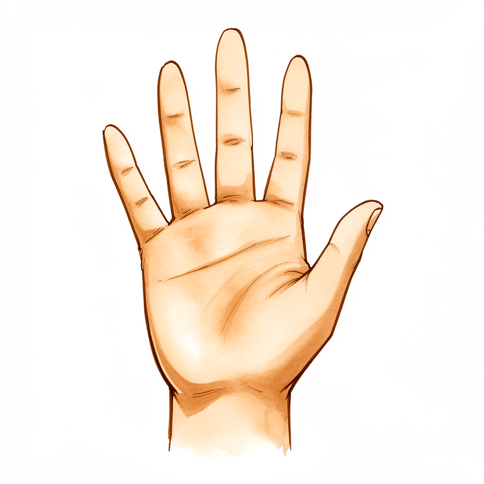
For patients: a plain-language version of this topic is available. See the patient guide.
Overview
Hamate body fractures and ring or little finger carpometacarpal (CMC) fracture-dislocations are uncommon injuries that are frequently underdiagnosed due to subtle radiographic findings [2, 12, 19]. These injuries result from longitudinally directed force along the fifth metacarpal axis, often leading to proximal and dorsal subluxation of the metacarpal base while a radial portion remains articulated [25]. The hamate articulates with the ring and small metacarpals via two concave facets separated by a ridge, a configuration that contributes to instability when disrupted [25]. Clinicians must maintain a high index of suspicion, as delayed diagnosis and treatment can lead to suboptimal outcomes and considerable morbidity [6, 9].
Diagnostic evaluation requires specific imaging protocols because routine x-rays often miss the extent of injury [25]. Recommended views include an anteroposterior projection with the forearm pronated 30 degrees from full supination, a Brewerton view, or CT scanning [25]. Three-dimensional CT reformatted views demonstrate higher interobserver and intraobserver reliability than standard two-dimensional CT [25]. Failure to detect these fractures risks nonunion, posttraumatic arthritis, decreased grip strength, and reduced range of motion [7]. Additionally, delayed ulnar-nerve palsy may occur due to extraneural fibrosis, necessitating close follow-up of nerve function [17].
Treatment principles emphasize the identification and management of associated CMC joint instability alongside the hamate fracture [5]. In cases involving multiple CMC dislocations with a hamate body fracture, open reduction and internal fixation (ORIF) of the hamate is recommended prior to reduction and pinning of the CMC joints [11]. For delayed presentations, ORIF is the recommended approach [36]. Surgical management of these complex fractures can provide good functional recovery, with operative treatment resulting in good-to-excellent outcomes despite relatively high complication rates [1, 13]. Nonoperative treatment may be successful if a concentric and stable reduction is achieved early, though it requires close follow-up during the first week [18]. Patients generally experience minimal functional deficits whether managed operatively or nonsurgically [15].
Anatomy & Pathophysiology
Bony Anatomy
The ring finger and small metacarpal bases share a common articulation with the hamate [33]. This carpometacarpal joint permits 15 to 30 degrees of mobility between the ring and small finger metacarpal bases and the hamate [33]. The base of the fifth metacarpal can slide radially if the entire fourth metacarpal is excised [33]. The lack of tendinous attachments to the fourth metacarpal base enhances the possibility of radial sliding of the fifth metacarpal base [33]. Fractures of the hamate, including hook fractures and those of the hamate body, account for 2–4% of all carpal fractures [24]. Hamate-metacarpal fracture-dislocations constitute 10–15% of carpometacarpal region fracture-dislocations [24]. Intra-articular fractures of the hamate are uncommon injuries [14]. Injuries to the body of the hamate, especially involving the little finger metacarpal articulation, are likely more common than reported [2].
Ligamentous Anatomy
Two distinct dorsal ligaments attach to the dorsal aspect of the fifth metacarpal: one extending from the ulnar base of the fifth metacarpal to the hamate, and another from the radial base of the fifth metacarpal to the hamate and sometimes to the fourth metacarpal ulnar base [48]. An intermetacarpal ligament attaches the radial base of the fifth metacarpal to the ulnar base of the fourth metacarpal [48]. One volar ligament attaches to the fifth metacarpal base and extends either to the hook of the hamate or to the ulnar base of the fourth metacarpal [48]. There are no intra-articular ligaments at the fifth CMC joint except for one located between the third/fourth metacarpal and the capitate/hamate [48]. The alignment of interosseous ligaments between the fourth and fifth metacarpals differs from those between the second-third and third-fourth metacarpals [48]. The fifth CMC joint allows approximately 25° of flexion/extension compared with approximately 15° at the fourth CMC joint [48]. When the metacarpals flex at the CMC joints, the dorsal interosseous ligament tightens and the anterior interosseous ligament relaxes [48]. When the metacarpals extend at the CMC joints, the anterior ligament tightens and the posterior ligament relaxes [48].
Pathomechanics
Articular fractures of the hamate-metacarpal joint are typically the result of a direct blow with a closed fist or high-energy trauma [25]. These injuries are usually associated with proximal and dorsal subluxation of the metacarpal, though palmar dislocations are also reported [25]. Displacement is accentuated by the pull of the extensor carpi ulnaris [25]. The pull of the abductor digiti minimi and the oblique slope of the hamate contribute to the instability of CMC fracture-dislocations of the small finger [25]. A force acting along the longitudinal axis of the fifth metacarpal causes a fracture dislocation at the base of the bone in a dorsal-ulnar direction due to joint anatomy and mechanical properties [79]. The most common mechanism of CMC dislocations with simultaneous hamate fracture is hyperflexion of the metacarpal heads [38]. These injuries usually require a great force [38].
Clinical Presentation and Diagnosis
The extent of the injury is frequently missed on routine x-rays [25]. Bora and Didizian recommended an anteroposterior view with the forearm pronated 30 degrees from the fully supinated position for diagnosis [25]. Three-dimensional CT reformatted views have higher interobserver and intraobserver reliability than standard two-dimensional CT [25]. Isolated palmar ulnar dislocation of the fifth CMC joint is a rare injury with subtle radiographic findings that may be easily overlooked [28]. Radiographs of the hand in 60° of pronation from the lateral are recommended if there is suspicion for a fifth CMC subluxation or dislocation [31]. The relative metacarpal shortening (RMS) is a radiographic measurement with almost perfect agreement between reviewers and is increased in fourth and fifth carpometacarpal fracture dislocations [35]. Carpometacarpal dislocations of the hand with a simultaneous fracture of the hamate occur in less than 1% of osseous hand injuries [38]. Diagnosis can be missed or delayed because of the high likelihood of other severe concomitant injuries [38]. Carpometacarpal dislocations are uncommon and prone to wrist instability if not treated promptly [40].
Classification
Epidemiology and Prevalence
Hamate fractures, encompassing both hook and body fractures, represent 2–4% of all carpal fractures [24]. Fractures of the hamate body are a more common presentation in accident and emergency departments than previously described, particularly among young adults [26]. In contrast, carpometacarpal (CMC) joint fracture dislocations involving the second through fifth fingers are extremely rare injuries [4].
Fracture Patterns and Mechanisms
Coronal hamate body fractures associated with CMC instability frequently present as dorsal oblique patterns following punching injuries [10]. These injuries are commonly associated with fractures of the fourth metacarpal base [10]. Four cases of unstable fracture-dislocation of the fifth CMC joint have been reported, comprising three isolated dorsal fracture-dislocations and one dorsal fracture-dislocation associated with a fracture of the fourth metacarpal bone [27].
Diagnostic Assessment
Thorough clinical examination and radiological assessment are required for CMC joint fracture dislocations from the second to fifth finger [4]. When a hamate fracture is suspected, an oblique x-ray view should be considered as part of the initial diagnostic investigations [21]. If there is suspicion for a fifth CMC subluxation or dislocation, radiographs of the hand in 60° of pronation from the lateral are recommended [31].
Classification Systems
Milch et al.: Established in 1934, this was the first classification of hamate fractures and does not include coronal fractures [24]. Cain: This classification concentrates on the dislocation of the fifth CMC and further co-fractures of the hamate [24]. Ebraheim: This classification focuses on the course of the fracture line through the hamate body [24].
Clinical Presentation
Epidemiology and Mechanism
Injuries to the body of the hamate, particularly those involving the little finger metacarpal articulation, are likely more common than reported [2]. CMC joint fracture dislocation from the second to fifth finger remains an extremely rare injury [4]. The most common mechanism for CMC dislocations with a simultaneous hamate fracture is hyperflexion of the metacarpal heads [38]. Articular fractures of the hamate-metacarpal joint typically result from either a direct blow with a closed fist or high-energy trauma [25]. An axial load to the fourth and fifth metacarpal heads secondary to a clenched-fist blow is often cited as a mechanism for these injuries [48]. Coronal hamate body fractures with CMC instability frequently present as dorsal oblique patterns following punching injuries [10].
Associated Injuries and Clinical Signs
Coronal hamate body fractures with CMC instability are commonly associated with fourth metacarpal base fractures [10]. CMC joint fracture dislocation from the second to fifth finger requires thorough clinical examination and radiological assessment [4]. The risks of not detecting a hamate body fracture include nonunion of the bone, posttraumatic arthritis, decreased grip strength, and decreased range of motion of the hand [7]. Delayed ulnar-nerve palsy can occur following a fracture of the hamate due to extraneural fibrosis [17]. It is important to assess ulnar-nerve function at the time of injury and follow patients closely for possible development of delayed symptoms [17]. Carpometacarpal dislocations are prone to wrist instability if not treated promptly [40].
Radiographic Assessment
Bora and Didizian recommended an anteroposterior (AP) view with the forearm pronated 30 degrees from the fully supinated position to diagnose these fractures [25]. The diagnosis of fifth CMC subluxation or dislocation can be made by CT or a Brewerton view [25]. Both the 2-5 IMA and the 3-5 IMA are useful screening measurements on lateral hand radiographs for detection of ulnar-sided carpometacarpal fracture-dislocations [55]. Careful evaluation of intraoperative and postoperative imaging, particularly CT, is important to detect rare persistent palmar trapezoid dislocations that may be missed on standard radiographs [57].
Investigations
Plain radiography: Fracture-dislocations of the metacarpal bases are frequently missed due to swelling and metacarpal overlap on standard lateral plain films [81]. A true lateral radiograph is required for accurate diagnosis because swelling can obscure the deformity [81]. On posteroanterior views, loss of parallel joint surfaces at the carpometacarpal articulations indicates fracture-dislocation [81]. For suspected fifth carpometacarpal subluxation or dislocation, radiographs of the hand in 60° of pronation from the lateral are recommended [31]. The 2-5 intermetacarpal angle (IMA) and the 3-5 IMA serve as useful screening measurements on lateral hand radiographs for detecting ulnar-sided carpometacarpal fracture-dislocations [55]. In cases of fifth carpometacarpal dislocation, measuring the angle between the long axes of the second and fifth metacarpals on a true lateral radiograph is advocated; this carpometacarpal angle is increased compared with controls (38.5° compared with 9.8°) [93]. Isolated palmar ulnar dislocation of the fifth carpometacarpal joint presents with subtle radiographic findings that may be easily overlooked [28].
CT: CT scans are beneficial for determining the extent of joint surface involvement and guiding appropriate intervention in carpometacarpal fracture-dislocations [81].
Other Considerations: This rare hand injury requires careful radiological assessment and meticulous, stable reduction [32]. Delayed diagnosis makes closed reduction difficult and is associated with less favorable radiographic outcome [60].
Treatment
Diagnostic Considerations for Treatment Planning
Accurate identification of hamate fractures and ring/little finger CMC fracture-dislocations requires specific imaging protocols. Bora and Didizian recommend an anteroposterior view with the forearm pronated 30 degrees from the fully supinated position to identify these fractures [25]. Kim et al. reported that three-dimensional CT reformatted views offer higher interobserver and intraobserver reliability than standard two-dimensional CT [25]. These injuries are rare; CMC dislocations of the hand with a simultaneous hamate fracture occur in less than 1% of osseous hand injuries [38]. Hamate fractures, including hook and body fractures, account for 2–4% of all carpal fractures [24]. The hamate-metacarpal fracture-dislocation constitutes 10–15% of these hamate injuries [24].
Non-Operative
Although operative treatment is generally recommended in the literature, conservative management through immediate reduction and splint immobilization can be sufficient for acute, uncomplicated ulnar CMC dislocations [95]. A patient with an ulnopalmar fifth carpometacarpal joint dislocation was successfully treated by nonoperative means [59]. Petrie and Lamb treated 14 fracture-dislocations of the fifth metacarpal-hamate joint by immediate, unrestricted motion and reviewed them at 4.5 years [25]. Despite persistent metacarpal shortening, incongruity in the articular surface, and widening of the joint, only 1 patient in the Petrie and Lamb series had pain significant enough to affect work [25].
Operative
Indications: Operative intervention is indicated for intra-articular hamate fractures, where open reduction and internal fixation with K-wires is recommended to reconstruct the articular surface [14]. Fracture-dislocations of the fourth and fifth metacarpal joints associated with comminuted dorsal hamate fractures or coronal fractures through the hamate are particularly unstable, making open reduction uniformly necessary [25].
Surgical Approach / Technique: Treatment options include open reduction and fixation of the hook of the hamate fracture with stabilization of the carpometacarpal joint [8]. For single-fragment hamate shear fractures, screw fixation of the hamate fragment to the body serves to treat both the fracture and the dislocation [25]. Dorsal buttress plating between the hamate and the capitate is an alternative technique for fifth CMC joint fracture-dislocations with avulsion fractures of the hamate [30]. In a case series of 11 patients, open reduction and buttress plate application successfully reduced the fracture while maintaining ulnar-sided CMC joint mobility by not securing the distal end of the plate to the metacarpal [25]. A suture button implant allows early motion from 2 weeks post-surgery, resulting in good motion of the ulnar CMC joints and increased hand grip [42]. In a rare case of open scaphoid dislocation with associated hamate and fourth metacarpal fractures, treatment involved open reduction, ligamentous repair, and augmentation using FiberTape as an internal brace [94].
Implant Selection: Stabilized arthroplasty provides better mobility than arthrodesis and restores metacarpal length better than nonstabilized resectional arthroplasty [58].
Complications and Outcomes: Five patients (45%) required a second surgery for hardware removal in the buttress plate case series [25]. Kjaer-Petersen et al. reported that regardless of the method of treatment (closed, percutaneous, or open), 19 of 50 (38%) patients had some symptoms at a median follow-up of 4.3 years [25]. Papaloizos et al. advocated open reduction and noted that pain directly correlated with degenerative arthritis, which was attributed to nonanatomic reduction in several cases [25]. Postoperatively, recovery of the wrist was rapid, though extension of the fingers remained poor for over 3 months [29].
Ring Finger Ray Amputation and Transposition
Transposition Techniques: Advocates for transposition of the small finger to the ring finger position aim to avoid risks associated with convergence of the long finger and small finger, such as impaired mobility and objects falling through the space [20]. Le Viet described a technique involving complete ring finger ray resection, an intracarpal wedge-shaped osteotomy, and a capitohamate arthrodesis for small finger translocation [20]. Le Viet proposed four main advantages: (1) convergence between the small finger and long finger is avoided, (2) the integrity of the ulnar side of the hand is maintained, (3) the fifth CMC joint is preserved, and (4) the action of the interossei is unchanged [20]. In a series of 17 patients, 14 were satisfied with preservation of motion at the CMC joint [20]. The author acknowledged a learning curve with this technique [20]. Others have also reported favorable results with intracarpal osteotomy [20]. A concern with small finger transposition is that the finger will be shorter than the ring finger [20]. Posner described a step-cut osteotomy that adds height to the small finger during transposition to the ring finger metacarpal base [20]. The small finger metacarpal is cut at a more proximal level than the ring finger metacarpal and can lengthen the small finger by 1.5 cm at the time of transposition [20]. This lessens the length discrepancy between the long finger and transposed small finger with improved hand function and cosmesis [20].
Anatomical Considerations for Ray Resection: In the ring finger, unlike in the index finger and long finger, there is 15 to 30 degrees of mobility between the ring and small finger metacarpal bases and the hamate at the CMC joint [33]. The ring finger and small metacarpal bases also share a common articulation with the hamate [33]. It is therefore possible for the base of the fifth metacarpal to slide radially if the entire fourth metacarpal is excised [33]. This possibility is enhanced by the lack of tendinous attachments to the fourth metacarpal base, unlike in the remaining metacarpals in the hand [33]. Transmetacarpal ray resection of the ring finger can leave an open space between the long finger and small finger as the remaining metacarpal base cannot converge [33]. This problem can be avoided by complete ray amputation, but complete deletion can lead to a loss of small finger abduction and possible scissoring [33].
Reconstruction and Stabilization: Steichen and Idler reported on patients who underwent central ray resection without transposition [33]. They noted that it was easier to close the intermetacarpal space between the long finger and small finger compared with the space between the index finger and ring finger after ray amputation [33]. Cosmetic and functional results were satisfactory in 12 of 13 patients in the Steichen and Idler series [33]. Morgan and colleagues conducted a cadaveric study comparing suture button stabilization versus repair of the transverse intermetacarpal ligaments after ring finger ray amputation and noted that the suture button was able to provide a more stable gap that permitted earlier range of motion [33]. The DTIL is imbricated in a pants-over-vest configuration with 3-0 nonabsorbable sutures [33]. This repair should be taut as stretching can occur over time [33]. Temporary transverse Kirschner wires can be placed between the long finger and small finger metacarpals [33]. The Kirschner wires are removed after 4 to 6 weeks [33].
Complications
Nerve palsy: Ulnar-nerve function must be assessed at the time of injury, and patients require close follow-up for the possible development of delayed symptoms following a hamate fracture [17].
Stiffness / Arthrofibrosis: Postoperatively, extension of the fingers may remain poor for over 3 months following treatment of an unusual carpometacarpal fracture-dislocation [29].
Other Considerations: Carpometacarpal joint dislocations and fracture dislocations can be very disabling if not recognized early [19]. Delay in diagnosis and treatment of lesser digit carpometacarpal joint fracture-dislocations can lead to a suboptimal outcome [9]. Inadequate reduction of fifth metacarpal-hamate joint injuries can result in weakness of grip as the major functional disability [25]. Pain in fifth metacarpal-hamate joint injuries directly correlates with degenerative arthritis, which has been attributed to nonanatomic reduction [25]. In a series of 17 patients undergoing small finger transposition to the ring finger position, 14 were satisfied with preservation of motion at the CMC joint [20]. A concern of small finger transposition is that the finger will be shorter than the ring finger [20]. Convergence of the long finger and small finger after ring finger ray amputation without transposition is associated with risks such as impaired mobility and objects falling through the space [20].
Recovery
Functional Outcomes: Surgical treatment of coronal plane hamate fractures associated with fourth and fifth metacarpal base fracture-dislocations provides good functional recovery [1]. Operative management of CMC fracture dislocations yields good-to-excellent functional outcomes, despite relatively high surgical complication rates [13]. In cases involving five CMC joint dislocations associated with a hamate body fracture, open reduction and internal fixation of the hamate fracture is recommended prior to reduction and pinning of the CMC joints [11]. Both cases reported in the literature achieved excellent anatomical and functional results following reduction and immobilization [22]. This specific approach was associated with an excellent short-term functional outcome [56].
Long-term Functional Status: Six months after injury, finger and wrist motions were equal bilaterally, and measured grip strength was equal in both hands [51]. Five years and two months postoperatively, radiographs showed anatomical positioning of the metacarpals and degenerative arthritis of the carpometacarpal joints was not observed [34].
Timing and Diagnostic Impact: Delay in diagnosis and treatment can lead to a suboptimal outcome for lesser digit carpometacarpal joint fracture-dislocations [9]. In patients with delayed presentation of CMC fracture dislocations, ORIF is recommended [36]. If the diagnosis of an ulnar CMC joint dislocation or fracture–dislocation is early accomplished and a concentric and stable reduction is initially achieved, nonoperative treatment may be a successful option requiring close follow-up for the first week [18].
Key Evidence
- [L4] The surgical treatment of coronal plane hamate fractures associated with the fourth and fifth metacarpal base fracture-dislocations can provide good functional recovery in these complex fractures. [1] (10.52312/jdrs.2025.1997)
- [L5] Injuries to the body of the hamate, especially involving the little finger metacarpal articulation, are likely more common than reported and should be recognized. [2] (10.1016/j.jhsa.2014.08.026)
- [Paper] The study showed favorable outcomes after open reduction and internal fixation of the hamate body fracture with interfragmentary screws, when combined with stabilization of the CMC dislocation with percutaneous Kirschner wires. [3] (10.1055/s-0039-1692326)
- [L4] CMC joint fracture dislocation from second to fifth finger is an extremely rare injury that needs thorough clinical examination and radiological assessment. [4] (10.4055/cios.2015.7.4.430)
- [L5] Successful treatment of coronal hamate fractures requires identification and treatment of associated disruption or instability of the ulnar 2 carpometacarpal joints. [5] (10.1016/j.hcl.2012.05.010)
- [Paper] It is important to diagnose and treat the Carpometacarpal joint dislocation to avoid considerable morbidity associated with it. [6] (10.1007/s12593-010-0018-3)
- [L5] The risks to not detecting a hamate body fracture are nonunion of the bone, posttraumatic arthritis, decreased grip strength, and decreased range of motion of the hand. [7] (10.1016/j.ajem.2014.03.050)
- [L5] It was successfully treated by open reduction and fixation of the hook of the hamate fracture and stabilization of the carpometacarpal joint. [8] (10.1016/s0266-7681(96)80043-x)
- [L5] Treating physicians need to maintain a high index of suspicion regarding lesser digit carpometacarpal joint fracture-dislocations as delay in diagnosis and treatment can lead to a suboptimal outcome. [9] (10.1016/j.jhsa.2026.02.016)
- [L4] Coronal hamate body fractures with CMC instability frequently presented as dorsal oblique patterns following punching injuries and were commonly associated with fourth metacarpal base fractures. [10] (10.1007/s00402-026-06366-5)
- [Case_report] In cases of five CMC joint dislocations associated with a hamate body fracture, the authors recommend addressing the hamate fracture with open reduction and internal fixation prior to proceeding with reduction and pinning of the CMC joints. [11] (10.1177/1558944716668860)
- [L4] Hamato-metacarpal injuries are uncommon and radiographic findings are often subtle, with the extent of injury often not appreciated. [12] (10.1016/s0266-7681(05)80063-4)
- [L4] Despite relatively high surgical complication rates, operative management of CMC fracture dislocations results in good-to-excellent functional outcomes. [13] (10.1016/j.jhsg.2024.11.003)
- [L5] Intra-articular fractures of the hamate are uncommon injuries and we would recommend open reduction and internal fixation with K-wires in an attempt to reconstruct the articular surface. [14] (10.1016/0020-1383(93)90187-b)
- [L4] Patients who sustain hamate fractures have minimal functional deficits whether managed operatively or nonsurgically. [15] (10.1016/j.jhsg.2024.02.002)
- [L4] Operative treatment with headless compression screws is a safe procedure with good clinical outcomes for acute dislocated hamate corpus fractures and nondisplaced fractures of the hook of the hamate bone. [16] (10.1055/s-0039-1695765)
- [L4] Delayed ulnar-nerve palsy can occur following a fracture of the hamate due to extraneural fibrosis, and it is important to assess ulnar-nerve function at the time of injury and follow patients closely for possible development of delayed symptoms. [17] (10.2106/00004623-196850030-00017)
- [L4] If the diagnosis of an ulnar CMC joint dislocation or fracture–dislocation is early accomplished and a concentric and stable reduction is initially achieved, the nonoperative treatment may be a successful option to take into account but requiring a close follow-up for the first week. [18] (10.1055/s-0039-1688468)
- [L5] Carpometacarpal joint dislocations and fracture dislocations are uncommon injuries that can be very disabling if not recognized early. [19] (10.5435/jaaos-d-25-00583)
- [Case_report] The authors believe that where a hamate fracture is suspected, an oblique x-ray view should be considered as part of the initial diagnostic investigations. [21] (10.1186/1749-799x-5-64)
- [L4] [24] (10.3205/iprs000131)
- [L4] Fracture of the body of the hamate is a more common injury presenting to accident and emergency departments than previously described, especially in young adults. [26] (10.1016/0020-1383(91)90129-3)
- [L4] Four cases of unstable fracture-dislocation of the fifth carpometacarpal joint are reported, including 3 isolated dorsal fracture-dislocations and 1 dorsal fracture-dislocation associated with a fracture of the fourth metacarpal bone. [27] (10.1016/0020-1383(77)90008-0)
- [L4] Isolated palmar ulnar dislocation of the fifth CMC joint is a rare injury with subtle radiographic findings that may be easily overlooked. [28] (10.1007/s004029900111)
- [L5] Postoperatively recovery of the wrist was rapid, though extension of the fingers remained poor for over 3 months. [29] (10.1016/0020-1383(94)90161-9)
- [Case_report] Dorsal buttress plating between the hamate and the capitate could be an alternative technique for the treatment of fracture-dislocation of the fifth CMC joint with avulsion fracture of the hamate. [30] (10.1007/s00402-018-3072-0)
- [L4] The authors recommend obtaining radiographs of the hand in 60° of pronation from the lateral if there is suspicion for a fifth CMC subluxation or dislocation. [31] (10.1016/j.jhsa.2018.04.012)
- [L5] This is a rare hand injury that requires careful radiological assessment and meticulous, stable reduction. [32] (10.1016/j.ijscr.2023.108417)
- [L5] Five years and two months postoperatively, radiographs showed anatomical positioning of the metacarpals and degenerative arthritis of the carpometacarpal joints was not observed. [34] (10.1016/s0020-1383(97)82145-6)
- [L4] The relative metacarpal shortening (RMS) is a radiographic measurement with almost perfect agreement between reviewers and is increased in fourth and fifth carpometacarpal fracture dislocations. [35] (10.1016/j.jhsa.2025.08.011)
- [L4] In patients with delayed presentation of CMC fracture dislocations, we recommend ORIF. [36] (10.1016/j.jhsa.2015.07.017)
- [L5] [38] (10.1155/2020/2861604)
- [L4] Carpometacarpal dislocations are uncommon and prone to wrist instability if not treated promptly. [40] (10.1016/j.otsr.2016.04.003)
- [L5] Dorsal drilling of coronal hamate fractures appears to be safe, as volar drill tips are well away from ulnar nerve motor and sensory branches. [41] (10.1016/j.jhsa.2022.04.023)
- [L4] The surgical treatment using a suture button implant allowed early motion from 2 weeks after surgery, which resulted in good motion of the ulnar CMC joints and increased hand grip. [42] (10.1055/s-0039-1693051)
- [Paper] [48] (10.1016/j.hcl.2011.05.010)
- [L5] Six months after injury, finger and wrist motions were equal bilaterally, and measured grip strength was equal in both hands. [51] (10.1016/s0363-5023(84)80020-9)
- [L4] Both the 2-5 IMA and the 3-5 IMA are useful screening measurements on lateral hand radiographs for detection of ulnar-sided carpometacarpal fracture-dislocations. [55] (10.1016/s0363-5023(11)60044-0)
- [Case_report] This approach was associated with an excellent short-term functional outcome. [56] (10.1007/s12593-015-0191-5)
- [L4] This report highlights the importance of careful evaluation of intraoperative and postoperative imaging, particularly CT, to detect rare persistent palmar trapezoid dislocations that may be missed on standard radiographs. [57] (10.1016/j.jhsg.2025.100769)
- [L5] Compared with the more commonly used operative procedures, stabilized arthroplasty provides a better mobility than arthrodesis and restores metacarpal length better than nonstabilized resectional arthroplasty. [58] (10.1097/bth.0b013e3181aa25c4)
- [L5] A patient with an ulnopalmar fifth carpometacarpal joint dislocation was successfully treated by nonoperative means. [59] (10.1016/s0363-5023(86)80189-7)
- [L4] Delayed diagnosis makes closed reduction difficult and was associated with less favorable radiographic outcome. [60] (10.1177/1558944719852743)
- [L5] A force acting along the longitudinal axis of the fifth metacarpal causes a fracture dislocation at the base of the bone in a dorsal-ulnar direction, because of the anatomy of the joint and the mechanical properties of the bone and the intermetacarpal ligament. [79] (10.1016/s0363-5023(79)80070-2)
- [L4] Measurement of the angle between the long axis of the second and fifth metacarpal bones on a true lateral radiograph, in cases of fifth carpometacarpal dislocation is advocated, as the carpometacarpal angle is increased compared with controls (38.5° compared with 9.8°). [93] (10.1016/s0020-1383(05)80042-7)
- [L4] The authors report a rare case of open scaphoid dislocation with associated hamate and fourth metacarpal fractures treated with open reduction, ligamentous repair, and augmentation using FiberTape as an internal brace. [94] (10.1016/j.jhsg.2023.10.004)
- [L4] Although operative treatment is recommended in the literature, these cases show that conservative treatment through immediate reduction and splint immobilization can be sufficient for acute, uncomplicated ulnar CMC dislocations. [95] (10.1007/s11552-011-9347-3)
- [L2] The clinically appropriate fixation method should be selected according to the experience of the surgeon and the degree and type of fracture and dislocation. [96] (10.1186/s13018-023-04225-2)
See Also
References
[1] Surgical treatment of coronal plane hamate fractures: Clinical and radiological outcomes. Joint Diseases and Related Surgery. 2024. DOI: 10.52312/jdrs.2025.1997
[2] Are Hamate Fractures Common? Letter Regarding “Carpal Fractures” Article. The Journal of Hand Surgery. 2014. DOI: 10.1016/j.jhsa.2014.08.026
[3] Carpometacarpal 4/5 Fracture Dislocations: Fracture Morphology and Surgical Treatment. Journal of Hand and Microsurgery. 2020. DOI: 10.1055/s-0039-1692326
[4] Carpometacarpal Joint Fracture Dislocation of Second to Fifth Finger. Clinics in Orthopedic Surgery. 2015. DOI: 10.4055/cios.2015.7.4.430
[5] Fractures of the Hamate and Pisiform Bones. Hand Clinics. 2012. DOI: 10.1016/j.hcl.2012.05.010
[6] Dislocation of Second and Third Carpometacarpal Joints Along with Fracture of First Metacarpal–Possible Mechanisms. Journal of Hand and Microsurgery. 2010. DOI: 10.1007/s12593-010-0018-3
[7] Hamate body and capitate fracture in punch injury. The American Journal of Emergency Medicine. 2014. DOI: 10.1016/j.ajem.2014.03.050
[8] Combined Fracture of the Hook of the Hamate and Palmar Dislocation of the Fifth Carpometacarpal Joint. Journal of Hand Surgery. 1996. DOI: 10.1016/s0266-7681(96)80043-x
[9] Current Concepts: Lesser Digit Carpometacarpal Joint Fracture-Dislocations. The Journal of Hand Surgery. 2026. DOI: 10.1016/j.jhsa.2026.02.016
[10] Coronal hamate body fractures with dorsal fourth and fifth carpometacarpal joint instability: fracture patterns, injury mechanisms, surgical strategies, and outcomes. Archives of Orthopaedic and Trauma Surgery. 2026. DOI: 10.1007/s00402-026-06366-5
[11] Coronal Hamate Fracture Associated With Carpometacarpal Dislocations of All of the Fingers: Review of the Literature and Case Report. HAND. 2016. DOI: 10.1177/1558944716668860
[12] Combined Fourth and Fifth Metacarpal Fracture and Fifth Carpometacarpal Joint Dislocation. Journal of Hand Surgery. 1995. DOI: 10.1016/s0266-7681(05)80063-4
[13] Operative Treatment of Non-Thumb Carpometacarpal Joint Fracture Dislocations. Journal of Hand Surgery Global Online. 2025. DOI: 10.1016/j.jhsg.2024.11.003
[14] Intra-articular fractures of the hamate. Injury. 1993. DOI: 10.1016/0020-1383(93)90187-b
[15] Study of Hamate Fractures in a Single Tertiary Hands Unit: A Retrospective Cohort Study and Literature Review. Journal of Hand Surgery Global Online. 2024. DOI: 10.1016/j.jhsg.2024.02.002
[16] Functional Outcome following Headless Compression Screw Fixation for Hamate Fractures. Journal of Wrist Surgery. 2019. DOI: 10.1055/s-0039-1695765
[17] Delayed Ulnar-Nerve Palsy Following a Fracture of the Hamate. The Journal of Bone & Joint Surgery. 1968. DOI: 10.2106/00004623-196850030-00017
[18] Nonoperative Treatment of Ulnar Carpometacarpal Fracture–Dislocations. Journal of Wrist Surgery. 2019. DOI: 10.1055/s-0039-1688468
[19] Carpometacarpal Joint Dislocations and Fracture Dislocations of the Index Through Small Digits. Journal of the American Academy of Orthopaedic Surgeons. 2025. DOI: 10.5435/jaaos-d-25-00583
[20] Green S Operative Hand Surgery. Ring Finger Ray Amputation With Transposition of the Fifth Metacarpal.
[21] Lessons to be learned from a missed case of Hamate fracture: a case report. Journal of Orthopaedic Surgery and Research. 2010. DOI: 10.1186/1749-799x-5-64
[22] CARPOMETACARPAL DISLOCATIONS: With Particular Reference to Simultaneous Dislocation of the Bases of the Fourth and Fifth Metacarpals.. The Journal of Bone and Joint Surgery. American Volume. 1948.
[24] Hamate's coronal fracture: diagnostic and therapeutic approaches based on a long-term follow-up. GMS Interdiscip Plast Reconstr Surg DGPW. 2019. DOI: 10.3205/iprs000131
[25] Green S Operative Hand Surgery. Metacarpal Base Fractures and Carpometacarpal Fracture-Dislocations > Fracture-Dislocations of the Small Finger Carpometacarpal Joint.
[26] Fractures of the body of the hamate. Injury. 1991. DOI: 10.1016/0020-1383(91)90129-3
[27] Unstable dorsal fracture-dislocation of the fifth carpometacarpal joint. Injury. 1977. DOI: 10.1016/0020-1383(77)90008-0
[28] Isolated palmar dislocation of the fifth carpometacarpal joint diagnosed by stress X-rays. Archives of Orthopaedic and Trauma Surgery. 2000. DOI: 10.1007/s004029900111
[29] An unusual carpometacarpal fracture—dislocation. Injury. 1994. DOI: 10.1016/0020-1383(94)90161-9
[30] Dorsal buttress plate fixation for the treatment of fracture–dislocation of the fifth carpometacarpal joint with avulsion fracture of the hamate: a case report. Archives of Orthopaedic and Trauma Surgery. 2018. DOI: 10.1007/s00402-018-3072-0
[31] Optimal Oblique Radiographs to Identify Fifth Carpometacarpal Dorsal Subluxations: A Cadaveric Study. The Journal of Hand Surgery. 2018. DOI: 10.1016/j.jhsa.2018.04.012
[32] Carpometacarpal dislocation second to fifth with associated hamate fracture: A case report of rare injury. International Journal of Surgery Case Reports. 2023. DOI: 10.1016/j.ijscr.2023.108417
[33] Green S Operative Hand Surgery. Ring Finger Ray Amputation Without Transposition.
[34] Open reduction and internal fixation of an unusual multiple carpometacarpal dislocation using one plate: a case report. Injury. 1997. DOI: 10.1016/s0020-1383(97)82145-6
[35] Relative Metacarpal Shortening as a Radiographic Measure of Fourth and Fifth Carpometacarpal Fracture Dislocation. The Journal of Hand Surgery. 2026. DOI: 10.1016/j.jhsa.2025.08.011
[36] The Effect of Timing on the Treatment and Outcome of Combined Fourth and Fifth Carpometacarpal Fracture Dislocations. The Journal of Hand Surgery. 2015. DOI: 10.1016/j.jhsa.2015.07.017
[38] Carpometacarpal Dislocation of the Third to Fifth Fingers and an Associated Fracture of the Hamate in a Military Paratrooper. Case Reports in Orthopedics. 2020. DOI: 10.1155/2020/2861604
[40] Rare carpometacarpal dislocations. Orthopaedics & Traumatology: Surgery & Research. 2016. DOI: 10.1016/j.otsr.2016.04.003
[41] Dorsal Fixation of Coronal Hamate and Fifth Metacarpal Base Fractures: An Anatomic Evaluation of the Ulnar Nerve. The Journal of Hand Surgery. 2024. DOI: 10.1016/j.jhsa.2022.04.023
[42] Surgical Treatment for Delayed Ulnar Carpometacarpal Fracture–Dislocations. Journal of Wrist Surgery. 2019. DOI: 10.1055/s-0039-1693051
[48] Arthroscopic Reduction and Percutaneous Fixation of Fifth Carpometacarpal Fracture Dislocations. Hand Clinics. 2011. DOI: 10.1016/j.hcl.2011.05.010
[51] Dorsal dislocation of the fourth and fifth carpometacarpal joints and simultaneous dislocation of the metacarpophalangeal joint of the small finger: A case report. The Journal of Hand Surgery. 1984. DOI: 10.1016/s0363-5023(84)80020-9
[55] The Interpetacarpal Angle: A Useful Screening Test for Ulnar-Sided Carpometacarpal Fracture-Dislocations. The Journal of Hand Surgery. 2011. DOI: 10.1016/s0363-5023(11)60044-0
[56] Floating Index Metacarpal Associated with Multiple Carpometacarpal Fracture-Dislocations: a Case Report. Journal of Hand and Microsurgery. 2015. DOI: 10.1007/s12593-015-0191-5
[57] Diagnosis and Management of Persistent Trapezoid Dislocation Following Surgical Treatment of Second through Fifth Carpometacarpal Fracture Dislocation. Journal of Hand Surgery Global Online. 2025. DOI: 10.1016/j.jhsg.2025.100769
[58] “Stabilized Arthroplasty” for Old Fracture Dislocations of the Fifth Carpometacarpal Joint. Techniques in Hand & Upper Extremity Surgery. 2009. DOI: 10.1097/bth.0b013e3181aa25c4
[59] Ulnopalmar dislocation of the fifth carpometacarpal joint—Successful closed reduction: Review of the literature and anatomic reevaluation. The Journal of Hand Surgery. 1986. DOI: 10.1016/s0363-5023(86)80189-7
[60] Carpometacarpal Fracture-Dislocations: A Retrospective Review of Injury Characteristics and Radiographic Outcomes. HAND. 2019. DOI: 10.1177/1558944719852743
[79] The mechanism of dorsal fracture dislocation of the fifth carpometacarpal joint. The Journal of Hand Surgery. 1979. DOI: 10.1016/s0363-5023(79)80070-2
[81] Campbell S Operative Orthopaedics 4 Volume Set. FRACTURES, DISLOCATIONS, AND LIGAMENTOUS INJURIES OF THE HAND AND WRIST > FINGER METACARPALS.
[93] Carpometacarpal dislocation: an aid to diagnosis. Injury. 1992. DOI: 10.1016/s0020-1383(05)80042-7
[94] Open Dislocation of the Scaphoid With an Associated Hamate Fracture and Fourth Metacarpal Fracture. Journal of Hand Surgery Global Online. 2024. DOI: 10.1016/j.jhsg.2023.10.004
[95] Acute Ulnar Carpometacarpal Dislocations. Can it be Treated Conservatively? A Review of Four Cases. HAND. 2011. DOI: 10.1007/s11552-011-9347-3
[96] A comparative study on the clinical efficacy of microplate trans-carpometacarpal joint fixation and non-trans-carpometacarpal joint fixation in treating fractures with dislocation or subluxation of the base of the fourth and fifth metacarpal bones. Journal of Orthopaedic Surgery and Research. 2023. DOI: 10.1186/s13018-023-04225-2