What you're feeling
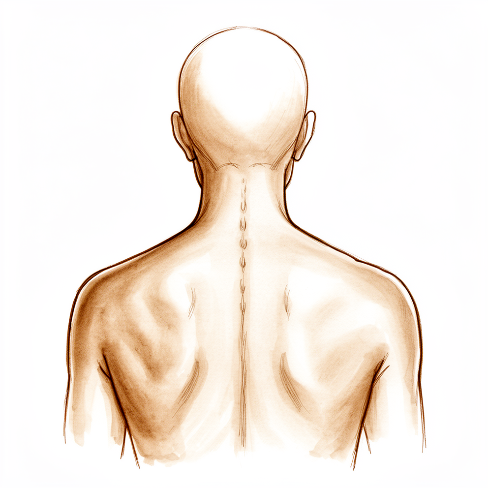
An osteoporotic fracture happens when a bone in your spine, called a vertebra, becomes so thin and weak that it cracks under everyday load. The pain usually sits in the middle of your back or where your mid back meets your lower back. It often comes on suddenly, sometimes after something as small as bending, lifting or a minor stumble.
The pain is usually sharp at first and settles into a dull ache. It tends to worsen when you stand or walk for a while, when you twist, or when you bend forward. Sitting up from lying down can be hard, and the pain often flares on waking or after you have been on your feet. Lying down may ease it. Everyday tasks that load your spine become difficult: carrying shopping, getting out of a low chair, making the bed, or climbing stairs.
If the fracture affects the lower part of a vertebra, it can sometimes irritate a nerve running down your leg. That can cause pain, tingling or numbness in that leg. Some people also strain the strong sheet of tissue that wraps around their lower back muscles, which adds to the ache.
These fractures can affect more than one vertebra, and they are easy to miss on plain X-rays. If you are over 60 and have sudden back pain, it is worth having it checked properly. A fracture also means your bones are weaker than they should be, so there is a real chance of another fracture, including a hip fracture, within the next few years. That risk is higher again if you have already had more than one spinal fracture.
Most of these fractures settle with time and do not need surgery. If your pain stays severe despite pain medication, there are procedures that can help, which we cover in the next sections.
What's actually happening
Your spine is a stack of bones called vertebrae, and each one has a solid block at the front that carries your weight. Healthy bone is a living scaffold, constantly being rebuilt as old bone is removed and new bone is laid down. With osteoporosis, that balance tips: bone is removed faster than it is replaced, so the scaffold becomes thin and porous, like a honeycomb with larger and larger holes.
The front block of each vertebra is designed to take the compressive load of your body, a bit like a brick carrying the weight of a wall above it. When that brick becomes porous, everyday loads such as bending or lifting can crush it. The bone compresses and wedges, so the vertebra loses height at the front and the spine bends forward slightly at that level. That crushing is the fracture you are feeling, and the pain comes from the cracked bone itself and from the strain it puts on the tissues around it.
These fractures happen most often where your mid back meets your lower back, because that junction takes more of the bending forces in your spine. If more than one vertebra is crushed, the forward bending can build up, which changes how your whole spine carries load and can add to the ache and stiffness you feel. The bone usually heals on its own over weeks to months, but because the bone is thin, the healed vertebra can keep slowly compressing, and the same weakness means another fracture elsewhere is possible.
Only about one in three people with these spinal fractures actually feel pain from them, which is why they are easy to miss. Most settle with time and pain medication alone. If your pain stays severe despite that, procedures exist that can steady the crushed bone and ease the pain, which we cover next.
What we can do about it
Most of these fractures heal without surgery. The first step is staying active. Gentle exercise and movement help the bone heal, stop further bone loss, and keep you doing the things you enjoy. Physiotherapy guides this: it aims to rebuild your strength and confidence while the fracture settles. Give it several weeks to months, because the bone usually heals on its own over that time. A brace may be used to support your back while it heals. Pain medication helps you keep moving through this period.
Alongside this, we look after the bone itself. Osteoporosis medicines can strengthen your bone and lower the chance of another fracture. Vitamin D deficiency is common with these fractures, and we check for it and correct it before any surgery. This bone health work matters: it improves healing, reduces complications, and lowers the risk of new fractures. It is often skipped, but it is an essential part of recovery after any fracture caused by osteoporosis.
If your pain is still severe after a trial of non-operative care, we may discuss a procedure called vertebral augmentation. This is a minimally invasive operation that steadies the crushed vertebra with bone cement. Two versions exist. Vertebroplasty injects the cement directly. Kyphoplasty first uses a small balloon to lift the collapsed bone, then fills the space with cement, which can also improve the forward bend caused by the fracture. Kyphoplasty is the option we favour when non-operative care has not helped enough. These procedures are done through small openings rather than a large incision, which keeps the risks low for older patients. Cement can sometimes leak out of the bone during the procedure, and this is more common with vertebroplasty, so we use techniques tailored to your fracture to prevent it. The operation has its own page, which explains what happens on the day and what recovery involves.
What to expect
For most people, the pain from one of these fractures settles over weeks to months as the bone heals on its own. The sharp pain usually eases first, and the dull ache fades more slowly. Staying active and keeping your strength up during this time helps, while long periods of rest can work against you by weakening your bones further and raising the chance of falls and new injuries.
If the pain is managed well, you can expect to get back to your normal activities as the fracture settles. If your pain is severe and does not ease with pain medication, a procedure to steady the bone can relieve that pain, and the benefit over medication alone can last through the first year. These procedures also aim to hold up the crushed vertebra so the spine does not keep bending forward at that level.
There are honest limits to know about. Because your bones are thin, a new fracture in another vertebra is possible afterwards, and the risk is higher if you are older, if you have fractured bones before, or if your bone density is low. The risk is lower when the osteoporosis itself is treated with medicines alongside the fracture care. Some people also find the collapsed vertebra slowly loses more height over time, which is part of how thin bone behaves rather than a sign the treatment has failed.
If a fracture is left alone and the forward bending builds up, especially with more than one fracture, that change in shape can affect how your spine carries load and can keep causing problems. Looking after your bone health, staying active, and following through on osteoporosis treatment give you the best chance of healing well and staying on your feet.
When to see someone
See your GP promptly if you are over 60 and get sudden back pain, even after a small stumble or a simple bend. These fractures are easy to miss on plain X-rays, so ask whether further imaging is needed. Go to an emergency department if you develop new weakness, numbness or tingling in your legs, or trouble controlling your bladder or bowel. Ask for a specialist review if your pain stays severe despite pain medication after several weeks, if you have already had one of these fractures and the pain returns or moves to another level, or if you have known thin bones and any new back pain at all.
Evidence & references
This is the clinical evidence summary written for health professionals. It is technical, and it lists the research this page was built from. You do not need to read it to understand your treatment or to make a decision about it.
Anatomy & Pathophysiology
Vertebral Column Anatomy
- The vertebral column comprises 33 vertebrae divided into five sections: 7 cervical, 12 thoracic, 5 lumbar, 5 sacral, and 4 coccygeal [16].
- The sacral and coccygeal vertebrae are fused, typically allowing for 24 mobile segments [16].
- A typical vertebra comprises an anterior body and a posterior arch that enclose the vertebral canal [16].
- The neural arch is composed of two pedicles laterally and two laminae posteriorly that are united to form the spinous process [16].
- To either side of the arch of the vertebral body is a transverse process and superior and inferior articular processes [16].
- The articular processes articulate with adjacent vertebrae to form synovial joints [16].
- The relative orientation of the articular processes accounts for the degree of flexion, extension, or rotation possible in each segment of the vertebral column [16].
- The spinous and transverse processes serve as levers for the numerous muscles attached to them [16].
- The length of the vertebral column averages 72 cm in men and 7 to 10 cm less in women [16].
- The vertebral canal extends throughout the length of the column and provides protection for the spinal cord, conus medullaris, and cauda equina [16].
Intervertebral Disk Anatomy
- The intervertebral disk connects adjacent vertebral bodies [6].
- The adjacent vertebral bodies, the disk, and the facet joints constitute the functional spinal unit that provides mechanical stability and allows physiologic motion [6].
- The intervertebral disks run between vertebral bodies from C2 to S1 and function to resist loads on the spine and provide stability [18].
- The disks contribute up to one-third of the height of the spinal column [18].
- The disk consists of the cartilaginous end plates of the vertebral bodies, the outer anulus fibrosus, and the inner nucleus pulposus [18].
- The end plate serves as a point of attachment of the disk to the superior and inferior surfaces of the vertebral bodies [18].
- The end plate is a thin layer of hyaline cartilage [18].
- The nucleus pulposus is centrally located and confined by the end plates and the anulus fibrosus [6].
- The nucleus pulposus resists compressive loads, dampens mechanical loads, and evenly distributes forces onto the end plates [6].
- The nucleus pulposus is the remnant of the embryonic notochord and comprises the gelatinous center of the intervertebral disk [18].
- The extracellular matrix of the nucleus pulposus is composed primarily of type II collagen and aggrecan, which makes it relatively hydrophilic [18].
- The nucleus pulposus serves to resist axial loads as well as provide height to the intervertebral disk [18].
- The anulus fibrosus is peripheral to the nucleus pulposus and confines the nucleus pulposus [6].
- The anulus fibrosus is designed to resist tensile loads, allow spinal motion, provide mechanical connection between the vertebrae, and confine the nucleus pulposus [6].
- The anulus fibrosus consists of concentrically layered fibrous cartilage lamellae primarily composed of type I collagen [18].
- The fibers of the anulus fibrosus run in alternating oblique trajectories [18].
- The anulus fibrosus resists tensile forces within the spine, including those due to the compression of the nucleus pulposus [18].
- The end plates form the interface between the vertebrae and the disk and define the upper and lower boundaries of the disk [21].
- The central portion of the end plate provides a major pathway for nutrients from the vertebral bodies to diffuse into the disk [21].
Disk Physiology and Pathophysiology
- In a normal healthy lumbar disk, large aggregating proteoglycans (aggrecan and versican) constitute a high percentage of the dry weight in the nucleus [21].
- Glycosaminoglycan molecules (keratan sulfate and chondroitin sulfate) decorate the aggrecan and versican core protein and are highly negatively charged [21].
- The highly negatively charged glycosaminoglycans create a highly hydrophilic matrix that attracts H2O molecules [21].
- The attraction of H2O molecules provides swelling pressure that counteracts the axial loads encountered by the disk [21].
- The matrix is viscoelastic and therefore dissipates mechanical energy and is subject to creep [21].
- Disk height is less at the end of each day due to creep [21].
- The adult intervertebral disk is avascular and derives its nutrition through diffusion from terminal capillaries in the vertebral bodies just below the end plates [18].
- In the adult, the blood supply ends at the bony end plate of the vertebral body and the outer anulus fibrosus [21].
- Most of the disk is considered immunologically isolated due to its avascularity [21].
- Nutrients are supplied to the disk cells primarily through diffusion [21].
- As the disk gets larger during development, the distances that nutrition must diffuse across become larger, further impeding nutritional supply to the disk cells [21].
- The decrease in nutritional transport is thought to contribute to disk degeneration [21].
- Innervation of the intervertebral disk is confined to the peripheral anulus fibrosus [21].
- The sinuvertebral nerve, which arises from the dorsal root ganglion, innervates the outer anulus fibrosus [21].
- In some degenerated disks with fissures, nerve fibers may be found deeper in the anulus fibrosus [21].
- The normal nucleus pulposus is not innervated [21].
- Pain sensation from the disk arises only from the anulus fibrosus [21].
- The nucleus pulposus can generate molecules such as cytokines and proteinases that can lead to pain [21].
- With normal ageing, glycosaminoglycan production diminishes, leading to gradual desiccation of the disc [19].
- The annulus fibrosus develops fissures and disc nuclear material may prolapse through during degeneration [19].
- The discs lose height and bulge beyond the margins of the vertebral bodies during degeneration [19].
- Disc protrusion against the ligaments causes formation of marginal osteophytes [19].
- Adjacent vertebral end plates ossify and become sclerotic while fatty change occurs in the subchondral bone marrow during spondylosis [19].
- Disc degeneration results in altered biomechanics and increased loading forces on the facet joints [19].
- Facet joint osteoarthritis may lead to osteophyte encroachment into the canal causing spinal lateral recess stenosis [19].
- The ligamentum flavum thickens, which contributes to stenosis, while the disc bulges from anteriorly into the spinal canal [19].
Osteoporosis Pathophysiology
- Osteoporosis is the most common metabolic bone disease resulting from imbalance between bone formation and bone resorption [2].
- Spinal fractures are the most common manifestation of osteoporosis [2].
- Only one in three patients with spinal fractures are symptomatic [2].
- In elderly patients, osteoporosis-related spinal fractures have similar morbidity and mortality as hip fractures [2].
- Osteoporosis and low bone mass are present in about 50% of elective spine patients older than 50 years [2].
- Osteoporosis and low bone mass negatively affect clinical outcomes and are associated with increased complications and revision surgery [2].
- Vitamin D deficiency is present in the majority of spine patients [2].
Clinical Presentation
- A history of fragility fracture significantly increases the risk of another fracture [15].
- The relative risk of a subsequent fracture is two to three times higher in women with a prior fragility fracture [15].
- The relative risk of a subsequent fracture is up to six times higher in men with a prior fragility fracture [15].
- A history of greater than a single vertebral fracture significantly increases the risk of further vertebral fractures [15].
- In a meta-analysis of nontreated patients enrolled in vertebroplasty studies, 18% had a secondary fracture within 12 months [15].
Investigations
Imaging Modalities
- Dual energy x-ray absorptiometry (DXA) is the benchmark to assess bone mineral density (BMD) [25].
- DXA measures areal BMD in g/cm² of the proximal femur, lumbar spine, and distal radius [25].
- DXA is accurate and precise when calibrated and performed properly [25].
- DXA involves a very low radiation dose [25].
- BMD is reported as a standard deviation difference to a reference standard [25].
- T-score is the reference standard for young healthy females [25].
- Z-score is the reference standard for age- and gender-matched subjects [25].
- Vertebral fracture assessment (VFA) can be performed at the time of DXA [25].
- VFA identifies occult vertebral fracture in 20% to 30% of cases [25].
- VFA is useful to change a diagnosis from low bone mass (osteopenia) to osteoporosis [25].
- Opportunistic CT uses CT data to estimate bone status [25].
- CT is based on the attenuation coefficient called the Hounsfield unit (HU) [25].
- Hounsfield units are related to BMD and can be used to estimate BMD and the presence of osteoporosis [25].
- PACS tools can calculate mean HU for any elliptical region of interest [25].
- Most osteoporotic vertebral fractures can be diagnosed by plain radiographs or CT [26].
- MRI can show edema in the vertebral body to indicate a recent fracture when age determination is required [26].
- An acute measured height change of greater than 1 inch is suggestive of an acute vertebral fracture [26].
Laboratory and Preoperative Assessment
- Greater than 75% of patients older than 50 years having elective spine surgery have vitamin D insufficiency or deficiency [14].
- Vitamin D deficiency is linked to increased pain severity in spinal stenosis [14].
- Ravindra demonstrated slower fusion and lower rates of success in vitamin D–deficient patients [14].
- Kim found that baseline low vitamin D was associated with worse Oswestry Disability scores at follow-up [14].
- Vitamin D deficiency is treatable but takes 6 weeks [14].
- Osteoporosis is linked to subsidence of cages, pedicle screw loosening, proximal junction failure, poorer fusion rates, increased spondylolisthesis after laminectomy, and higher rates of revision surgery [14].
- Bjerke reviewed 140 lumbar fusion patients and found that 10 were osteoporotic and 59% had low bone mass [14].
- Nonunion was 50% in osteoporotic patients compared with 18% in those with low bone mass or normal BMD [14].
- Osteoporosis-related complications occurred in 23% of patients with normal BMD, 28% of patients with low bone mass, and 46% of osteoporotic patients [14].
- Complication rates worsened relative to BMD when more than single-level fusion was performed [14].
- The AOA suggests using the same principles as outlined for secondary fracture prevention for preoperative bone health optimization [14].
- Inclusion criteria for preoperative bone health optimization assessment include all patients having thoracolumbar surgery who are older than 50 years [14].
- Preoperative patients are recommended to consume vitamin D3 2,000 U daily and 1,200 mg calcium in the diet [14].
- Preoperative patients should be assessed for fall risk and considered for physical therapy if warranted [14].
- Patients who do not need DXA or who screen negative for DXA should undergo surgery [14].
- Patients with low bone mass or osteoporosis should be considered for preoperative bone health optimization [14].
- If medically indicated, patients are treated with diphosphonate or anabolic medication before surgery [14].
- The duration of preoperative treatment is unknown, but effects of bone strength and physiology are seen within 2 months [14].
- A minimum of 3 months of preoperative treatment is recommended if clinically possible before surgery [14].
- Longer treatment up to 9 months should be considered for multilevel or high-risk cases such as osteotomy [14].
- Antiosteoporosis medications should be continued postoperatively [14].
- Multiple RCTs comparing biphosphonates and placebo in patients undergoing lumbar spine fusion show improved clinical outcomes, radiologic outcomes, and lower risk of complications [14].
- Biphosphonates do not appear to be associated with any impairment of bone health [14].
- One RCT and multiple cohort studies show that teriparatide can lead to improved clinical outcomes, radiologic outcomes, and lower risk of complications in spinal fusion patients [14].
Treatment
Non-Operative Management
- The initial treatment of osteoporotic vertebral fractures is nonsurgical [2].
- Secondary fracture prevention is performed in less than 20% of cases following osteoporosis-related fractures [2].
- Secondary fracture prevention includes education, assessment of bone health, nutritional supplements, weight-bearing exercise, fall prevention, and pharmaceutical medications if indicated [2].
- A history of fragility fracture increases the relative risk of another fracture by two to three times in women and up to six times in men [15].
- Secondary treatment was given in 10% of patients in a Medicare database analysis but was associated with a reduced risk of fracture by 40% [15].
- The American Orthopaedic Association (AOA) Own the Bone program is located in all states and 150 sites in the United States [15].
- The AOA Own the Bone program significantly improves compliance with treatment recommendations after fragility fracture [15].
- A prospective randomized controlled non-inferiority trial found that rigid brace, soft brace, and no brace treatments for osteoporotic compression fractures without neurologic injury had comparable outcomes [1].
- Vitamin D deficiency is present in the majority of spine patients and should be corrected after fracture and before elective surgery [2].
- Routine use of vitamin D (1,000 to 2,000 IU/day) and calcium supplementation for all spine fusion patients aged 65 years or older may help avoid nonunion and infection [28].
- Target serum 25-hydroxyvitamin D levels are 30 ng/mL [28].
Operative Management: Cement Augmentation
- Vertebroplasty or kyphoplasty is considered for osteoporotic vertebral fracture in patients who are hospitalized for pain, those who do not improve with nonoperative care, or those who have progressive collapse [2].
- A 2009 multicenter trial by Kallmes et al. found that vertebroplasty and a simulated procedure without cement resulted in similar improvements in pain [29].
- A 2009 multicenter, randomized, double-blinded placebo-controlled trial by Buchbinder et al. identified no clinical benefit to vertebroplasty compared to a simulated procedure at follow-up visits ranging from 1 week to 6 months [29].
- A multicenter European randomized control trial by Klazen et al. concluded that vertebroplasty was safe and provided immediate and sustained pain relief and improvement in quality of life to a significantly greater degree than nonsurgical treatment [29].
- A randomized control study by Boonen et al. showed that kyphoplasty and vertebroplasty significantly reduced pain, improved early mobility, and enhanced short-term quality of life compared to noninvasive management options [29].
- A systematic review by Taylor et al. showed that kyphoplasty and vertebroplasty significantly reduced pain, improved early mobility, and enhanced short-term quality of life compared to noninvasive management options [29].
- Balloon kyphoplasty may correct the loss of vertebral body height, a predictor of progressive kyphosis, but this has only been confirmed on a limited basis for kyphoplasty and not for vertebroplasty [29].
- Vertebroplasty and kyphoplasty are largely equivalent in their ability to provide pain relief and functional improvement, with little advantage gained by performing kyphoplasty given its significantly greater cost [29].
- A Medicare database analysis by Edidin et al. identified lower mortality (39% vs. 50%) in patients treated with vertebroplasty compared to patients treated noninvasively [29].
- A 2018 Cochrane Review by Buchbinder et al. analyzing 21 randomized and quasi-randomized controlled trials concluded that current high- to moderate-quality evidence does not support a role for vertebroplasty in the routine treatment of acute or subacute osteoporotic vertebral fractures [29].
- The 2018 Cochrane Review by Buchbinder et al. could not identify sufficient evidence to support any clinically relevant benefit to vertebroplasty compared to placebo [29].
- Subgroup analyses in the 2018 Cochrane Review showed that results were not affected by whether the duration of pain was less than or greater than 6 weeks [29].
- Adverse events identified in the 2018 Cochrane Review included thromboembolic events, cement leakage, spinal cord compression, neurologic injury, respiratory failure, and osteomyelitis [29].
- A randomized study by Firanescu et al. comparing vertebroplasty with a placebo (simulated procedure) control group found a similar, statistically significant reduction in pain as measured by visual analog scale (VAS) scores for both groups during 12-month follow-up [29].
Preoperative Bone Health Optimization
- Greater than 50% of patients older than 50 years having elective spine surgery have low bone mass or osteoporosis [14].
- Vitamin D deficiency is treatable but will take 6 weeks [14].
- In a review of 140 lumbar fusion patients by Bjerke, nonunion was 50% in osteoporotic patients compared with 18% in those with low bone mass or normal BMD [14].
- In a review of 140 lumbar fusion patients by Bjerke, osteoporosis-related complications occurred in 23% of patients with normal BMD, 28% in patients with low bone mass, and 46% in osteoporotic patients [14].
- Inclusion criteria for preoperative bone health optimization include all patients having thoracolumbar surgery who are older than 50 years of age [14].
- Preoperative patients are recommended to consume vitamin D3 2,000 U daily and 1,200 mg calcium in diet [14].
- Preoperative patients should be assessed for fall risk and consider physical therapy if warranted [14].
- Patients who have low bone mass or osteoporosis should be considered for preoperative bone health optimization [14].
- The author recommends a minimum of 3 months of preoperative treatment if clinically possible, as effects of bone strength and physiology are seen within 2 months [14].
- For multilevel or high-risk cases such as osteotomy, longer treatment up to 9 months should be considered [14].
Complications
- Osteoporosis and vitamin D deficiency are associated with poorer outcomes, recurrent fractures, and higher complication risks [2].
- Osteoporosis and low bone mass negatively affect clinical outcomes and are associated with increased complications and revision surgery in elective spine patients older than 50 years [2].
- Preoperative and postoperative treatment of osteoporosis in spine surgery patients has been shown to improve outcomes and reduce complications [2].
References
[1] Aaos Comprehensive Orthopaedic Review 3. Assessment and Treatment of Osteoporosis and Vertebral Fractures* > XV. Conclusion > Bibliography.
[2] Aaos Comprehensive Orthopaedic Review 3. Assessment and Treatment of Osteoporosis and Vertebral Fractures* > XV. Conclusion.
[6] Aaos Comprehensive Orthopaedic Review 3. Intervertebral Disk > I. Function.
[14] Aaos Comprehensive Orthopaedic Review 3. Assessment and Treatment of Osteoporosis and Vertebral Fractures* > XIV. Preoperative Bone Health Optimization.
[15] Aaos Comprehensive Orthopaedic Review 3. Assessment and Treatment of Osteoporosis and Vertebral Fractures* > XII. Secondary Fracture Prevention.
[16] Campbell S Operative Orthopaedics 4 Volume Set. RECONSTRUCTION OF THE PATELLOFEMORAL AND PATELLOTIBIAL LIGAMENTS WITH A SEMITENDINOSUS TENDON GRAFT > ANATOMY OF VERTEBRAL COLUMN.
[18] Orthopaedic Knowledge Update 13 Ebook Without Multimedia. Spinal Anatomy > Intervertebral Disk.
[19] Apley And Solomon S Concise System Of Orthopaedics And Trauma. INTERVERTEBRAL DISC LESIONS.
[21] Aaos Comprehensive Orthopaedic Review 3. Intervertebral Disk > II. Anatomy.
[25] Aaos Comprehensive Orthopaedic Review 3. Assessment and Treatment of Osteoporosis and Vertebral Fractures* > V. Diagnosis of Osteoporosis.
[26] Aaos Comprehensive Orthopaedic Review 3. Assessment and Treatment of Osteoporosis and Vertebral Fractures* > XIII. Osteoporotic Vertebral Fractures.
[28] Rockwood And Green S Fractures In Adults. Imaging of Cervical Spine Fractures and Dislocations > Special Considerations in Thoracolumbar Spine Fractures and Dislocations > Treatment Options for Osteoporotic Vertebral Fractures.
[29] Rockwood And Green S Fractures In Adults. Imaging of Cervical Spine Fractures and Dislocations > Operative Treatment of Osteoporotic Vertebral Fractures > Cement Augmentation.