Clinicians › Spine
Adult spinal deformity
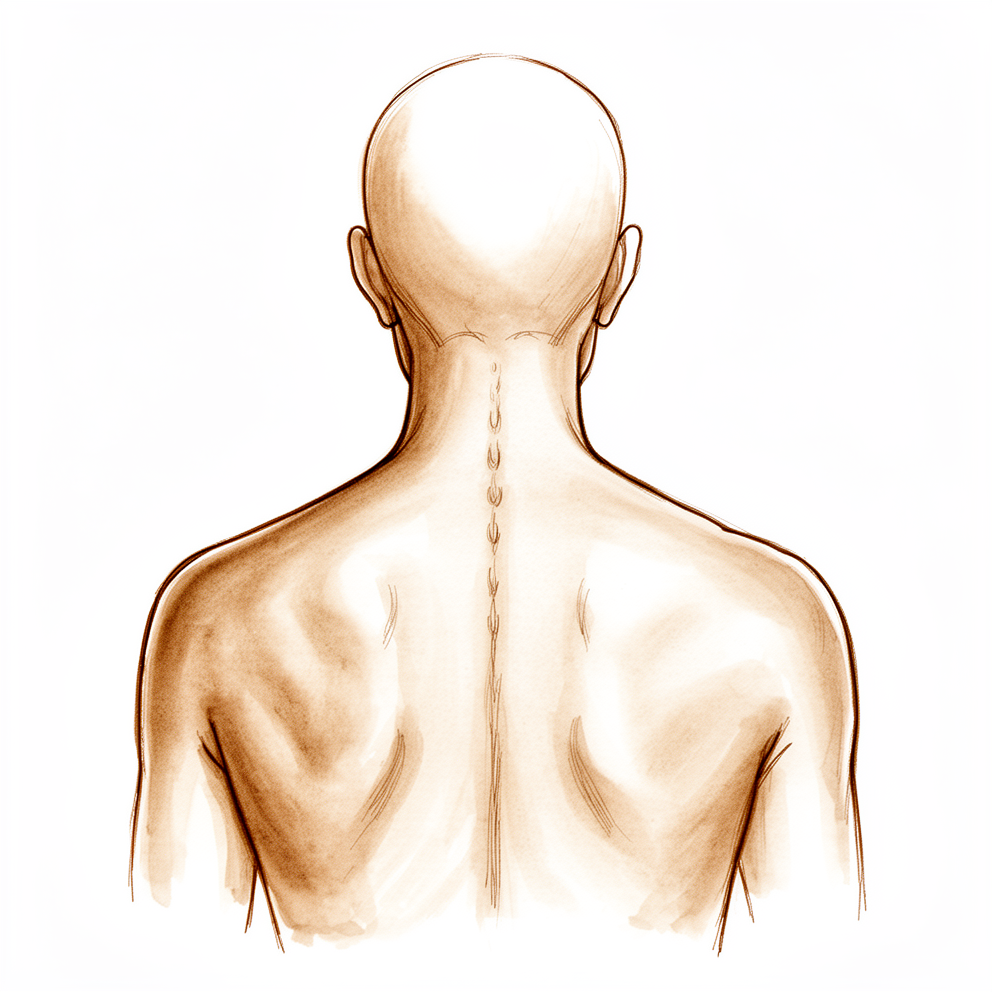
For patients: a plain-language version of this topic is available. See the patient guide.
Overview¶
Adult spinal deformity encompasses lateral curvatures in the frontal plane and sagittal plane abnormalities, including kyphosis and lordosis, which may be idiopathic, congenital, or secondary to neuromuscular disorders [13]. Scoliosis is defined by a lateral curve best appreciated on anteroposterior radiographs and physical examination, while kyphosis represents a forward curvature in the sagittal plane [13]. True scoliosis inherently includes a rotational component that may not be fully visible on radiographs, and curvatures are measured using the Cobb method to determine the degree of tilt between the most affected vertebral endplates [13]. The etiology varies widely; adolescent idiopathic scoliosis accounts for 80% of idiopathic cases, whereas neuromuscular conditions such as Duchenne muscular dystrophy and cerebral palsy typically present with long, sweeping C-shaped curves associated with pelvic obliquity [13, 2].
Surgical intervention is indicated for progressive deformity in the coronal or sagittal plane, neurological compression causing claudication or radiculopathy unresponsive to nonsurgical treatment, or disability preventing activities of daily living [25]. The primary goals of surgery include decompression of neural elements, reestablishment of coronal and sagittal balance, and restoration of horizontal gaze without compensatory mechanisms [25]. Reestablishment of global balance is prioritized over absolute Cobb angle correction [25]. Contraindications include cardiopulmonary comorbidities, profound osteoporosis that precludes durable fixation, or physical or mental conditions impairing surgical preparation or recovery [25].
Surgical outcomes are influenced by patient frailty and comorbidities, with patients over 50 facing a 44% complication rate and a 50% risk of requiring a second operation at six years [15, 18]. Proximal junctional kyphosis is a common complication ranging from asymptomatic recurrence to proximal junctional failure requiring revision [7]. While decompression alone may be sufficient for stable deformities with radiculopathy, it risks iatrogenic instability [25]. Long-segment fusion with deformity correction is indicated for severe subluxation or large curves, necessitating careful selection of the upper instrumented vertebra and consideration of distal fusion extent to S1 [25].
Anatomy & Pathophysiology¶
Embryology and Development¶
Spinal development initiates as the primitive streak deepens to form the primitive groove, which folds onto itself to create the neural tube [102]. During this process, the neural crest forms dorsally while the notochord forms ventrally [102]. The neural tube develops into the spinal cord, whereas the notochord gives rise to the vertebral bodies and intervertebral disks [102]. The neural crest ultimately forms the peripheral nervous system, spinal ganglia, and the sympathetic trunk [102]. Vertebrae develop from somites surrounding the notochord and neural tube, with each vertebra forming from the caudal portion of one sclerotome and the rostral portion of the subjacent level [102]. The intervertebral disk forms from the space between segments, with the nucleus pulposus deriving from notochordal cells and the anulus fibrosus developing from sclerotomal cells and mesenchymal stem cells surrounding the notochord [102].
Developmental failures result in specific congenital anomalies. Failure of the neural tube to close cranially causes anencephaly, while caudal failure results in spina bifida, meningocele, or myelomeningocele [102]. Failure of somite segmentation may produce block vertebrae or unsegmented bars, and failure of formation leads to congenital hemivertebrae [102]. Diastematomyelia is believed to result from the persistence of the neurenteric canal, present during the third and fourth weeks of gestation [104]. The most aggressive congenital scoliosis is associated with a hemivertebra on one side and an unsegmented bar on the other [104]. Klippel-Feil syndrome represents a defect of cervical segmentation characterized by congenital brevicollis [104].
Osseous Anatomy¶
The bony spine consists of 7 cervical, 12 thoracic, 5 lumbar, 5 fused sacral, and 4 or 5 fused coccygeal vertebrae [64]. The vertebral body is a cylindrical mass of bone connected by pedicles to the posterior arch, which comprises the lamina and spinous process [64]. The spinal canal is formed anteriorly by the vertebral body, posteriorly by the lamina, and laterally by the pedicles [64]. Vertebral bodies primarily bear weight and transfer forces to the pelvis and hips, while posterior elements protect neural structures and function as a tension band [64].
The thoracic spine represents two transitional zones: from the highly mobile cervical spine to the rigid thoracic region, and back to the more mobile lumbar spine [96]. In conjunction with ribs and sternum, the thoracic spine forms a bony "cube" that is inherently stable [96]. Thoracic vertebral bodies are larger than cervical but smaller than lumbar vertebrae [96]. Thoracic pedicles arise more superiorly from the posterior vertebral body than in the cervical or lumbar spine and project obliquely from superodorsal to inferoventral [96]. The spinal canal is narrowest in the thoracic region [96]. Spinous processes of the upper four thoracic vertebrae project horizontally with slight inferior angulation; in the midthoracic spine, they project sharply obliquely, overlapping inferior structures, while from T10 to T12 they transition to a horizontal projection consistent with lumbar vertebrae [96]. Superior articular facets project cranially from the junction of laminae and pedicles and are oriented coronally, while inferior facets are contiguous with the ventral aspect of the lamina [96]. This facet orientation permits only a small arc of motion [96]. Rib heads articulate with the lateral aspect of vertebral bodies, with a shared articulation at the disk space level where the rib head articulates with the superolateral aspect of the named vertebral body and the inferior aspect of the level above [96]. The first, eleventh, and twelfth vertebral bodies have only a single articulation for the same-numbered rib head [96]. Thoracic transverse processes project obliquely superolaterally, with the costotransverse joint located along the ventral aspect where the rib articulates with the same-numbered transverse process [96]. There is no costotransverse articulation at T11 or T12 [96].
Lumbar vertebral bodies are large, with a transverse diameter greater than the anterior-posterior diameter [103]. Lumbar pedicles arise from the superior aspect of the vertebral bodies and project more horizontally than thoracic pedicles [103]. L1 pedicles are minimally medially angled, while orientation becomes more medial as one progresses down the lumbar spine, particularly at L5 [103]. Lumbar transverse processes project more perpendicular relative to the vertebral body, with upper lumbar processes being large and flat, while L4 and L5 processes are often smaller [103]. Lumbar spinous processes are thick and project straight dorsally [103]. The superior articular facet arises at the junction of the pedicle and lamina and faces dorsomedially, while the inferior facet extends down from the lamina and nestles on the medial side of the superior facet [103]. The sagittal orientation of lumbar facet joints allows flexion and extension while providing resistance to axial rotation and translation [103].
Ligaments¶
The anterior longitudinal ligament is strong, thickest at the center of the vertebral body, and thinnest at the periphery [107]. It is characterized by separate fibers extending from one to five levels and resists hyperextension [107]. The posterior longitudinal ligament is weaker than the anterior longitudinal ligament and extends from the occiput (tectorial membrane) to the posterior sacrum [107]. It is separated from the center of the vertebral body by a space allowing passage of dorsal branches of the spinal artery and veins [107]. The posterior longitudinal ligament is hourglass-shaped, with wider yet thinner sections located over the discs [107]. Ruptured discs tend to be lateral to the expansions of the posterior longitudinal ligament, and ossification of this ligament is associated with increased risk of dural tears [107].
The ligamentum flavum is a strong yellow elastic ligament connecting the laminae, running from the anterior surface of the superior lamina to the posterior surface of the inferior lamina and constantly in tension [107]. Hypertrophy of the ligamentum flavum may contribute to nerve root compression [107]. Supraspinous, interspinous, and intertransverse ligaments form ligamentous capsules overlying the zygapophyseal joints [107]. Intertransverse ligaments contribute little to interspinous stability [107]. The supraspinous ligament lies dorsal to the spinous processes and begins at C7, in continuity with the ligamentum nuchae, which runs from C7 to the occiput [108]. The interspinous ligament lies between the spinous processes [108]. Integrity of the posterior ligamentous complex has implications for operative versus nonoperative treatment [108].
Spinal Cord Anatomy¶
Dorsal cells within the spinal cord are primarily sensory, while ventral cells are primarily motor [100]. The dorsal columns transfer vibration, deep pressure, and proprioception [100]. The lateral spinothalamic tract lies anterolaterally and transmits pain and temperature sensation, while the ventral spinothalamic tract transmits light touch [100]. Efferent voluntary motor function is transmitted along the lateral corticospinal tracts [100]. Fibers of the upper extremities are located deeper within the spinal column, with those related to the torso and lower extremities located sequentially more superficially [100].
At each level, anterior and posterior roots leave the cord separately then unite to form a common segmental nerve root with both motor and sensory components [100]. The dorsal root ganglion lies along the posterior root and is the point where peripheral sensory nerves synapse with afferent spinal nerves [100]. At birth, the conus medullaris lies around the L3 level, but by adulthood it lies around the L1-L2 level [100]. The neurologic level of the spinal cord does not necessarily correspond with the vertebral level [100]. There are 31 pairs of spinal nerves: 8 cervical, 12 thoracic, 5 lumbar, 5 sacral, and 1 coccygeal [100]. Lumbar, sacral, and coccygeal roots exit the cord at levels corresponding to the lower thoracic or upper lumbar vertebrae and form the cauda equina within the canal before exiting the spine [100].
Nerve roots exit the canal through the neuroforamen, formed by pedicles superiorly and inferiorly, the intervertebral disk anteriorly, and the facet joint and capsule posteriorly [100]. In the cervical spine, nerve roots exit above the same-numbered pedicle, with the eighth cervical nerve root exiting under the C7 pedicle [100]. From T1 distally, nerve roots exit the spine below the same-numbered pedicle [100].
A typical mixed spinal nerve has three distinct components: motor, sensory, and sympathetic [99]. Motor rootlets leave the anterolateral sulcus of the spinal cord and unite to form each motor root, with motor fibers arising from anterior horn cells and innervating skeletal muscles [99]. Sensory fibers arise from pain, thermal, tactile, and stretch receptors, with cell bodies located within the dorsal root ganglia [99]. Sensory axons enter the posterolateral sulcus of the cord via several rootlets [99]. Fibers conveying joint or position sensibility and some tactile fibers turn cephalad in the dorsal columns and do not synapse before reaching the gracile and cuneate nuclei at the cervicomedullary junction [99]. Pain and temperature fibers synapse in the substantia gelatinosa and cross to ascend in the dorsal spinothalamic tract, while tactile fibers enter, synapse, and cross to ascend in the ventral spinothalamic tract [99].
The sympathetic component of all 31 mixed spinal nerves leaves the spinal cord along only 14 motor roots [99]. Sympathetic cells of origin are in the intermediolateral cell column extending throughout the thoracic and upper lumbar cord segments [99]. Sympathetic fibers exit the cord with the 12 thoracic and first two lumbar motor roots [99]. White rami pass anteriorly to the corresponding sympathetic ganglion, and postganglionic fibers pass back to the mixed spinal nerve as a gray ramus [99]. Mixed spinal nerves receive their sympathetic component and promptly branch into anterior and posterior primary rami after leaving the intervertebral foramina [99]. Posterior primary rami supply the paraspinal musculature and the skin along the posterior aspect of the trunk, neck, and head, with the upper three cervical posterior rami being larger than their corresponding anterior rami [99]. Anterior primary rami of all cervical, the first thoracic, and all lumbosacral nerves join in the formation of plexuses [99]. The upper four cervical anterior rami form the cervical plexus, the lower four cervical and first thoracic anterior rami form the brachial plexus, the first three and a part of the fourth lumbar anterior rami form the lumbar plexus, and sacral anterior rami along with the fifth lumbar and a part of the fourth join to form the lumbosacral plexus [99]. The area of skin supplied by the fibers of a single spinal root is called a dermatome, with segmental dermatomal patterns well preserved in the thoracic region but not in the limbs [99].
Vascular Anatomy¶
Thoracic and lumbar levels are supplied by paired segmental arteries originating directly from the aorta along its posterior surface [98]. Segmental arteries run posteriorly along the midportion of the vertebral body, with branches supplying the vertebral body, paraspinal musculature, and spinal cord [98]. The cervical spine derives its circulation primarily from the vertebral arteries, which arise from the subclavian arteries and course superiorly [98]. Vertebral arteries typically enter the transverse foramen at the C6 level and run proximally through the transverse foramina to C1 [98]. They course posteriorly over the superior aspect of the C1 ring before turning proximally to enter the foramen magnum, where they merge to form the basilar artery [98]. Segmental branches to each cervical vertebra arise from the vertebral artery and the deep cervical branch of the costocervical trunk [98]. One side of the vertebral artery is typically more dominant, having a larger diameter than the other [98]. The vertebral artery may enter through the transverse foramen of C7 rather than C6, and anomalous courses, such as looping through a cervical vertebral body, are not uncommon [98].
The vascular supply of the spinal cord is primarily from medullary branches of the segmental spinal arteries [98]. Medullary branches merge to feed the anterior spinal artery, which supplies approximately 80% of the vascular supply to the spinal cord [98]. Three anterior medullary arteries typically supply the cervical region, one or two supply the thoracic region, and one typically supplies the lumbosacral spinal cord [98]. The arteria medullaris magna (AMM), also known as the arteria radicularis magna or artery of Adamkiewicz, is the largest anterior segmental artery [98]. The AMM typically arises on the left side anywhere between the T8 and L1 level, though right-sided origins are not uncommon [98].
The arterial supply to the spinal cord depends on three vessels: the anterior median longitudinal arterial trunk and a pair of posterolateral trunks near the posterior nerve rootlets [101]. Longitudinal arterial trunks are largest in the cervical and lumbar regions near ganglionic enlargements and much smaller in the thoracic region [101]. The metabolic demands of gray matter are greater than those of white matter [101]. There are 2 to 17 anterior medullary feeder (radicular) arteries and 6 to 25 posterior medullary feeder arteries [101]. Vertebral arteries supply 80% of the radicular arteries in the neck, while arteries in the thoracic and lumbar areas arise from the aorta [101]. The lateral sacral, fifth lumbar, iliolumbar, and middle sacral arteries are important in the sacral region [101]. Vertebral and posterior inferior cerebellar arteries are important sources of arterial supply to the spinal cord [101]. Sacral medullary feeders arise from lateral sacral arteries and accompany the distal roots of the cauda equina [101]. Segmental arteries divide into numerous branches at the intervertebral foramen, termed the distribution point [101]. A second anastomotic network lies within the spinal
Classification¶
General Definitions and Etiology¶
Adult spinal deformity is defined as a coronal Cobb angle of greater than 10° in a skeletally mature person [71]. These deformities are often three-dimensional, with abnormalities present in the coronal, sagittal, and axial planes [71]. The primary categories of adult spinal deformity are degenerative (de novo) and idiopathic [71]. Scoliosis is a lateral spinal curvature in the frontal plane that always includes a rotational component and generally includes a lordotic component [13]. Kyphosis is a forward (flexed) curvature of the spine in the sagittal plane, which may present with a posterior prominence called a gibbus if acutely angular [13].
Congenital spinal deformities result from defects of formation or segmentation [20]. The natural history and treatment of these conditions correspond to the three major patterns of lordosis, kyphosis, and scoliosis [20]. Congenital kyphosis is classified into three types: failure of formation (type I), failure of segmentation (type II), and mixed abnormalities (type III) [2, 3]. Type I congenital kyphosis (failure of formation) is the most common type, carries the worst prognosis, and presents the highest risk for neurologic complications [2, 3].
Congenital Scoliosis Classification¶
Progression risk in congenital scoliosis is highest for a unilateral unsegmented bar with a contralateral hemivertebra, characterized by rapid and relentless progression [2, 3]. A unilateral unsegmented bar alone poses a high risk for rapid progression [2, 3]. A fully segmented hemivertebra is associated with steady progression [2, 3]. A partially segmented hemivertebra is associated with less rapid progression, with curves usually less than 40 degrees at maturity [2, 3]. An incarcerated hemivertebra may slowly progress [2, 3]. A nonsegmented hemivertebra is associated with little progression [2, 3].
Neuromuscular Scoliosis Classification¶
Neuromuscular scoliosis is characterized by long, sweeping C-shaped curves and associated pelvic obliquity [2, 3]. This condition can be rapidly progressive, especially in patients who use a wheelchair [2, 3].
Adult Spinal Deformity Classification Systems¶
SRS–Schwab: The Scoliosis Research Society (SRS)–Schwab classification system describes coronal curve type plus sagittal modifiers and reflects the radiographic severity of the deformity [71].
Cervical Spinal Deformity: A proposed classification system for cervical spinal deformity includes a deformity descriptor defining the apex of the deformity and five modifiers [71].
King: The current two-dimensional King classification is inadequate for describing spinal deformities in three dimensions due to significant variability in sagittal configurations within each class [109].
T4-L1-Hip Axis: The T4-L1-Hip axis is conceptually aligned with the description of spinal shapes in the Roussouly classification but utilizes continuous measures of spinal alignment [87].
Roussouly: The Roussouly classification provides only a rough estimate of optimal spinopelvic alignment [167].
Degenerative Thoracolumbar Kyphosis: A novel classification for degenerative thoracolumbar kyphosis based on sagittal spino-pelvic alignment includes four types followed by targeted treatment strategies [133].
Thoracic Adolescent Idiopathic Scoliosis: The sagittal alignment of thoracic adolescent idiopathic scoliosis patients can be classified into three types based on sacral slope and maximum lumbar lordosis [175].
Lenke 1: A new three-dimensional classification for Lenke 1 adolescent idiopathic scoliosis can identify subtypes without the need for quantitative 3D image post-processing [176].
Trauma and Fracture Classification¶
AO/OTA: The AO Classification system has largely replaced the contemporary TLICS and Denis classification scales for thoracolumbar injuries [38]. Spinal fractures, dislocations, and fracture-dislocations are classified using the comprehensive AO/Orthopaedic Trauma Association classification system, which is based on an alphanumeric classification [48]. The AO thoracolumbar injury classification system categorizes injury morphology into type A (compression injuries), type B (tension band injuries), and type C (displacement/translational injuries) [38]. In the AO thoracolumbar classification, Type A and Type B injuries are subclassified more granularly [38].
TLICS: The TLICS scale assigns 1 point for compression morphology, +1 for burst, 3 for translation/rotation, and 4 for distraction [38]. It assigns 0 points for an intact posterior ligamentous complex, 2 points for suspected/indeterminate disruption, and 3 points for injury [38]. The scale assigns 0 points for intact neurologic status, 2 points for nerve root or complete cord conus medullaris injury, and 3 points for incomplete cord conus medullaris or cauda equina injury [38].
ASIA: The American Spinal Injury Association (ASIA) Impairment Scale classifies spinal cord injuries into complete (ASIA grade A) or incomplete (ASIA grades B-D), with ASIA grade E reflecting a normal neurologic status [48].
Other Fracture Classifications: A reliable classification for assessing the stability of a healed vertebra after thoracolumbar burst fractures has been developed [125]. A new morphological classification system for chronic symptomatic osteoporotic thoracolumbar fracture demonstrated excellent reliability in initial assessment [177].
Other Considerations¶
In thoracolumbar disc herniation, type I involves accelerated disc degeneration by regional kyphosis, while type II involves excessive mechanical stress directly loaded at the thoracolumbar apex [35]. Patients with adult spinal deformity demonstrate a distinct Coronal Plane Alignment of the Knee (CPAK) profile [41].
Clinical Presentation¶
History and General Assessment¶
Spinal curvature presents with variable findings and may be idiopathic, congenital, or associated with neuromuscular disorders, tumors, and infections [13]. Because spinal curvature is sometimes the first clue to important underlying disease, a comprehensive history is essential [13]. The history must include symptoms such as changes in body habitus indicating progressive deformity, gait disturbance due to myelopathy or claudication, and axial or radicular pain [70]. Comorbidities, including bone mineral density and neuromuscular disease, must be documented [70]. Prior surgeries, including decompressions and associated complications, should be reviewed [70]. The patient's normal activity level and any changes to that level should be determined [166]. The degree to which spinal deformity or pain interferes with physical activities should be assessed [166]. Neurologic symptoms such as radiating pain or loss of bowel or bladder control require investigation [166]. Family history of scoliosis, connective tissue disease (e.g., Marfan syndrome, neurofibromatosis), or neuromuscular disease (particularly muscular dystrophy) should be obtained [166].
Physical Examination¶
Physical examination begins with an assessment of patient stance for trunk shift, shoulder or pelvic asymmetry, and overall coronal and sagittal alignment [70]. The examiner should check the level of the pelvis and look for obvious asymmetry of the rib, scapula, neck, and shoulder height [13]. A plumb line held over the base of the occiput or the C7 spinous process can aid in clinical assessment of trunk shift [166]. The patient is examined from behind while standing evenly on both feet with knees straight to look for waistline, scapular, or paraspinal asymmetry [166]. The level of the posterior sacral dimples is checked to ensure leg length inequality is not creating apparent scoliosis [166]. The relative position of the scapulae on the posterior chest wall is determined to rule out an associated or isolated Sprengel deformity [166].
Scoliosis is a lateral spinal curvature in the frontal plane, best appreciated by physical examination from the patient’s back and by anteroposterior radiographs [13]. Lateral curvature is often undetected externally, and the rotation of vertebrae is the physical feature that allows clinical detection [13]. The patient should bend forward to note any asymmetric prominence of the lumbar paraspinous muscle, rib cage, or scapula, which suggests the rotational portion of scoliosis [13]. Kyphosis is a forward (flexed) curvature of the spine in the sagittal plane, best appreciated from the side and by lateral radiographs [13]. Kyphosis is assessed from the side for prominence of the spine in both upright and forward-bending positions [13].
Supine positioning is used to evaluate sagittal plane rigidity and hip flexion contractures via the Thomas test [70]. Leg length discrepancy and pelvic obliquity should be evaluated during physical examination [70]. The neck range of motion should be checked while looking for evidence of facial, neckline, or scapular asymmetry [166]. A high-arched palate may be seen in patients with Marfan syndrome [166]. Upper extremities should be examined for restricted range of motion and muscle wasting, which may indicate peripheral neuropathy or atrophy caused by syringomyelia [166]. Finger lengths should be checked for signs of arachnodactyly, an indication of Marfan syndrome [166].
Skin over the spine is inspected for pigmented spots, hairy patches, and deep pits that might overlie external openings of sinus tracts extending to the spinal cord [166]. Café au lait spots and neurofibromata should be noted during skin inspection [166]. Flattening of the buttocks with apparent loss of lumbar lordosis may indicate the presence of spondylolisthesis [166]. Defects of the vertebral bodies may be palpated by running fingers along the spine to detect stiff curvature or defects in the spinous process [166]. The spine should be percussed for areas of tenderness if the patient complains of pain [166].
The patient should walk on heels and toes and hop on each foot to provide an indication of general strength, muscle tone, and coordination [166]. Formal testing of joint range of motion, muscle strength, and reflexes is performed with the patient on the examination table [166]. The straight-leg raising test is performed during the physical examination [166]. Abdominal reflexes should be checked for asymmetry and hypesthesia to light touch in the concavity of the deformity if scoliosis and possible syringomyelia are present [166]. A complete neurological examination is required [70].
Radiographic Evaluation¶
Radiographs are used to assess type, severity, and location of the curvature and to look for underlying lesions [13]. AP and lateral 36-inch cassette views are used to visualize the entire spine, iliac crests, and clavicles [70]. AP and lateral whole-body views are used to visualize deformity compensatory mechanisms such as pelvic retroversion, hip flexion, and knee flexion [70]. Upright and supine films are used to assess flexibility [70].
Sagittal plane deformity is a prime driver of disability [70]. Key sagittal parameters include: C7 sagittal vertical axis (C7 SVA): The horizontal distance between the C7 plumb line and the posterior superior aspect of S1, with a normal range of <5 cm [70]. T1 pelvic angle (T1PA): The angle drawn from the center of T1, to the center of the femoral heads, then to the midpoint of the S1 end plate, with a surgical goal of <14° [70]. Lumbar lordosis (LL): The T12–S1 or L1–S1 sagittal Cobb angle, with a normal value around 60° [70]. Loss of lumbar lordosis correlates with pain and functional disability [70]. Pelvic incidence (PI): The angle subtended by a perpendicular from the midpoint of the S1 end plate and a line to the center of the femoral heads [70]. Pelvic incidence is a constant, anatomic parameter that is independent of pelvic positioning [70]. It is intimately related to lumbar lordosis, with a surgical goal of lumbar lordosis within 10° of pelvic incidence [70]. Pelvic tilt (PT): The angle drawn from the midpoint of the S1 end plate, to the center of the femoral heads, then to the vertical, with a normal range of <20° [70]. Pelvic tilt varies with pelvic position and increasing pelvic tilt is a compensatory mechanism for sagittal plane malalignment [70]. Sacral slope (SS): The angle subtended by the superior end plate of S1 and the horizontal [70]. Sacral slope varies with pelvic position and Pelvic incidence equals Pelvic tilt plus sacral slope (PI = PT + SS) [70].
Coronal and cervical parameters include: Coronal alignment (CA): The horizontal distance between the C7 plumb line and a vertical line drawn from the center of the sacrum, with a surgical goal of <4 cm [70]. Fractional curve: The angle between the lower end vertebra of the lumbar curve (usually L4) and the superior end plate of S1, which is key to overall coronal alignment [70]. Cervical lordosis (CL): The C1–C7 or C2–C7 sagittal Cobb angle, with an average of around −40° in patients without cervical spine deformity [70]. Cervical hyperlordosis may be compensating for sagittal malalignment of the remaining spine [70]. Cervical sagittal vertical axis: The sagittal horizontal distance between the C2 plumb line and the posterior superior margin of C7, with a normal range of <40 mm [70]. T1 slope (T1S): The sagittal angle between the superior end plate of T1 and the horizontal drawn from the superior end plate of T1 [70]. The normal range of T1S minus C2–C7 cervical lordosis is <15° [70]. If T1S is high, full spine radiographs should be obtained to check for concomitant thoracolumbar deformity [70]. Chin-brow vertical angle (CBVA): The sagittal angle formed between a line drawn between the chin and eyebrow, and the vertical drawn from the eyebrow, with a normal range of −10° to +20° [70].
MRI assesses central canal and foraminal stenosis, facet hypertrophy, spinal cord and neural compression, and degenerative disk disease [70]. CT assesses bony anatomy better than MRI and may be useful for evaluating pedicle anatomy and planning surgical fixation [70]. Dual-energy radiograph absorptiometry measures bone density [70].
Specific Etiologies and Conditions¶
In Noonan syndrome, spinal deformities tend to develop early and are relatively severe, requiring clinical and radiographic assessment with careful follow-up for early detection [1]. Congenital kyphosis type I (failure of formation) is the most common type, has the worst prognosis, and carries the highest risk for neurologic complications [2]. Severe congenital kyphosis type I is an immediate indication for surgery [2]. Congenital kyphosis type II (failure of segmentation) can be monitored to document progression, but progressive curves should be fused posteriorly [2]. Congenital kyphosis type III involves mixed abnormalities [2].
In congenital scoliosis, unilateral unsegmented bar with contralateral hemivertebra poses the highest risk for progression, characterized by rapid and relentless curve progression [2]. Unilateral unsegmented bar poses a high risk for progression, characterized by rapid curve progression [2]. Fully segmented hemivertebra in congenital scoliosis is associated with steady curve progression [2]. Partially segmented hemivertebra in congenital scoliosis is associated with less rapid progression, with curves usually <40 degrees at maturity [2]. Incarcerated hemivertebra in congenital scoliosis may slowly progress [2]. Nonsegmented hemivertebra in congenital scoliosis has little progression [2].
Neuromuscular scoliosis typically presents with long, sweeping C-shaped curves and associated pelvic obliquity [2]. Most patients with neuromuscular scoliosis have some pulmonary involvement secondary to the underlying condition and detrimental contribution from the scoliosis [2]. Bracing for neuromuscular scoliosis is controversial and not typically used, but may be used to delay surgical treatment [2]. In Duchenne muscular dystrophy, bracing is ineffective for scoliosis [2]. In Friedrich ataxia, bracing is ineffective for scoliosis [2]. In spinal muscular atrophy, bracing is useful to delay fusion in young patients with curves between 25 and 45 degrees [2]. In spina bifida (myelomeningocele), bracing is useful to delay fusion in young patients with curves between 25 and 45 degrees [2]. In cerebral palsy, bracing is ineffective for scoliosis [2]. In neurofibromatosis, bracing is used for nondystrophic curves between 25 and 40 degrees [2]. In arthrogryposis, bracing is ineffective for scoliosis [2]. Patients with WBC counts less than 1500 cells/µL and albumin levels lower than 3.5 g/dL have higher infection rates and longer hospital stays in the context of neuromuscular scoliosis evaluation [2].
Surgical indications for neuromuscular scoliosis vary by condition: Duchenne muscular dystrophy: Surgery is indicated when the curve is progressive and more than 25 to 30 degrees in patients whose forced vital capacity is affected [2]. Friedrich ataxia: Fusion is indicated if the curve is >50 degrees or progressive [2]. Spinal muscular atrophy: Fusion is indicated if the curve is >50 degrees or progressive [2]. Spina bifida (myelomeningocele): Fusion is indicated if the curve is >50 degrees or progressive [2]. Cerebral palsy: Surgery is indicated for >50 degrees in ambulatory patients, progressive curves >50 degrees in communicative and aware patients, or curves interfering with seating and nursing with family desire for surgery [2]. Neurofibromatosis: Fusion is indicated if the curve is >40 degrees or progressive [2]. Arthrogryposis: Fusion is indicated if the curve is >50 degrees or progressive [2].
Scoliosis in familial dysautonomia is managed operatively, with correction and stabilization considered beneficial despite results [4]. Late kyphotic deformity was noted in 50 per cent of patients with anterior decompression of burst fractures with neurological deficit [6]. In rare cases of severe congenital scoliosis deformity, Harrington instrumentation may be used to gain correction provided a preoperative myelogram excludes spinal cord anomalies and cord function is monitored [8]. In scoliosis caused by benign osteoblastoma, correction and stabilization may be required in the event of late diagnosis with structural changes or spinal instability due to the extent of excision [9]. Early recognition of the diagnosis and early spine fusion are recommended treatment for congenital thoracic lordosis, which is a severe and progressive deformity [11]. Severity of kyphosis at initial presentation may predict progression of thoracolumbar deformity in patients with mucopolysaccharidoses [12]. Congenital spinal deformities are caused by defects of formation or segmentation, with the natural history and treatment corresponding to the three major patterns of lordosis, kyphosis, and scoliosis [20]. Cervical kyphosis in diastrophic dwarfism, though rare, can be severe and require surgical fusion [24]. While progression of angular deformity often occurs after rod removal in thoracic and lumbar spine fractures, most patients had few or no complaints despite increasing kyphosis [27]. Early identification of patients likely to develop severe deformity in tuberculosis of the spine allows for radical excision and bone-grafting to prevent severe kyphosis [30]. Successful management of idiopathic scoliosis requires understanding the etiology, natural history, evaluation, and available nonsurgical and surgical management options [31]. cMIS provides for good clinical and radiographic outcomes for moderate (30°–75°) adult idiopathic scoliosis [32]. Despite the increase in angular deformity in juvenile and idiopathic kyphosis, all patients managed fairly well with only two complaining of distressing back pain [33]. Solid fusion was achieved in all but one patient, and no patient had residual spinal deformity following Harrington instrumentation and spine fusion [33].
Investigations¶
General Spinal Deformity Assessment¶
Physical examination serves as the primary method for detecting spinal curvatures, while radiographs assess the type, severity, and location of deformity [13]. The Cobb method measures spinal curvature by determining the degree of tilt between the most affected vertebral endplates [13]. Bending radiographs distinguish structural curvatures from flexible compensations [13]. A thorough neurologic examination, including upper and lower extremity reflexes, is part of the standard spinal examination protocol [13]. Spinal curvature may be the first clue to important underlying disease [13]. In Noonan syndrome, clinical and radiographic assessment with careful follow-up is recommended for early detection and treatment [1].
Imaging Modalities¶
Plain radiography: Plain radiographs have limited utility in the acute setting for spinal trauma due to high false-negative and false-positive rates [113]. When the segmental sagittal angle on plain radiography exceeds 17°, a significant discrepancy may exist between plain radiography and supine computed tomography for thoracolumbar fractures [217].
CT: CT remains the most useful advanced imaging technique for spinal trauma due to inherent bone contrast and unmatched spatial resolution [147]. Multidetector CT has a sensitivity of 97% to 100% for detecting cervical spine injuries in patients with neck tenderness and pain [113]. Whole-body CT scans with two-dimensional and three-dimensional reconstructions have largely replaced conventional radiographs for the initial assessment of multiply injured patients [48]. Postoperative computed tomography is indispensable for clarifying spinal canal width, position of posterior wall fragments, and exact fracture morphology in thoracolumbar spine fractures [236]. CT scan provides better delineation of bony retropulsion, subchondral sclerosis, and vertebral end plate erosion than MRI in vertebral osteomyelitis [46]. CT scans were not generally used to evaluate fusion in minimally invasive surgical techniques for adult degenerative spinal deformity [53].
MRI: MRI is helpful in patients with suspected spinal cord injury, epidural hematoma, or traumatic disc herniation [147]. MRI can reliably identify ligamentous injuries of the cervical spine that may not be clearly identifiable on multidetector CT [113]. MRI with and without gadolinium contrast is indicated for all patients with suspected osteomyelitis of the spine to assess local spread, epidural abscess, and chronicity [46]. Gadolinium-enhanced MRI allows differentiation between epidural abscess and cerebrospinal fluid in spinal infections [81]. MRI is sensitive for detecting spinal infection and specific in differentiating infection from tumor [81].
Bone scan: Bone scanning is sensitive for detecting a destructive process in pyogenic vertebral osteomyelitis [81].
Specific Pathology Imaging¶
In symptomatic thoracolumbar disc herniation, type I disc degeneration is accelerated by regional kyphosis, while type II involves excessive mechanical stress at the thoracolumbar apex [35]. Asymptomatic radiologic cervical and thoracic stenosis is common in elderly patients with symptomatic lumbar stenosis [228]. Cervical kyphosis is among the most prevalent morphological alterations in patients with adolescent idiopathic scoliosis and is closely associated with curve severity [42]. Vertebral wedging is present in mild scoliosis and increases as the scoliosis progresses [69]. The pattern and sequence of vertebral and intervertebral disc wedging are related to the location of the curve rather than the presence of curve progression in adolescent idiopathic scoliosis [233]. Apical wedging and coronal imbalance may identify patients with adolescent idiopathic scoliosis suited for closer monitoring and early spinal fusion [231].
In diastrophic dwarfism, cervical spina bifida occulta is universal, and cervical kyphosis can be severe and require surgical fusion [24]. In congenital thoracic lordosis, early recognition of the diagnosis is recommended for treatment planning [11]. In rare cases of severe congenital scoliosis, a preoperative myelogram is used to exclude spinal cord anomalies before Harrington instrumentation [8]. In scoliosis caused by benign osteoblastoma, correction and stabilization may be required if late diagnosis reveals structural changes or spinal instability [9].
Proximal junctional kyphosis is a common complication following adult spinal deformity surgery, ranging from asymptomatic deformity recurrence to proximal junctional failure [7]. Late kyphotic deformity was noted in 50% of patients with anterior decompression of burst fractures with neurological deficit [6]. While progression of angular deformity often occurs after rod removal for thoracic and lumbar spine fractures, most patients had few or no complaints despite increasing kyphosis [27]. Solid fusion was achieved in all but one patient treated with Harrington instrumentation and spine fusion for unstable thoracic and lumbar spine fractures, with no residual spinal deformity [34].
Correcting spinal deformity in severe early-onset scoliosis permitted longer-term improvement in pulmonary function [36]. Combined anterior-posterior surgery is effective for correcting osteoporotic spinal deformities with global sagittal imbalance, with 94% of patients reporting subjective improvement at 2 years [26]. cMIS provides good clinical and radiographic outcomes for moderate (30°–75°) adult idiopathic scoliosis [32]. Surgical stabilisation of the spine can be reserved for severe progressive deformities unresponsive to conservative treatment in infantile developmental thoracolumbar kyphosis [51]. Early identification of patients likely to develop severe deformity in spinal tuberculosis allows for radical excision and bone-grafting to prevent severe kyphosis [30].
The cervical spine morphology of asymptomatic adolescents varies widely, from lordotic to kyphotic [94]. Spinal RDOA is characterized by quickly developing edema of the vertebral endplates followed by destruction of the disk space within months from the first diagnosis [237]. Radiologically visible fusion has a minimal effect on correction loss after antero-posterior spondylodesis of traumatic vertebral body fracture [238]. A novel pre-contoured V-shaped rod in one-level pedicle subtraction osteotomy obtains satisfactory radiographic and clinical outcomes for ankylosing spondylitis patients without necessitating additional surgery [239]. MIAS leads to good clinical results with minimal spine-related impairment at the latest follow-up for unstable incomplete burst fractures of the thoracolumbar junction [234]. A short lumbosacral curve accompanied by a long thoracic or thoracolumbar curve toward the opposite side and a relatively straight sagittal profile have been noted in adolescents with lumbar disc herniation presenting with scoliotic posture [235]. Sagittal parameter changes from underarm bracing for adolescent idiopathic scoliosis do not appear to be associated with worse quality of life [224]. Despite significant changes during skeletal maturity, modifications in spinal curvatures are not large enough to impact surgical planning [61].
Congenital spinal deformities are caused by defects of formation or segmentation, with natural history and treatment corresponding to patterns of lordosis, kyphosis, and scoliosis [20]. The purpose of the review on spinal manifestations of skeletal dysplasia was to provide a practical guide for diagnosing skeletal dysplasia by recognizing spinal manifestations [67]. This case highlights the importance of attention to the sagittal plane and patient symptoms in adolescents with congenital thoracic kyphoscoliosis and compensatory thoracolumbar lordosis [72]. This rare case involving multiple complex spinal conditions provides valuable insights into the development of thoracic myelopathy [74]. Correction and stabilization of spinal deformity in patients with familial dysautonomia is considered beneficial by the authors [4].
Treatment¶
Non-Operative¶
Nonsurgical treatment for adult spinal deformity is associated with neither improvement nor worsening of patient function [80]. However, health-related quality of life (HRQOL) improved more by surgical treatment than by nonsurgical treatment [80]. For specific etiologies such as Duchenne muscular dystrophy, Friedrich ataxia, cerebral palsy, and arthrogryposis, bracing is generally ineffective [2, 79]. In arthrogryposis, bracing is commonly believed to be ineffective because curve magnitudes often exceed 30 degrees at prescription in nonambulatory patients [79]. Conversely, bracing may be useful for controlling compensatory curves and delaying surgery in congenital scoliosis [28, 29], and for delaying fusion in young patients with spinal muscular atrophy or spina bifida who have curves between 25 and 45 degrees [2]. For neurofibromatosis, bracing is used for nondystrophic curves between 25 and 40 degrees [2]. Conservative treatment should be the first-line treatment for most cases of early-onset scoliosis because it has fewer complications associated with it than surgery [86].
Operative¶
Indications: Indications for surgical treatment of adult spinal deformity include inability to perform activities of daily living due to deformity-associated disability, progressive deformity in the coronal or sagittal plane, and neurological compression causing claudication or radiculopathy not responsive to nonsurgical treatment [25]. Contraindications include cardiopulmonary conditions or associated comorbidities, profound osteoporosis that may prevent durable and effective fixation, and physical or mental conditions that would impair surgical preparation or recovery [25]. For late posttraumatic deformity, surgery is recommended for patients with significant pain, progressive neurologic deficits, or postural difficulties in the presence of focal kyphosis of 20 degrees or greater [55]. In patients with myelomeningocele, spinal fusion should be considered for curves greater than 55 degrees unless the patient is a community ambulator [17]. For Duchenne muscular dystrophy, surgery is indicated early at 25–30 degrees to delay pulmonary function deterioration [2]. For Friedrich ataxia, fusion is indicated if the curve is >50 degrees or progressive [2]. For spinal muscular atrophy and spina bifida, fusion is indicated if the curve is >50 degrees or progressive [2]. For cerebral palsy, surgery is indicated for >50 degrees in ambulatory patients, progressive curves >50 degrees in communicative and aware patients, or curves interfering with seating and nursing with family desire for surgery [2]. For neurofibromatosis, fusion is indicated if the curve is >40 degrees or progressive [2]. For arthrogryposis, fusion is indicated if the curve is >50 degrees or progressive [2].
Surgical Approach / Technique: The goals of surgery are decompression of involved neural elements, reestablishment of coronal and sagittal balance, and reestablishment of horizontal gaze without compensatory mechanisms [25]. Reestablishment of coronal and sagittal balance is considered more important than absolute Cobb angle correction [25]. Decompression alone is indicated for patients presenting with radiculopathy, stable deformity, and central or lateral recess stenosis, but it could result in increased postoperative deformity or iatrogenic instability [25]. Decompression with limited fusion is indicated for patients with the same indications as decompression alone but who show apical progression on radiographs, have a symptomatic lumbosacral fractional curve, or cannot undergo a long fusion [25]. Decompression and long fusion with deformity correction is indicated for lumbar degenerative scoliosis, large scoliosis curves, and severe subluxation of the apical vertebra [25]. In the setting of mild degenerative scoliosis (<30°), decompression and limited fusion of the decompressed segments appears to be a more effective treatment for patients 65 years or older with symptoms of stenosis [49].
Osteotomy selection depends on the required correction. Schwab Grade 2 osteotomy involves complete facet joint resection and provides approximately 10° of correction per level [25]. Schwab Grades 3 and 4 osteotomies involve pedicle and partial body resection and provide 25° to 35° of correction per level [25]. Schwab Grades 5 and 6 osteotomies involve complete vertebra and disc resection (vertebral column resection) and allow high biplanar angular correction [25]. Smith–Peterson or Ponte osteotomies may achieve correction of approximately 10 to 15 degrees per level and require a mobile anterior intervertebral segment [55]. A pedicle subtraction osteotomy may provide correction of more than 30 degrees and is very effective in the case of a deformity with a rigid anterior column, as in malunited burst fractures [55].
Minimally invasive and specific technique outcomes vary by condition. Single-staged posterior minimally invasive surgery effectively corrects global alignment in adult spinal deformities, yielding positive clinical outcomes with a low re-operation rate [68]. Circumferential minimally invasive surgery provides good clinical and radiographic outcomes for moderate (30°–75°) adult idiopathic scoliosis [32]. Vertebral column decancellation is a safe and effective method in treating rigid kyphotic deformity secondary to ankylosing spondylitis [116]. Posterior vertebral column resection effectively improves the kyphosis correction rate for thoracolumbar spinal tuberculous angular kyphosis without increasing perioperative complications [121]. Transpedicular bi-vertebrae wedge osteotomy is a safe and effective treatment option for post-tubercular spinal deformity [149]. Modified bone-disc-bone osteotomy demonstrates stability of correction effect and clinical efficacy for treating kyphosis caused by old thoracolumbar vertebral fractures [131]. Surgical treatment of severe congenital thoracolumbar kyphosis through a single posterior approach is feasible, safe, and effective [110]. For congenital kyphosis, posterior arthrodesis was highly effective, giving better eventual correction than when both anterior and posterior arthrodesis was done [76]. Anterior spinal fusion achieved satisfactory deformity correction with high perioperative complication rates, but no long-term sequelae among children with high level myelomeningocele [47]. Surgical treatment of neuropathic scoliosis achieves effective and permanent morphologic correction [132]. Early outcome studies show that growth modulation with vertebral body tethering is safe, can achieve good results, and preserve motion in select patients [136].
Implant Selection: Use of recombinant human bone morphogenetic protein-2 is associated with reduced risk of pseudarthrosis [80].
Alignment / Balancing Strategy: The proximal construct should not stop within kyphotic regions [25]. Surgical risk factors for proximal junctional kyphosis or failure include posterior soft-tissue injury, combined anterior-posterior fusion, fusion to the sacrum or ilium, and thoracoplasty [25]. Techniques for preventing proximal junctional kyphosis or failure include preserving supra-adjacent facets and ligaments, reducing instrumentation stiffness, augmenting the UIV or UIV+1 with cement, and choosing age-appropriate spinopelvic alignment goals [25]. Extending distal fusion to S1 rather than stopping at L5 is controversial; advantages include improved maintenance of correction and avoidance of subsequent L5–S1 disk degeneration, while disadvantages include increased operative time, complication rate, revision rate, and risk of pseudarthrosis [25]. Indications for extending fusion to S1 include existing pathology or symptomatic degenerative changes at L5–S1, L5 involvement in the fractional curve with lumbosacral obliquity, or incomplete correction of global sagittal imbalance [25].
Pain Management: No specific analgesic regimens are detailed in the provided evidence.
Adjuncts: Intraoperative neurological monitoring is mandatory in adult spinal deformity surgeries [80]. Tranexamic acid is associated with reduced intraoperative blood loss [80]. Intrawound vancomycin powder may reduce the rate of surgical site infection [80].
Setting of Care: No specific outpatient versus inpatient setting data is provided.
Revision: Despite a high complication rate of 51 percent, osteotomy of the fusion mass may be indicated in patients with a previously fused but progressively unbalanced, painful deformity of the spine [60].
Other Considerations: Overall complication rates for surgical treatment of adult spinal deformity are 72%, with radiographic complications at 29%, neurological at 28%, implant at 26%, surgical at 26%, cardiopulmonary at 22%, and infection at 15% [80]. Perioperative complication rates are 44%, with surgical complications at 26%, cardiopulmonary at 20%, neurological at 16%, infection at 11%, and gastrointestinal at 9% [80]. Late complication rates are 54%, with radiographic complications at 24%, implant at 23%, neurological at 12%, infection at 5%, and cardiopulmonary at 2% [80]. For complex adult spinal deformity surgery patients, neurologic and nonneurologic complications predict lower 2-year health-related quality of life [80].
Early complications for cervical deformity surgical treatment occur in 44% of cases, including dysphagia (12%), C5 palsy (6%), surgical site infection (6%), respiratory failure (5%), and mortality (1%) [80]. Long-term complications for cervical deformity surgical treatment include major medical complications in 3% to 44% of cases, neurological complications around 14%, and mortality in 3% to 7% [80]. Cervical deformity surgical treatment has a high patient satisfaction rate despite complications [80].
Specific etiology considerations include the following. One-stage posterior spinal fusion resulted in superior correction and fewer complications than growing rod treatment in 9- to 11-year-old children with congenital scoliosis, with similar health-related quality of life scores at final follow-up [50]. Surgical treatment of congenital upper thoracic scoliosis is viable as a primary treatment modality, though there is a high risk of complications, especially distal adding-on [58]. In patients with myelomeningocele, spinal fusion to the pelvis should be delayed until the patient becomes largely wheelchair-reliant or the curve is significantly worse if they are community ambulators [17]. Patients with myelomeningocele who undergo spinal surgery are particularly likely to experience peri- and postoperative complications, including pressure sores, urinary tract infections, wound breakdown, deep infections, pseudarthrosis, and progression of the deformity [17]. Patients with WBC counts less than 1500 cells/µL and albumin levels lower than 3.5 g/dL have higher infection rates and longer hospital stays [2].
In arthrogryposis, patients younger than 8 years with early-onset deformity are managed with techniques that preserve spinal and thoracic growth, while patients older than 10 years undergo conventional spinal fusion with instrumentation [79]. In arthrogryposis, fusion to the pelvis must take into consideration how sitting will be accommodated postoperatively, with hip releases or flexion osteotomies planned as necessary [79]. Some arthrogryptic patients with thoracic insufficiency may be best treated with expansion thoracoplasty procedures because of the severity of the respiratory compromise [79].
Surgery is effective in correcting kyphosis and providing sustained improvements over time for burst fractures in children and adolescents [16]. Solid fusion was achieved in all but one patient, and no patient had residual spinal deformity following Harrington instrumentation and spine fusion for unstable fractures and fracture-dislocations of the thoracic and lumbar spine [34]. Late posttraumatic deformity is the most common long-term complication associated with fractures of the spinal column [55]. The primary aim of surgical treatment for kyphotic deformity is to restore normal sagittal balance, decompress neurologic structures, and stabilize the spine to improve pain, posture, and neurologic symptoms [55]. The overall complication rate is approximately 10% when correcting posttraumatic deformities [55]. Mild loss of correction mainly occurred in the global kyphosis in both pedicle subtraction osteotomy and vertebral column decancellation techniques for thoracolumbar kyphosis secondary to ankylosing spondylitis, with no significant difference between the two [140]. Although immediate postoperative outcomes were comparable with those of unaffected individuals, tailored management strategies are needed for spine trauma patients with ankylosing spondylitis and diffuse idiopathic skeletal hyperostosis [212]. Non-operative treatment for stable thoracolumbar burst fractures and uninstrumented fusion for low-grade lytic spondylolisthesis demonstrate established clinical proficiency with excellent long-term outcomes and lower complication rates compared to instrumented approaches [223].
In the event of late diagnosis and the presence of structural changes in the spine, or when the spine becomes unstable due to the extent of the excision for benign osteoblastoma, correction and stabilization may be required [9]. Correction and stabilization of spinal deformity in patients with familial dysautonomia is beneficial, and operative management is planned to continue [4]. Early recognition of the diagnosis and early spine fusion are recommended treatment for congenital thoracic lordosis [11]. Since spinal deformities in Noonan syndrome tend to develop early and are relatively severe, a clinical and, if necessary, radiographic assessment of the spine with careful follow-up should be performed for early detection and treatment [1]. Surgical stabilisation of the spine can be reserved for severe progressive deformities unresponsive to conservative treatment in infantile developmental thoracolumbar kyphosis with segmental subluxation [51]. If anterior and posterior fusions are combined, surgical treatment of this type of scoliosis is effective [93]. Despite the increase in angular deformity, all patients with juvenile and idiopathic kyphosis managed fairly well with only two complaining of distressing back pain [33].
Complications¶
Adult Spinal Deformity Surgery¶
Overall Complication Rates: Surgical treatment for adult lumbar and thoracolumbar scoliosis in patients over 50 is associated with a complication rate of 44% [15]. The overall complication rate for adult spinal deformity surgery is 72% [80]. This comprises a perioperative complication rate of 44% and a late complication rate of 54% [80]. Most complications related to adult spinal deformity surgery are recoverable and have minimal detrimental effect on health-related quality of life [80].
Complication Categories: Radiographic complications account for 29% of overall complications, neurological complications for 28%, implant complications for 26%, surgical complications for 26%, cardiopulmonary complications for 22%, and infection for 15% [80]. Among perioperative complications, surgical complications account for 26%, cardiopulmonary for 20%, neurological for 16%, infection for 11%, and gastrointestinal for 9% [80]. Among late complications, radiographic complications account for 24%, implant complications for 23%, neurological for 12%, infection for 5%, and cardiopulmonary for 2% [80].
Specific Complications: Proximal junctional kyphosis is a common complication following adult spinal deformity surgery, ranging from asymptomatic deformity recurrence to proximal junctional failure requiring revision [7]. Postoperative neurologic deficit and short-term implant malposition are essential causes of unintended return to the operating room [134]. Surgical treatment of adult idiopathic and degenerative scoliosis is more likely to have complications such as dural tears, nonunion, implant breakage, and wound infection than typical adolescent idiopathic scoliosis treatment [39].
Operative Factors: Decompression alone for adult spinal deformity could result in increased postoperative deformity or iatrogenic instability [25]. Extending distal fusion to S1 rather than stopping at L5 increases operative time, complication rate, revision rate, and risk of pseudarthrosis [25]. Pseudarthrosis is the most common level for complications when distal fusion is extended to S1 [25].
Other Considerations: Surgical treatment for adult lumbar and thoracolumbar scoliosis in patients over 50 carries a risk of requiring a second operation of 50% at 6 years [15]. The overall complication rate for correcting posttraumatic spinal deformities is approximately 10% [55]. Osteotomy of the fusion mass in scoliosis has a complication rate of 51 percent [60]. Surgical treatment of congenital upper thoracic scoliosis carries a high risk of complications, especially distal adding-on [58]. The absence of major neurological complications supports the viability of surgical treatment for congenital upper thoracic scoliosis [58].
Cervical Deformity Surgery¶
Early Complications: The overall early complication rate for cervical deformity surgical treatment is 44% [80]. Major early complications occur in 24% of cases, while minor early complications occur in 28% of cases [80]. Dysphagia occurs in 12% of early complications, C5 palsy in 6%, surgical site infection in 6%, respiratory failure in 5%, and mortality in 1% [80].
Long-term Complications: Major medical complications for long-term cervical deformity surgical treatment range from 3% to 44% [80]. Neurological complications occur in around 14% of cases [80]. Mortality ranges from 3% to 7% [80].
Pediatric and Congenital Spinal Deformity¶
Congenital Scoliosis: One-stage posterior spinal fusion resulted in fewer complications than growing rod treatment for congenital scoliosis in 9- to 11-year-old children [50]. Type I congenital kyphosis carries the highest risk for neurologic complications [2]. Severe kyphosis subsequently developed in both cases of dorsal vertebrae resection in infants, which proved to be a worse deformity than the original scoliosis [198].
Myelomeningocele: Patients with myelomeningocele who undergo spinal surgery are particularly likely to experience peri- and postoperative complications [17]. Pressure sores, urinary tract infections, wound breakdown, deep infections, pseudarthrosis, and progression of the deformity are more frequent in patients with myelomeningocele undergoing spinal surgery than in other patient populations with spinal deformities [17]. Surgical complication rates for spinal deformity in myelomeningocele patients are as high as 53% [169]. The infection risk for spinal deformity surgery in myelomeningocele patients is 33.3% [169]. Anterior spinal fusion for children with high level myelomeningocele is associated with high perioperative complication rates [47].
Spinal Cord Injury: Scoliosis is the most common complication of spinal cord injury in children [73]. The incidence of scoliosis in children with spinal cord injury injured before the adolescent growth spurt has been reported to be between 85% and 100% [73]. Syringomyelia has been noted frequently after spinal cord injury, developing in the first few months or decades later [73]. Syringomyelia after spinal cord injury is more common in patients with complete lesions [73]. Late kyphotic deformity was noted in 50 per cent of patients treated for burst fractures with neurological deficit [6].
Thromboembolism: Deep vein thrombosis developed in 6 of 28,692 pediatric trauma patients reviewed [73]. Pulmonary embolism developed in 2 of 28,692 pediatric trauma patients reviewed [73]. The overall incidence of pulmonary embolism in the reviewed pediatric trauma population was 0.000069% [73]. The incidence of pulmonary embolism in pediatric patients with spinal cord injury was 1.85% [73].
Neuromuscular and Systemic Conditions¶
Tuberculosis: Neurological deterioration might occur in patients with severe angular kyphosis from tuberculosis after many years of an apparently disease-free period [62]. The outcome of surgical decompression and fusion for tuberculosis of the spine was poor once neurological symptoms appeared [62]. Prevention of severe angular kyphosis is the key to prevent late onset neurological deterioration and restrictive lung disease in tuberculosis of the spine [62].
Recovery¶
Light activity (weeks): The provided evidence does not specify a typical week range for desk work, driving, or light activities of daily living.
Full activity (months): The provided evidence does not specify a month range for the return to manual work, sport, or full range of motion and strength.
Complete recovery / outcome plateau (months): The provided evidence does not specify a month range for when pain, strength, and final functional outcomes stabilise.
Rehabilitation protocol: The provided evidence does not detail specific physiotherapy phasing, immobilisation duration, weight-bearing progression, or brace removal timing.
Functional milestones: At mid-term follow-up, PLIF and OLIF provided sustained improvement in pain and function [161]. Surgery is effective in correcting kyphosis and providing sustained improvements over time [16]. Correction of the spinal deformity has a positive impact on quality of life, functional ability, and provision of nursing care [164]. Correcting the spinal deformity permitted longer-term improvement in pulmonary function in patients with sEOS [36]. One-stage posterior spinal fusion resulted in superior correction and fewer complications than growing rod treatment, with similar health-related quality of life scores at the final follow-up visit [50]. Only long-term follow-up of functional results on quality-of-life scales combined with radiological results will demonstrate superiority of correction over in situ fusion surgery [158].
Other Considerations: Long-term follow-up after final fusion is necessary to determine true final results [77]. The natural history of thoracolumbar burst fractures without neurology would appear to be benign [85]. While progression of angular deformity often occurs after rod removal, most patients had few or no complaints despite increasing kyphosis [27]. Both posterior long-segment and short-segment fixation have significant effects in correcting kyphosis and improving neurological function, but long-segment fixation is superior in the correction of kyphosis and maintenance of spinal stability, especially in preventing long-term correction loss [84]. Despite the increase in angular deformity, all patients managed fairly well with only two complaining of distressing back pain [33]. The results suggest that the natural history of idiopathic scoliosis may be becoming more benign spontaneously [78]. It is currently impossible to state that bracing effectively alters the natural history of scoliosis in immature patients who are at high risk for progression [83]. We determine that 18.4 months was a threshold of preoperative symptom duration that shifted the risk of unfavorable PROs after spinal corrective surgery, and there was a dose-response relationship [232]. Approximately 18% of early-onset scoliosis patients experienced proximal junctional kyphosis after traditional growing rods treatment [200]. Late kyphotic deformity was noted in 50 per cent of our patients [6]. Neurological deterioration might occur in patients with severe angular kyphosis after many years of an apparently disease-free period [62]. The outcome of surgical decompression and fusion was poor once neurological symptoms appeared [62]. Prevention of severe angular kyphosis is the key to prevent late onset neurological deterioration and restrictive lung disease [62].
Key Evidence¶
- [L4] Since the deformities tend to develop early and are relatively severe, a clinical and, if necessary, radiographic assessment of the spine with careful follow-up should be performed for early detection and treatment of spinal deformity. [1] (10.2106/00004623-200110000-00006)
- [L4] Despite these results, the authors believe correction and stabilization of spinal deformity in these patients is beneficial and plan to continue operative management. [4] (10.2106/00004623-199509000-00012)
- [L3] The study describes the longitudinal cumulative outcome after adult spinal deformity surgery, utilizing patient-reported outcome measures to assess quality of life and disability over time. [5] (10.1186/s12891-025-08927-y)
- [L4] Late kyphotic deformity was noted in 50 per cent of our patients. [6] (10.1016/0020-1383(90)90127-g)
- [L4] Proximal junctional kyphosis (PJK) is a common complication following adult spinal deformity surgery with a broad spectrum of disease ranging from asymptomatic deformity recurrence to proximal junctional failure requiring revision. [7] (10.5435/jaaos-d-14-00393)
- [L4] In rare cases of severe deformity, Harrington instrumentation may be used to gain correction provided a preoperative myelogram excludes spinal cord anomalies and cord function is monitored. [8] (10.2106/00004623-198163040-00011)
- [L4] In the event of late diagnosis and the presence of structural changes in the spine, or when the spine becomes unstable due to the extent of the excision, correction and stabilization may be required. [9] (10.2106/00004623-198163070-00012)
- [L4] Early recognition of the diagnosis and early spine fusion are recommended treatment for this severe and progressive deformity. [11] (10.2106/00004623-197860060-00014)
- [L3] Severity of kyphosis at initial presentation may predict progression of thoracolumbar deformity. [12] (10.1302/0301-620x.98b2.36144)
- [L4] Surgical treatment for adult lumbar and thoracolumbar scoliosis in patients over 50 is associated with a high rate of complications (44%) and a significant risk of requiring a second operation (50% at 6 years). [15] (10.1016/j.otsr.2012.04.014)
- [L4] Surgery is effective in correcting kyphosis and providing sustained improvements over time. [16] (10.1016/j.otsr.2019.08.021)
- [L3] Frail patients are at risk of a poor outcome after surgery for adult spinal deformity due to their comorbidities. [18] (10.1302/0301-620x.106b11.bjj-2024-0369.r2)
- [L5] Congenital spinal deformities are caused by defects of formation or segmentation, with the natural history and treatment corresponding to the three major patterns of lordosis, kyphosis, and scoliosis. [20] (10.2106/00004623-199602000-00020)
- [L4] The literature does not support routine definitive fusion of thoracic spinal deformity at an early age in children with scoliosis. [21] (10.1007/s11999-010-1622-z)
- [L4] Cervical spina bifida occulta is universal in this population, and cervical kyphosis, though rare, can be severe and require surgical fusion. [24] (10.2106/00004623-197860020-00006)
- [L4] Combined anterior-posterior surgery is effective for correcting osteoporotic spinal deformities with global sagittal imbalance, with 94% of patients reporting subjective improvement at 2 years. [26] (10.1097/01.blo.0000200237.22308.93)
- [L4] While progression of angular deformity often occurs after rod removal, most patients had few or no complaints despite increasing kyphosis. [27] (10.1007/bf00422835)
- [L1] Early identification of patients likely to develop severe deformity allows for radical excision and bone-grafting to prevent severe kyphosis. [30] (10.2106/00004623-198769040-00005)
- [L5] Successful management of idiopathic scoliosis requires understanding the etiology, natural history, evaluation, and available nonsurgical and surgical management options for these patients. [31] (10.5435/00124635-200602000-00005)
- [L4] cMIS provides for good clinical and radiographic outcomes for moderate (30°–75°) adult idiopathic scoliosis. [32] (10.1007/s11999-014-3565-2)
- [L4] Despite the increase in angular deformity, all patients managed fairly well with only two complaining of distressing back pain. [33] (10.1007/bf00395802)
- [L4] Solid fusion was achieved in all but one patient, and no patient had residual spinal deformity. [34] (10.2106/00004623-197759020-00001)
- [L4] In type I, disc degeneration was accelerated by regional kyphosis, while in type II, excessive mechanical stress was directly loaded at the thoracolumbar apex. [35] (10.1186/s12891-021-04033-x)
- [L4] Correcting the spinal deformity permitted longer-term improvement in pulmonary function in patients with sEOS. [36] (10.2106/jbjs.22.01088)
- [L3] Patients with adult spinal deformity demonstrate a distinct CPAK profile. [41] (10.1016/j.arth.2026.03.041)
- [L4] Cervical kyphosis is among the most prevalent morphological alterations in patients with AIS and is closely associated with curve severity. [42] (10.1186/s12891-026-10014-9)
- [L5] Treatment options are limited, and additional surgery may be indicated for patients with problematic deformity or risk of progression. [45] (10.5435/jaaos-d-16-00584)
- [L4] Anterior spinal fusion achieved satisfactory deformity correction with high perioperative complication rates, but no long-term sequelae among children with high level myelomeningocele. [47] (10.1302/0301-620x.103b6.bjj-2020-2158.r1)
- [L3] Decompression and limited fusion of the decompressed segments in the setting of mild degenerative scoliosis (<30°) appears to be a more effective treatment for patients 65 years or older with symptoms of stenosis. [49] (10.1055/s-0032-1328140)
- [L5] One-stage posterior spinal fusion resulted in superior correction and fewer complications than growing rod treatment, with similar health-related quality of life scores at the final follow-up visit. [50] (10.1097/corr.0000000000001452)
- [L4] Surgical stabilisation of the spine can be reserved for severe progressive deformities unresponsive to conservative treatment. [51] (10.1302/0301-620x.97b7.35665)
- [L5] Recent research provides updated criteria to determine optimal candidates for selective fusion, which must be weighed against patient goals and patient-specific factors to limit complications and maximize chances of successful deformity correction. [52] (10.5435/jaaos-d-21-01175)
- [L4] The literature on these techniques is scanty; only two of the 13 studies that met inclusion criteria were considered high quality; CT scans were not generally used to evaluate fusion, deformity correction was inconsistent, and complication rates were high. [53] (10.1007/s11999-013-3441-5)
- [L4] The absence of major neurological complications supports its viability as a primary treatment modality for this complex deformity, though there is a high risk of complications, especially distal adding-on. [58] (10.1186/s12891-025-09364-7)
- [L4] Despite a high complication rate of 51 percent, the procedure may be indicated in patients with a previously fused but progressively unbalanced, painful deformity of the spine. [60] (10.2106/00004623-198264090-00005)
- [L4] Despite significant changes during skeletal maturity, the modifications in spinal curvatures are not large enough to be considered in clinical practice and to impact surgical planning. [61] (10.2106/jbjs.22.00977)
- [L4] [62] (10.1302/0301-620x.99b10.bjj-2017-0148.r1)
- [L5] The purpose of this review was to provide a practical guide and pictorial reference for diagnosing skeletal dysplasia for patients by recognizing spinal manifestations of a suspected skeletal dysplasia. [67] (10.5435/jaaos-d-23-00974)
- [L3] Single-staged posterior MIS effectively corrects global alignment in adult spinal deformities, satisfying patient demand and yielding positive clinical outcome with low re-operation rate. [68] (10.1186/s12891-025-08550-x)
- [L4] Vertebral wedging was present in mild scoliosis and increased as the scoliosis progressed. [69] (10.1371/journal.pone.0071504)
- [L5] This case highlights the importance of attention to the sagittal plane and patient symptoms in adolescents with congenital thoracic kyphoscoliosis and compensatory thoracolumbar lordosis. [72] (10.5435/jaaosglobal-d-24-00343)
- [L5] This rare case, involving multiple complex spinal conditions, provides valuable insights into the development of thoracic myelopathy. [74] (10.5435/jaaosglobal-d-24-00232)
- [L4] For congenital kyphosis, posterior arthrodesis was highly effective, giving better eventual correction than when both anterior and posterior arthrodesis was done. [76] (10.2106/00004623-198264030-00013)
- [L4] Long-term follow-up after final fusion is necessary to determine true final results. [77] (10.2106/jbjs.15.01334)
- [L4] The results suggest that the natural history of idiopathic scoliosis may be becoming more benign spontaneously. [78] (10.2106/00004623-199609000-00006)
- [L4] It is currently impossible to state that bracing effectively alters the natural history of scoliosis in immature patients who are at high risk for progression. [83] (10.2106/00004623-199604000-00009)
- [L3] Both posterior long-segment and short-segment fixation have significant effects in correcting kyphosis and improving neurological function, but long-segment fixation is superior in the correction of kyphosis and maintenance of spinal stability, especially in preventing long-term correction loss. [84] (10.1007/s00264-018-3807-0)
- [L4] The natural history of thoracolumbar burst fractures without neurology would appear to be benign. [85] (10.1302/0301-620x.98b1.36121)
- [L4] Conservative treatment should be the first-line treatment for most cases of early-onset scoliosis because it has fewer complications associated with it than surgery. [86] (10.1016/j.otsr.2014.06.032)
- [L2] The T4-L1-Hip axis is conceptually aligned with the description of spinal shapes in the Roussouly classification but with the advantage of utilizing continuous measures of spinal alignment. [87] (10.2106/jbjs.24.01489)
- [L3] It is necessary to implement an ERAS protocol for adult patients with spinal deformity undergoing long-segment fusion surgery. [88] (10.1186/s13018-024-05399-z)
- [L4] If anterior and posterior fusions are combined, surgical treatment of this type of scoliosis is effective. [93] (10.2106/00004623-199312000-00028)
- [L4] The cervical spine morphology of asymptomatic adolescents varies widely, from lordotic to kyphotic. [94] (10.1186/s12891-022-05792-x)
- [L4] The study establishes that the current two-dimensional King classification is inadequate for describing spinal deformities in three dimensions due to significant variability in sagittal configurations within each class. [109] (10.1097/01.blo.0000072462.53786.96)
- [L4] Surgical treatment of severe congenital thoracolumbar kyphosis through a single posterior approach is feasible, safe and effective. [110] (10.1302/0301-620x.95b11.31376)
- [L3] VCD is a safe and effective method in treating rigid kyphotic deformity secondary to AS. [116] (10.1016/j.wneu.2019.04.011)
- [L4] It could effectively improve the kyphosis correction rate while not increasing the perioperative complications. [121] (10.1186/s13018-015-0195-7)
- [L3] A reliable classification for assessing the stability of a healed vertebra was developed. [125] (10.1186/s12891-020-03386-z)
- [L4] The study demonstrates the stability of the correction effect and the clinical efficacy and safety of this modified technique for treating kyphosis caused by old thoracolumbar vertebral fractures. [131] (10.1186/s13018-026-06867-4)
- [L4] Morphologic results confirm that effective and permanent correction can be achieved by surgical intervention in neuropathic scoliosis. [132] (10.1007/bf00433992)
- [L3] The study proposed a novel classification with four types of degenerative thoracolumbar kyphosis based on thoracolumbar kyphosis and balance, followed by targeted treatment strategies for various types. [133] (10.1186/s13018-025-05713-3)
- [L3] Postoperative neurologic deficit and short-term implant malposition are essential causes of unintended return to the operating room in adult spinal deformity patients. [134] (10.1186/s13018-021-02385-7)
- [L4] Early outcome studies show that growth modulation with vertebral body tethering is safe, can achieve good results, and preserve motion in select patients. [136] (10.5435/jaaos-d-23-00312)
- [L3] Mild loss of correction mainly occurred in the global kyphosis in both techniques with no significant difference. [140] (10.1186/s13018-019-1170-5)
- [L4] Transpedicular bi-vertebrae wedge osteotomy is a safe and effective treatment option for post-tubercular spinal deformity. [149] (10.1186/s12891-021-04220-w)
- [L4] Only long-term follow-up of functional results on quality-of-life scales combined with radiological results will demonstrate superiority of correction over in situ fusion surgery. [158] (10.1016/j.otsr.2015.03.021)
- [L3] At mid-term follow-up, PLIF and OLIF provided sustained improvement in pain and function. [161] (10.1186/s13018-026-06818-z)
- [L4] Correction of the spinal deformity has a positive impact on quality of life, functional ability, and provision of nursing care. [164] (10.1302/0301-620x.96b6.33020)
- [L3] Roussouly classification could only be a rough estimate of optimal spinopelvic alignment. [167] (10.1186/s13018-021-02786-8)
- [L5] Additional research will be needed to determine the ideal patient for minimally invasive adult spinal deformity surgery and whether prone or lateral single-position confers the best outcomes. [173] (10.5435/jaaos-d-22-01037)
- [L4] Kyphectomy was associated with a high risk of complications and reoperations and did not seem to deliver a substantial clinical benefit for patients who underwent the procedure. [174] (10.1097/corr.0000000000000976)
- [L3] The sagittal alignment of thoracic AIS patients could be classified into three types based on sacral slope and maximum lumbar lordosis. [175] (10.1186/s12891-022-05379-6)
- [L4] The new 3D classification has the potential to identify the subtypes of the Lenke 1 AIS without a need for quantitative 3D image post-processing. [176] (10.1186/s12891-020-03798-x)
- [L4] The new classification system for CSOTF demonstrated excellent reliability in this initial assessment. [177] (10.1186/s13018-020-01882-5)
- [L4] Harrington compression instrumentation and spine fusion is indicated in selected cases of roundback deformity. [183] (10.2106/00004623-197961040-00002)
- [L4] Surgery can be of benefit in this group of severely handicapped patients, with functional improvement and stabilization of the spine. [190] (10.2106/00004623-198365010-00007)
- [L4] Each technique should improve flexibility, allow a more gradual reduction of the deformity to safely maintain the integrity of the spinal cord, and improve the final correction. [193] (10.5435/jaaos-d-16-00282)
- [L4] The author concludes that while dorsal vertebrae can be removed from infants, the procedure is currently not recommended because a severe kyphosis subsequently developed in both cases, which proved to be a worse deformity than the original scoliosis. [198] (10.2106/00004623-195133010-00013)
- [L3] Approximately 18% of early-onset scoliosis patients experienced proximal junctional kyphosis after traditional growing rods treatment. [200] (10.1186/s12891-022-05564-7)
- [L3] Although immediate postoperative outcomes were comparable with those of unaffected individuals, these findings underscore the need for tailored management strategies for spine trauma patients with AS and DISH. [212] (10.5435/jaaos-d-25-00785)
- [L3] When the segmental sagittal angle on plain radiography is more than 17°, there might be a significant discrepancy between the two imaging modalities. [217] (10.1007/s00402-018-2889-x)
- [L5] Non-operative treatment for stable thoracolumbar burst fractures and uninstrumented fusion for low-grade lytic spondylolisthesis demonstrate established clinical proficiency with excellent long-term outcomes and lower complication rates compared to instrumented approaches. [223] (10.1302/0301-620x.98b1.37508)
- [L3] Nevertheless, these sagittal parameter changes do not appear to be associated with worse quality of life. [224] (10.1302/0301-620x.101b11.bjj-2019-0515.r1)
- [L4] At an average follow-up of 3.6 years, 68 per cent of patients were free of pain and a solid fusion was obtained in all but six patients. [227] (10.2106/00004623-198163020-00013)
- [L4] Asymptomatic radiologic cervical and thoracic stenosis is common in elderly patients with symptomatic lumbar stenosis. [228] (10.1055/s-0035-1549031)
- [L3] Upon validation in larger cohorts, apical wedging and coronal imbalance may identify patients suited for closer monitoring and early spinal fusion. [231] (10.2106/jbjs.22.00939)
- [L3] We determine that 18.4 months was a threshold of preoperative symptom duration that shifted the risk of unfavorable PROs after spinal corrective surgery, and there was a dose-response relationship. [232] (10.1186/s13018-025-05818-9)
- [L2] Pattern and sequence of vertebral and IVD wedging were related to the location of the curve rather than the presence of curve progression. [233] (10.1186/s12891-022-05863-z)
- [L3] MIAS leads to good clinical results with—in majority—minimal spine-related impairment at the latest follow-up. [234] (10.1186/s13018-020-01807-2)
- [L4] A short lumbosacral curve accompanied with a long thoracic or thoracolumbar curve toward the opposite side, and a relatively straight sagittal profile have been noted in all the patients. [235] (10.1186/1471-2474-12-216)
- [Paper] Postoperative computed tomography is still indispensable for the clarification of the spinal canal width and the position of the posterior wall fragments, as well as for the evaluation of the exact fracture morphology. [236] (10.1016/j.injury.2010.02.006)
- [L5] Spinal RDOA is characterized by a quickly developing edema of the vertebral endplates followed by destruction of the disk space within months from the first diagnosis. [237] (10.1186/s12891-022-05686-y)
- [L4] The importance of radiologically visible fusion is however questionable since the extent of fusion only has a minimal effect on correction loss. [238] (10.1016/j.otsr.2014.12.012)
- [L4] This alternative technique obtains satisfactory radiographic and clinical outcomes for AS patients without necessitating additional surgery or elevating the risk of complications such as sagittal translation. [239] (10.1186/s12891-025-08872-w)
See Also¶
References¶
[1] Spinal Deformities in Noonan Syndrome. The Journal of Bone and Joint Surgery-American Volume. 2001. DOI: 10.2106/00004623-200110000-00006
[2] Miller S Review Of Orthopaedics. SECTION 16 PATELLAR TRACKING IN TOTAL KNEE ARTHROPLASTY > CONGENITAL SPINAL DEFORMITIES > 2. Congenital kyphosis (Fig. 3.29).
[3] Miller S Review Of Orthopaedics. CONGENITAL SPINAL DEFORMITIES > 2. Congenital kyphosis (Fig. 3.29).
[4] Scoliosis in familial dysautonomia. Operative treatment.. The Journal of Bone & Joint Surgery. 1995. DOI: 10.2106/00004623-199509000-00012
[5] Longitudinal cumulative outcome after adult spinal deformity surgery. BMC Musculoskeletal Disorders. 2025. DOI: 10.1186/s12891-025-08927-y
[6] Anterior decompression of burst fractures with neurological deficit. Injury. 1990. DOI: 10.1016/0020-1383(90)90127-g
[7] Proximal Junctional Kyphosis. Journal of the American Academy of Orthopaedic Surgeons. 2016. DOI: 10.5435/jaaos-d-14-00393
[8] Surgical treatment of congenital scoliosis with or without Harrington instrumentation.. The Journal of Bone & Joint Surgery. 1981. DOI: 10.2106/00004623-198163040-00011
[9] Scoliosis caused by benign osteoblastoma of the thoracic or lumbar spine.. The Journal of Bone & Joint Surgery. 1981. DOI: 10.2106/00004623-198163070-00012
[11] Congenital thoracic lordosis.. The Journal of Bone & Joint Surgery. 1978. DOI: 10.2106/00004623-197860060-00014
[12] Thoracolumbar kyphosis in patients with mucopolysaccharidoses. The Bone & Joint Journal. 2016. DOI: 10.1302/0301-620x.98b2.36144
[13] A Lange Medical Book Current Diagnosis Treatment In Orthopedics Fifth Edition. 10Pediatric Orthopedic Surgery > SPINAL CURVATURE.
[15] Surgery of lumbar and thoracolumbar scolioses in adults over 50. Morbidity and survival in a multicenter retrospective cohort of 180 patients with a mean follow-up of 4.5 years. Orthopaedics & Traumatology: Surgery & Research. 2012. DOI: 10.1016/j.otsr.2012.04.014
[16] Surgical management of burst fractures in children and adolescents: A Multicentre Retrospective Study. Orthopaedics & Traumatology: Surgery & Research. 2020. DOI: 10.1016/j.otsr.2019.08.021
[17] Tachdjian S Pediatric Orthopaedics From The Texas Scottish Rite Hospital For Children E Book. Pigmented Villonodular Synovitis and Giant Cell Tumor of the Tendon Sheath > Spinal Deformities > General Management of the Spine.
[18] Frail patients require instrumentation of a more proximal vertebra for a successful outcome after surgery for adult spine deformity. The Bone & Joint Journal. 2024. DOI: 10.1302/0301-620x.106b11.bjj-2024-0369.r2
[20] Instructional Course Lectures, The American Academy of Orthopaedic Surgeons - Congenital Spinal Deformity†. The Journal of Bone & Joint Surgery*. 1996. DOI: 10.2106/00004623-199602000-00020
[21] Early Definitive Spinal Fusion in Young Children: What We Have Learned. Clinical Orthopaedics & Related Research. 2011. DOI: 10.1007/s11999-010-1622-z
[24] The spinal disorders in diastrophic dwarfism.. The Journal of Bone & Joint Surgery. 1978. DOI: 10.2106/00004623-197860020-00006
[25] Aaos Comprehensive Orthopaedic Review 3. Adult Spinal Deformity* > V Surgical Treatment.
[26] Correction of Osteoporotic Fracture Deformities with Global Sagittal Imbalance. Clinical Orthopaedics & Related Research. 2006. DOI: 10.1097/01.blo.0000200237.22308.93
[27] Treatment of fractures and dislocations of the thoracic and lumbar spine by fusion and Harrington instrumentation. Archives of Orthopaedic and Trauma Surgery. 1995. DOI: 10.1007/bf00422835
[28] Miller S Review Of Orthopaedics. SECTION 16 PATELLAR TRACKING IN TOTAL KNEE ARTHROPLASTY > CONGENITAL SPINAL DEFORMITIES > 1. Congenital scoliosis.
[29] Miller S Review Of Orthopaedics. CONGENITAL SPINAL DEFORMITIES > 1. Congenital scoliosis.
[30] Prediction of the angle of gibbus deformity in tuberculosis of the spine.. The Journal of Bone & Joint Surgery. 1987. DOI: 10.2106/00004623-198769040-00005
[31] Early Onset Idiopathic Scoliosis. Journal of the American Academy of Orthopaedic Surgeons. 2006. DOI: 10.5435/00124635-200602000-00005
[32] Is Circumferential Minimally Invasive Surgery Effective in the Treatment of Moderate Adult Idiopathic Scoliosis?. Clinical Orthopaedics & Related Research. 2014. DOI: 10.1007/s11999-014-3565-2
[33] Juvenile and idiopathic kyphosis. Archives of Orthopaedic and Trauma Surgery. 1991. DOI: 10.1007/bf00395802
[34] Harrington instrumentation and spine fusion for unstable fractures and fracture-dislocations of the thoracic and lumbar spine. The Journal of Bone & Joint Surgery. 1977. DOI: 10.2106/00004623-197759020-00001
[35] Analysis of sagittal profile and radiographic parameters in symptomatic thoracolumbar disc herniation patients. BMC Musculoskeletal Disorders. 2021. DOI: 10.1186/s12891-021-04033-x
[36] Long-Term Radiographic and Pulmonary Function Outcomes After Dual Growing-Rod Treatment for Severe Early-Onset Scoliosis. Journal of Bone and Joint Surgery. 2023. DOI: 10.2106/jbjs.22.01088
[38] Aaos Comprehensive Orthopaedic Review 3. Spinal Trauma > IV. Thoracolumbar Fractures.
[39] Campbell S Operative Orthopaedics 4 Volume Set. OVERVIEW OF LUMBAR AND THORACIC DISC DEGENERATION AND HERNIATION > ADULT SPINAL DEFORMITY.
[41] Coronal Knee Alignment in Adult Spinal Deformity Patients: Implications for Total Knee Arthroplasty Planning and Coronal Plane Alignment of the Knee Classification Reliability. The Journal of Arthroplasty. 2026. DOI: 10.1016/j.arth.2026.03.041
[42] Morphological changes of the cervical spine in adolescent idiopathic scoliosis and correlation analysis. BMC Musculoskeletal Disorders. 2026. DOI: 10.1186/s12891-026-10014-9
[45] The Crankshaft Phenomenon. Journal of the American Academy of Orthopaedic Surgeons. 2017. DOI: 10.5435/jaaos-d-16-00584
[46] Orthopaedic Knowledge Update 13 Ebook Without Multimedia. Spinal Column Infections > Osteomyelitis/Diskitis > Diagnosis.
[47] The surgical treatment of spinal deformity in children with non-ambulatory myelomeningocele. The Bone & Joint Journal. 2021. DOI: 10.1302/0301-620x.103b6.bjj-2020-2158.r1
[48] Orthopaedic Knowledge Update Trauma. Treatment of Patients With Polytrauma and Indications for Damage Control Orthopaedic Care > Treatment of Patients With Polytrauma > Spinal Injuries.
[49] Decompression alone versus decompression with limited fusion for treatment of degenerative lumbar scoliosis in the elderly patient. Evidence-Based Spine-Care Journal. 2013. DOI: 10.1055/s-0032-1328140
[50] CORR Insights®: Is Growth-friendly Surgical Treatment Superior to One-stage Posterior Spinal Fusion in 9- to 11-year-old Children with Congenital Scoliosis?. Clinical Orthopaedics & Related Research. 2020. DOI: 10.1097/corr.0000000000001452
[51] Infantile developmental thoracolumbar kyphosis with segmental subluxation of the spine. The Bone & Joint Journal. 2015. DOI: 10.1302/0301-620x.97b7.35665
[52] Updated Criteria for Fusion Level Selection in Adolescent Idiopathic Scoliosis Including Use of Three-Dimensional Analysis. Journal of the American Academy of Orthopaedic Surgeons. 2023. DOI: 10.5435/jaaos-d-21-01175
[53] Minimally Invasive Surgical Techniques in Adult Degenerative Spinal Deformity: A Systematic Review. Clinical Orthopaedics & Related Research. 2014. DOI: 10.1007/s11999-013-3441-5
[55] Rockwood And Green S Fractures In Adults. Imaging of Cervical Spine Fractures and Dislocations > Spinal Deformity.
[58] Surgical treatment of congenital upper thoracic scoliosis: at minimum 2-year follow-up. BMC Musculoskeletal Disorders. 2025. DOI: 10.1186/s12891-025-09364-7
[60] Osteotomy of the fusion mass in scoliosis.. The Journal of Bone & Joint Surgery. 1982. DOI: 10.2106/00004623-198264090-00005
[61] Spinal Sagittal Alignment Changes During Childhood. Journal of Bone and Joint Surgery. 2023. DOI: 10.2106/jbjs.22.00977
[62] Tuberculosis of the spine with severe angular kyphosis. The Bone & Joint Journal. 2017. DOI: 10.1302/0301-620x.99b10.bjj-2017-0148.r1
[64] Orthopaedic Knowledge Update 13 Ebook Without Multimedia. Spinal Anatomy > Osseous Anatomy.
[67] Spinal Manifestations of Skeletal Dysplasia: A Practical Guide for Clinical Diagnosis. Journal of the American Academy of Orthopaedic Surgeons. 2024. DOI: 10.5435/jaaos-d-23-00974
[68] Outcome and complication following single-staged posterior minimally invasive surgery in adult spinal deformity. BMC Musculoskeletal Disorders. 2025. DOI: 10.1186/s12891-025-08550-x
[69] Three-Dimensional Vertebral Wedging in Mild and Moderate Adolescent Idiopathic Scoliosis. PLoS ONE. 2013. DOI: 10.1371/journal.pone.0071504
[70] Aaos Comprehensive Orthopaedic Review 3. Adult Spinal Deformity* > III Patient Evaluation.
[71] Aaos Comprehensive Orthopaedic Review 3. Adult Spinal Deformity* > I Introduction.
[72] Surgical Sagittal Correction of Symptomatic Sweeping Thoracolumbar Lordosis in an Adolescent With Congenital Kyphoscoliosis. JAAOS: Global Research and Reviews. 2025. DOI: 10.5435/jaaosglobal-d-24-00343
[73] Tachdjian S Pediatric Orthopaedics From The Texas Scottish Rite Hospital For Children E Book. Pigmented Villonodular Synovitis and Giant Cell Tumor of the Tendon Sheath > Complications After Spinal Cord Injury.
[74] Thoracic Myelopathy Caused by Thoracic Degenerative Spondylolisthesis and Lumbar Scoliosis. JAAOS: Global Research and Reviews. 2025. DOI: 10.5435/jaaosglobal-d-24-00232
[76] The results of spinal arthrodesis for congenital spinal deformity in patients younger than five years old.. The Journal of Bone & Joint Surgery. 1982. DOI: 10.2106/00004623-198264030-00013
[77] Final Fusion After Growing-Rod Treatment for Early Onset Scoliosis. Journal of Bone and Joint Surgery. 2016. DOI: 10.2106/jbjs.15.01334
[78] Late-Onset Idiopathic Scoliosis in Children Six to Fourteen Years Old. A Cross-Sectional Prevalence Study. The Journal of Bone & Joint Surgery*. 1996. DOI: 10.2106/00004623-199609000-00006
[79] Tachdjian S Pediatric Orthopaedics From The Texas Scottish Rite Hospital For Children E Book. Plate 35.2 Scapulocostal Stabilization for Scapular Winging (Ketenjian Technique) > Hand > Scoliosis and Spinal Deformity.
[80] Aaos Comprehensive Orthopaedic Review 3. Adult Spinal Deformity* > VI Outcomes.
[81] Miller S Review Of Orthopaedics. SECTION 16 PATELLAR TRACKING IN TOTAL KNEE ARTHROPLASTY > SPINAL INFECTIONS AND INFLAMMATORY ARTHRITIDES > 2. Pyogenic vertebral osteomyelitis.
[83] Use of the Milwaukee Brace for Progressive Idiopathic Scoliosis. The Journal of Bone & Joint Surgery*. 1996. DOI: 10.2106/00004623-199604000-00009
[84] A comparative study of single-stage transpedicular debridement, fusion, and posterior long-segment versus short-segment fixation for the treatment of thoracolumbar spinal tuberculosis in adults: minimum five year follow-up outcomes. International Orthopaedics. 2018. DOI: 10.1007/s00264-018-3807-0
[85] Early mobilisation of thoracolumbar burst fractures without neurology. The Bone & Joint Journal. 2016. DOI: 10.1302/0301-620x.98b1.36121
[86] Early-onset scoliosis – Current treatment. Orthopaedics & Traumatology: Surgery & Research. 2015. DOI: 10.1016/j.otsr.2014.06.032
[87] The T4-L1-Hip Axis Objectifies the Roussouly Classification Using Continuous Measures. Journal of Bone and Joint Surgery. 2025. DOI: 10.2106/jbjs.24.01489
[88] Enhanced recovery pathway in adult patients with spinal deformity undergoing open thoracolumbar surgery. Journal of Orthopaedic Surgery and Research. 2025. DOI: 10.1186/s13018-024-05399-z
[90] Low Backache and Sciatica: Results of Surgical Treatment: PART I. SPINE FUSION ONLY.. The Journal of Bone and Joint Surgery. American Volume. 1964.
[93] Thoracolumbar Spine Fractures.. The Journal of Bone & Joint Surgery. 1993. DOI: 10.2106/00004623-199312000-00028
[94] Sagittal alignment of the cervical spine: radiographic analysis of 111 asymptomatic adolescents, a retrospective observational study. BMC Musculoskeletal Disorders. 2022. DOI: 10.1186/s12891-022-05792-x
[96] Orthopaedic Knowledge Update 13 Ebook Without Multimedia. Spinal Anatomy > Osseous Anatomy > Thoracic Vertebrae.
[98] Orthopaedic Knowledge Update 13 Ebook Without Multimedia. Spinal Anatomy > Vascular Anatomy.
[99] Campbell S Operative Orthopaedics 4 Volume Set. PERIPHERAL NERVE INJURIES OF THE UPPER AND LOWER EXTREMITIES > ANATOMY OF THE SPINAL NERVES > COMPONENTS OF MIXED SPINAL NERVES.
[100] Orthopaedic Knowledge Update 13 Ebook Without Multimedia. Spinal Anatomy > Spinal Cord.
[101] Campbell S Operative Orthopaedics 4 Volume Set. RECONSTRUCTION OF THE PATELLOFEMORAL AND PATELLOTIBIAL LIGAMENTS WITH A SEMITENDINOSUS TENDON GRAFT > CIRCULATION OF SPINAL CORD.
[102] Orthopaedic Knowledge Update 13 Ebook Without Multimedia. Spinal Anatomy > Embryology and Development.
[103] Orthopaedic Knowledge Update 13 Ebook Without Multimedia. Spinal Anatomy > Osseous Anatomy > Lumbar Vertebrae.
[104] Aaos Comprehensive Orthopaedic Review 3. Anatomy of the Spine* > I. Nervous System and Spinal Cord Anatomy.
[107] Miller S Review Of Orthopaedics. Genetics of musculoskeletal conditions and abnormalities are summarized in Table 1.27 > ARTHROLOGY > 1. Spinal ligaments (Fig. 2.105).
[108] Miller S Review Of Orthopaedics. SECTION 16 PATELLAR TRACKING IN TOTAL KNEE ARTHROPLASTY > ARTHROLOGY > 1. Spinal ligaments (Fig. 2.105).
[109] Evidence of Three-Dimensional Variability in Scoliotic Curves. Clinical Orthopaedics & Related Research. 2003. DOI: 10.1097/01.blo.0000072462.53786.96
[110] Surgical treatment of severe congenital thoracolumbar kyphosis through a single posterior approach. The Bone & Joint Journal. 2013. DOI: 10.1302/0301-620x.95b11.31376
[113] Orthopaedic Knowledge Update 13 Ebook Without Multimedia. Spinal Trauma > Initial Management of Spinal Trauma > Diagnostic Imaging.
[116] Comparison of 2 Surgeries in Correction of Severe Kyphotic Deformity Caused by Ankylosing Spondylitis: Vertebral Column Decancellation and Pedicle Subtraction Osteotomy. World Neurosurgery. 2019. DOI: 10.1016/j.wneu.2019.04.011
[121] Analysis of 17 cases of posterior vertebral column resection in treating thoracolumbar spinal tuberculous angular kyphosis. Journal of Orthopaedic Surgery and Research. 2015. DOI: 10.1186/s13018-015-0195-7
[125] Healing pattern classification for thoracolumbar burst fractures after posterior short-segment fixation. BMC Musculoskeletal Disorders. 2020. DOI: 10.1186/s12891-020-03386-z
[131] Modified bone-disc-bone osteotomy (MBDBO) for the treatment of spinal kyphotic deformity secondary to old thoracolumbar vertebral fracture: a retrospective study with 2-year follow-up. Journal of Orthopaedic Surgery and Research. 2026. DOI: 10.1186/s13018-026-06867-4
[132] Surgical treatment of neuropathic scoliosis: morphologic and functional outcome. Archives of Orthopaedic and Trauma Surgery. 1997. DOI: 10.1007/bf00433992
[133] A novel classification on degenerative thoracolumbar kyphosis based on sagittal spino-pelvic alignment: should the thoracolumbar segments be intervened?. Journal of Orthopaedic Surgery and Research. 2025. DOI: 10.1186/s13018-025-05713-3
[134] Risk factors of unintended return to the operating room in adult spinal deformity. Journal of Orthopaedic Surgery and Research. 2021. DOI: 10.1186/s13018-021-02385-7
[136] Anterior Vertebral Body Tethering: A Review of the Available Evidence. Journal of the American Academy of Orthopaedic Surgeons. 2024. DOI: 10.5435/jaaos-d-23-00312
[140] Comparison of loss of correction between PSO and VCD technique in treating thoracolumbar kyphosis secondary to ankylosing spondylitis, a minimum 2 years follow-up. Journal of Orthopaedic Surgery and Research. 2019. DOI: 10.1186/s13018-019-1170-5
[147] Campbell S Operative Orthopaedics 4 Volume Set. SPINAL TRAUMA.
[149] Transpedicular bi‐vertebrae wedge osteotomy in treatment of post‐tubercular spinal deformity: a retrospective study. BMC Musculoskeletal Disorders. 2021. DOI: 10.1186/s12891-021-04220-w
[158] L5S1 spondylolisthesis in children and adolescents. Orthopaedics & Traumatology: Surgery & Research. 2016. DOI: 10.1016/j.otsr.2015.03.021
[161] Mid-term effects of posterior versus oblique lumbar interbody fusion on spinopelvic alignment and clinical outcomes in lumbar spinal stenosis: a retrospective comparative cohort study. Journal of Orthopaedic Surgery and Research. 2026. DOI: 10.1186/s13018-026-06818-z
[164] The surgical treatment of lordoscoliosis and hyperlordosis in patients with quadriplegic cerebral palsy. The Bone & Joint Journal. 2014. DOI: 10.1302/0301-620x.96b6.33020
[166] Tachdjian S Pediatric Orthopaedics From The Texas Scottish Rite Hospital For Children E Book. Spinal Deformity.
[167] Which sagittal evaluation system can effectively predict mechanical complications in the treatment of elderly patients with adult degenerative scoliosis? Roussouly classification or Global Alignment and Proportion (GAP) Score. Journal of Orthopaedic Surgery and Research. 2021. DOI: 10.1186/s13018-021-02786-8
[169] Orthopaedic Knowledge Update 13 Ebook Without Multimedia. Neuromuscular Disorders in Children > Myelomeningocele > Spinal Deformity.
[173] Single-position Minimally Invasive Surgery for Correction of Adult Spinal Deformity. Journal of the American Academy of Orthopaedic Surgeons. 2023. DOI: 10.5435/jaaos-d-22-01037
[174] Does Kyphectomy Improve the Quality of Life of Patients With Myelomeningocele?. Clinical Orthopaedics & Related Research. 2019. DOI: 10.1097/corr.0000000000000976
[175] Thoracic kyphosis in light of lumbosacral alignment in thoracic adolescent idiopathic scoliosis: recognition of thoracic hypokyphosis and therapeutic implications. BMC Musculoskeletal Disorders. 2022. DOI: 10.1186/s12891-022-05379-6
[176] Three-dimensional classification of the Lenke 1 adolescent idiopathic scoliosis using coronal and lateral spinal radiographs. BMC Musculoskeletal Disorders. 2020. DOI: 10.1186/s12891-020-03798-x
[177] Reliability and application of the new morphological classification system for chronic symptomatic osteoporotic thoracolumbar fracture. Journal of Orthopaedic Surgery and Research. 2020. DOI: 10.1186/s13018-020-01882-5
[183] Surgical management of thoracic kyphosis in adolescents.. The Journal of Bone & Joint Surgery. 1979. DOI: 10.2106/00004623-197961040-00002
[190] Operative treatment of spinal deformities in patients with cerebral palsy or mental retardation. An analysis of one hundred and seven cases.. The Journal of Bone & Joint Surgery. 1983. DOI: 10.2106/00004623-198365010-00007
[193] Strategies for Surgical Management of Large, Stiff Spinal Deformities in Children. Journal of the American Academy of Orthopaedic Surgeons. 2017. DOI: 10.5435/jaaos-d-16-00282
[198] RESECTION OF DORSAL VERTEBRAE IN CONGENITAL SCOLIOSIS. The Journal of Bone & Joint Surgery. 1951. DOI: 10.2106/00004623-195133010-00013
[200] Unmatched rod contouring at the proximal end predisposes to occurrence of junctional kyphosis in early-onset scoliosis patients undergoing traditional growing rods treatment. BMC Musculoskeletal Disorders. 2022. DOI: 10.1186/s12891-022-05564-7
[212] Thoracolumbar Spine Trauma Outcomes: Effect of Ankylosing Spondylitis and Diffuse Idiopathic Skeletal Hyperostosis. Journal of the American Academy of Orthopaedic Surgeons. 2025. DOI: 10.5435/jaaos-d-25-00785
[217] Comparison of sagittal values between lateral decubitus plain radiography and supine computed tomography in thoracolumbar fractures: a greater degree of kyphosis is observed in plain radiography than CT. Archives of Orthopaedic and Trauma Surgery. 2018. DOI: 10.1007/s00402-018-2889-x
[223] Thoracolumbar spinal treatment without screws. The Bone & Joint Journal. 2016. DOI: 10.1302/0301-620x.98b1.37508
[224] Underarm bracing for adolescent idiopathic scoliosis leads to flatback deformity. The Bone & Joint Journal. 2019. DOI: 10.1302/0301-620x.101b11.bjj-2019-0515.r1
[227] Surgical treatment of adult scoliosis. A review of two hundred and twenty-two cases.. The Journal of Bone & Joint Surgery. 1981. DOI: 10.2106/00004623-198163020-00013
[228] Asymptomatic Stenosis in the Cervical and Thoracic Spines of Patients with Symptomatic Lumbar Stenosis. Global Spine Journal. 2015. DOI: 10.1055/s-0035-1549031
[231] Post-Maturity Progression in Adolescent Idiopathic Scoliosis Curves of 40° to 50°. Journal of Bone and Joint Surgery. 2023. DOI: 10.2106/jbjs.22.00939
[232] Dose-response relationship between symptom duration and postoperative patient-reported outcomes in patients with adult spinal deformity. Journal of Orthopaedic Surgery and Research. 2025. DOI: 10.1186/s13018-025-05818-9
[233] Contribution of coronal vertebral and IVD wedging to Cobb angle changes in adolescent idiopathic scoliosis during growth. BMC Musculoskeletal Disorders. 2022. DOI: 10.1186/s12891-022-05863-z
[234] Long-term results after thoracoscopic anterior spondylodesis with or without posterior stabilization of unstable incomplete burst fractures of the thoracolumbar junction: a prospective cohort study. Journal of Orthopaedic Surgery and Research. 2020. DOI: 10.1186/s13018-020-01807-2
[235] Scoliotic posture as the initial symptom in adolescents with lumbar disc herniation: its curve pattern and natural history after lumbar discectomy. BMC Musculoskeletal Disorders. 2011. DOI: 10.1186/1471-2474-12-216
[236] Which is the ideal point of time to perform intraoperative 3D imaging in dorsal stabilisation of thoracolumbar spine fractures? A matched pair analysis. Injury. 2010. DOI: 10.1016/j.injury.2010.02.006
[237] Rapidly destructive osteoarthritis of the spine: lessons learned from the first reported case. BMC Musculoskeletal Disorders. 2022. DOI: 10.1186/s12891-022-05686-y
[238] Relationship between autologous bone graft osteointegration and correction loss after antero-posterior spondylodesis of traumatic vertebral body fracture. Orthopaedics & Traumatology: Surgery & Research. 2015. DOI: 10.1016/j.otsr.2014.12.012
[239] A novel pre-contoured V-shaped rod in one-level pedicle subtraction osteotomy for the treatment of rigid lumbar kyphosis caused by ankylosing spondylitis: technical note and case series. BMC Musculoskeletal Disorders. 2025. DOI: 10.1186/s12891-025-08872-w