Why this operation has been suggested
Vertebroplasty and kyphoplasty are minimally invasive procedures for painful compression fractures in the middle and lower back. Bone cement is placed inside a cracked vertebral body, one of the building blocks of the spine, to steady it and settle pain. Vertebroplasty injects the cement directly. Kyphoplasty first makes a small space inside the bone, then fills it.
We usually suggest these operations when a fracture is still painful after non-operative care has not given enough improvement. That care might include rest, a brace, pain relief and gradually returning to movement. Some people come to surgery sooner if the fracture is unstable or pressing on nerves.
The aim is pain relief and getting you moving again. Vertebroplasty gives quick pain relief in about 90% of people with osteoporotic fractures, meaning fractures caused by thin, weakened bone. Both procedures can ease pain and restore movement faster than non-operative treatment alone.
Before the operation
You will need some imaging before we can plan your procedure. This may include X-rays, an MRI scan or a CT scan. These scans confirm the fracture and show us the shape of the broken bone. We also take a small sample of bone tissue, called a biopsy, before every cement procedure. This helps us confirm what we are treating.
In the days before surgery, keep taking your usual medications unless we tell you otherwise. You will need to stop eating and drinking seven hours beforehand. We ask for seven hours so we can bring you forward if the theatre list runs early. Arrange for someone to drive you home afterwards. Wear loose, comfortable clothing and bring a list of your current medications. If you have other medical conditions, you may need blood tests or a review with the anaesthetist, the doctor who gives your anaesthetic.
On the day
You will arrive at the hospital's surgical admissions unit, where you are checked in and prepared for theatre. You will then meet the anaesthetist, the doctor who gives your anaesthetic. This operation is done under general anaesthetic. A regional nerve block is sometimes added for post-operative pain relief; the anaesthetist will discuss this with you on the day.
You are then taken into the operating theatre, where the operation is performed. Afterwards, you wake up in the recovery area, where nurses monitor you while the anaesthetic wears off. Once you are stable, you either go to the ward or go home, depending on the procedure and your recovery.
What the operation involves
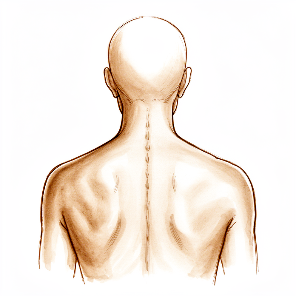
Both vertebroplasty and kyphoplasty are keyhole procedures done through a small cut in your back, over the broken bone. Your surgeon works with X-ray guidance so the instruments reach the right spot inside the spine.
In vertebroplasty, bone cement is placed directly into the cracked vertebral body to steady it. In kyphoplasty, your surgeon first makes a small space inside the bone, often using a tiny balloon that is inflated and then removed, before filling that space with cement. The cement sets firmly within the bone and holds the fracture from the inside.
The cut is closed with stitches and covered with a dressing. Because the procedure works through such a small opening, there is no large incision to heal.
What can go wrong
These procedures are generally safe, and complications are relatively infrequent. The most common is cement leaking slightly outside the fractured bone. This happens more often with vertebroplasty than with kyphoplasty, and more often when a fracture is caused by cancer spread rather than thin bone. Most leaks cause no symptoms at all.
Any operation on the spine carries some general risks, including infection, bleeding and reactions to the anaesthetic. There is also a small chance of a new fracture in a nearby vertebral body later on. The strongest factors behind new fractures are how thin your bone is and whether the spine stays bent forward at the fracture site. Having one of these procedures does not raise the risk of a fracture in new levels compared with non-operative care.
We will talk through all of these risks with you before you decide, and you can ask questions at any point. The complications table on this page lists typical rates if you want the specifics.
After the operation
You will wake up in the recovery area, where nurses watch you as the anaesthetic wears off. Pain relief is part of your care plan, and we will keep you comfortable while the cement settles and the small cut in your back begins to heal. Most people are up and moving soon after the procedure, often on the same day. Someone should stay with you for the first 24 hours after you get home. Your team will tell you whether you go home the same day or stay one night in hospital. We leave the dressing on for about 10 days; please do not take it off before then unless we tell you to. We change or remove it when we see you.
Recovery
Most people are up and moving soon after the procedure, often on the same day. The small cut in your back may be tender for a few days. Some bruising around the site is normal and settles on its own. Simple pain relief and short, gentle walks help. Keep the dressing dry and leave it on for about 10 days; we will change or remove it when we see you.
In the early days you will do short walks around the house, resting as you need. Your physiotherapist will guide you through gentle movement and build this up as your comfort allows. You can sleep in whatever position feels comfortable. Avoid heavy lifting, bending and twisting until we tell you these are safe. Once the pain has settled, day-to-day tasks like cooking and showering become easier.
The cement sets within the bone during the operation itself, so the fracture is steadied from the inside straight away. As your pain eases, you will find you can stand and walk for longer. Getting back to your usual activities happens in stages, and your own surgeon will clear you for each one as you reach it. For driving, what matters is being off strong pain medication, being able to sit in the driving position long enough for the journey without pain that distracts you, being able to twist and turn enough to check your mirrors and blind spots, and being able to brake hard without hesitating. If a back brace or movement restrictions have been given to you, those come first.
Recovery varies between people. Your timeline may differ, and your surgeon and physiotherapist will guide you at each review.
When to call us
Call us if you have a fever, or the skin around the small cut in your back becomes more red, swollen or starts to leak fluid. Call us if you notice new pain, numbness or weakness, or if the pain from your fracture suddenly gets much worse. Call us too if the pain comes back or your spine seems to be bending forward more than before, which can mean the treated bone has collapsed again and lost some of the height that was restored. Contact the clinic rather than waiting for your next review. Go to emergency if you have calf swelling or pain, shortness of breath, chest pain, loss of feeling in your legs, or trouble moving them. These symptoms need checking straight away.
Evidence & references
This is the clinical evidence summary written for health professionals. It is technical, and it lists the research this page was built from. You do not need to read it to understand your treatment or to make a decision about it.
Anatomy & Pathophysiology
Osseous Anatomy
- The bony anatomy of the spine consists of 7 cervical vertebrae, 12 thoracic vertebrae, 5 lumbar vertebrae, 5 fused sacral vertebrae, and 4 or 5 fused coccygeal vertebrae [3].
- The vertebral body consists of a fairly cylindrical mass of bone connected by the pedicles to the posterior arch of the vertebra, which consists of the lamina and spinous process [3].
- The spinal canal is created by the vertebral body anteriorly, the lamina posteriorly, and the pedicles laterally [3].
- The vertebral bodies function primarily to bear weight and transfer forces to the pelvis and hips [3].
- The posterior elements provide protection to the neural structures and function as a tension band [3].
- The thoracic spine represents two transitional zones: from the highly mobile cervical spine into the more rigid thoracic region, and then back to the more mobile lumbar spine [5].
- The thoracic spine, in conjunction with the ribs and sternum, forms a bony “cube” that is an inherently stable structure providing protection to the heart and lungs [5].
- Thoracic vertebral bodies are larger than cervical vertebrae but smaller than lumbar vertebrae [5].
- Thoracic pedicles arise more superiorly from the posterior vertebral body than in the cervical or lumbar spine and project obliquely from superodorsal to inferoventral [5].
- The spinal canal is narrowest in the thoracic region of the spine [5].
- The spinous processes of the midthoracic spine project sharply obliquely, overlapping the lamina and spinous processes inferiorly [5].
- The superior articular facets of the thoracic spine project cranially from the junction of the laminae and pedicles and are oriented coronally [5].
- The rib heads articulate with the lateral aspect of the vertebral bodies, with a shared articulation at the level of the disk space referred to as a demifacet [5].
- The transverse processes of the thoracic spine project obliquely superolaterally, with the costotransverse joint located along their ventral aspect [5].
- There is no costotransverse articulation at T11 or T12 [5].
- The vertebral body is composed of an inner region of cancellous bone surrounded by a thin shell of cortical bone [8].
- Each vertebra is composed of an anterior portion (vertebral body) and a posterior arch formed by the pedicle, facet, lamina, and spinous process [8].
- The cervical spine is composed of seven vertebrae and assumes a lordotic curvature [8].
- The thoracic spine is composed of 12 vertebrae and assumes a kyphotic curvature [8].
- The lumbar spine is composed of five vertebrae and assumes a lordotic curvature [8].
- The five fused sacral vertebrae form a portion of the pelvis [8].
- Four small, fused vertebrae form the coccyx at the most caudal extent of the spinal column [8].
- The vertebral column comprises 33 vertebrae divided into five sections: 7 cervical, 12 thoracic, 5 lumbar, 5 sacral, and 4 coccygeal [11].
- The sacral and coccygeal vertebrae are fused, which typically allows for 24 mobile segments [11].
- The length of the vertebral column averages 72 cm in men and 7 to 10 cm less in women [11].
- The vertebral canal extends throughout the length of the column and provides protection for the spinal cord, conus medullaris, and cauda equina [11].
- The cervical endplates have a cup-in-saucer configuration, distinct from the normally flat endplates of the thoracic and lumbar vertebrae [20].
- The posterior aspect of the cervical transverse process guides the cervical spinal nerves as they exit the spinal canal, with the spinal nerves lying posterior to the vertebral artery [20].
Intervertebral Disc Anatomy
- The intervertebral disc (IVD) separates each successive vertebral body except between the atlas (C1) and the axis (C2) [8].
- The IVD is composed of an inner nucleus pulposus (NP) and an outer ring termed the anulus fibrosus (AF) [8].
- The nucleus pulposus serves as an osmotic pump to attract water and generate hydraulic pressure when subjected to significant loads [8].
- The anulus fibrosus encapsulates the gelatinous nucleus pulposus and provides mechanical support to contain NP pressure and constrain intervertebral rotations [8].
- The outer anulus fibrosus is integrated with the vertebral rim via a fibrocartilage enthesis that consists of a thin layer of calcified cartilage, or “tidemark” [8].
- The end plate is a bilayer of cartilage and bone that separates the disk from adjacent vertebrae [8].
- The cartilage end plate integrates with the inner anulus fibrosus to fully encapsulate the nucleus pulposus [8].
- The end plate must be strong and thick to resist significant loads but also permeable to favor chemical transport and disk cellular vitality [8].
Ligaments and Soft Tissue
- Additional soft-tissue structures providing passive support include the anterior longitudinal ligament, posterior longitudinal ligament, ligamentum flavum, facet joint capsule, interspinous ligament, and supraspinous ligaments [8].
- The erector spinae muscle runs longitudinally on the dorsal surface of the spinal column and functions to extend the spine [8].
- The psoas muscle runs longitudinally on the ventrolateral surface of the spinal column and serves to flex the hip or laterally bend the trunk [8].
- The multifidus muscle connects intersegmentally to stabilize the spine by acting like a bowstring to maintain lordosis [8].
Biomechanics and Alignment
- Normal cervical alignment is approximately 15° of lordosis [7].
- The thoracic spine generally ranges from 20° to 40° of kyphosis [7].
- The lumbar spine has approximately 40° to 50° of lordosis [7].
- Kyphotic segments (thoracic, sacral) are considered “primary” curvatures as they are present in utero and at birth [7].
- The lordotic curvatures of the cervical and lumbar spine develop secondarily later in life to allow the growing child to develop an upright posture [7].
- The center of gravity of the spinal column runs from the odontoid process proximally through the sacral promontory caudally [7].
- Changes in sagittal balance that shift the center of gravity too far ventrally can result in significant pain and disability [7].
- The functional spinal unit consists of two vertebrae, the disk between them, and the facet joints (and their capsules) [7].
- The vertebral bodies bear 70% to 90% of the static axial load of the spine [7].
- The facet joints support 10% to 20% of axial load in a standing, neutral alignment [7].
- In extension, the facet joints may bear up to 30% of the axial load [7].
- In flexion, the facet joints may be burdened with up to 50% of the anterior shear load [7].
- As compressive forces are applied to the disk, the nucleus pulposus deforms, redistributing axial forces radially [7].
- The radial pressure generated by the nucleus pulposus is resisted by the tensile properties of the alternating bands of fibers within the anulus fibrosus [7].
- The spinous processes and transverse processes act as lever arms, providing mechanical advantage for the muscles that insert along their surfaces [7].
Vascular Anatomy
- The thoracic and lumbar levels are supplied by paired segmental arteries which originate directly from the aorta along its posterior surface [12].
- Branches of the segmental arteries supply the vertebral body, the paraspinal musculature, and the spinal cord [12].
- The cervical spine derives its circulation primarily from the vertebral arteries [12].
- The vertebral arteries typically enter the transverse foramen at the C6 level and run proximally through the transverse foramina to C1 [12].
- The vertebral arteries course posteriorly over the superior aspect of the C1 ring before turning proximally again and entering the foramen magnum [12].
- The vertebral arteries merge to form the basilar artery at the foramen magnum [12].
- The vascular supply of the spinal cord is primarily from the medullary branches of the segmental spinal arteries [12].
- The anterior spinal artery is responsible for supplying approximately 80% of the vascular supply to the spinal cord [12].
- The arteria medullaris magna (AMM), also known as the arteria radicularis magna or artery of Adamkiewicz, is the largest anterior segmental artery [12].
- The AMM typically arises on the left side anywhere between the T8 and L1 level [12].
Neural Anatomy
- A typical mixed spinal nerve has three distinct components: motor, sensory, and sympathetic [13].
- Motor rootlets leave the anterolateral sulcus of the spinal cord and unite to form each motor root, with fibers arising from anterior horn cells to innervate skeletal muscles [13].
- Sensory fibers arise from pain, thermal, tactile, and stretch receptors, with cell bodies located within the dorsal root ganglia [13].
- The sympathetic component of all 31 mixed spinal nerves leaves the spinal cord along only 14 motor roots [13].
- The cells of origin for the sympathetic component are in the intermediolateral cell column that extends throughout the thoracic and upper lumbar cord segments [13].
- Mixed spinal nerves, having left the intervertebral foramina, receive their sympathetic component and promptly branch into anterior and posterior primary rami [13].
- The posterior primary rami are directed posteriorly and supply the paraspinal musculature and the skin along the posterior aspect of the trunk, neck, and head [13].
- The anterior primary rami of all the cervical, the first thoracic, and all the lumbosacral nerves join in the formation of plexuses [13].
- The upper four cervical anterior rami form the cervical plexus [13].
- The lower four cervical and first thoracic anterior rami form the brachial plexus [13].
- The first three and a part of the fourth lumbar anterior rami form the lumbar plexus [13].
- The sacral anterior rami along with the fifth lumbar and a part of the fourth join to form the lumbosacral plexus [13].
- The area of skin supplied by the fibers of a single spinal root is called a dermatome [13].
- Segmental dermatomal patterns are well preserved in the thoracic region but not in the limbs [13].
Pathophysiology of Degeneration and Stenosis
- Degeneration of the disc occurs with disc narrowing and subsequent ligamentous redundancy, which compromises the spinal canal area [17].
- Instability resulting from disc degeneration may precipitate the formation of facet overgrowth and ligamentous hypertrophy [17].
- The ligamentum flavum may be markedly thickened into the lateral recess where it attaches to the facet capsule, causing nerve root compression [17].
- Central spinal stenosis denotes involvement of the area between the facet joints, which is occupied by the dura and its contents [17].
- Symptomatic central spinal stenosis results in neurogenic claudication with generalized leg pain [17].
- The lateral recess, also known as “Lee’s entrance zone,” begins at the medial border of the superior articular process and extends to the medial border of the pedicle [17].
- Facet arthritis most frequently causes stenosis in the lateral recess zone, along with vertebral body spurring and disc or anulus pathology [17].
- “Lee’s midzone” describes the foraminal region, which lies ventral to the pars [17].
- The dorsal root ganglion and ventral motor root occupy 30% of the space in the foraminal region [17].
- The exit zone is identified as the area lateral to the facet joint [17].
- The most common type of spinal stenosis is caused by degenerative arthritis of the spine, characterized by hyperostosis and spinal rigidity in elderly patients [17].
- The L4-5 level is the most commonly involved in degenerative spinal stenosis, followed by L5-S1 and L3-4 [17].
- Congenital spinal stenosis usually is central and is evident on imaging studies [17].
- In achondroplasia, the canal is narrowed in the anteroposterior plane owing to shortened pedicles and in lateral diameter because of diminished interpedicular distance [17].
Investigations
Magnetic Resonance Imaging (MRI)
- MRI is the standard for advanced imaging of the spine and is superior to CT in most circumstances, particularly for the identification of infections, tumors, and degenerative changes within the discs [23].
- MRI is superior to CT for imaging the intervertebral disc and directly imaging neural structures [23].
- MRI typically shows the entire region of the spine (cervical, thoracic, or lumbar) [23].
- MRI allows for the imaging of the nerve root in the foramen, which is difficult even with postmyelography CT because the subarachnoid space and contrast agent do not extend fully through the foramen [23].
- In the lumbar and thoracic spine, MRI has supplanted CT myelography because it is noninvasive and less expensive [22].
- The combination of high soft-tissue contrast and high resolution in MRI allows ideal evaluation of the intervertebral discs, nerve roots, posterior longitudinal ligament, and intervertebral foramen [22].
- MRI provides excellent assessment of the spinal cord [22].
- A normal intervertebral disc exhibits signal hyperintensity on T2-weighted images due to its high water content [22].
- The aging process results in a gradual desiccation of the disc material and a loss of T2-weighted signal hyperintensity [22].
- Disc herniations or extrusions appear as convex or polypoid masses extending posteriorly into the ventral epidural space, frequently maintaining a signal intensity similar to that of the disc of origin [22].
- Sagittal T2-weighted or gradient-echo images create a “myelographic” effect useful for evaluating compromise of the subarachnoid space [22].
- Sagittal T1-weighted images should be closely examined to identify narrowing of the neuroforamina [22].
- Normal T1-weighted hyperintense perineural fat in the foramina provides excellent contrast to darker displaced disc material [22].
- Far lateral disc herniations are best seen on selected axial images localized through disc levels [22].
- Free disc fragments appear discontinuous with the intervertebral disc and usually exhibit intermediate T1-weighted signal in contrast to the hypointense cerebrospinal fluid [22].
- MRI can detect significant spinal cord compromise, with edema within the cord readily demonstrated as hyperintensity on T2-weighted images [22].
- MRI evidence of disc degeneration has been reported in the cervical spine in 25% of patients younger than 40 years and in 60% of patients 60 years and older [23].
- Lumbar disc degeneration has been found in 35% of patients aged 20 to 39 years and in 100% of patients older than 50 years [23].
- The best way to obtain meaningful clinical information from MRI is to have a specific question derived from the patient’s history and physical examination, posed using the parameters of neural compression, instability, and deformity [23].
- Only abnormalities in the categories of neural compression, instability, or deformity are important for operative treatment [23].
- Diffusion tensor imaging is an MRI technique based on the diffusion rate of water in tissue that has been reported to demonstrate spinal cord impairment in patients with early stage cervical spondylosis before it is visible on plain MRI scans [26].
Computed Tomography (CT)
- CT has largely supplanted plain radiographs as the initial screening study of choice for spine injuries due to its combination of high sensitivity and specificity [24].
- CT allows for the identification of subtler fractures that might have remained undiagnosed on plain radiographs and provides additional three-dimensional detail [24].
- CT is particularly useful in differentiating compression fractures from burst fractures [24].
- CT identifies subtle features of injury such as the presence of facet widening [24].
- CT evaluation is essential in determining the stability of thoracic and lumbar spine fractures [24].
- The primary disadvantage of CT imaging in comparison to MRI is that it does not provide as good a visualization of the soft tissues [24].
- CT myelography is invasive and more costly than MRI and is reserved for patients who have contraindications to MRI or who have equivocal MRI examinations [22].
Diagnostic Principles
- Proper diagnosis of a spine tumor with a biopsy is the critical first step in devising proper treatment for a patient who presents with a spine tumor [2].
- Careful identification of the tumor type by direct biopsy decreases the chance of misdiagnosis and performing unnecessary or incorrect surgery [2].
- MRI findings must be carefully correlated with the clinical impression because MRI shows anatomy that is abnormal but may be asymptomatic [23].
- The specific location of an abnormality should be suspected before MRI and confirmed with the study [23].
References
[2] Orthopaedic Knowledge Update 13 Ebook Without Multimedia. Concepts in Primary Benign, Primary Malignant, and Metastatic Tumors of the Spine > Summary.
[3] Orthopaedic Knowledge Update 13 Ebook Without Multimedia. Spinal Anatomy > Osseous Anatomy.
[5] Orthopaedic Knowledge Update 13 Ebook Without Multimedia. Spinal Anatomy > Osseous Anatomy > Thoracic Vertebrae.
[7] Orthopaedic Knowledge Update 13 Ebook Without Multimedia. Spinal Anatomy > Biomechanics.
[8] Orthopaedic Basic Science Fifth Edition Print Ebook. Biology and Mechanics of the Skeletal Extracellular Matrix > Anatomy.
[11] Campbell S Operative Orthopaedics 4 Volume Set. RECONSTRUCTION OF THE PATELLOFEMORAL AND PATELLOTIBIAL LIGAMENTS WITH A SEMITENDINOSUS TENDON GRAFT > ANATOMY OF VERTEBRAL COLUMN.
[12] Orthopaedic Knowledge Update 13 Ebook Without Multimedia. Spinal Anatomy > Vascular Anatomy.
[13] Campbell S Operative Orthopaedics 4 Volume Set. PERIPHERAL NERVE INJURIES OF THE UPPER AND LOWER EXTREMITIES > ANATOMY OF THE SPINAL NERVES > COMPONENTS OF MIXED SPINAL NERVES.
[17] Campbell S Operative Orthopaedics 4 Volume Set. OVERVIEW OF LUMBAR AND THORACIC DISC DEGENERATION AND HERNIATION > STENOSIS OF THE THORACIC AND LUMBAR SPINE > ANATOMY.
[20] Rockwood And Green S Fractures In Adults. Imaging of Cervical Spine Fractures and Dislocations > Lower Cervical Spine (C3–C7).
[22] Campbell S Operative Orthopaedics 4 Volume Set. INTERVERTEBRAL DISC DISEASE.
[23] Campbell S Operative Orthopaedics 4 Volume Set. OVERVIEW OF LUMBAR AND THORACIC DISC DEGENERATION AND HERNIATION > MAGNETIC RESONANCE IMAGING.
[24] Rockwood And Green S Fractures In Adults. Imaging of Cervical Spine Fractures and Dislocations > Computed Tomography.
[26] Campbell S Operative Orthopaedics 4 Volume Set. POSTERIOR APPROACH TO THE LUMBAR SPINE, L1 TO L5 > MAGNETIC RESONANCE IMAGING.