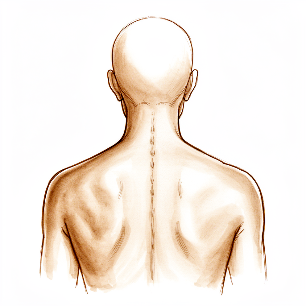
What you're feeling
Cervical radiculopathy is the name for pain, numbness or tingling that starts in your neck and travels down into your arm. It happens when a nerve root in your neck gets squeezed or irritated, often by a bulging disc or by wear-and-tear arthritis in the small joints of the spine. The pain often feels sharp, shocking or electric, and it can shoot from your neck into your shoulder and arm. Many people also feel pins and needles or numbness in part of the arm or hand.
The pain usually follows a pattern. You might notice aching near your shoulder blade and in your arm, with tingling further down towards your fingers. Turning or bending your neck can make it worse, especially if you tilt your head towards the sore side. Resting your hand on top of your head can ease the pain, because this takes pressure off the nerve. Some people also get headaches at the back of the head that spread forward.
Symptoms often start with neck pain, and the arm symptoms follow later. The onset can be sudden or build up over weeks. Night-time can be uncomfortable, and tasks that need you to hold your arms up or turn your head, like reversing the car or reaching a high shelf, can become hard. Shoulder pain is common too, and it can be hard to tell whether the trouble is in your neck or your shoulder. Pain that worsens when you move your neck points to the neck, while pain worse with shoulder movement points to the shoulder.
Most people improve without surgery. Up to 75% of people with a recent flare-up get better on their own, and when symptoms have been present for about 6 weeks, 75% respond well to further non-surgical treatment over the next 6 weeks. A small percentage of people need surgery. If you notice clumsiness in your hands, or trouble with walking or balance, tell your surgeon, as these symptoms need a closer look at your neck.
What's actually happening
Your spine is made up of bones stacked one on top of another, with a cushioning disc between each pair. Think of the disc as a shock absorber with a tough outer wall and a soft, jelly-like centre. With age, these discs lose water and height, and the outer wall can weaken and bulge or even tear, letting some of the soft centre push out. The small joints at the back of your neck also wear down, and bony spurs form around them.
As the disc settles and spurs grow, the spaces where nerves leave the spine get narrower. A nerve root can then be squeezed by the bulging disc, a spur, or both. An irritated nerve becomes inflamed and more sensitive, which is why the pain, tingling and numbness travel down your arm in the pattern described above. The most common levels for this are C6-C7 and C5-C6, in the middle of your neck. Bending your neck backwards narrows these spaces further, which is why looking up can make symptoms worse.
Most of the time this is a wear-and-tear problem that builds up slowly. Sometimes a disc tears suddenly and the soft centre pushes out quickly, causing intense pain that starts without warning. The nerve itself is usually not badly damaged, which is why most people improve without surgery. If the squeezing becomes severe, or if it starts to press on the spinal cord itself rather than just a nerve root, symptoms such as clumsy hands or unsteady walking can appear. Those signs mean the problem needs closer attention, and surgery may be considered to take the pressure off.
What we can do about it
The first step is simple movement changes and exercises you can do at home. These include isometric exercises, where you tighten your neck muscles without moving your head. Some people are helped by traction, a gentle pull on the neck that eases pressure on the nerve, or by wearing a soft collar for a short time. As you read earlier, most flare-ups settle on their own, so we often give things time to improve before doing more.
Pain medicine can help you stay active while the nerve settles. Anti-inflammatory tablets, called NSAIDs, are a common choice. If tablets are not enough, a cervical epidural injection can be offered. This places anti-inflammatory medicine close to the irritated nerve root in your neck to settle the pain.
Surgery is considered when there is progressive weakness in the arm, or when pain stays severe and disabling despite the steps above. It may also be advised when scans show the nerve or spinal cord is being squeezed and your symptoms match. The aim of surgery is to take the pressure off the nerve. This can be done from the front of the neck, where the bulging disc and any bony spurs are removed and the bones are then joined together with a graft. In some cases a damaged disc can be replaced with an artificial one instead of fusing the bones. Some problems are better reached from the back of the neck through a small window in the bone, which frees the nerve without fusing the spine. Which option suits you depends on where and how the nerve is squeezed, and we will talk that through with you as a shared decision.
What to expect
The outlook for this condition is often good. Most people improve without surgery, and a recent flare-up often settles on its own. When symptoms have been present for about 6 weeks, most people respond well to further non-surgical care over the next 6 weeks, as you read earlier. Nonsurgical treatment is the usual starting point, and it works for the majority of people.
Recovery is usually gradual rather than sudden. The pain tends to ease first, and the tingling and numbness can take longer to fade. Some numbness or mild weakness may linger even after the pain has gone, because an irritated nerve settles slowly. Over weeks to months you should notice the arm pain becoming less sharp and less frequent, and everyday tasks like reaching and driving becoming easier.
If non-surgical care does not settle things, surgery is an option for a small percentage of people. The aim is to take pressure off the nerve, and the results of different surgical approaches are broadly similar for this condition. Whether the neck is fused or the disc is replaced, measures of disability, pain and quality of life have not differed at 5 years. Surgery from the back of the neck works as well as surgery from the front at 2 years, although neck pain can be more noticeable early on after back-of-neck surgery before it improves.
A few honest cautions. The size of a disc bulge on your scan does not predict how bad your symptoms are, nor how well you will do after surgery. Some people continue to have some neck or arm discomfort even after treatment has worked well overall. And if symptoms are left alone, most still improve, but a small number go on to need surgery anyway. If you notice worsening hand clumsiness or trouble with balance at any point, contact your surgeon rather than waiting for the next appointment.
When to see someone
See your GP if you have arm pain, numbness or tingling that has not started to settle after a few weeks, or if it keeps coming back. Ask for a specialist review if the pain is severe, if it stops you sleeping or working, or if your arm is getting weaker. Weakness that is getting worse matters most, because it can mean the nerve is under real pressure. Go to an emergency department if you suddenly become clumsy with your hands, or if you develop trouble with walking or balance. Those signs suggest the spinal cord itself may be squeezed, and they need checking the same day rather than waiting.
Evidence & references
This is the clinical evidence summary written for health professionals. It is technical, and it lists the research this page was built from. You do not need to read it to understand your treatment or to make a decision about it.
Anatomy & Pathophysiology
Bony Anatomy
- The cervical vertebral body is an oblong structure with a coronal diameter larger than its sagittal diameter [18].
- Cervical endplates possess a cup-in-saucer configuration, distinct from the flat endplates of the thoracic and lumbar vertebrae [18].
- The posterior aspect of the cervical transverse process guides the spinal nerves as they exit the spinal canal [18].
- The spinal nerves lie posterior to the vertebral artery within the transverse process [18].
- The transverse process forms a half-pipe configuration that cradles the exiting spinal nerve [18].
- The cervical pedicles project from the vertebral body in an orientation running posterolateral to anteromedial [22].
- The cervical pedicles form the posteromedial border of the transverse foramina and the anterolateral aspect of the spinal canal [22].
- The facet joints are highly mobile diarthrodial joints formed by the interaction of superior and inferior articular processes from adjacent vertebrae [22].
- The articular surfaces of the cervical facet joints are angled approximately 45 degrees in relation to the transverse axis of each segment [22].
- The pillar of bone between the superior and inferior articular processes is referred to as the lateral mass [22].
- The laminae arise from the posteromedial border of the lateral masses and project posteriorly toward the midline to form bifid spinous processes between C2 and C6 [22].
- The subaxial cervical spine includes the C3–C7 vertebral segments, which maintain a relatively uniform anatomical configuration analogous to the thoracic and lumbar spine [27].
Ligamentous Anatomy
- The ligamentum flavum spans each interlaminar space and is noncontiguous in nature [22].
- The ligamentum flavum, interspinous ligaments, and supraspinous ligaments (ligamentum nuchae) form the posterior ligamentous complex [22].
- Disruption of the posterior ligamentous complex structures can result in mechanical instability [22].
- The tectorial membrane extends from the posterior border of the foramen magnum to the superior surface of the C1 ring and is analogous to the posterior longitudinal ligament in the lower cervical spine [27].
- The posterior atlantooccipital membrane spans between the lower occiput and the posterior C1 ring and is analogous to the ligamentum flavum at other levels [27].
- The vertebral artery enters the posterior atlantooccipital membrane approximately 1.5 cm from the posterior midline [27].
- The ligamentum nuchae is a thick condensation of supraspinous fibrous bands that overlays the spinous processes of the cervical vertebrae and extends from the inion to C7 [27].
Intervertebral Disc Anatomy
- The intervertebral disc contains a central gelatinous nucleus pulposus surrounded by a fibrous anulus fibrosus [23].
- The nucleus pulposus consists mainly of a high concentration of proteoglycans and water surrounded by a loose type II collagen network [23].
- The anulus fibrosus has a low proteoglycan and water content and a high concentration of type I collagens organized into concentric lamellae [23].
- The anulus fibrosus is described as possessing 20 to 25 lamellae rich in collagen fibrils arranged in a parallel fashion [23].
- In each adjacent lamella of the anulus fibrosus, the collagen fibrils along the axis are fashioned in the opposite direction to create an alternating pattern [23].
- The content of water and proteoglycan concentration within the disc increases when progressing from the anulus fibrosus to the nucleus pulposus [23].
- The content of collagen within the disc decreases from the outer anulus to the nucleus [23].
- With increasing age, the proteoglycan and water content of the nucleus decrease [23].
- The collagen content of the nucleus is highest in the cervical disks and lowest in the lumbar disks [23].
- The nucleus pulposus is avascular in adults and receives nutrients through perforations in the cartilaginous end plates of the intervertebral discs [24].
- Microscopic studies found that in adults older than 30 years of age there is no direct vascular supply to the disc [24].
- The microvasculature of the vertebral bony end plates contains vessels oriented obliquely that originate from circumferential vessels and nearby metaphyseal marrow vessels [24].
Spinal Cord Anatomy
- The anterior corticospinal tract mediates skilled movement on the opposite side of the body [20].
- The lateral corticospinal (pyramidal) tract mediates skilled movement on the same side of the body [20].
- The dorsolateral fasciculus mediates pain and temperature sensation bidirectionally [20].
- The fasciculus gracilis mediates position and fine touch sensation on the same side of the body [20].
- The fasciculus cuneatus mediates position and fine touch sensation on the same side of the body [20].
- The lateral spinothalamic tract mediates pain and temperature sensation on the opposite side of the body [20].
- The anterior spinothalamic tract mediates light touch sensation on the opposite side of the body [20].
Pathophysiology
- Cervical spondylosis is defined as a generalized disease process affecting the entire cervical spine and related to chronic disk degeneration [4].
- In approximately 90% of men older than 50 years and 90% of women older than 60 years, degeneration of the cervical spine can be demonstrated by radiographs [4].
- Initial disk changes in cervical spondylosis are followed by facet arthropathy, osteophyte formation, and ligamentous instability [4].
- Disk degeneration starts with tears in the posterolateral region of the annulus [4].
- Loss of water content and proteoglycans in the nucleus leads to a decrease of disk height [4].
- Longitudinal ligaments degenerate and form bony spurs at their insertion into the vertebral body [4].
- The most frequently involved levels in cervical spondylosis are the more mobile segments: C5-C6, C6-C7, and C4-C5 [4].
- Converging of the cervical disk space may result in buckling of the ligamentum flavum, with further narrowing of the spinal canal [4].
- Segmental instability results in hypertrophic formation of osteophytes by the uncovertebral joint of Luschka and by the facet joints [4].
- The sagittal cervical canal diameter was appreciably smaller (3 mm on average) in the myelopathic spondylotic spine than in the normal spine [4].
- The anterior-posterior dimensions of the cervical spinal canal measure between 17 and 18 mm in normal individuals [4].
- Spinal canal stenosis is present when the canal diameter becomes less than 13 mm [4].
- With extension of the neck, both the spinal canal diameter and the neuroforaminal diameter decrease [4].
- Cervical degenerative disc disease is a pathophysiologic process that incorporates a spectrum of disease states ranging from neck pain and headache to cervical radiculopathy and/or myelopathy [13].
- The degenerative spinal cascade in the cervical spine is the result of the interplay of the intervertebral disc and four other articulations: two uncovertebral joints (of Luschka) and two facet joints [5].
- Facet joint capsules are known to have sensory receptors that may play a role in pain and proprioceptive sensation in the cervical spine [5].
- Progressive collapse of cervical discs results in loss of normal lordosis of the cervical spine and chronic anterior cord compression across the kyphotic spine or anterior chondroosseous/discoosteophytic spurs [5].
- Subsequent loading of facet and uncovertebral joints results in spondylotic changes in foramina that may restrict motion and lead to spinal cord and/or nerve root compression [5].
- "Soft" disc herniation is a nonspecific term describing herniation of the intervertebral disc without bony osteophytes [5].
- Soft disc herniations are usually posterolateral, between the posterior edge of the uncinate process and the lateral edge of the posterior longitudinal ligament [5].
- "Hard" disc herniation is a nonspecific term describing herniation with an associated discoosteophytic spur [5].
- Cervical nerve roots exit above their corresponding vertebrae [7].
- Disc herniation at C5 to C6 involves the C6 nerve root [7].
- Disc herniation at C7 to T1 involves the C8 root [7].
- Overlapping findings in cervical radiculopathy occur because of intraneural intersegmental connections of sensory nerve roots [7].
- Cord compression can cause myelopathy by an ischemic effect secondary to compression of the anterior spinal artery or by a direct mechanical effect on cord function [15].
- The natural history of cervical spondylotic myelopathy typically includes stable periods punctuated by unpredictable stepwise progression [15].
- Stepwise deterioration in symptomatology followed by a period of stability is the most common presentation of cervical spondylotic myelopathy, occurring in 65%–80% of cases [7].
- Slowly progressive decline over months to years occurs in 20%–25% of cervical spondylotic myelopathy cases [7].
- Rapidly progressive decline over days to weeks occurs in 3%–5% of cervical spondylotic myelopathy cases [7].
- Dorsal column (proprioceptive) dysfunction occurs with advanced cervical myelopathy and carries a poor prognosis [15].
- Patients with concomitant myelopathy and peripheral nerve disease from conditions such as diabetes, hypothyroidism, peripheral neuropathy, or severe multilevel cervical foraminal stenosis can have diminished or absent reflexes [15].
- Patients with cervical myelopathy who have coexisting lumbar stenosis may exhibit brisk upper extremity reflexes consistent with upper motor neuron findings yet diminished lower extremity reflexes because of root level compression in the lumbar spine [15].
- Anterior structures such as bulging, ossified, or herniated disks and osteophytic anterior spurs are the usual cause of cord compression in cervical spondylotic myelopathy [15].
- Less commonly, conditions involving posterior structures such as ligamentum flavum hypertrophy or ossification of the ligamentum flavum may contribute to cervical spondylotic myelopathy [15].
- Degenerative spondylolisthesis can exacerbate or cause compression in cervical spondylotic myelopathy [15].
- Cervical spondylotic myelopathy commonly arises in the setting of a congenitally narrowed cervical canal [15].
- Cervical spondylotic myelopathy often does not become symptomatic until the later decades of life because the cord may have sufficient space to avoid compression until a threshold amount of space-occupying degenerative changes accumulate [15].
- A compression ratio of less than 0.4 is associated with a poor prognosis in cervical myelopathy [15].
- An increase in the compression ratio to more than 0.4 postoperatively correlates with clinical recovery in cervical myelopathy [15].
- The genetic influence on disc degeneration may be attributed to a small effect from each of multiple genes or possibly a relatively large effect of a smaller number of genes [13].
- Specific gene loci associated with disc degeneration include the aggrecan gene, metalloproteinase-3 gene, collagen type IX, and alpha 2 and 3 gene forms [13].
- Potential genetic factors for degenerative disk disease include those related to MMP-2 and collagen IX [12].
- Potential genetic factors for ossification of the posterior longitudinal ligament include collagen VI and XI [12].
- Congenital anomalies including spinal stenosis, Down syndrome, and Klippel-Feil syndrome may predispose to the development of cervical disk degeneration [12].
- Occupational hazards, including transportation of goods by weight bearing on top of the head, may accelerate cervical disk degeneration [12].
- Myelopathy results from static compression, spinal malalignment leading to altered cord tension and vascular supply, and dynamic injury mechanisms [12].
- The cascade of events after compression of the spinal cord includes ischemia, destruction of the blood–spinal cord barrier, demyelination, and neuronal apoptosis [12].
- Extension of the cervical spine often exacerbates the symptoms of spinal cord and nerve root compression [4].
- Neck extension results in cord compression between the degenerative disc and spondylotic bar anteriorly and the hypertrophic facets and infolded ligamentum flavum posteriorly [5].
- Neck flexion results in a slight increase in canal diameter and relief of cord compression [5].
- Normal cervical spinal canal diameter is 14 mm or greater [5].
- Relative cervical spinal canal stenosis is defined as a diameter less than 14 mm (10–13 mm) [5].
- Absolute cervical spinal canal stenosis is defined as a diameter less than 10 mm [5].
- The Pavlov (Torg) ratio normal value should be 1.0 [5].
- A Pavlov (Torg) ratio less than 0.8 is considered abnormal and may be a risk factor for later neurologic involvement, though clinical significance is debated [5].
- Cervical spondylosis is the most common cause of cervical myelopathy in people aged 55 years and older [11].
- Cervical spondylosis is the most common cause of cervical myelopathy in patients older than 50 years [15].
- The incidence of cervical myelopathy is twice as great in men as in women [4].
- People older than 60 years are more likely to have multi-segmental cervical disease [4].
- Cervical degenerative disc disease peaks between age 40 and 50 years [5].
- Men are affected more than women by cervical degenerative disc disease [5].
- The C5 to C6 level is most frequently involved in cervical degenerative disc disease, followed by C6 to C7 [5].
- Risk factors for cervical degenerative disc disease include frequent lifting, cigarette smoking, and history of excessive driving [5].
- Approximately 80% of individuals are affected by nonspecific axial pain at some time in their lives [13].
- Significant abnormalities shown by imaging studies in asymptomatic matched controls occur in 76% of cases [13].
- Axial spine pain is attributed to disc degeneration, but disc degeneration does not always cause pain [13].
- Nonanatomic factors, specifically work perception and psychosocial factors, are intimately intertwined with physical complaints in spinal disorders [13].
- Cervical disk herniation can lead to symptoms of radiculopathy or myelopathy depending on if the disk compresses the nerve roots or spinal cord, respectively [11].
- Progressive degeneration of the disks and weakening of the muscles and soft tissues leads to deformity of the cervical spine with spinal cord and nerve root impingement [11].
- Cervical deformity can present as cervical kyphosis (sagittal plane deformity) or scoliosis (coronal plane deformity) [11].
- The rate and degree of neurologic deterioration in cervical spondylotic myelopathy are variable [11].
- Early recognition and treatment of cervical spondylotic myelopathy is critical before the onset of spinal cord damage [11].
- Early recognition and treatment, before the onset of irreversible spinal cord damage, is essential for optimal outcomes in cervical myelopathy [15].
- Patients with cervical canal stenosis and cord compression secondary to spondylosis, without clinical evidence of myelopathy, who present with clinical or electrophysiological evidence of cervical radicular dysfunction or central conduction deficits seem to be at higher risk for developing myelopathy [12].
- Cervical motion segment disorders are considered to be multifactorial, with developmental size of the canal and foramina, pathological encroachment, biomechanical effects, and circulatory deficiencies always present to some degree [12].
- Static and dynamic factors should be considered for the development of myelopathy in patients with cervical spondylotic spinal cord compression [12].
Clinical Presentation
History and Symptoms
- Cervical degenerative conditions can be a major cause of disability, particularly in elderly patients [1].
- The clinical presentation and natural history of cervical degenerative conditions are variable because of the many ways these conditions can manifest [1].
- Patients with disk degeneration may complain of axial neck pain that is often chronic and insidious in nature [3].
- Patients with a cervical disk herniation will complain of a sudden and acute pain that is very intense [3].
- Headache may be the presenting symptom of cervical spondylosis, usually worse in the morning and improving throughout the day [4].
- The headache associated with cervical spondylosis is commonly located in the occipital region and radiates toward the frontal area [4].
- Cervical radiculopathy symptoms may initially manifest as neck pain before radicular symptoms develop [7, 8].
- Common symptoms of cervical radiculopathy include shoulder and arm pain, paresthesias, and numbness [7, 8].
- Pain, numbness, or paresthesia in a dermatomal distribution to the upper extremity is a symptom of cervical radiculopathy [7, 8].
- In cervical spondylotic radiculopathy, sensory involvement in the form of paresthesias or hyperesthesia is more common than motor or reflex changes [4].
- The chief complaint in cervical spondylotic radiculopathy is radiation of pain into the interscapular area and into the arm [4].
- Patients with cervical spondylotic radiculopathy typically have proximal arm pain and distal paresthesias [4].
- Cervical myelopathy presents with a variety of subtle neurologic findings that can present insidiously [3].
- Characteristic signs and symptoms of myelopathy include loss of manual dexterity in the hands, weakness, stiffness, urinary symptoms, spasticity in extremities, and gait disturbance [3].
- Patients with myelopathy may report difficulty with buttons, a change in their handwriting, or dropping objects [3].
- Patients with cervical myelopathy may report a generalized feeling of clumsiness of the arms and hands and dropping things [15].
- Patients with cervical myelopathy may report an inability to manipulate fine objects such as coins or buttons [15].
- Patients with cervical myelopathy may report trouble with handwriting [15].
- Patients with cervical myelopathy may report diffuse, typically nondermatomal, numbness [15].
- Patients with cervical myelopathy may report gait instability, a sense of imbalance, and bumping into walls when walking [15].
- Patients with severe cord compression may report the Lhermitte phenomenon: electric shock–like sensations that radiate down the spine or into the extremities with certain offending positions of the neck [15].
- Subjective weakness, bowel and bladder symptoms, loss of motor strength, neck pain, and radicular symptoms may occur late or not at all in cervical myelopathy [15].
- Neck pain may be absent despite advanced degrees of spondylosis in patients with cervical myelopathy [15].
- Many patients with cervical myelopathy do not have radicular symptoms or signs [15].
- Pain that radiates and worsens with neck motion suggests a cervical spine etiology [14].
- Any myelopathic symptom, such as clumsiness or gait or balance disturbance, should alert the surgeon to closely evaluate the cervical spine [14].
Physical Examination
- The neurologic examination will be benign in most cases of disk degeneration [3].
- Depending on the level of herniation, patients with a cervical disk herniation will have loss of sensation and motor weakness in a distribution consistent with the nerve root affected [3].
- A comprehensive neurologic examination should be performed for cervical degenerative disorders [3].
- The motor examination may be completely normal even in cases of nerve root or spinal cord compression [3].
- When upper extremity weakness is present, it often presents as diminished grip and/or intrinsic strength [3].
- The finding of severe weakness of major muscle groups in the upper or lower extremities is relatively uncommon [3].
- Sensory examination findings are often subtle [3].
- The neurologic examination should include an assessment for gait instability [3].
- Hyperreflexia may be present in the upper and/or lower extremities and is suggestive of spinal cord compression with upper motor neuron signs [3].
- Upper motor neuron signs can be masked or diminished in patients who have concomitant diabetes mellitus, peripheral neuropathy, or lumbar stenosis [3].
- Spinal cord compression with myelopathy can manifest with abnormal upper motor neuron signs such as Hoffman’s sign, inverted radial reflex, pathological clonus, and Babinski’s sign [3].
- The Hoffman’s sign is described as quick flexion of both the thumb and index finger when the middle finger nail is snapped [3].
- Clonus is a series of abnormal reflex movements of the foot in plantar flexion induced by sudden dorsiflexion [3].
- The Babinski reflex occurs after the sole of the foot has been firmly stroked, resulting in the big toe moving upward while the other toes fan out [3].
- The inverted radial reflex is noted by flexion of the fingers without flexion of the forearm when the distal end of the radius is tapped [3].
- Lhermitte’s sign is an electric shock-like sensation that runs down the center of the patient’s back and enters the limbs during flexion of the neck [3].
- Myeloradiculopathy is associated with spinal stenosis with concurrent compression of the neuroforaminal contents, producing lower motor neuron signs at the level of the cervical cord lesion and upper motor neuron signs caudal to the level of compression [3].
- Extension of the neck with rotation toward the side of neural impingement (Spurling sign) can reproduce the patient’s pain pattern in cervical spondylotic radiculopathy [4].
- Signs of cervical spondylosis include decreased range of motion, crepitus, or both [4].
- Physical examination for cervical radiculopathy should assess motor function, where weakness is uncommon but, when present, is associated with the myotome [7, 8].
- Physical examination for cervical radiculopathy should assess sensory function, where pain, numbness, or dysesthesias along dermatomal distribution is common [7, 8].
- Reflexes in cervical radiculopathy are typically normal or below normal (hyporeflexia) [7, 8].
- The Spurling test involves rotation and lateral bend of the neck with vertical compression on the head; the occurrence of radicular symptoms during this test suggests nerve root pain [7, 8].
- The shoulder abduction sign, defined as relief of radicular pain with shoulder abduction, is suggestive of a cervical etiology [7, 8].
- Physical examination for cervical myelopathy includes upper motor neuron findings [7, 8].
- The myelopathy hand and the finger escape sign, where the small finger spontaneously abducts because of weak intrinsic muscles, are physical examination findings in cervical myelopathy [7, 8].
- Hyperreflexia, Hoffmann sign, clonus, or Babinski sign are physical examination findings in cervical myelopathy [7, 8].
- The inverted radial reflex is defined as ipsilateral finger flexion when the brachioradialis reflex is being elicited [7, 8].
- Funicular pain, described as central burning and stinging with or without Lhermitte sign, is a physical examination finding in cervical myelopathy [7, 8].
- Upper motor neuron findings are not always present in all patients with cervical myelopathy [7, 8].
- Upper extremities may have radicular (lower motor neuron) signs along with evidence of distal myelopathy [7, 8].
- Severe weakness of the major muscle groups in the upper or lower extremities is uncommon in cervical myelopathy [15].
- Hyperreflexia, which may be present in the upper and/or lower extremities, suggests spinal cord compression [15].
- Patients with cervical myelopathy who have coexisting lumbar stenosis may exhibit brisk upper extremity reflexes consistent with upper motor neuron findings yet diminished lower extremity reflexes because of the root level compression in the lumbar spine [15].
- Physical examination for cervical radiculitis should include examination of the neck with attention to loss of cervical lordosis or posturing from paravertebral muscle spasm [14].
- C5 and C6 lesions can produce shoulder muscle atrophy [14].
- Lack of point tenderness in the shoulder girdle area and point tenderness over the posterior cervical spine usually imply cervical spine pathology [14].
- Painful neck motion with full and painless shoulder motion suggests cervical problems [14].
- Painful neck motion with restricted passive shoulder motion can signify a concurrent frozen shoulder [14].
Investigations
Plain Radiography
- Inadequate cervical spine imaging can miss over 20% of cervical injuries [32].
- The lateral cervical spine view is the most important view in radiographic imaging of the cervical spine [32].
- When all five views (AP, right oblique, left oblique, open-mouth, and cross-table lateral) are taken for a trauma series, sensitivity is 92% [32].
- The upper limits for the prevertebral space are 10 mm at C1; 5 mm at C2; 7 mm at C3 and C4; and 20 mm at C5, C6, and C7 [32].
- The atlanto-dental interval (ADI) normally measures less than 3 mm in adults and less than 4 mm in children [32].
- Vertical widening of the interspinous distance at a given level greater than 1.5 times the level above and below indicates a hyperflexion injury with posterior instability or interlocking of the posterior facets [32].
- A combined overhang of C1 lateral masses on C2 exceeding 6.9 mm is highly correlated with insufficiency of the transverse ligament and C1-C2 sagittal instability [32].
Computed Tomography (CT)
- CT scans allow excellent visualization of the bony architecture and the paravertebral soft tissues of the cervical spine [32].
- CT with myelography or intrathecal contrast enhancement permits visualization of the spinal canal contents [32].
- CT is an appropriate modality for evaluating congenital variations and malformations, including spinal canal stenosis and spina bifida [32].
- Pars defects, atlantoaxial joint diseases, inflammatory changes, primary tumors, and metastatic carcinoma are well appreciated with CT [32].
- Although cervical disk disease is detectable when thin cuts and contrast enhancement are used with CT, it is better visualized with MRI [32].
- In the trauma patient with questionable findings on plain radiographs, CT is integral in evaluating possible fractures or instability [32].
- With the advent of MRI, CT is now reserved for the assessment of the bony architecture, which it does better than MRI [32].
- The CT scan has largely supplanted plain radiographs as the initial screening study of choice due to its combination of high sensitivity and specificity [30].
- A dedicated spine CT consists of 2- to 3-mm wide axial slices of the thoracic and lumbar spine, typically reformatted into sagittal and coronal images [30].
- Compared to plain radiographs, CT allows for identification of subtler fractures that might have remained undiagnosed on plain radiographs [30].
- CT provides additional three-dimensional detail, including the degree of canal compromise and the amount of fracture comminution [30].
- CT is particularly useful in differentiating compression fractures from burst fractures [30].
- CT evaluation is essential in determining the stability of thoracic and lumbar spine fractures [30].
- The primary disadvantage of CT imaging in comparison to magnetic resonance imaging (MRI) is that it does not provide as good a visualization of the soft tissues [30].
- In patients with hyperostotic disease, CT is invaluable in delineating injuries [38].
Magnetic Resonance Imaging (MRI)
- MRI allows for a more detailed assessment of the integrity of the posterior ligamentous complex (PLC), anterior longitudinal ligament (ALL), posterior longitudinal ligament (PLL), intervertebral disc, and other surrounding soft tissues [35].
- MRI plays an important role in characterizing the degree of spinal cord injury by providing a better assessment of the degree of edema, compression, and continuity of the spinal cord and nerve roots [35].
- MRI can demonstrate the presence and degree of fluid in the spinal canal, including the presence of compressive epidural hematoma, and cerebrospinal fluid in the event of a traumatic durotomy [35].
- The presence and extent of spinal cord signal and hematoma within the spinal cord are helpful in determining the prognosis for neurologic recovery [35].
- The relatively low specificity of MRI in distinguishing clinically relevant tension band injuries from less worrisome soft tissue injuries means that indiscriminately relying on MRI findings may result in overestimating the degree of spinal instability and an increased risk of recommending unnecessary surgery [35].
- Because of its inferiority to CT in evaluating osseous injuries, MRI is used primarily as a complementary study to CT [35].
- MRI is particularly indicated when there is a need to evaluate spinal cord integrity or compression, when the neurologic examination is inconsistent with CT findings, or when the integrity of the posterior tension band is unclear [35].
- Additional limitations to the use of MRI in the routine evaluation of thoracic and lumbar spine fractures include its increased cost, the length of time required to obtain the images, and lower availability relative to CT [35].
- MRI has the additional advantage of demonstrating spinal cord contusion, cord edema, and epidural hematoma in patients with hyperostotic disease [38].
- CT demonstrates excellent bony detail, and MRI allows assessment of the spinal cord and disks [32].
Diagnostic Criteria and Measurements
Injection Studies
- Pressure-controlled discography has been studied for its ability to predict surgical and nonsurgical outcomes [2].
- CT discography has been evaluated as a useful adjuvant in the precise diagnosis of pure foraminal-type cervical disc herniation [2].
- The value of magnetic resonance imaging and discography in determining the level of cervical discectomy and fusion has been investigated [2].
- Complications and side effects of cervical and lumbosacral selective nerve root injections have been documented [2].
- Incidence of intravascular penetration in transforaminal cervical epidural steroid injections has been reported [2].
- Complications of fluoroscopically guided extraforaminal cervical nerve blocks have been analyzed in a study of 1036 injections [2].
Treatment
Non-Operative Management
- Nonsurgical options may be tried with careful observation for mild cervical degenerative conditions [1].
- Nonsurgical treatment for cervical radiculopathy includes nonsteroidal anti-inflammatory drugs (NSAIDs), cervical epidural injections, isometric exercises, traction, and, occasionally, temporary collar immobilization [7].
- Nonsurgical treatment for cervical myelopathy includes NSAIDs, cervical epidural injections, isometric exercises, traction, and occasionally temporary collar immobilization [7].
- Most cervical degenerative disc disease pathologies can be managed nonoperatively [13].
Surgical Indications
- Surgical intervention has shown to be superior for conditions where there is symptomatic moderate to severe spinal cord and nerve root compression [1].
- Surgical indications for cervical radiculopathy include progressive motor weakness and persistent disabling pain despite conservative measures [7].
- Surgical decompression is frequently indicated for cervical myelopathy because the natural history of myelopathy is typically progressive [7].
- Patients with cervical canal stenosis and cord compression secondary to spondylosis, without clinical evidence of myelopathy, who present with clinical or electrophysiological evidence of cervical radicular dysfunction or central conduction deficits seem to be at higher risk for developing myelopathy and should be counseled to consider surgical treatment [12].
- Surgical intervention may be required if symptoms and signs of cervical degenerative disc disease are found to be persistent or progressive [13].
Operative Procedures
- The goal of surgery is to decompress any spinal cord or nerve root compression, correct any deformity, and stabilization to maintain correction or prevent deformity [1].
- Anterior cervical discectomy and fusion (ACDF) involves removal of the herniated disc with excision of associated osteophytes followed by strut graft fusion with or without instrumentation [7].
- ACDF can be performed as a single-level or multilevel procedure [7].
- Anterior cervical corpectomy and fusion (ACCF) is necessary if neural compression is due to pathology behind the vertebral body, involving removal of the vertebral body with subsequent bony fusion with or without instrumentation [7].
- Application of anterior plating may increase the fusion rate in multilevel discectomies with fusion and will protect a strut graft in multilevel corpectomies [7].
- The goal of cervical total disc replacement is to maintain normal kinematics at the index level, thereby reducing the stress experienced at adjacent segments [7].
- Indications for cervical total disc replacement include patients with cervical radiculopathy and/or myelopathy secondary to single-level spondylosis [7].
- The US Food and Drug Administration recently approved Simplify (Nuvasive) for two-level disease in cervical total disc replacement [7].
- Contraindications for cervical total disc replacement include cervical deformity, segmental spinal instability, facet arthropathy, and inability to adequately visualize the implant on intraoperative fluoroscopy [7].
- Complications of cervical total disc replacement are similar to ACDF; however, formation of heterotopic bone is specific to disc arthroplasty and occurs in between 7.3% and 69.2% of cases [7].
- In appropriately selected patients, cervical disc replacement demonstrated equivalent patient-reported outcomes and lower reoperation rates compared to ACDF at 7- and 10-year follow-up [7].
- Posterior keyhole laminoforaminotomy is an option for radiculopathy secondary to posterior compression (facet hypertrophy) or for lateral soft disc herniations [7].
- Central disc herniations are a contraindication to posterior keyhole laminoforaminotomy because of the inability to safely access the herniation [7].
- Anterior procedures for cervical myelopathy include ACDF versus ACCF or a combination (hybrid) approach [7].
Surgical Considerations and Outcomes
- The optimal surgical approach is still under debate and can vary depending on the location of the spinal cord compression, number of levels involved, sagittal alignment, instability, and patient comorbidities [1].
- The success of surgical or conservative management of cervical degenerative conditions is multifactorial and high-quality studies are lacking [1].
- Further high-quality randomized clinical studies with long-term follow up are still needed to further define the natural history and help predict the most ideal surgical strategy [1].
Diagnostic Procedures
- A graduated spinal block technique has been used for the diagnosis of pain [2].
- Computed tomographic discography has been used in the evaluation of extreme lateral disc herniation [2].
- Pressure-controlled discography has been evaluated for its ability to predict surgical and nonsurgical outcomes [2].
- CT discography has been characterized as a useful adjuvant in the precise diagnosis of pure foraminal-type cervical disc herniation [2].
- Magnetic resonance imaging and discography have been evaluated for their value in determining the level of cervical discectomy and fusion [2].
- A novel technique for the delivery of epidural steroids and diagnosing the level of nerve root pathology has been described [2].
Therapeutic Injections
- Epidural and intrathecal steroids have been used in the management of discogenic pain [2].
- Spinal injections of local anesthetics, spinal and systemic steroids have been reviewed for the treatment of backache [2].
- Cervical epidural steroid injections have been used for symptomatic disc herniations [2].
- Fluoroscopically guided extraforaminal cervical nerve blocks have been analyzed in a series of 1036 injections [2].
- Complications and side effects of cervical and lumbosacral selective nerve root injections have been reported [2].
- The incidence of intravascular penetration in transforaminal cervical epidural steroid injections has been reported [2].
Recovery
- For mild cervical degenerative conditions, nonsurgical options may be tried with careful observation [1].
- The success of surgical or conservative management of cervical degenerative conditions is multifactorial [1].
- High-quality studies regarding the success of surgical or conservative management of cervical degenerative conditions are lacking [1].
- Further high-quality randomized clinical studies with long-term follow-up are needed to define the natural history and predict the most ideal surgical strategy [1].
- Patients with a cervical disk herniation will have loss of sensation and motor weakness in a distribution consistent with the nerve root affected [3].
- Severe weakness of major muscle groups in the upper or lower extremities is relatively uncommon [3].
- Hyperreflexia findings can be masked or diminished in patients who have concomitant diabetes mellitus, peripheral neuropathy, or lumbar stenosis [3].
- Myelopathy can often be accompanied by radicular findings in some patients [3].
- Myeloradiculopathy is associated with spinal stenosis with concurrent compression of the neuroforaminal contents [3].
- Myeloradiculopathy produces lower motor neuron signs at the level of the cervical cord lesion and upper motor neuron signs caudal to the level of compression [3].
References
[1] Orthopaedic Knowledge Update 13 Ebook Without Multimedia. Cervical Degenerative Conditions > Summary.
[2] Campbell S Operative Orthopaedics 4 Volume Set. FIBULAR STRUT GRAFT IN CERVICAL SPINE ARTHRODESIS WITH CORPECTOMY > INJECTION STUDIES.
[3] Orthopaedic Knowledge Update 13 Ebook Without Multimedia. Cervical Degenerative Conditions > Evaluation.
[4] A Lange Medical Book Current Diagnosis Treatment In Orthopedics Fifth Edition. 4Disorders, Diseases, and Injuries of the Spine > CERVICAL SPONDYLOSIS.
[5] Miller S Review Of Orthopaedics. CERVICAL SPINE.
[7] Miller S Review Of Orthopaedics. 2. Discogenic neck pain > 3. Cervical radiculopathy.
[8] Miller S Review Of Orthopaedics. SECTION 16 PATELLAR TRACKING IN TOTAL KNEE ARTHROPLASTY > 2. Discogenic neck pain > 3. Cervical radiculopathy.
[11] Orthopaedic Knowledge Update 13 Ebook Without Multimedia. Cervical Degenerative Conditions > Introduction.
[12] Orthopaedic Knowledge Update 13 Ebook Without Multimedia. Cervical Degenerative Conditions > Annotated References.
[13] Campbell S Operative Orthopaedics 4 Volume Set. POSTERIOR APPROACH TO THE LUMBAR SPINE, L1 TO L5 > OVERVIEW OF DISC DEGENERATION AND HERNIATION IN THE CERVICAL SPINE.
[14] Rockwood And Matsen S The Shoulder. Risk Factors for Failure of Arthroscopic Stabilization > Cervical Radiculitis.
[15] Aaos Comprehensive Orthopaedic Review 3. Degenerative Conditions of the Cervical Spine > IV. Cervical Myelopathy.
[18] Rockwood And Green S Fractures In Adults. Imaging of Cervical Spine Fractures and Dislocations > Lower Cervical Spine (C3–C7).
[20] Campbell S Operative Orthopaedics 4 Volume Set. RECONSTRUCTION OF THE PATELLOFEMORAL AND PATELLOTIBIAL LIGAMENTS WITH A SEMITENDINOSUS TENDON GRAFT > ANATOMY OF CERVICAL, THORACIC, AND LUMBAR PEDICLES.
[22] Rockwood And Green S Fractures In Adults. Imaging of Cervical Spine Fractures and Dislocations > Posterior Elements.
[23] Orthopaedic Basic Science Fifth Edition Print Ebook. Lumbar Spondylosis, Degenerative Disk Disease, and Radiculopathy > Lumbar Degenerative Disk.
[24] Campbell S Operative Orthopaedics 4 Volume Set. ANTERIOR CERVICAL DISCECTOMY AND FUSION WITH PLATING > INFECTIONS OF THE SPINE.
[27] Rockwood And Green S Fractures In Adults. Imaging of Cervical Spine Fractures and Dislocations > Pathoanatomy and Applied Anatomy of Cervical Spine Fractures and Dislocations.
[30] Rockwood And Green S Fractures In Adults. Imaging of Cervical Spine Fractures and Dislocations > Computed Tomography.
[32] A Lange Medical Book Current Diagnosis Treatment In Orthopedics Fifth Edition. 4Disorders, Diseases, and Injuries of the Spine > Image DISEASES AND DISORDERS OF THE CERVICAL SPINE.
[35] Rockwood And Green S Fractures In Adults. Imaging of Cervical Spine Fractures and Dislocations > Magnetic Resonance Imaging.
[38] Rockwood And Green S Fractures In Adults. Imaging of Cervical Spine Fractures and Dislocations > Cervical Injuries of the Ankylosed and Spondylotic Spine.