Patients › Elbow
کہنی کے برسا کی سوزش (Olecranon Bursitis)
Olecranon bursitis — causes, symptoms, and when to seek urgent medical attention for infection.
آپ کو کیا محسوس ہوتا ہے
کہنی کے برسا کی سوزش (olecranon bursitis) کا مطلب ہے کہ آپ کی کہنی کی نوک پر موجود گدی کا کام دینے والی چھوٹی تھیلی سوج گئی ہے۔ یہ تھیلی، جسے برسا (سیال سے بھری چھوٹی تھیلی) کہتے ہیں، عام طور پر جلد کو ہڈی کے اوپر آسانی سے پھسلنے دیتی ہے۔ جب اس میں سوزش بھڑک اٹھتی ہے، تو آپ کو کہنی کی نوک کے بالکل اوپر ایک نرم گانٹھ محسوس ہوتی ہے، جسے بعض اوقات چھونے پر درد ہوتا ہے۔
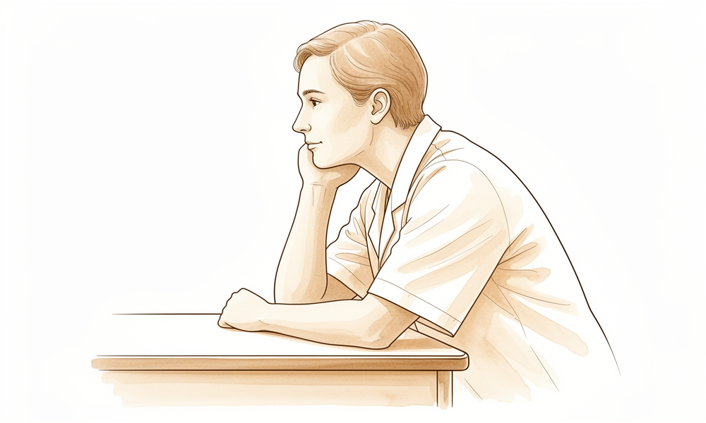
عام طور پر سوجن ہی وہ چیز ہے جو آپ سب سے پہلے دیکھتے ہیں۔ یہ ایک دو دن میں تیزی سے، یا ہفتوں میں آہستہ آہستہ ابھر سکتی ہے۔ اس کہنی پر ٹیک لگانے سے یہ بڑھ جاتی ہے، اس لیے دفتری کام، کرسی کے بازو پر بازو رکھنا، یا کہنی کو اپنے نیچے موڑ کر سونا، یہ سب اسے بگاڑ سکتے ہیں۔ بہت سے لوگوں کو رات کے وقت اس میں دکھن محسوس ہوتی ہے، خاص طور پر اگر وہ اسی طرف سوتے ہوں۔ بازو کو سیدھا کرنے اور کہنی کی نوک سے دباؤ ہٹانے سے یہ عموماً پرسکون ہو جاتی ہے۔
روزمرہ کے وہ کام مشکل ہو جاتے ہیں جن میں کہنی کو زور لگا کر موڑنا پڑے یا نوک پر دباؤ پڑے۔ نیچی کرسی سے سہارا لے کر اٹھنا، ڈش واشر میں برتن رکھنا، خریداری کے تھیلے بازو کے نچلے حصے (forearm) سے لگا کر اٹھانا، یا پڑھتے وقت کہنی میز پر ٹکانا، یہ سب تکلیف دہ ہو سکتے ہیں۔ سر سے اوپر ہاتھ بڑھانے سے بھی سوجے ہوئے حصے پر کھنچاؤ پڑ سکتا ہے۔
یہ سوجن ہمیشہ صرف عام گھساؤ کی وجہ سے نہیں ہوتی۔ بعض اوقات یہ تھیلی میں انفیکشن کی وجہ سے ہوتی ہے، جس سے عام طور پر کہنی کے اوپر کی جلد سرخ، گرم اور زیادہ دردناک ہو جاتی ہے۔ بعض اوقات یہ جوڑ میں کرسٹلز (crystals) کی وجہ سے ہوتی ہے، جسے گاؤٹ (gout) کہتے ہیں، خاص طور پر اگر آپ کو پہلے بھی ایسے دورے پڑ چکے ہوں یا دوسری کہنی بھی متاثر ہو۔ یہاں کی سوجن جوڑوں کی سوزش والی بیماریوں سے بھی جڑی ہو سکتی ہے، جیسے ریومیٹائیڈ آرتھرائٹس، جو عام طور پر جسم کے دونوں طرف کئی جوڑوں کو ایک ساتھ متاثر کرتا ہے۔
چونکہ یہ وجوہات ایک جیسی نظر آ سکتی ہیں، اس لیے باہر سے دیکھ کر انہیں الگ پہچاننا ہمیشہ آسان نہیں ہوتا۔ اگر آپ کی کہنی گرم، سرخ یا بہت دردناک ہے، یا سوجن ہفتوں سے بغیر کم ہوئے موجود ہے، تو بہتر ہے کہ اس کا صحیح طرح معائنہ کروایا جائے تاکہ درست علاج شروع کیا جا سکے۔
دراصل کیا ہو رہا ہے
آپ کی کہنی کی نوک پر سیال کی ایک چھوٹی تھیلی ہوتی ہے جسے برسا کہتے ہیں۔ اسے ایک پتلی گدی سمجھیں، جیسے جلد اور ہڈی کے درمیان ایک گدی دار تھیلی۔ اس کا کام یہ ہے کہ ہر بار جب آپ بازو موڑیں یا سیدھا کریں تو جلد ہڈی کے اوپر آسانی سے پھسلے۔
کہنی کے برسا کی سوزش میں یہ گدی سوزش کا شکار ہو جاتی ہے اور اضافی سیال سے سوج جاتی ہے۔ یہ عام طور پر کہنی کی نوک پر بار بار چھوٹی چوٹوں یا دباؤ کے بعد ہوتا ہے، اس قسم کا دباؤ جو روز بہ روز سخت سطحوں پر ٹیک لگانے سے پڑتا ہے۔ وقت کے ساتھ رگڑ سے بھی یہ تھیلی بس بڑی ہو سکتی ہے۔ جو گانٹھ آپ دیکھ اور محسوس کر سکتے ہیں وہ خود سوجی ہوئی تھیلی ہے، اور چھونے پر درد اس کے ارد گرد کے سوزش زدہ ٹشو سے ہوتا ہے۔
بعض اوقات یہ سوجن صرف جلن (irritation) نہیں ہوتی۔ تھیلی میں انفیکشن ہو سکتا ہے، اسی لیے انفیکشن والا برسا عموماً اس برسا کے مقابلے میں زیادہ گرم، سرخ اور دردناک ہوتا ہے جس میں صرف سوزش ہو۔ گاؤٹ کے کرسٹلز تھیلی میں جم سکتے ہیں، اور ریومیٹائیڈ آرتھرائٹس جیسی سوزش والی بیماریاں اسی جگہ پر سوجن اور چھوٹی سخت گانٹھیں پیدا کر سکتی ہیں۔ چونکہ یہ وجوہات باہر سے بہت ملتی جلتی نظر آتی ہیں، اس لیے انہیں الگ پہچاننے کے لیے باقاعدہ معائنہ درکار ہو سکتا ہے۔
کہنی کی نوک والی ہڈی، جسے کہنی کی نوک (olecranon) کہتے ہیں، جلد کے بالکل نیچے ہوتی ہے اور اس کے ارد گرد بہت کم گدی ہوتی ہے۔ یہی کھلی جگہ وجہ ہے کہ اسے اتنی آسانی سے چوٹ لگتی ہے، اور اس کے اوپر کا برسا اتنا زیادہ گھساؤ کیوں برداشت کرتا ہے۔ یہی ہڈی بازو کے پچھلے حصے کے اس پٹھے کے ٹینڈن (tendon)، یعنی وہ مضبوط ڈوری جو پٹھے کو ہڈی سے جوڑتی ہے، کو بھی تھامے رکھتی ہے جو آپ کی کہنی کو سیدھا کرتا ہے، اس لیے اس چھوٹے سے حصے کے مسائل پوری کہنی کے احساس اور حرکت پر اثر ڈال سکتے ہیں۔
ہم اس کے لیے کیا کر سکتے ہیں
ڈاکٹر کیرن ہِرپرا، جو Mater Private Hospital Rockhampton میں بازو اور ہاتھ کے سرجن (upper-limb surgeon) ہیں، سب سے کم مداخلت والے ان طریقوں سے شروع کرتے ہیں جو آپ کی حالت کے مطابق ہوں۔ مریضوں کو عام طور پر ان کے فیملی ڈاکٹر (GP) ہمارے کلینک بھیجتے ہیں؛ اگر کسی فزیوتھراپسٹ نے آپ کو ہمیں دکھانے کا مشورہ دیا ہے، تب بھی میڈیکیئر (Medicare) کی رقم کی واپسی کا حقدار ہونے کے لیے آپ کو اپنے فیملی ڈاکٹر سے ریفرل (referral) لینا ہوگا۔ پہلی ملاقات میں ہم آپ کی تفصیلات لیتے ہیں، آپ کی کہنی کا معائنہ کرتے ہیں اور اگر مددگار ہو تو الٹراساؤنڈ یا ایم آر آئی جیسی امیجنگ کا انتظام کرتے ہیں۔ ایم آر آئی اسکین مقناطیس کے ذریعے نرم ٹشوز کی تفصیلی تصاویر بناتا ہے، اور یہ دکھانے میں اچھا ہے کہ تھیلی کتنی سوجی ہوئی ہے اور اس میں کتنا سیال ہے۔ امیجنگ سوجن کے پیچھے چھپے دوسرے مسائل بھی دکھا سکتی ہے، جو اہم ہے کیونکہ مختلف وجوہات کو مختلف علاج کی ضرورت ہوتی ہے۔
زیادہ تر پرانے کیسز میں ہم سادہ معاون دیکھ بھال سے شروع کرتے ہیں۔ اس کا مطلب ہے برف، پٹی یا آستین (sleeve) سے دباؤ دینا، کہنی کی نوک پر حفاظتی گدی، اور ان سرگرمیوں کو بدلنا جو اسے بار بار بگاڑتی ہیں۔ کہنی کی نوک سے دباؤ ہٹانا اکثر وہ واحد سب سے مفید تبدیلی ہے جو آپ کر سکتے ہیں۔ فزیوتھراپی کا مقصد جلن کو کم کرنا اور سوجن بھڑکائے بغیر آرام دہ حرکت دوبارہ بحال کرنا ہے۔ کچھ اور سوچنے سے پہلے ہم اس طریقے کو پورا موقع دیتے ہیں، کیونکہ بغیر آپریشن کا علاج زیادہ تر لوگوں کے لیے اچھا کام کرتا ہے اور سوئیوں یا سرجری سے جڑے خطرات سے بچاتا ہے۔
اگر تھیلی میں انفیکشن ہو تو ہم براہِ راست اس انفیکشن کا علاج کرتے ہیں۔ اس تھیلی کا سادہ (پیچیدگی کے بغیر) انفیکشن عام طور پر صرف دوا سے ٹھیک ہو سکتا ہے، تھیلی نکالنے کی ضرورت کے بغیر۔ ہم ریومیٹائیڈ آرتھرائٹس والے لوگوں پر بھی گہری نظر رکھتے ہیں، کیونکہ کبھی کبھار برسا کے ساتھ ساتھ خود کہنی کا جوڑ بھی متاثر ہو سکتا ہے، اور اس سے علاج بدل جاتا ہے۔
اس مسئلے کے لیے سرجری ہمارا معمول کا پہلا قدم نہیں ہے۔ زیادہ تر سرجن جلد آپریشن کرنے سے گریز کرتے ہیں، کیونکہ اس ابھری ہوئی ہڈی پر زخم آہستہ بھر سکتا ہے اور بعض اوقات ایک سائنس (sinus)، یعنی ایک چھوٹی نالی جس سے مسلسل رطوبت رستی رہتی ہے، چھوڑ سکتا ہے۔ جب بغیر آپریشن کے علاج سے کافی بہتری نہ ہو اور سوجن بار بار واپس آتی رہے، تو ہم سوزش والی تھیلی کو نکالنے پر غور کرتے ہیں، جس آپریشن کو برسا نکالنا (bursectomy) کہتے ہیں۔ جہاں مناسب ہو، ہم یہ ایک ننھے کیمرے کی مدد سے چند چھوٹے چیروں کے ذریعے کر سکتے ہیں، اس طریقے کو دوربین کے ذریعے آپریشن (endoscopic surgery) کہتے ہیں۔ اس سے کہنی کی حساس نوک کے عین اوپر لمبے زخم سے بچا جا سکتا ہے۔ سرجری آپ کے لیے صحیح ہے یا نہیں، یہ فیصلہ ہم آپ کے ساتھ مل کر کرتے ہیں، یہ دیکھتے ہوئے کہ آپ کن کاموں میں واپس جانا چاہتے ہیں اور ہر راستے میں کیا کیا شامل ہے۔
کیا توقع رکھیں
زیادہ تر لوگوں کے لیے یہ سوجن سادہ دیکھ بھال سے ٹھیک ہو جاتی ہے۔ کہنی کی نوک سے دباؤ ہٹانا، اسے گدی سے محفوظ رکھنا اور بگاڑنے والی سرگرمیاں کم کرنا تھیلی کو پرسکون ہونے کا موقع دیتا ہے۔ اس مسئلے میں بغیر آپریشن کا علاج عام طور پر سرجری سے زیادہ مؤثر اور محفوظ ہے، اور زیادہ تر لوگوں کو کبھی آپریشن کی ضرورت نہیں پڑتی۔ اگر تھیلی میں انفیکشن ہو لیکن انفیکشن سادہ ہو، تو صرف دوا سے اس کا علاج عام طور پر کامیاب رہتا ہے، اور سوجن کو اکثر سیال نکالے یا تھیلی ہٹائے بغیر سنبھالا جا سکتا ہے۔
اس دیکھ بھال کے بغیر، یا اگر وہی دباؤ جاری رہیں، تو سوجن برقرار رہنے یا بار بار واپس آنے کا رجحان رکھتی ہے۔ کچھ لوگوں کو لگتا ہے کہ یہ ہفتوں تک بغیر کم ہوئے رہتی ہے۔ اگر آپ کی کہنی لمبے عرصے تک سوجی رہے، تو دوبارہ معائنہ کروانا بہتر ہے، کیونکہ جو سوجن ٹھیک نہ ہو وہ کبھی کبھار کسی کم عام وجہ کی طرف اشارہ کر سکتی ہے جسے اپنے الگ علاج کی ضرورت ہو۔
جب بغیر آپریشن کے علاج کو پورا موقع مل چکا ہو اور سوجن پھر بھی بار بار واپس آتی رہے، تو سوزش والی تھیلی کو نکالنے کی سرجری ایک راستہ بن جاتی ہے۔ یہ فیصلہ ہلکے میں نہیں لیا جاتا، اور عام طور پر اسے اس وقت تک روکا جاتا ہے جب تک سادہ تدابیر ناکام نہ ہو جائیں۔ جہاں اس ضدی قسم کی سوجن کے لیے سرجری کی جاتی ہے، وہاں لوگ عموماً اپنی کہنی کے احساس اور کارکردگی میں حقیقی بہتری دیکھتے ہیں۔ ننھے کیمرے کے ساتھ چند چھوٹے چیروں کے ذریعے تھیلی نکالنے کو آپریشن کے بعد بہت کم درد اور تیز صحت یابی سے جوڑا گیا ہے، اور رپورٹ کیے گئے مریضوں میں نہ تو سوجن دوبارہ ہوئی اور نہ زخم بھرنے کی ایسی کوئی پیچیدگی ہوئی جس کے لیے دوبارہ آپریشن تھیٹر جانا پڑا ہو۔ اس کے باوجود یہاں سرجری بے خطا نہیں ہے: جن لوگوں کی تھیلی نکالی جاتی ہے ان میں سے تقریباً 11.5 فیصد کو بعد میں مزید آپریشن کی ضرورت پڑتی ہے۔ اس بات کا بھی تھوڑا امکان ہے کہ سرجری کے بعد بھی سوجن واپس آ جائے۔
سرجری کے بعد صحت یابی عام طور پر آسان ہوتی ہے، لیکن اس ہڈی والی نوک پر زخم آہستہ بھر سکتا ہے، اور یہ ایک وجہ ہے کہ سرجن جلد آپریشن کرنے سے گریز کرنا پسند کرتے ہیں۔ زیادہ تر لوگ مہینوں کے بجائے ہفتوں میں کہنی کے آرام دہ، روزمرہ استعمال پر واپس آ جاتے ہیں۔ اگر کبھی سرجری منصوبے کا حصہ بنے تو آپ کے سرجن آپ کو بتائیں گے کہ آپ کے لیے صحت یابی کیسی ہوگی۔
ڈاکٹر کو کب دکھائیں
کہنی کی نوک پر زیادہ تر سوجن آرام اور سادہ دیکھ بھال سے ٹھیک ہو جاتی ہے۔ اپنے فیملی ڈاکٹر کو دکھائیں اگر گانٹھ بڑھتی رہے، یا کہنی سے چند ہفتے دباؤ ہٹانے کے بعد بھی کم نہ ہوئی ہو۔
کچھ وجوہات کو جلدی سلجھانا ضروری ہے۔ اگر کہنی کے اوپر کی جلد گرم، سرخ اور بہت دردناک ہو تو ہسپتال کی ایمرجنسی جائیں، کیونکہ اس کا مطلب ہو سکتا ہے کہ تھیلی میں انفیکشن ہے اور یہاں انفیکشن کا اسی دن معائنہ ضروری ہے۔ کسی ماہر (specialist) سے معائنے کی درخواست کریں اگر آپ کو پہلے گاؤٹ کے دورے پڑ چکے ہوں، اگر دوسری کہنی بھی سوجی ہوئی ہو، یا اگر آپ کے جسم کے دونوں طرف کئی جوڑ متاثر ہوں، کیونکہ یہ سادہ جلن کے بجائے گاؤٹ یا ریومیٹائیڈ آرتھرائٹس جیسی سوزش والی بیماری کی طرف اشارہ کرتے ہیں۔ باہر سے انفیکشن اور گاؤٹ میں فرق کرنا مشکل ہو سکتا ہے، اس لیے خود اس کا اندازہ لگانے کی کوشش نہ کریں۔
مزید تفصیل
Advanced reading: the deeper science (optional)
یہ حصہ آپ کے اپنے علاج کے فیصلوں کے لیے ضروری معلومات سے آگے جاتا ہے۔ کہنی کے برسا کی سوزش کے بارے میں اضافی مطالعہ اس لیے مفید ہے کہ اس کے لیے جو دو کام سب سے زیادہ فطری طور پر کیے جاتے ہیں، یعنی سوئی سے سوجن کا سیال نکالنا اور اس میں اسٹیرائیڈ کا انجکشن لگانا، ان دونوں کے خلاف شواہد موجود ہیں۔
سیال نکالنا ہی مسئلے کی وجہ بن سکتا ہے
دل یہی کہتا ہے: سیال ہے، تو سیال نکال دو۔ تحقیق کے اعداد و شمار الٹی طرف اشارہ کرتے ہیں۔ سادہ (پیچیدگی کے بغیر) انفیکشن والے کہنی کے برسا کی سوزش کے علاج کے طریقوں کے ایک موازنے میں، سیال نکالے بغیر (without aspiration) اندازے کی بنیاد پر کیا گیا علاج مؤثر رہا، اور کسی مریض کو برسا نکالنے کے آپریشن کی ضرورت نہیں پڑی، جبکہ روایتی طور پر سیال نکالنے والے گروپ میں 11 میں سے 8 مریضوں کو بعد میں سرجری کے ذریعے برسا نکلوانا پڑا [1]۔
ایک چھوٹی تحقیق میں یہ ایک نمایاں فرق ہے، اور اسے اسی احتیاط کے ساتھ پڑھنا چاہیے۔ لیکن اس کا طریقۂ کار پراسرار نہیں ہے۔ برسا ایک ہڈی والی نوک پر پتلی جلد کے بالکل نیچے ہوتا ہے جو ہر چوٹ اور میز پر ہر ٹیک کا بوجھ اٹھاتی ہے۔ اس جلد میں سوئی ڈالنے سے ایک راستہ (tract) بن جاتا ہے، اور دباؤ والی جگہ پر بنا راستہ آہستہ بند ہوتا ہے اور اس سے رطوبت رستی رہ سکتی ہے۔
عمومی جائزے سمت پر متفق ہیں۔ بنیادی طور پر درجہ چار (level IV) کے شواہد کی بنیاد پر، بغیر سرجری کا علاج سرجری کے مقابلے میں نمایاں طور پر زیادہ مؤثر اور محفوظ ہے [2]۔
اسٹیرائیڈ کا انجکشن نتیجہ بہتر کیے بغیر خطرہ رکھتا ہے
یہ بات طبی تحقیق میں غیر معمولی طور پر صاف الفاظ میں کہی گئی ہے۔ بغیر انفیکشن والی برسا کی سوزش (aseptic bursitis) میں، کورٹیکوسٹیرائیڈ کے انجکشن کے ساتھ نمایاں خطرات جڑے ہیں اور اس سے نتیجہ بہتر نہیں ہوتا [2]۔
یہ خطرات اس جگہ سے مخصوص ہیں: جلد کے بالکل نیچے ابھری ہوئی ہڈی کے اوپر جلد کا پتلا ہو جانا اور اس کا رنگ اڑ جانا، اور ایسی جگہ میں انفیکشن داخل ہو جانا جسے صاف کرنا مشکل ہے۔ بازو اور ہاتھ کے دوسرے حصوں میں، جب سادہ تدابیر ناکام ہو جائیں تو اسٹیرائیڈ کا انجکشن ایک معقول قدم ہے۔ یہاں شواہد اس کی حمایت نہیں کرتے، اور اگر آپ کو یہ پیش کیا جائے تو یہ فرق جاننا مفید ہے۔
سرجری واقعی آخری راستہ ہے، اور اس کی ناکامی کی شرح ناپی جا سکتی ہے
جہاں برسا نکالنے کا آپریشن کیا جاتا ہے، وہاں یہ قابلِ اعتماد طور پر معاملہ ختم نہیں کرتا۔ برسا نکالنے کے بعد دوبارہ آپریشن کی شرح 11.5 فیصد تھی، اور دوبارہ آپریشن ان مریضوں میں زیادہ ہوا جنہیں ریومیٹائیڈ آرتھرائٹس، ذیابیطس ہو، جنہیں کسی بھی کہنی میں پہلے برسا کی سوزش ہو چکی ہو، اور خواتین میں [3]۔
یہ خطرے کے عوامل ان حالات کو بیان کرتے ہیں جن میں برسا کے دوبارہ بن جانے کا امکان ہوتا ہے: سوزش یا جسم کے کیمیائی نظام (metabolic) سے جڑی کوئی وجہ جسے سرجری ختم نہیں کرتی، یا اس شخص کے ٹشو میں یہ مسئلہ پیدا کرنے کا ثابت شدہ رجحان۔ بار بار ہونے والے کیسز کے لیے، برسا کے اندر ڈوکسی سائیکلین سے سکلیروتھراپی (intrabursal doxycycline sclerotherapy)، یعنی برسا کو کاٹ کر نکالنے کے بجائے جان بوجھ کر اس جگہ میں زخم کا ریشہ بنا کر اسے بند کر دینا، کو 45 کہنیوں کی ایک تحقیق میں ایک متبادل کے طور پر بیان کیا گیا ہے [4]۔
اصل میں کیا چیز اسے ٹھیک کرتی ہے
سادہ سا جواب ہے حفاظت اور وقت۔ چونکہ برسا میں جلن کہنی کی نوک پر دباؤ سے مکینیکی طور پر ہوتی ہے، اس لیے جو علاج اصل وجہ کو دور کرتا ہے وہ اس دباؤ کو ہٹانا ہے: گدی، اور کہنی پر ٹیک لگانے کی عادت بدلنا، اور یہ اس سے زیادہ عرصے تک جاری رکھنا جتنا ضروری محسوس ہو۔ زیادہ تر کیسز ٹھیک ہو جاتے ہیں [5]، اور اوپر بیان کی گئی مداخلتیں زیادہ تر ایک ایسے مرض کی مدت کم کرنے کی کوششیں ہیں جو ویسے بھی ٹھیک ہو جاتا ہے، اور ٹھیک اسی وجہ سے یہاں اپنا خطرہ رکھنے والی کسی بھی مداخلت کو ایک اونچا معیار پورا کرنا چاہیے۔
حوالہ جات
[1] Deal JB, Vaslow AS, Bickley RJ, Verwiebe EG, Ryan PM. Empirical treatment of uncomplicated septic olecranon bursitis without aspiration. J Hand Surg Am. 2020;45(1):20-5. https://doi.org/10.1016/j.jhsa.2019.06.012
[2] Sayegh ET, Strauch RJ. Treatment of olecranon bursitis: a systematic review. Arch Orthop Trauma Surg. 2014;134(11):1517-36. https://doi.org/10.1007/s00402-014-2088-3
[3] Germawi L, Westenberg RF, Wang F, Schep NW, Chen NC, Eberlin KR. Factors associated with revision surgery for olecranon bursitis after bursectomy. J Shoulder Elbow Surg. 2021;30(5):1135-41. https://doi.org/10.1016/j.jse.2020.09.033
[4] McDermott D, Wakefield D, Kowalsky M, Sethi P, Vitale MA, Morrey BF. Intrabursal doxycycline sclerotherapy for recurrent olecranon bursitis of the elbow. J Hand Surg Glob Online. 2024;6(4):504-9. https://doi.org/10.1016/j.jhsg.2024.03.006
[5] Nchinda NN, Wolf JM. Clinical management of olecranon bursitis: a review. J Hand Surg Am. 2021;46(6):501-6. https://doi.org/10.1016/j.jhsa.2021.02.006
Evidence & references
This is the clinical evidence summary written for health professionals. It is technical, and it lists the research this page was built from. You do not need to read it to understand your treatment or to make a decision about it.
Overview
- Surgical resection of a chronic olecranon bursitis should not be considered lightly [1].
- The revision rate after bursectomy for olecranon bursitis was 11.5% [2].
- The operation appears to give satisfactory results for chronic olecranon bursitis [3].
- Based primarily on level IV evidence, nonsurgical management of olecranon bursitis is significantly more effective and safer than surgical management [4].
- Nontuberculous mycobacterial olecranon bursitis should be considered in any patient with a swollen bursa and protracted course, regardless of immune status [5].
- Patients who underwent endoscopic olecranon bursectomy experienced no recurrences or wound-healing complications necessitating return to the operating room [6].
- Empirical management of uncomplicated septic olecranon bursitis was found to be effective with no patients requiring bursectomy [7].
- In the traditional aspiration group for uncomplicated septic olecranon bursitis, 8 of 11 patients required bursectomy [7].
- Intrabursal doxycycline sclerotherapy may be an effective alternative to surgical bursectomy for patients with recurrent olecranon bursitis refractory to conservative management [8].
- Patients with refractory olecranon bursitis, particularly if aseptic, tend to gain significant physical health benefits from open bursectomy [9].
- Endoscopic bursectomies are a useful treatment option for patients with recalcitrant olecranon bursitis [10].
- Bursal suture repair is a viable alternative to bursectomy in selected patients with chronic traumatic olecranon bursitis [11].
- Bursal suture repair combines functional and cosmetic benefits in selected patients with chronic traumatic olecranon bursitis [11].
- Deferring aspiration in uncomplicated septic olecranon bursitis is a reasonable treatment option [16].
Anatomy & Pathophysiology
Bony Anatomy
- The olecranon and the coronoid process form the greater sigmoid notch, which articulates with the trochlea of the distal humerus [19].
- The intrinsic anatomy of the greater sigmoid notch allows flexion/extension movement of the elbow joint and provides stability for the elbow [19].
- The olecranon serves as the insertion for the triceps tendon, which blends with the periosteum of the proximal ulna [19].
- The exposed position of the olecranon renders it vulnerable to direct trauma and violent muscular contractions from the triceps [19].
- In children, the olecranon is predominantly cartilage, particularly in younger children, which reduces the chance of fracture occurring with a direct blow [20].
- The thick periosteum and relatively thin metaphyseal cortex of the olecranon in children predispose it to minimally displaced greenstick fractures [20].
Pathophysiology
- Olecranon bursitis is a condition where the bursal cavity, superficial to the olecranon, becomes inflamed [13].
- Olecranon bursitis can occur with or without infection [13].
- Olecranon bursitis is often predisposed by repeated minor trauma from external pressure [13].
- The olecranon bursa sometimes becomes enlarged as a result of pressure or friction [27].
- When the olecranon bursa is painful, the cause is more likely to be infection, gout, or rheumatoid arthritis [27].
- Gout is suspected in olecranon bursitis if there is a history of previous attacks, if the condition is bilateral, if there are tophi, or if X-ray shows calcification in the bursa [27].
- Rheumatoid arthritis causes both swelling and nodularity over the olecranon [27].
- In almost all cases of rheumatoid arthritis affecting the olecranon, it is associated with a typical symmetrical polyarthritis [27].
- In the late stages of rheumatoid arthritis, erosion of the elbow joint may cause marked instability [27].
Classification
- Olecranon bursitis has been given pseudonyms relating to the repeated minor trauma from external pressure that often predisposes [13].
- Traumatic lesions of the olecranon bursa are common injuries associated with a high risk of complications [14].
Clinical Presentation
- Olecranon bursitis is a common condition where the bursal cavity, superficial to the olecranon, becomes inflamed [13].
- Olecranon bursitis can occur either with or without infection [13].
- Due to multiple aetiologies, olecranon bursitis can present to any medical specialty with reasonable frequency [13].
- When the olecranon bursa is also painful, the cause is more likely to be infection, gout, or rheumatoid arthritis [27].
- Gout is suspected if there is a history of previous attacks, if the condition is bilateral, if there are tophi, or if X-ray shows calcification in the bursa [27].
- In almost all cases of rheumatoid arthritis-associated olecranon bursitis, the condition is associated with a typical symmetrical polyarthritis [27].
- Pyoderma gangrenosum must be considered in the differential diagnosis whenever a patient presents with ulcerative cutaneous lesions that resemble an infectious process such as olecranon bursitis [38].
Investigations
- MR imaging is probably the method of choice for determining both the development of the bursae and their fluid content [44].
Treatment
Non-Operative Management
- The first treatment line for olecranon bursitis is conservative, including ice, rest, anti-inflammatory and analgesic drugs and, occasionally, bursal fluid aspiration [25].
- More recent literature demonstrates adverse effects of intrabursal injections and surgery compared with noninvasive management for initial treatment of nonseptic olecranon bursitis [18].
- Hydrothermal ablation at temperatures between 50C and 52C is a safe treatment option for recurrent or chronic olecranon bursitis [34].
- Hydrothermal ablation for recurrent or chronic olecranon bursitis has fewer complications than open bursectomy and comparable efficacy [34].
Operative Management
- Open bursectomy appears to give satisfactory results for chronic olecranon bursitis [3].
- Endoscopic olecranon bursectomy provides a satisfactory view with less morbidity than the open method while avoiding a wound over the sensitive point of the olecranon [15].
- Bursal suture repair is a viable alternative to bursectomy in selected patients with chronic traumatic olecranon bursitis, combining functional and cosmetic benefits [11].
- Excision has been curative for all lesions of the olecranon bursa, whereas multiple medications have been tried for cutaneous and systemic infections without clear-cut success [17].
Surgical Complications and Outcomes
- More recent literature demonstrates adverse effects of surgery compared with noninvasive management for initial treatment of nonseptic olecranon bursitis [18].
- Surgeons are reluctant to perform surgical excision as first-line treatment for chronic olecranon bursitis due to the fear of causing a chronic draining sinus or infection [45].
Infection and Atypical Etiologies
Recovery
- Nonsurgical management of olecranon bursitis is significantly more effective and safer than surgical management [4].
- 8 of 11 patients in the traditional aspiration group required bursectomy [7].
- Bursal suture repair combines functional and cosmetic benefits [11].
- The extrabursal endoscopic approach provides a satisfactory view with less morbidity than the open method [15].
- The extrabursal endoscopic approach avoids a wound over the sensitive point of the olecranon [15].
- Excision has been curative for all lesions of the olecranon bursa [17].
- Multiple medications have been tried for cutaneous and systemic infections without clear-cut success [17].
Key Evidence
- [L3] The revision rate after bursectomy for olecranon bursitis was 11.5%. [2] (10.1016/j.jse.2020.09.033)
- [L4] The operation appears to give satisfactory results for chronic olecranon bursitis. [3] (10.1016/s0020-1383(77)80050-8)
- [L4] Based primarily on level IV evidence, nonsurgical management of olecranon bursitis is significantly more effective and safer than surgical management. [4] (10.1007/s00402-014-2088-3)
- [L4] Nontuberculous mycobacterial olecranon bursitis should be considered in any patient with a swollen bursa and protracted course, regardless of immune status. [5] (10.1016/j.jse.2008.07.009)
- [L4] In this population, patients who underwent endoscopic olecranon bursectomy experienced no recurrences or wound-healing complications necessitating return to the operating room. [6] (10.1016/j.asmr.2023.100832)
- [L4] Empirical management of uncomplicated septic olecranon bursitis was found to be effective with no patients requiring bursectomy, whereas 8 of 11 patients in the traditional aspiration group required bursectomy. [7] (10.1016/j.jhsa.2019.06.012)
- [L4] This may be an effective alternative to surgical bursectomy for patients with recurrent olecranon bursitis refractory to conservative management. [8] (10.1016/j.jhsg.2024.03.006)
- [L4] Patients with refractory olecranon bursitis, particularly if aseptic, tend to gain significant physical health benefits from open bursectomy. [9] (10.7759/cureus.43696)
- [L5] Endoscopic bursectomies are a useful treatment option for patients with recalcitrant olecranon bursitis. [10] (10.1016/j.eats.2023.09.004)
- [L5] Bursal suture repair is a viable alternative to bursectomy in selected patients with chronic traumatic olecranon bursitis, combining functional and cosmetic benefits. [11] (10.1016/j.xrrt.2025.100597)
- [L4] [13] (10.1177/1758573214532787)
- [Paper] Traumatic lesions of the olecranon bursa or prepatellar bursa are common injuries associated with a high risk of complications. [14] (10.1007/s00402-017-2690-2)
- [L4] This approach provides a satisfactory view with less morbidity than the open method while avoiding a wound over the sensitive point of the olecranon. [15] (10.1097/bth.0b013e31829c0535)
- [L4] Deferring aspiration in uncomplicated septic olecranon bursitis is a reasonable treatment option. [16] (10.1016/j.jhsa.2018.06.059)
- [Case_report] Excision has been curative for all lesions of the olecranon bursa, whereas multiple medications have been tried for cutaneous and systemic infections without clear-cut success. [17] (10.2106/00004623-198062050-00024)
- [L5] Older studies showed resolution with injections and surgery, but more recent literature demonstrates adverse effects of intrabursal injections and surgery compared with noninvasive management for initial treatment of nonseptic olecranon bursitis. [18] (10.1016/j.jhsa.2021.02.006)
- [L4] The first treatment line for olecranon bursitis is conservative, including ice, rest, anti-inflammatory and analgesic drugs and, occasionally, bursal fluid aspiration. [25] (10.1016/j.surge.2012.02.002)
- [L4] Hydrothermal ablation at temperatures between 50C and 52C is a safe treatment option for recurrent or chronic olecranon bursitis with fewer complications than open bursectomy and a comparable efficacy. [34] (10.1016/j.jse.2024.03.021)
- [Case_report] PG must be considered in the differential diagnosis whenever a patient presents with ulcerative cutaneous lesions that resemble an infectious process such as olecranon bursitis. [38] (10.1016/j.jse.2014.06.032)
- [L4] MR imaging is probably the method of choice for determining both the development of the bursae and their fluid content. [44] (10.1007/s002560050117)
- [L3] [45] (10.1016/j.jhsa.2010.12.030)
References
[1] Complications following resection of the olecranon bursa. Acta Orthopaedica Belgica. 2006.
[2] Factors associated with revision surgery for olecranon bursitis after bursectomy. Journal of Shoulder and Elbow Surgery. 2021. DOI: 10.1016/j.jse.2020.09.033
[3] A useful procedure in the treatment of chronic olecranon bursitis. Injury. 1977. DOI: 10.1016/s0020-1383(77)80050-8
[4] Treatment of olecranon bursitis: a systematic review. Archives of Orthopaedic and Trauma Surgery. 2014. DOI: 10.1007/s00402-014-2088-3
[5] Nontuberculous mycobacterial olecranon bursitis: Case reports and literature review. Journal of Shoulder and Elbow Surgery. 2009. DOI: 10.1016/j.jse.2008.07.009
[6] No Wound Healing Complications or Recurrences Were Seen and a High Level of Satisfaction Was Reported in Patients Who Underwent Endoscopic Olecranon Bursectomy for Recalcitrant Olecranon Bursitis. Arthroscopy, Sports Medicine, and Rehabilitation. 2024. DOI: 10.1016/j.asmr.2023.100832
[7] Empirical Treatment of Uncomplicated Septic Olecranon Bursitis Without Aspiration. The Journal of Hand Surgery. 2020. DOI: 10.1016/j.jhsa.2019.06.012
[8] Intrabursal Doxycycline Sclerotherapy for Recurrent Olecranon Bursitis of the Elbow: A Case Control Study. Journal of Hand Surgery Global Online. 2024. DOI: 10.1016/j.jhsg.2024.03.006
[9] Clinical Outcomes Following Open Olecranon Bursa Excision for Septic and Aseptic Olecranon Bursitis: An Observational Study. Cureus. 2023. DOI: 10.7759/cureus.43696
[10] Endoscopic Olecranon Bursectomy in the Treatment of Recalcitrant Olecranon Bursitis: Patient Selection and Operative Technique. Arthroscopy Techniques. 2023. DOI: 10.1016/j.eats.2023.09.004
[11] Olecranon bursal repair for chronic traumatic bursitis: a surgical technique. JSES Reviews, Reports, and Techniques. 2026. DOI: 10.1016/j.xrrt.2025.100597
[13] Olecranon bursitis: a systematic overview. Shoulder & Elbow. 2014. DOI: 10.1177/1758573214532787
[14] Treatment and outcome with traumatic lesions of the olecranon and prepatellar bursa: a literature review apropos a retrospective analysis including 552 cases. Archives of Orthopaedic and Trauma Surgery. 2017. DOI: 10.1007/s00402-017-2690-2
[15] Olecranon Extrabursal Endoscopic Bursectomy. Techniques in Hand & Upper Extremity Surgery. 2013. DOI: 10.1097/bth.0b013e31829c0535
[16] Empiric Treatment of Uncomplicated Septic Olecranon Bursitis Without Aspiration. The Journal of Hand Surgery. 2018. DOI: 10.1016/j.jhsa.2018.06.059
[17] Protothecal olecranon bursitis. A case report and review of the literature.. The Journal of Bone & Joint Surgery. 1980. DOI: 10.2106/00004623-198062050-00024
[18] Clinical Management of Olecranon Bursitis: A Review. The Journal of Hand Surgery. 2021. DOI: 10.1016/j.jhsa.2021.02.006
[19] Aaos Comprehensive Orthopaedic Review 3. Fractures of the Elbow > II. Olecranon Fractures.
[20] Tachdjian S Pediatric Orthopaedics From The Texas Scottish Rite Hospital For Children E Book. Pigmented Villonodular Synovitis and Giant Cell Tumor of the Tendon Sheath > Olecranon Fractures.
[25] Diagnosis and management of olecranon bursitis. The Surgeon. 2012. DOI: 10.1016/j.surge.2012.02.002
[27] Apley And Solomon S Concise System Of Orthopaedics And Trauma. OLECRANON BURSITIS.
[34] Hydrothermal ablation in recurrent or chronic olecranon bursitis: a prospective study. Journal of Shoulder and Elbow Surgery. 2024. DOI: 10.1016/j.jse.2024.03.021
[38] Case report: misdiagnosed olecranon bursitis: pyoderma gangrenosum. Journal of Shoulder and Elbow Surgery. 2014. DOI: 10.1016/j.jse.2014.06.032
[44] The US,CT and MR findings of cubital bursitis: a report of five cases. Skeletal Radiology. 1996. DOI: 10.1007/s002560050117
[45] Chronic Olecranon Bursitis. The Journal of Hand Surgery. 2011. DOI: 10.1016/j.jhsa.2010.12.030