Patients › Rehabilitation
ریڈیل ٹنل ریلیز
Post-operative exercises and precautions after radial tunnel release, including radial nerve glides.
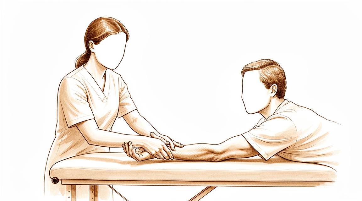
یہ پروٹوکول (protocol، یعنی بحالی کا مرحلہ وار منصوبہ) Mater Private Hospital Rockhampton میں ڈاکٹر کیرن ہِرپرا کے ہاتھوں ریڈیل ٹنل ریلیز (radial tunnel release، کہنی کے پاس ریڈیل عصب پر دباؤ ختم کرنے کا آپریشن) کے بعد آپ کی صحت یابی میں رہنمائی کرتا ہے۔ یہ بتاتا ہے کہ سرجری کے بعد کے ہفتوں میں کیا توقع رکھیں، اور آپریشن کے بعد دیے گئے آپ کے ہدایت نامے (handout) سے ورزشوں کا پروگرام پیش کرتا ہے۔ فزیوتھراپی یا ہینڈ تھراپی کی اپنی پہلی ملاقات پر یہ صفحہ یا اس کی پی ڈی ایف ساتھ لائیں تاکہ آپ کی بحالی کا علاج مربوط رہے۔ آپ کا تھراپسٹ آپ کی صحت یابی کی رفتار کے مطابق اس منصوبے میں تبدیلی کر سکتا ہے۔
اگر سرجری کے بعد آپ کو اپنے زخم کے بارے میں کوئی تشویش ہو تو کلینک سے رابطہ کریں۔ اکثر یہ مددگار ہوتا ہے کہ زخم کی تصویر لے کر جائزے کے لیے ای میل کر دیں۔
کیا توقع رکھیں
زخم کی دیکھ بھال کا طریقہ پریکٹس کی زخم کی دیکھ بھال کی ہدایات میں بتایا گیا ہے۔ جیسے جیسے آپ کا زخم بھرتا ہے، جس عصب (nerve) کو آزاد کیا گیا ہے وہ آس پاس کے ٹشو سے چپک سکتا ہے؛ نیچے دی گئی عصب کو سرکانے (gliding) والی ورزشیں اسے آزادانہ حرکت میں رکھنے اور بندھ جانے سے روکنے کے لیے بہت اہم ہیں۔
بعض اوقات زخم حساس ہو سکتا ہے۔ یہ معمول کی بات ہے، اور روزانہ حساسیت کم کرنے کی مشق (desensitisation) شروع کر کے اسے روکا یا کم کیا جا سکتا ہے: یعنی سرجری کے فوراً بعد سے زخم (یا پٹی) کے اوپر آہستہ سے تھپتھپانا اور سہلانا۔ اس طرح کا "حسی ردعمل" (sensory feedback) عصب کو چھونے اور بناوٹ کے احساس کو معمول پر لانے میں مدد دیتا ہے۔
جب زخم پوری طرح بھر جائے تو زخم کے نشان کی مالش شروع کریں: چیرے کے اوپر مضبوطی سے دائروں میں۔ زخم کے نشان کی دیکھ بھال کے بارے میں مزید معلومات کے لیے زخم کی دیکھ بھال کی ہدایات دیکھیں۔
صحت یابی کے بارے میں حقیقت پسندانہ توقعات رکھنا ضروری ہے۔ بازو کی عام حرکتوں کے دوران ریڈیل عصب (radial nerve) کو ایک قابل پیمائش حد تک سرکنا اور کھنچنا پڑتا ہے، اس لیے اسے جلد حرکت میں رکھنا ہی اسے بھرتے ہوئے ٹشوز سے چپکنے سے روکتا ہے [1]۔ اس کے باوجود ریڈیل ٹنل ریلیز کے بعد درد میں آرام اکثر فوری نہیں بلکہ آہستہ آہستہ آتا ہے، اور کچھ لوگوں میں یہ صرف جزوی ہوتا ہے۔ سچ یہ ہے کہ یہ آپریشن عصب پر دباؤ ختم کرنے کے کچھ دوسرے آپریشنوں کے مقابلے میں ایسے نتائج دیتا ہے جن کا پہلے سے اندازہ لگانا زیادہ مشکل ہے: ریڈیل ٹنل سنڈروم (radial tunnel syndrome) کی پورے یقین سے تشخیص مشکل ہو سکتی ہے اور یہ اکثر ٹینس ایلبو (کہنی کے باہر کی طرف درد) کے ساتھ ملتا جلتا ہوتا ہے، اور نتائج میں فرق کی ایک وجہ یہی ہے۔ شائع شدہ طویل مدتی تحقیقات مجموعی طور پر تقریباً دو تہائی مریضوں میں اچھے نتائج بتاتی ہیں، اور بہترین نتائج ان لوگوں میں ہوتے ہیں جن میں صرف ریڈیل ٹنل کی علامات ہوں [2][3]۔ جب ساتھ میں ٹینس ایلبو (lateral epicondylitis) بھی ہو، ایک ہی بازو میں ایک سے زیادہ جگہ عصب دبا ہو، یا کارکنوں کے معاوضے (workers' compensation) کا دعویٰ ہو، تو صحت یابی عموماً سست اور کم مکمل ہوتی ہے [2][4]۔ عصب کو سرکانے والا پروگرام اور درجہ بدرجہ حساسیت کم کرنے کی مشق بحالی کے وہ حصے ہیں جو سب سے زیادہ آپ کے اپنے اختیار میں ہیں، اور روزانہ باقاعدہ مشق عصب کو ٹھیک ہونے کا بہترین موقع دیتی ہے۔
احتیاطیں اور پابندیاں
روزمرہ زندگی کے کاموں، جیسے اپنی دیکھ بھال، کھانا کھانا، کپڑے پہننا، لکھنا اور ٹائپ کرنا، کے لیے ہاتھ کے ہلکے استعمال کی حوصلہ افزائی کی جاتی ہے۔ اس کے علاوہ پابندیاں سادہ ہیں: سرجری کے بعد 6 ہفتے تک نہ کچھ اٹھائیں، نہ کچھ زور سے پکڑیں، نہ ہاتھ پر وزن ڈالیں، اور نہ ہی تھرتھراہٹ پیدا کرنے والی مشینیں (مثال کے طور پر بجلی کے اوزار یا گھاس کاٹنے کی مشین) استعمال کریں، اور پہلے 2 سے 3 ہفتوں تک گاڑی چلانا محدود ہے۔
آپ کے فزیوتھراپسٹ کے لیے:
اہداف
- آزاد کیے گئے عصب کو بھرتے ہوئے زخم سے چپکنے سے روکنا (عصب کو سرکانے کا پروگرام)
- درجہ بدرجہ حساسیت کم کرنے کی مشق سے زخم کی حساسیت کم کرنا
- کلائی، بازو کے نچلے حصے اور کہنی کی حرکت کی حد برقرار رکھنا
- روزمرہ زندگی کے کاموں کے لیے ہاتھ کے ہلکے استعمال میں مدد دینا
علاج کا طریقہ
- روزانہ حساسیت کم کرنے کی مشق: زخم (پٹی) کے اوپر آہستہ سے تھپتھپانا / سہلانا، آپریشن کے فوراً بعد سے شروع
- زخم پوری طرح بھر جانے کے بعد زخم کے نشان کی مالش (چیرے کے اوپر مضبوطی سے دائروں میں)
- نیچے دیے گئے کارڈز کے مطابق گھر پر ورزشوں کا پروگرام: کلائی کو موڑنے / پیچھے کرنے کا اسٹریچ؛ ہتھیلی اوپر / نیچے گھمانے کا اسٹریچ؛ کہنی کو موڑنا / سیدھا کرنا؛ ریڈیل عصب کو سرکانے کی ورزش
- آخری حد تک زور دار کھنچاؤ (tensioning) کے بجائے ہلکی سرکانے والی ("slider") عصبی ورزشوں کو ترجیح دیں: سرکانے والی تکنیکوں سے عصب کہیں کم کھنچاؤ کے ساتھ کافی زیادہ حرکت کرتا ہے، جو حال ہی میں دباؤ سے آزاد کیے گئے عصب کے آس پاس بہتر برداشت ہوتا ہے [1][5]
- پروگرام کے ساتھ اضافی طور پر عصب کو متحرک کرنے (nerve mobilisation) پر غور کیا جا سکتا ہے؛ عصب سے متعلق بیماریوں میں عصب کو متحرک کرنے (neural mobilisation) کے شواہد حمایت کرتے ہیں مگر ان کے قابلِ اعتماد ہونے کی سطح مختلف ہے، اس لیے آگے بڑھنا علامات کی رہنمائی میں ہونا چاہیے [6]
احتیاطیں
- ہاتھ کا صرف ہلکا روزمرہ استعمال (اپنی دیکھ بھال، کھانا کھانا، کپڑے پہننا، لکھنا، ٹائپ کرنا)
- آپریشن کے بعد 6 ہفتے تک نہ کچھ اٹھانا، نہ زور سے پکڑنا، نہ ہاتھ پر وزن ڈالنا، اور نہ تھرتھراہٹ پیدا کرنے والی مشینیں (مثلاً بجلی کے اوزار، گھاس کاٹنے کی مشین) استعمال کرنا
- پہلے 2 سے 3 ہفتوں تک گاڑی چلانا محدود ہے
- عصب کو سرکانے اور اسٹریچ کی ورزشیں ہلکی اور بنیادی طور پر درد کے بغیر ہونی چاہییں؛ زبردستی ایسی حد تک نہ لے جائیں جس سے آپریشن سے پہلے والا عصبی درد دوبارہ پیدا ہو
یہ آپریشن کے بعد دیے گئے آپ کے ہدایت نامے کی ورزشیں ہیں، جو سرجری کے بعد شروع کی جاتی ہیں اور گھر پر آپ کے فزیوتھراپسٹ یا ہینڈ تھراپسٹ (ہاتھ کی بحالی کے ماہر) کی رہنمائی کے مطابق جاری رکھی جاتی ہیں۔ ہر کارڈ پر دہرانے کی تعداد، روکے رکھنے کا وقت اور کتنی بار کرنی ہے، درج ہے۔
آپ کی ورزشیں
Kieran Hirpara 4.0
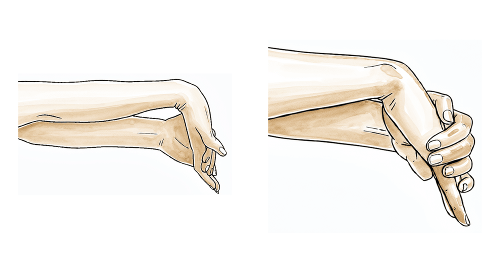
کلائی کو آگے موڑنے / پیچھے کرنے کا اسٹریچ
اپنی کہنی میز پر رکھیں اور کلائی کو آہستہ سے آگے پیچھے جھلائیں (یا تصویر کی طرح اسے میز یا کرسی کے بازو کے کنارے پر رکھیں)۔ جب زیادہ آرام محسوس ہو تو اپنے دوسرے ہاتھ کو ہتھیلی پر رکھ کر کلائی کو پیچھے کی طرف دبائیں، پھر آگے کی طرف، اور انگلیوں کو ڈھیلا رکھیں۔ ہر اسٹریچ کو 15 سیکنڈ تک روکے رکھیں؛ ہر سمت میں 5 بار دہرائیں۔
10 بار دہرائیں، روزانہ 4 سے 5 مرتبہ
Kieran Hirpara 4.0
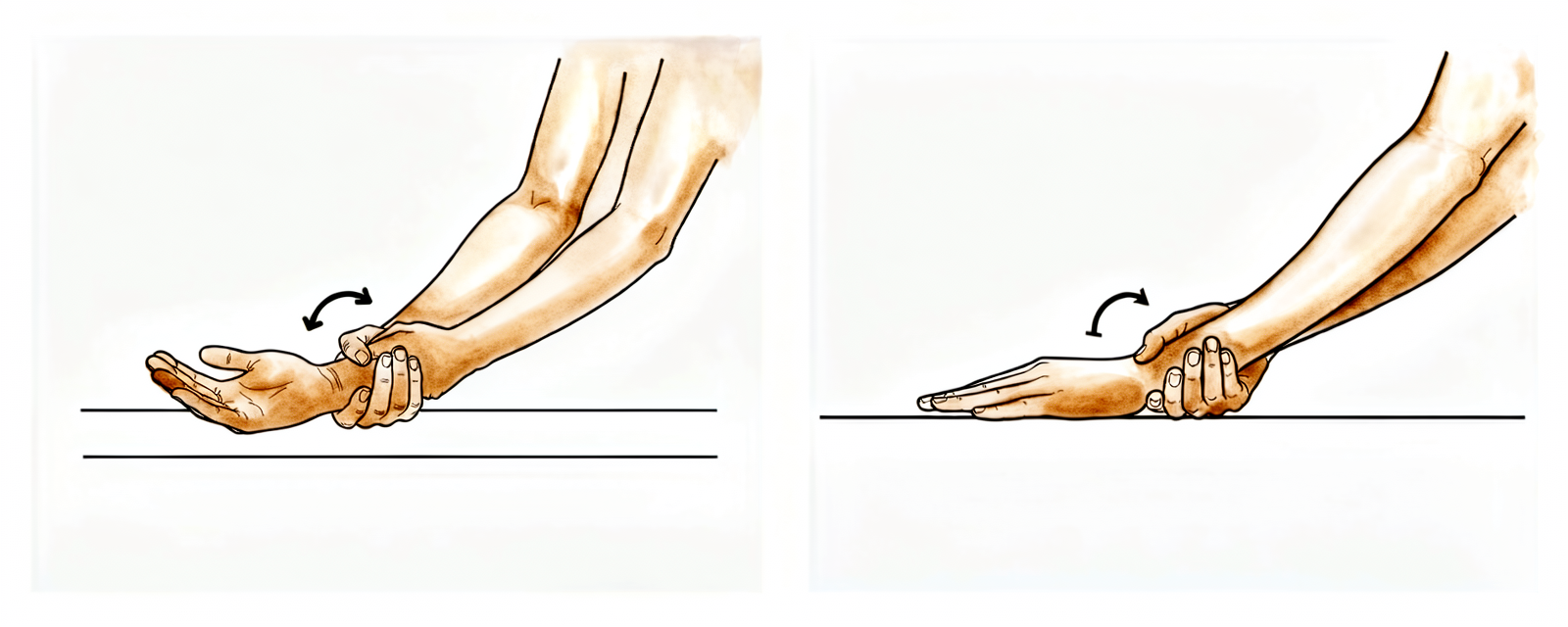
ہتھیلی اوپر / نیچے گھمانے کا اسٹریچ
کہنی کو جسم کے پہلو میں موڑیں، ہتھیلی کا رخ چھت کی طرف ہو۔ ہاتھ کو ہتھیلی اوپر کی طرف گھمائیں اور 15 سیکنڈ روکے رکھیں، پھر ہتھیلی نیچے کی طرف اور 15 سیکنڈ روکے رکھیں؛ 5 بار دہرائیں۔ جیسے جیسے آرام بڑھے، اپنے دوسرے ہاتھ سے — ہاتھ کو نہیں بلکہ اپنی کلائی کو پکڑ کر — ہر حالت میں گھماؤ کو آہستہ سے مزید آگے دھکیلیں یہاں تک کہ کھنچاؤ محسوس ہو۔
5 بار دہرائیں، دن میں 4 مرتبہ، ہفتے میں 5 سے 7 دن
Kieran Hirpara 4.0
کہنی کو موڑنا / سیدھا کرنا
بازو کو اپنے پہلو میں رکھ کر، بازو کے نچلے حصے اور کہنی کو جتنا ہو سکے آہستہ سے سیدھا کریں۔ آخری حد کے اس اسٹریچ کو 3 سے 5 سیکنڈ روکے رکھیں۔ پھر الٹی سمت میں دہرائیں، بازو کے نچلے حصے کو اوپر موڑتے ہوئے اپنے ہاتھ سے کندھے کو چھونے کی کوشش کریں۔
10 بار دہرائیں، دن میں 2 مرتبہ، روزانہ
Kieran Hirpara 4.0
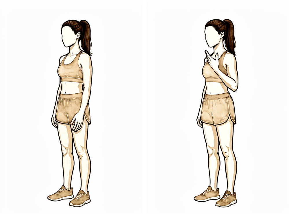
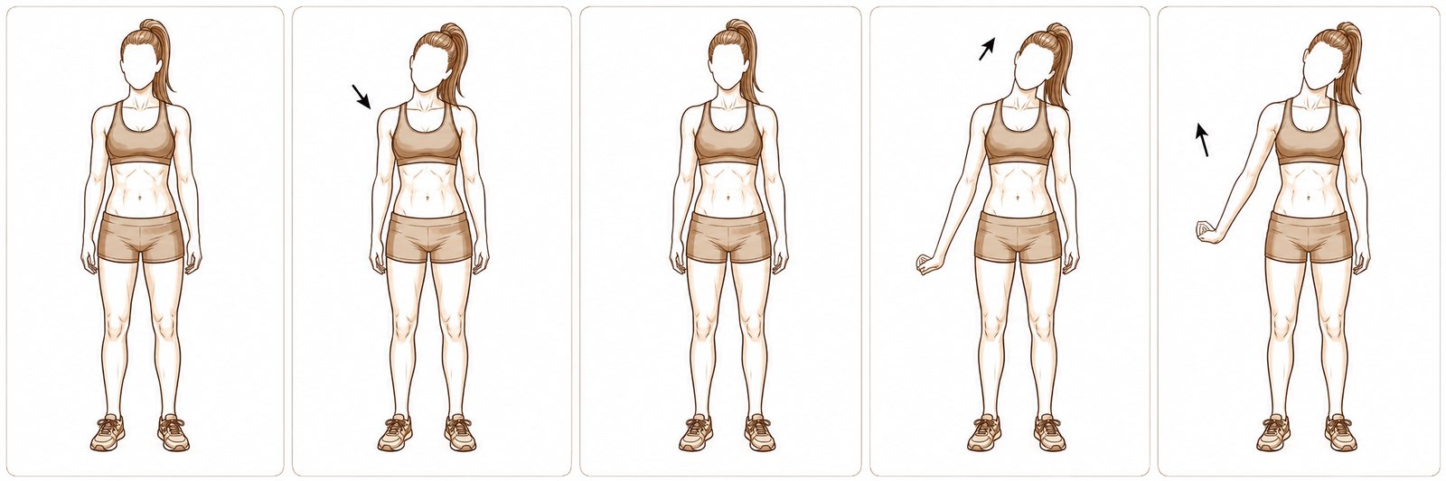
ریڈیل عصب کو سرکانے کی ورزش
بازوؤں کو پہلوؤں میں ڈھیلا چھوڑ کر کھڑے ہو جائیں۔ اپنا کندھا نیچے گرائیں اور انگلیوں کو فرش کی طرف بڑھائیں۔ بازو کو اندر کی طرف گھمائیں (انگوٹھا جسم کی طرف) اور کلائی کو موڑیں، ہتھیلی اوپر۔ جس طرف اسٹریچ کر رہے ہیں، سر کو آہستہ سے اس کی مخالف طرف جھکائیں، پھر بازو کو اوپر اور جسم سے دور اٹھائیں۔ اس ورزش کی ہر حالت کو 3 سے 5 سیکنڈ روکے رکھیں۔
5 سے 8 بار دہرائیں، دن میں 2 سے 4 مرتبہ، ہفتے میں 6 سے 7 دن
ورزشوں کا یہ پروگرام Sarah Farrell کے تعاون سے لکھا گیا ہے، جو بیچلر آف آکیوپیشنل تھراپی (Bachelor of Occupational Therapy, BOccThy) اور تسلیم شدہ ہینڈ تھراپسٹ (Accredited Hand Therapist, AHT) ہیں۔
آپ کے پروٹوکول کے بعد
یہ پروٹوکول پریکٹس کے صحت یابی کے عمومی مشوروں کے ساتھ مل کر کام کرتا ہے؛ دیکھیں آپریشن کے بعد درد پر قابو، زخم کی دیکھ بھال اور ہینڈ تھراپی کی بنیادی باتیں۔ خود آپریشن اور جس بیماری کا یہ علاج کرتا ہے، اس کے بارے میں ریڈیل ٹنل ریلیز اور ریڈیل ٹنل سنڈروم دیکھیں۔
حوالہ جات
[1] Wright TW, Glowczewskie F, Cowin D, Wheeler DL. Radial nerve excursion and strain at the elbow and wrist associated with upper-extremity motion. J Hand Surg Am. 2005;30(5):990–996. https://pubmed.ncbi.nlm.nih.gov/16182056/ [2] Lee JT, Azari K, Jones NF. Long term results of radial tunnel release — the effect of co-existing tennis elbow, multiple compression syndromes and workers' compensation. J Plast Reconstr Aesthet Surg. 2008;61(9):1095–1099. https://www.sciencedirect.com/science/article/abs/pii/S1748681507004044 [3] Sotereanos DG, Varitimidis SE, Giannakopoulos PN, Westkaemper JG. Results of surgical treatment for radial tunnel syndrome. J Hand Surg Am. 1999;24(3):566–570. https://pubmed.ncbi.nlm.nih.gov/10357537/ [4] Naam NH, Nemani S. Radial tunnel syndrome. Orthop Clin North Am. 2012;43(4):529–536. (Radial Tunnel Syndrome, StatPearls.) https://www.ncbi.nlm.nih.gov/books/NBK555937/ [5] Coppieters MW, Butler DS. Do "sliders" slide and "tensioners" tension? An analysis of neurodynamic techniques and considerations regarding their application. Man Ther. 2008;13(3):213–221. https://pubmed.ncbi.nlm.nih.gov/17398140/ [6] Basson A, Olivier B, Ellis R, Coppieters M, Stewart A, Mudzi W. The effectiveness of neural mobilization for neuromusculoskeletal conditions: a systematic review and meta-analysis. J Orthop Sports Phys Ther. 2017;47(9):593–615. https://pubmed.ncbi.nlm.nih.gov/28704626/
Evidence & references
This is the clinical evidence summary written for health professionals. It is technical, and it lists the research this page was built from. You do not need to read it to understand your treatment or to make a decision about it.
Radial Tunnel Release — Evidence Brief & Post-operative Rehabilitation
Topic scope: post-operative rehabilitation after surgical decompression / neurolysis of the posterior interosseous nerve (deep branch of the radial nerve) in the radial tunnel of the proximal forearm, performed for radial tunnel syndrome (RTS). This is an elbow / proximal-forearm topic — anatomically and clinically distinct from carpal-tunnel and cubital-tunnel decompression. Like other nerve decompressions it is an early-motion pathway (early elbow/forearm/wrist motion, radial-nerve glides, oedema and scar care). The scope deliberately foregrounds the diagnostic controversy and the more variable, lower success rates that distinguish RTS release from the better-validated carpal-tunnel and cubital-tunnel operations.
Defining principle of the rehab here: a decompressed nerve does not create a healing construct that needs months of protection — it needs early, gentle movement to stop it adhering to the operative bed and to restore its glide. So the rehab is an early-motion programme: light functional hand use from day 1, radial-nerve sliders, graded desensitisation and (once healed) scar massage; heavier loading deferred to ~6 weeks. But two honesty caveats sit over the whole topic. First, RTS is a contested diagnosis — there is no confirmatory imaging or electrodiagnostic test, it is a diagnosis of exclusion, and a substantial body of opinion regards it as a variant of recalcitrant lateral epicondylitis. Second, outcomes after release are more variable and on average lower than carpal- or cubital-tunnel release — good results cluster around two-thirds overall, and fall further with co-existing tennis elbow, multiple compression sites, or a workers'-compensation context. Patient expectations should be set accordingly.
A. THE DIAGNOSTIC CONTROVERSY (read first — it frames everything)
RTS is among the most contested entities in upper-limb surgery, and the rehab brief is incomplete without it:
- No confirmatory test. Electromyography and nerve-conduction studies are characteristically normal in RTS (compression is intermittent/dynamic and predominantly of a motor nerve carrying few pain fibres), and MRI is frequently negative — denervation oedema in supinator/extensors is suggestive but inconsistent, and a normal scan does not exclude the diagnosis. RTS is therefore a clinical diagnosis of exclusion, resting on point tenderness ~4 cm distal to the lateral epicondyle (over the radial tunnel rather than the epicondyle), pain on resisted supination / resisted long-finger extension, and — for some surgeons — temporary relief from a diagnostic local-anaesthetic block at the radial tunnel.
- Overlap with lateral epicondylitis (tennis elbow). The two coexist frequently and share the lateral-elbow pain territory. A recognised school of thought holds that "RTS" is often severe, recalcitrant lateral epicondylitis rather than a discrete compression neuropathy. Importantly, routine PIN release added to lateral-epicondylitis surgery has not been shown to improve outcomes, so the diagnosis should be secure before a decompression is planned.
- Practical consequence. Surgery is a last resort after prolonged failed conservative care (activity modification, splinting, anti-inflammatories, sometimes a steroid injection), and is best reserved for patients with proximal-forearm pain and no better explanation. This uncertainty is the single most important reason post-operative expectations must be framed honestly.
B. RELEASE OUTCOMES (variable — and why)
- Headline success ~two-thirds. Across the older long-term series, roughly 67% good, 15% fair, 18% poor after radial tunnel decompression — markedly more variable than carpal- or cubital-tunnel release. A 2008 long-term series (Lee, Azari, Jones) and a 1999 series (Sotereanos et al.) both document this spread; the Sotereanos cohort reported good/excellent results in only ~39% by objective assessment (though ~64% by patient self-rating), underscoring how outcome depends on the metric used.
- Co-existing lateral epicondylitis lowers success. Success falls to roughly 40% when tennis elbow coexists, versus far higher with isolated RTS.
- Multiple compression sites and workers'-compensation context lower success — reported ~58% success in compensation cases vs ~73% without. These are the same modifiers named in the patient protocol.
- 2025 systematic review (Raymond et al., HAND). 11 studies, 401 limbs (381 patients). Outcomes were heterogeneous; a dorsal approach between ECRB and EDC was associated with the most favourable Roles-and-Maudsley scores and satisfaction. The review's central message is that the overall evidence is low-grade (observational), the diagnosis non-standardised, and the effectiveness of conservative treatment essentially untested — a "tendency" toward benefit rather than proof.
- Resorption-style "spontaneous improvement" does not apply here — unlike calcific tendinitis, RTS does not self-resolve through a biological cycle; conservative care manages symptoms rather than curing a deposit.
C. SURGICAL APPROACH (shapes the early rehab)
- What is done. Complete neurolysis of the radial nerve at its bifurcation, decompressing the deep branch (PIN) and superficial sensory branch, releasing the arcade of Frohse (the proximal supinator edge), the leash of Henry (radial recurrent vessels), the ECRB fascial edge, and the distal supinator border. Any constrictive bands or vessels are divided.
- Approaches. Dorsal (Thompson, between ECRB/EDC or the brachioradialis–ECRL interval), volar/anterior (Henry), or transmuscular. Anatomical studies map the trade-offs; the dorsal ECRB–EDC interval performed best in the 2025 review. The superficial radial branch matters — it is a recognised source of post-operative dysaesthesia if irritated.
- Rehab implication. A muscle-splitting/dorsal exposure through the extensor mass means early gentle forearm rotation and wrist motion are encouraged but heavy resisted supination/extension is deferred; the incision sits over a mobile, frequently sensitive area, so desensitisation and scar care carry real weight here.
D. POST-OP THERAPY ROLE (nerve/tendon glides, oedema, scar)
The decompressed nerve must glide, not adhere. The mechanical rationale is well quantified: the radial nerve translates and stretches a measurable amount across the elbow and wrist during ordinary arm motion (Wright et al. 2005), so early motion is what keeps it free of the healing bed.
- Early motion, immediately. Early active elbow, forearm and wrist movement within pain limits from the first post-op days; most protocols use no rigid splinting (or a removable splint for comfort/night only).
- Radial-nerve glides — favour "sliders" over "tensioners". Sliding (slider) neurodynamic techniques achieve substantially greater nerve excursion at much lower nerve strain than end-range tensioners — preferable around a freshly decompressed nerve. Neural-mobilisation evidence across neuromusculoskeletal conditions is supportive but of variable certainty, so progression is symptom-guided and essentially pain-free; mechanism work (e.g., the MONET protocol) is still maturing.
- Oedema and desensitisation. Graded desensitisation (tapping/rubbing over the dressing) from day 1 normalises touch and pre-empts a sensitive scar — particularly relevant given superficial- radial-branch proximity.
- Scar management once healed. Massage, pressure, and silicone are advocated to loosen skin–tissue adhesions and aid remodelling, started once the wound is closed/sutures out.
- Strengthening deferred. Light functional ADL use throughout; resisted strengthening of wrist/ elbow and fine-motor work introduced from ~6 weeks. Heavy work and vibration tools avoided to ~6–8 weeks.
Phased post-op timeline (maps to the patient protocol phases)
| Phase | Window | Splint | Motion / nerve work | Load / strengthening | Notes |
|---|---|---|---|---|---|
| I — Protect & glide | Day 0–2 wk | None, or removable for comfort/night | Early pain-free active elbow/forearm/wrist ROM; radial-nerve sliders; desensitisation from day 1 | Light functional ADL use only (self-care, feeding, dressing, writing, typing) | Stop the nerve adhering; settle the wound. No lifting/gripping/weight-bearing/vibration tools. Driving limited first 1–2 wk |
| II — Restore motion | 2–6 wk | Off | Progress full active + gentle assisted ROM; continue sliders; scar massage once healed | Still no resisted loading; ADL use continues | Sensitivity/dysaesthesia common and usually settles; keep glides gentle |
| III — Strengthen & return | ~6 wk onward | Off | Full ROM goal; sliders as needed | Begin graded wrist/elbow strengthening + fine-motor work from ~6 wk; advance work/heavy tasks thereafter | Vibration tools/heavy work resume ~6–8 wk. Pain relief is often gradual and may be partial — counsel accordingly |
E. COMPLICATIONS / DOWNSIDES
- Incomplete or no pain relief — the dominant "complication," tied directly to diagnostic uncertainty; relief is frequently gradual and sometimes partial.
- Superficial-radial-branch dysaesthesia / scar sensitivity — recognised; desensitisation and careful technique mitigate it.
- Transient PIN weakness (finger/thumb extension) from retraction — usually recovers.
- Adhesion/recurrence of symptoms if early glide is neglected.
- Standard wound risks (infection, haematoma) — uncommon.
F. KEY CONTROVERSIES / EVIDENCE QUALITY
- Does RTS exist as a discrete entity? Genuinely contested. No confirmatory test; substantial opinion equates much of it with recalcitrant lateral epicondylitis. This is the defining controversy and must shape consent and expectation-setting. Unresolved — expert opinion divided.
- Patient selection drives outcome more than technique. Isolated RTS does best; coexisting tennis elbow, multiple compressions, and compensation context predict worse results. Moderate (consistent across cohorts).
- Approach choice. A dorsal ECRB–EDC interval was favoured in the 2025 SR, but the evidence is observational and confounded by diagnostic heterogeneity. Weak–moderate.
- The rehab protocol itself is consensus/expert — drawn from surgeon and hand-therapy guidance (early motion, sliders, desensitisation, scar care), not from a rehab RCT. Phase timings are typical, not trial-derived. Weak / consensus.
- Conservative-treatment efficacy is essentially untested — the 2025 SR notes no usable trials of non-operative care, so "failed conservative management" before surgery rests on practice convention. Weak.
G. EVIDENCE STRENGTH FLAGS (summary)
- STRONG: the mechanical rationale for early nerve glide — quantified radial-nerve excursion/ strain across elbow and wrist (Wright et al. 2005); slider-vs-tensioner excursion/strain physiology.
- MODERATE: patient-selection modifiers of outcome (lateral epicondylitis, multiple compressions, workers' compensation lower success); ~two-thirds overall good-result rate from long-term cohorts; dorsal-approach signal from the 2025 systematic review (low-grade studies).
- WEAK / CONSENSUS: the existence and diagnostic criteria of RTS (no confirmatory test; overlap with lateral epicondylitis); the post-operative rehabilitation protocol (surgeon/ hand-therapy guidance, no rehab RCT); neural-mobilisation certainty (supportive but variable); efficacy of conservative care (essentially untested).
CITATIONS
RAG corpus (180,000+ Orthopaedic articles)
- Posterior Interosseous Nerve Compression in the Forearm, AKA Radial Tunnel Syndrome. HAND. 2022. DOI: 10.1177/15589447221122822
- Radial Tunnel Syndrome: Emphasis on the Superficial Branch of the Radial Nerve. J Hand Surg Eur. 2009. DOI: 10.1177/1753193408099832
- Anatomical Study of the Surgical Approaches to the Radial Tunnel. J Hand Surg Am. 2015. DOI: 10.1016/j.jhsa.2015.03.009
- MR Imaging Features of Radial Tunnel Syndrome: Initial Experience. Radiology. 2006. DOI: 10.1148/radiol.2401050028
- Management of Lateral Epicondylitis: Current Concepts. J Am Acad Orthop Surg (JAAOS). 2008. DOI: 10.5435/00124635-200801000-00004
- Uncommon Nerve Compression Syndromes of the Upper Extremity. J Am Acad Orthop Surg (JAAOS). 1998. DOI: 10.5435/00124635-199811000-00006
- Radial Nerve Excursion and Strain at the Elbow and Wrist Associated With Upper-Extremity Motion. J Hand Surg Am. 2005. DOI: 10.1016/j.jhsa.2005.06.008
- Evidence and Techniques in Rehabilitation Following Nerve Injuries. Hand Clin. 2013. DOI: 10.1016/j.hcl.2013.04.012
- Preventive Strategies, Exercises and Rehabilitation of Hand Compression Neuropathies. J Hand Ther. 2022. DOI: 10.1016/j.jht.2021.11.003
- Mechanisms of Neurodynamic Treatments (MONET): a protocol for a mechanistic study. BMC Musculoskelet Disord. 2024. DOI: 10.1186/s12891-024-07713-6
Radial-tunnel literature (URLs)
- Clinical Outcomes of Operative Management for Radial Tunnel Syndrome According to Surgical Approach: a Systematic Review. HAND. 2025. https://journals.sagepub.com/doi/10.1177/15589447251315761
- The Epidemiology of Radial Tunnel Syndrome and Its Overlap With Lateral Epicondylitis. J Hand Surg Am. 2023. https://www.jhandsurg.org/article/S0363-5023(23)00138-7/abstract
- Lee JT, Azari K, Jones NF. Long-term results of radial tunnel release — the effect of co-existing tennis elbow, multiple compression syndromes and workers' compensation. J Plast Reconstr Aesthet Surg. 2008. https://www.sciencedirect.com/science/article/abs/pii/S1748681507004044
- Sotereanos DG, et al. Results of surgical treatment for radial tunnel syndrome. J Hand Surg Am. 1999. https://pubmed.ncbi.nlm.nih.gov/10357537/
- Interventions for treating the radial tunnel syndrome: a systematic review of observational studies (DARE). https://www.ncbi.nlm.nih.gov/books/NBK75403/
- Radial Tunnel Syndrome (StatPearls). https://www.ncbi.nlm.nih.gov/books/NBK555937/
- Orthopedic Management of Radial Tunnel Syndrome: A Diagnostic and Treatment Dilemma. PMC. https://pmc.ncbi.nlm.nih.gov/articles/PMC10081130/
- Radial Tunnel Syndrome: Case Report and Comprehensive Critical Review of a Compression Neuropathy Surrounded by Controversy. PMC. https://pmc.ncbi.nlm.nih.gov/articles/PMC9896270/
Published rehab protocols (patient-guidance — basis for the phase structure)
- Radial Tunnel Release post-op protocol (Santa Barbara Orthopedic / Mencias). https://www.sbortho.com/wp-content/uploads/2023/09/radial-tunnel-release-new.pdf
- Radial Tunnel Syndrome — conservative and post-operative rehabilitation. Physiopedia. https://www.physio-pedia.com/Radial_Tunnel_Syndrome
- Basson A, et al. The effectiveness of neural mobilization for neuromusculoskeletal conditions: a systematic review and meta-analysis. J Orthop Sports Phys Ther. 2017. https://pubmed.ncbi.nlm.nih.gov/28704626/
- Coppieters MW, Butler DS. Do "sliders" slide and "tensioners" tension? Man Ther. 2008. https://pubmed.ncbi.nlm.nih.gov/17398140/