آپ کو کیا محسوس ہوتا ہے
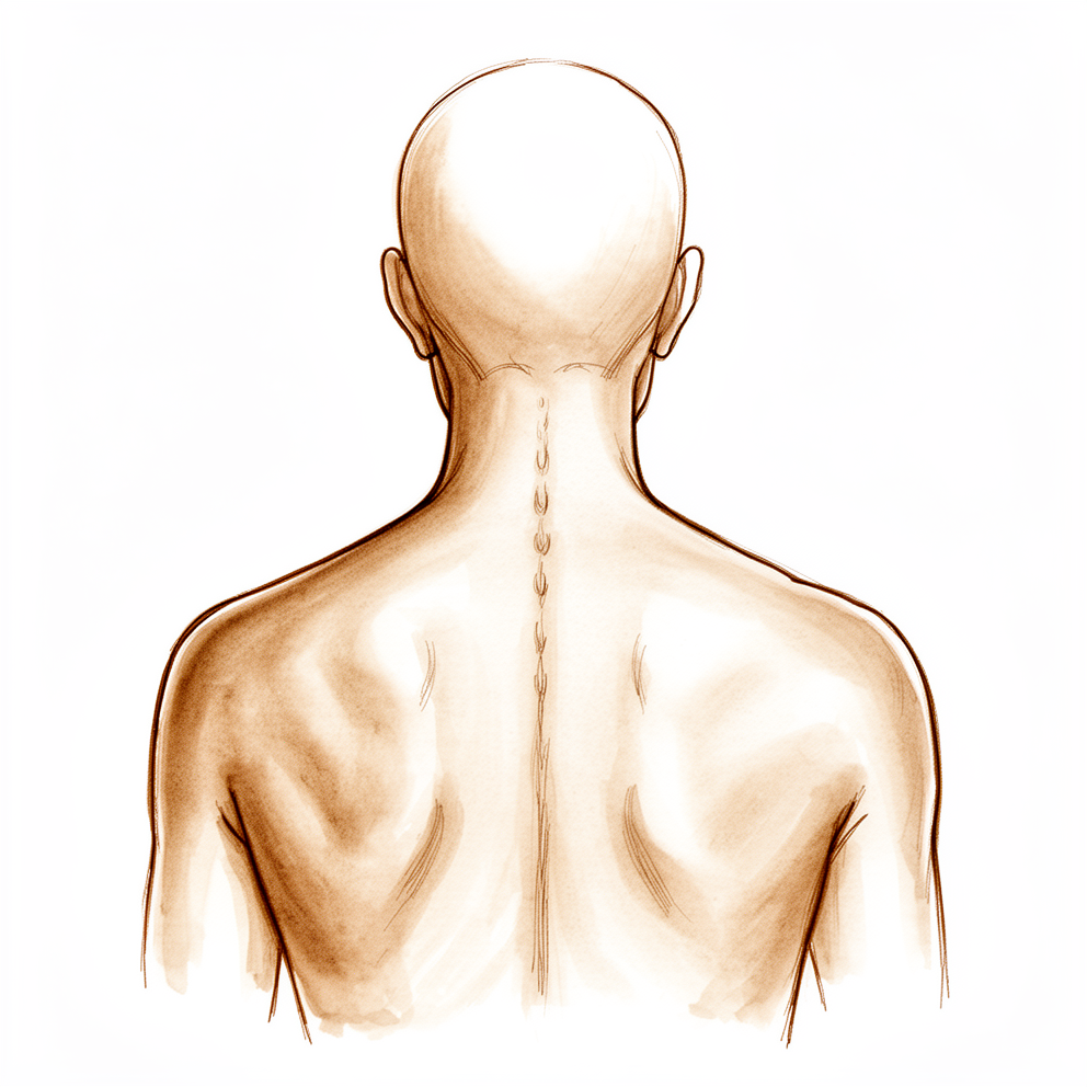
ریڑھ کی ہڈی کے انفیکشن کی بنیادی علامت ایسا درد ہے جو ٹھیک نہیں ہوتا۔ یہ عام طور پر آپ کی کمر یا گردن کے کسی ایک حصے میں گہرائی میں ہوتا ہے، اور دن بہ دن وہیں رہتا ہے۔ آرام، سینک یا وہ درد کش دوائیں جو آپ عام طور پر لیتے ہیں، اسے دور نہیں کرتیں۔ اس جگہ کو اکثر چھونے پر درد ہوتا ہے، اور اس کے آس پاس کے پٹھے کھنچے ہوئے یا اینٹھے ہوئے محسوس ہو سکتے ہیں۔ ریڑھ کی ہڈی کے اس حصے کو حرکت دینا مشکل ہو جاتا ہے، اس لیے بستر میں کروٹ لینا، کرسی سے اٹھنا یا کندھے کے اوپر سے پیچھے دیکھنا، ان سب میں واقعی زور لگ سکتا ہے۔
درد اکثر رات کو بھڑک اٹھتا ہے اور آپ کو نیند سے جگا سکتا ہے۔ یہ سرگرمی کے بعد یا صبح سویرے بھی بڑھ سکتا ہے۔ کچھ لوگوں کو بخار یا عمومی طور پر طبیعت خراب محسوس ہوتی ہے، لیکن بہت سے لوگوں کو نہیں، اور یہی ایک وجہ ہے کہ یہ بیماری آسانی سے نظر انداز ہو جاتی ہے۔ بخار کے بغیر کمر کا درد ابتدائی ایکسرے پر کمر کے کسی سادہ مسئلے جیسا لگ سکتا ہے، جو تشخیص میں اکثر تاخیر کی ایک وجہ ہے۔ اوسطاً پہلی علامات سے تشخیص تک دو سے چھ ماہ لگتے ہیں، اور 6 سے 12 ہفتے کی تاخیر عام ہے۔
روزمرہ زندگی میں یہ درد عموماً ہر اس کام کو محدود کرتا ہے جس سے ریڑھ کی ہڈی پر بوجھ پڑے۔ کھانے کے دوران بیٹھے رہنا، کھانا پکانے کے لیے کاؤنٹر کے پاس کھڑے ہونا، دھلے کپڑے اٹھانا یا تھوڑے فاصلے سے زیادہ گاڑی چلانا، یہ سب مشکل ہو سکتے ہیں۔ کچھ لوگ بیٹھنا بالکل چھوڑ دیتے ہیں اور لیٹنا یا کھڑا رہنا پسند کرتے ہیں۔
اگر انفیکشن آپ کی ریڑھ کی ہڈی کے اعصاب پر دباؤ ڈالنے لگے تو نئی علامات ظاہر ہو سکتی ہیں۔ ان میں بازو یا ٹانگ میں نیچے کی طرف جانے والا درد یا سوئیاں چبھنے جیسا احساس، کسی بازو یا ٹانگ میں کمزوری، یا پیشاب پر قابو رکھنے میں دشواری شامل ہیں۔ اس بیماری میں مبتلا تقریباً ہر تین میں سے ایک شخص کو اعصاب سے متعلق کوئی مسئلہ ہو جاتا ہے۔ نئی کمزوری، سُن پن یا پیشاب میں تبدیلیوں پر فوری توجہ کی ضرورت ہوتی ہے، اس لیے اگر یہ ظاہر ہوں تو فوراً اپنے سرجن یا ہسپتال سے رابطہ کریں۔
اگر آپ کو ذیابیطس (شوگر) ہے، آپ تمباکو نوشی کرتے ہیں، آپ کے جسم میں کہیں اور کوئی انفیکشن ہے، پہلے ریڑھ کی ہڈی کی سرجری ہو چکی ہے، یا آپ کا مدافعتی نظام کمزور ہے، تو آپ کو ریڑھ کی ہڈی کا انفیکشن ہونے کا امکان زیادہ ہے۔
دراصل کیا ہو رہا ہے
ریڑھ کی ہڈی کا انفیکشن اس وقت ہوتا ہے جب بیکٹیریا آپ کی ریڑھ کی ہڈیوں میں یا ان کے درمیان کی گدی میں داخل ہو جاتے ہیں۔ کمر کی دو ہڈیوں کے درمیان کی گدی کو ڈسک (disc) کہتے ہیں۔ یہ کچھ حد تک جھٹکا جذب کرنے والے پرزے کی طرح کام کرتی ہے، اور آپ کی ریڑھ کی ہڈی کو جھکنے اور بوجھ اٹھانے دیتی ہے۔ ڈسک کی اپنی خون کی فراہمی بہت کم ہوتی ہے، اس لیے یہ اپنی غذائیت ساتھ والی ہڈی سے جذب کر کے حاصل کرتی ہے۔ یہی سست فراہمی انفیکشن کے پھیلنے کا راستہ بھی ہے: بیکٹیریا عام طور پر کسی مہرے کی ہڈی میں شروع ہوتے ہیں، پھر رس کر ڈسک میں اور دوسری طرف کی ہڈی میں پہنچ جاتے ہیں۔ ڈاکٹر ہڈی کے انفیکشن کو اوسٹیومائلائٹس (osteomyelitis) اور ڈسک کے انفیکشن کو ڈسکائٹس (discitis) کہتے ہیں۔
بیکٹیریا عام طور پر آپ کے جسم میں کسی اور جگہ سے خون کے ذریعے پہنچتے ہیں، اور زیادہ تر یہ جلد کا ایک عام جراثیم ہوتا ہے جسے Staphylococcus aureus کہتے ہیں۔ کم صورتوں میں یہ براہِ راست داخل ہوتے ہیں، مثلاً کسی زخم یا ناسور کے ذریعے، یا ریڑھ کی ہڈی کی سرجری یا انجکشن کے دوران۔ چونکہ انفیکشن ہڈی کے اندر گہرائی میں ہوتا ہے، آپ کا جسم اسے آسانی سے گھیر کر الگ نہیں کر سکتا۔ ہڈی اور ڈسک میں سوزش ہو جاتی ہے اور وہ ٹوٹنا شروع ہو جاتی ہیں، اسی لیے درد مسلسل رہتا ہے، رات کو ہوتا ہے اور آرام سے کم نہیں ہوتا، جیسا کہ اوپر بیان کیا گیا ہے۔
زیادہ تر انفیکشن ریڑھ کی ہڈی کے اگلے حصے میں رہتا ہے، یعنی وہ ہڈیاں اور ڈسکیں جو بوجھ اٹھاتی ہیں۔ لیکن یہ پھیل سکتا ہے۔ اعصاب کے آس پاس کی جگہ میں حرام مغز (spinal cord) کے ساتھ پیپ جمع ہو سکتی ہے، جس سے انفیکشن کی ایک تھیلی بن جاتی ہے جسے ایپیڈورل ایبسیس (epidural abscess، پیپ کی تھیلی) کہتے ہیں۔ یہ تھیلی اعصاب پر دباؤ ڈالتی ہے، اور بازو یا ٹانگ میں نئی کمزوری، سُن پن یا پیشاب کا مسئلہ اسی سے پیدا ہوتا ہے۔ ان علامات پر فوری توجہ کی ضرورت ہوتی ہے۔
اچھی خبر یہ ہے کہ ریڑھ کی ہڈی کے زیادہ تر انفیکشن اینٹی بائیوٹک اور کمر کے سہارے سے، بغیر کسی آپریشن کے، ٹھیک ہو جاتے ہیں۔ سرجری مخصوص حالات کے لیے رکھی جاتی ہے: پیپ کی تھیلی جو اعصاب کو دبا رہی ہو، ایسا انفیکشن جو اینٹی بائیوٹک سے ٹھیک نہ ہو، ریڑھ کی ہڈی کا غیر مستحکم یا ٹیڑھا ہو جانا، یا جب جراثیم کی شناخت کے لیے ٹشو کے نمونوں کی ضرورت ہو۔
ہم اس کے لیے کیا کر سکتے ہیں
علاج کا پہلا قدم ڈرپ کے ذریعے دی جانے والی اینٹی بائیوٹک ہے، عام طور پر کم از کم 6 ہفتے تک، اور پھر گولیاں۔ اس دوران آپ اٹھتے اور چلتے پھرتے رہتے ہیں۔ اکثر ساتھ میں بریس یا کمر کا سہارا بھی لگایا جاتا ہے، تاکہ ٹھیک ہونے کے دوران ریڑھ کی ہڈی ساکن رہے۔ ریڑھ کی ہڈی کے نچلے حصے کے انفیکشن کے لیے یہ عام طور پر کمر کے نچلے حصے کا سہارا (lumbosacral support) ہوتا ہے، اور کمر کے درمیانی حصے کے لیے کمر کے درمیانی اور نچلے حصے کا بریس (thoracolumbar brace) یا جیویٹ ایکسٹینشن بریس (Jewett extension brace)۔ صحیح اینٹی بائیوٹک اور کمر کے سہارے سے زیادہ تر انفیکشن بغیر آپریشن کے ٹھیک ہو جاتے ہیں۔ اگر اعصاب کے ساتھ پیپ کی تھیلی بن گئی ہو لیکن وہ ان پر دباؤ نہ ڈال رہی ہو تو یہی منصوبہ لاگو ہوتا ہے: کم از کم 6 ہفتے ڈرپ کے ذریعے اینٹی بائیوٹک، آپ کے خون کے ٹیسٹوں کی قریبی نگرانی اور آپ کے اعصاب کی کارکردگی کا بار بار معائنہ، اور ساتھ میں سہارے کے لیے بریس۔
سرجری پر اس وقت غور کیا جاتا ہے جب اینٹی بائیوٹک کارگر نہ ہوئی ہو، جب جراثیم کی شناخت کے لیے ٹشو کے نمونوں کی ضرورت ہو، جب ریڑھ کی ہڈی غیر مستحکم یا ٹیڑھی ہو رہی ہو، یا جب اعصاب کا کوئی مسئلہ ہو جیسے کمزوری، سُن پن یا پیشاب میں تبدیلیاں۔ اعصاب کو دبانے والی پیپ کی تھیلی کا علاج بھی سرجری سے کیا جاتا ہے۔ مقصد پیپ نکالنا، متاثرہ ہڈی نکالنا اور ٹھیک ہونے تک ریڑھ کی ہڈی کو ساکن رکھنا ہے۔ بعض اوقات پیچھے رہ جانے والی خالی جگہ کو بھرنے کے لیے آپ کے جسم کی کسی اور جگہ سے لی گئی ہڈی، عطیہ کی گئی ہڈی یا کیج (cage) استعمال کیا جاتا ہے، اور پیچ اور راڈ (rods) اوپر اور نیچے کے مہروں کو ساکن رکھتے ہیں۔ اس بیماری میں مبتلا تقریباً آدھے لوگوں کو آخرکار آپریشن کی ضرورت پڑتی ہے۔ جب سرجری کی ضرورت ہو تو اس سے آپ بعد میں جلدی اٹھ کر چلنے پھرنے کے قابل بھی ہو جاتے ہیں۔
کیا توقع رکھیں
ریڑھ کی ہڈی کے زیادہ تر انفیکشن علاج سے ٹھیک ہو جاتے ہیں۔ زیادہ تر لوگوں کے لیے اینٹی بائیوٹک اور کمر کا سہارا کافی ہوتے ہیں، اور ریڑھ کی ہڈی مستحکم رہتی ہے اور اپنی شکل برقرار رکھتی ہے۔ اگر آپ کی عمر ساٹھ سال سے کم ہے، آپ کا مدافعتی نظام اچھی طرح کام کر رہا ہے، اور خون میں سوزش کی نشاندہی کرنے والے ٹیسٹ کم ہونا شروع ہو جائیں، تو صرف اینٹی بائیوٹک سے امکانات بہتر ہوتے ہیں۔
صحت یابی سست ہوتی ہے، اور یہ بات شروع میں ہی جان لینا مفید ہے۔ انفیکشن ہڈی کے اندر گہرائی میں ہوتا ہے، اس لیے درد کم ہونے اور ہڈی کے دوبارہ جڑنے میں ہفتوں سے مہینوں تک لگتے ہیں۔ آپ کم از کم 6 ہفتے اینٹی بائیوٹک لیں گے، اکثر اس سے زیادہ، اور آپ کے باقاعدگی سے خون کے ٹیسٹ اور معائنے ہوں گے تاکہ ہم دیکھ سکیں کہ انفیکشن علاج پر کیسا ردِ عمل دے رہا ہے۔ بہت سے لوگ محسوس کرتے ہیں کہ اس عرصے میں درد آہستہ آہستہ کم ہو جاتا ہے، اگرچہ چھونے پر کچھ درد اور اکڑاؤ باقی رہ سکتا ہے۔
اس بیماری میں مبتلا تقریباً آدھے لوگوں کو آخرکار آپریشن کی ضرورت پڑتی ہے۔ جب سرجری جلد کی جائے تو لوگ ایک سال بعد صرف اینٹی بائیوٹک سے علاج کروانے والوں کے مقابلے میں کمر کا کم درد اور روزمرہ کی بہتر کارکردگی بتاتے ہیں۔ سرجری سے آپ بعد میں جلدی اٹھ کر چلنے پھرنے کے قابل بھی ہو جاتے ہیں۔
اگر انفیکشن کا علاج نہ کیا جائے تو یہ خود بخود ختم نہیں ہوتا۔ یہ ہڈی اور ڈسک کو تباہ کرتا رہ سکتا ہے، ریڑھ کی ہڈی غیر مستحکم یا ٹیڑھی ہو سکتی ہے، اور انفیکشن پھیل سکتا ہے۔ اعصاب کے ساتھ بننے والی پیپ کی تھیلی دیرپا کمزوری یا فالج کا سبب بن سکتی ہے، اور جب خود حرام مغز کو نقصان پہنچ جائے تو وہ نقصان واپس ٹھیک نہیں ہوتا۔ علاج کے بغیر یہ بیماری جان لیوا بھی ہو سکتی ہے۔ اس بیماری میں مبتلا تقریباً 2 سے 17 فیصد لوگ اس کی وجہ سے ہسپتال میں فوت ہو جاتے ہیں، اور جب اعصاب کا مسئلہ ہو یا انفیکشن سینے کی طرف پھیل رہا ہو تو خطرہ بڑھ جاتا ہے۔
کچھ چیزیں اعصاب کے سنگین مسائل کا امکان بڑھاتی ہیں: ذیابیطس، ریومیٹائیڈ آرتھرائٹس، زیادہ عمر، اور ریڑھ کی ہڈی کے اوپری حصے میں انفیکشن۔ اگر کسی بھی وقت آپ کو نئی کمزوری، سُن پن یا پیشاب میں تبدیلیاں محسوس ہوں تو فوراً اپنے سرجن یا ہسپتال سے رابطہ کریں۔ اگر اسے جلد پکڑ لیا جائے اور صحیح علاج کیا جائے تو زیادہ تر لوگ مستحکم ریڑھ کی ہڈی کے ساتھ اور اعصاب کو کسی دیرپا نقصان کے بغیر اس سے نکل آتے ہیں۔
ڈاکٹر کو کب دکھائیں
اگر آپ کو کمر یا گردن کا ایسا درد ہے جو ٹھیک نہیں ہو رہا، خاص طور پر اگر آپ کو بخار یا عمومی طور پر طبیعت خراب بھی محسوس ہو، تو اپنے فیملی ڈاکٹر (GP) کو دکھائیں۔ یہ اور بھی اہم ہے اگر آپ کو ذیابیطس ہے، آپ تمباکو نوشی کرتے ہیں، آپ کے جسم میں کہیں اور کوئی انفیکشن ہے، یا حال ہی میں آپ کی ریڑھ کی ہڈی کی سرجری ہوئی ہے یا ریڑھ کی ہڈی کے قریب انجکشن لگا ہے۔ اگر آپ کے فیملی ڈاکٹر کو ریڑھ کی ہڈی کے انفیکشن کا شبہ ہو تو ماہر ڈاکٹر سے معائنے کی درخواست کریں، کیونکہ یہ جتنی جلدی پکڑا جائے علاج اتنا ہی آسان ہوتا ہے۔
اگر آپ کے بازو یا ٹانگ میں نئی کمزوری پیدا ہو، نیا سُن پن ہو، یا پیشاب یا پاخانے پر قابو رکھنے میں دشواری ہو تو فوراً ہسپتال کی ایمرجنسی جائیں۔ ان کا مطلب ہو سکتا ہے کہ انفیکشن آپ کے حرام مغز یا اعصاب پر دباؤ ڈال رہا ہے، اور اس کا اسی دن معائنہ ضروری ہے۔ اعصاب کے ساتھ بننے والی پیپ کی تھیلی دیرپا کمزوری یا فالج کا سبب بن سکتی ہے، اور خود حرام مغز کو پہنچنے والا نقصان واپس ٹھیک نہیں ہو سکتا، اس لیے یہ دیکھنے کے لیے انتظار نہ کریں کہ یہ بہتر ہوتا ہے یا نہیں۔
اگر درد شدید ہو جائے اور پہلے کبھی ہونے والے کسی بھی درد سے مختلف ہو، یا اگر آپ ٹانگ پر وزن نہ ڈال سکیں یا بازو کو معمول کے مطابق استعمال نہ کر سکیں تو بھی فوری طبی مدد حاصل کریں۔
Evidence & references
This is the clinical evidence summary written for health professionals. It is technical, and it lists the research this page was built from. You do not need to read it to understand your treatment or to make a decision about it.
Anatomy & Pathophysiology
Spinal Anatomy & Vascular Supply
- The bony anatomy of the spine consists of 7 cervical vertebrae, 12 thoracic vertebrae, 5 lumbar vertebrae, 5 fused sacral vertebrae, and 4 or 5 fused coccygeal vertebrae [17].
- The vertebral body is a cylindrical mass of bone connected by pedicles to the posterior arch, which consists of the lamina and spinous process [17].
- The spinal canal is formed by the vertebral body anteriorly, the lamina posteriorly, and the pedicles laterally [17].
- Vertebral bodies function primarily to bear weight and transfer forces to the pelvis and hips, while posterior elements protect neural structures and function as a tension band [17].
- The thoracic spine is characterized by rigidity, forming a bony "cube" with the ribs and sternum that provides protection to the heart and lungs [26].
- The spinal canal is narrowest in the thoracic region of the spine [26].
- The lumbar vertebral bodies are large, with a transverse diameter greater than the anterior-posterior diameter [33].
- The sagittal orientation of lumbar facet joints allows flexion and extension while providing resistance to axial rotation and translation [33].
- The intervertebral disk has a limited blood supply, with the majority of nutrient delivery occurring via diffusion from the vertebral body [7].
- The nucleus pulposus derives from notochordal cells, whereas the anulus fibrosus develops from sclerotomal cells [32].
- The nucleus pulposus is hydrophilic, composed of type II collagen, and functions to maintain disk space height and resist compressive loads [35].
- The anulus fibrosus is composed of laminae of obliquely oriented type I collagen fibrils and functions to resist tensile loads [35].
- The anterior longitudinal ligament is strong, thickest at the center of the vertebral body, and resists hyperextension [38].
- The posterior longitudinal ligament is weaker than the anterior longitudinal ligament and is hourglass-shaped, with wider sections located over the discs [38].
- The ligamentum flavum is a strong yellow elastic ligament connecting the laminae that is constantly in tension [38].
- The thoracic and lumbar levels are supplied by paired segmental arteries originating from the aorta, which run posteriorly along the midportion of the vertebral body [28].
- The cervical spine derives its circulation primarily from the vertebral arteries, which typically enter the transverse foramen at the C6 level [28].
- The vascular supply of the spinal cord is primarily from medullary branches of segmental spinal arteries that merge to feed the anterior spinal artery [28].
- The anterior spinal artery is responsible for supplying approximately 80% of the vascular supply to the spinal cord [28].
- The arteria medullaris magna (artery of Adamkiewicz) is the largest anterior segmental artery, typically arising on the left side between T8 and L1 [28].
- The blood supply to the spinal cord is poorest at T4-T9, which is considered the critical vascular zone where interference with circulation is most likely to result in paraplegia [31].
- The spinal cord changes position within the spinal canal with growth; at birth the conus medullaris lies around the L3 level, but by adulthood it lies around the L1-L2 level [30].
- Within the spinal cord, dorsal cells are primarily sensory and ventral cells are primarily motor [30].
- The dorsal columns are responsible for the transfer of vibration, deep pressure, and proprioception [30].
- The lateral spinothalamic tract lies anterolaterally and transmits pain and temperature sensation [30].
- The ventral spinothalamic tract transmits light touch [30].
- Efferent voluntary motor function is transmitted along the lateral corticospinal tracts [30].
- Nerve fibers of the upper extremities are located deeper within the spinal column, with those related to the torso and lower extremities located sequentially more superficially [30].
Pathogenesis & Microbiology
- The pathogenesis of spinal osteomyelitis/diskitis involves either direct inoculation of the spinal column or hematogenous spread from another organ site [47].
- Hematogenous spread is more common than direct inoculation and accounts for the majority of cases of osteomyelitis/diskitis [47].
- Direct inoculation of the spinal column can occur secondary to skin compromise, such as chronic sacral decubitus ulcers exposing the bony sacrum [47].
- Direct inoculation can also occur in patients undergoing spinal surgery or procedures such as epidural injection or diskography due to iatrogenic contamination [47].
- Once a vertebral body is inoculated with a bacterial pathogen, the pathogen may spread to the adjacent disk space via diffusion and cause diskitis [47].
- Staphylococcus aureus is the most common source of bacterial osteomyelitis/diskitis [47].
- In a systematic review of 1,008 patients with pyogenic vertebral osteomyelitis, S. aureus was the most frequently found organism, followed by Streptococcus [47].
- Gram-negative species are a frequent source of osteomyelitis, with common pathogens including Escherichia coli and Klebsiella pneumonia [47].
- Pseudomonas aeruginosa has been described as a common pyogenic vertebral osteomyelitis pathogen in patients with intravenous drug abuse [47].
- Osteomyelitis almost always affects the anterior spinal column and rarely involves the posterior elements [7].
- Diskitis is an infection of the disk space between vertebral bodies [7].
- For an infection to invade the disk space, it usually originates from the vertebral body due to the limited blood supply of the intervertebral disk [7].
- Bloodborne infection can primarily invade the disc space in children [10].
- In children, Staphylococcus aureus is the most common offender, but gram-negative organisms are common in older patients [10].
- Tuberculosis spondylitis originates in the metaphysis of the vertebral body and spreads under the anterior longitudinal ligament [8].
- The spread of tuberculosis under the anterior longitudinal ligament leads to destruction of several contiguous levels, skip lesions, or abscess formation [8].
- Tuberculosis spondylitis is more likely to spread along the anterior longitudinal ligament to involve adjacent vertebral bodies compared to pyogenic infections [5].
- In tuberculosis spondylitis, disc spaces are typically preserved, distinguishing it from pyogenic infection [5].
- Tuberculosis spondylitis is more typically associated with large paravertebral abscess or phlegmon than pyogenic infections [5].
- Associated spinal deformity, typically kyphosis, is more common in tuberculosis spondylitis than in pyogenic infections [5].
Clinical Presentation & Risk Factors
- Patients with osteomyelitis/diskitis of the spine often have an indolent clinical course in which low-grade back pain increases in severity over several weeks to months [1].
- Back pain is the most common presenting report for osteomyelitis/diskitis, followed by fever [1].
- In a systematic review by Mylona et al, 34% of patients with pyogenic vertebral osteomyelitis presented with some type of neurologic issue, ranging from radiculopathy to urinary incontinence [1].
- As pyogenic infection spreads, it can cause neurologic compromise secondary to bony retropulsion or extension into the epidural space causing an epidural abscess [1].
- Resultant central or foraminal stenosis from bony retropulsion or epidural abscess can result in neurologic deficits [1].
- The incidence of osteomyelitis of the spine is 2.2/100,000 people [7].
- Risk factors for osteomyelitis of the spine include diabetes, smoking, immunocompromise secondary to infections such as HIV or hepatitis C, infections in other parts of the body, previous spine surgery, and skin compromise [7].
- The median age of patients with pyogenic vertebral osteomyelitis is 59 years [7].
- There is a male predominance (62%) of individuals affected by pyogenic vertebral osteomyelitis [7].
- In patients with pyogenic vertebral osteomyelitis, 24% had diabetes mellitus and 11% used intravenous drugs [7].
- The lumbar vertebrae were affected in 59% of patients with pyogenic vertebral osteomyelitis, followed by the thoracic vertebrae in 30% and the cervical vertebrae in 11% [7].
- The lumbar spine is the most common region involved in pyogenic vertebral osteomyelitis, accounting for 50% of cases [2].
- Pyogenic vertebral osteomyelitis is commonly associated with a significant delay in diagnosis of 6 to 12 weeks [2].
- Intravenous drug users are at increased risk for pyogenic vertebral osteomyelitis [2].
- Patients with a history of pneumonia, urinary tract infection, skin infection, or immunologic compromise are more common in pyogenic vertebral osteomyelitis [2].
- Fungal spondylitis can be seen in patients with immunologic compromise [2].
- Neurologic deficits in pyogenic vertebral osteomyelitis are seen in older patients, patients with infections at more cephalic levels of the spine, patients with debilitating systemic illnesses such as diabetes or rheumatoid arthritis, and those with delayed diagnoses [2].
- Spinal epidural abscess (SEA) is a serious condition with high morbidity and mortality if left untreated [15].
- The incidence of SEA is estimated to be 2 to 5/10,000 hospital admissions [15].
- The most common age for SEA is 50 to 70 years, and males are more frequently affected than females [15].
- Intravenous drug use is a significant risk factor for SEA [15].
- Recent trauma and alcohol use have been shown to increase the risk of SEA [15].
- Patients who have undergone a recent spinal epidural or facet injection or spine surgery are at risk for SEA secondary to direct bacterial inoculation [15].
- Medical comorbidities that cause immunocompromise, such as diabetes and HIV, place patients at elevated risk for SEA [15].
- S. aureus is the most common bacterial pathogen that causes SEA [15].
- MSSA SEA is the most common SEA pathogen in several studies [15].
- In a review of 128 patients with SEA, the most common location was the lumbar spine (54.7%) followed by the thoracic spine (39.1%) [15].
- The most common risk factors for SEA in a review of 128 patients were IV drug use (39.1%) and diabetes (21.9%) [15].
- In a systematic review of 1,099 patients with SEA, the lumbar spine was the most common location (48%), males were affected more frequently (62.5%), and S. aureus was the most common pathogen (63.6%) [15].
- Patients with discitis are often younger, with a mean age of 2.8 years compared to 7.5 years for vertebral osteomyelitis [13].
- Discitis is most commonly seen at the L3 to L4 and L4 to L5 disc spaces [13].
- S. aureus is the most common organism in pediatric infectious spondylitis, though K. kingae, Mycobacterium tuberculosis, Bartonella henselae, and Salmonella are also seen [13].
- Children with infectious spondylitis present with acute back pain and refusal to sit or bear weight [13].
- Tuberculosis is the second most common cause of infectious mortality worldwide, behind HIV [48].
- A recent WHO report estimated 10.4 million new cases of tuberculosis worldwide, or 142 cases per 100,000 people [48].
- Extrapulmonary tuberculosis affects the musculoskeletal system in 10% of cases, with the spinal column being the most common site of osseous involvement [48].
- The spinal column is involved in 50% of TB cases that spread to the musculoskeletal system [48].
- The thoracic and lumbar spine are the most commonly infected areas of the spinal column in tuberculosis [48].
- Patients with HIV have suppressed CD4-related immunity, increasing the ability of TB to penetrate their immune defenses [48].
- In regions such as Sub-Saharan Africa, 50% of all TB cases were estimated to occur in patients who also had HIV [48].
- Spinal tuberculosis may be seen in the HIV-positive population with CD4+ counts of 50 to 200 cells/µL [8].
- The diagnosis of spinal TB should be suspected in any individual with a diagnosis of tuberculosis who develops worsening back pain [24].
- A kyphotic deformity of the spinal column on physical examination should raise suspicions for spinal TB [24].
- Patients with spinal TB can present with motor and/or sensory neurologic deficits depending on the extent and location of the infection [24].
- Cervical TB patients can develop quadriplegia and have been noted to suffer from retropharyngeal abscesses [24].
- Lumbar TB patients can have restriction of hip flexion secondary to large psoas abscesses rather than true neurologic compromise [24].
- Severe kyphosis, sinus formation, and Pott paraplegia are late sequelae of spinal tuberculosis [8].
- Spinal cord injury in spinal tuberculosis may occur secondary to direct pressure from the abscess, bony sequestra, or rarely meningomyelitis [8].
Laboratory & Imaging Findings
- Bloodwork for osteomyelitis/diskitis can show a normal or elevated white blood cell count [1].
- Patients with osteomyelitis/diskitis will have an elevated erythrocyte sedimentation rate (ESR) and C-reactive protein (CRP) secondary to the inflammatory response [1].
- Blood cultures should be obtained to assess for disseminated infection and identification of the microbial pathogen in osteomyelitis/diskitis [1].
- Radiographic examination of patients with osteomyelitis of the spine usually demonstrates changes in the architecture of the vertebral body, such as scalloping of end plates and sclerosis of the subchondral bone [1].
- Changes in the osteology of the posterior elements of the spine are rarely seen in osteomyelitis because of the predilection of infectious pathogens for the vertebral body [1].
- Radiographic changes in the vertebrae usually take several weeks to develop and may not be seen in a patient with acute vertebral osteomyelitis [1].
- In patients with chronic osteomyelitis, loss of bone commonly causes focal kyphosis [1].
- Noncontrast CT scan can show bony morphologic changes in greater detail than plain radiographs [1].
- Bony retropulsion into the spinal canal, subchondral sclerosis, and erosion of the vertebral end plates are better delineated with CT scan imaging than with MRI [1].
- MRI of the affected area of the spine with and without gadolinium contrast allows for detailed imaging of the soft-tissue structures and should be obtained in all patients with suspected osteomyelitis [1].
- On T1-weighted imaging, a patient with vertebral osteomyelitis/diskitis will have hypointense signal at the affected end plate and disk [1].
- T2-weighted imaging will demonstrate hyperintense signal in the vertebral body and disk space in vertebral osteomyelitis/diskitis [1].
- If the infectious process has spread to the spinal canal, MRI will demonstrate any associated epidural phlegmon or epidural abscess [1].
- The addition of gadolinium contrast allows for improved visualization of the infectious process, as the contrast is taken up at the site of infection [1].
- Plain radiographic findings in pyogenic vertebral osteomyelitis include osteopenia, paraspinous soft tissue swelling (loss of a psoas shadow), erosion of the vertebral end plates, and disc destruction [2].
- Disc space is preserved in metastatic disease, distinguishing it from infectious disc destruction [2].
- Bone scanning is sensitive for a destructive process in pyogenic vertebral osteomyelitis [2].
- MRI is sensitive for detecting infection and specific in differentiating infection from tumor in pyogenic vertebral osteomyelitis [2].
- Gadolinium enhances MRI sensitivity for detecting pyogenic vertebral osteomyelitis [2].
- Tissue diagnosis via blood cultures or aspiration of the infection is mandatory for pyogenic vertebral osteomyelitis [2].
- Radiographs are often normal in osteodiscitis, with loss of lumbar lordosis and disc space narrowing being the earliest findings [5].
- MRI with gadolinium is the diagnostic modality of choice for osteodiscitis [10].
- Radiographic findings in osteodiscitis include loss of normal lumbar lordosis (range 20–80 degrees), disc space narrowing, and end plate erosion [10].
- Radiographic findings in osteodiscitis do not occur until 10 days to 3 weeks after onset, and their absence is unreliable [10].
- MRI is the modality of choice for spinal epidural abscess, and supplementation with gadolinium allows differentiation between epidural abscess and CSF [2].
- Abscess and CSF have high signal intensity on T2-weighted images [2].
- Gadolinium enhances the pus on T1-weighted images, whereas CSF remains low-signal [2].
- On early plain radiographs of spinal tuberculosis, anterior vertebral body destruction with preservation of the disc distinguishes tuberculosis from pyogenic infection [8].
- About two-thirds of patients with
Clinical Presentation
General Presentation and Risk Factors
- Back pain is the most common presenting report for spinal osteomyelitis/diskitis, followed by fever [1].
- A history of unremitting spinal pain at any level is characteristic of pyogenic vertebral osteomyelitis, accompanied by tenderness, spasm, and loss of motion [2].
- Risk factors for spinal osteomyelitis include diabetes, smoking, immunocompromise secondary to infections such as HIV or hepatitis C, infections in other parts of the body, previous spine surgery, and skin compromise [7].
- Patients with a history of pneumonia, urinary tract infection, skin infection, or immunologic compromise (transplantation, RA, diabetes mellitus, HIV positivity with CD4+ counts <200 cells/μL) are more commonly affected by pyogenic vertebral osteomyelitis [2].
- In a systematic review of 1,008 patients with pyogenic vertebral osteomyelitis, the median age was 59 years with a male predominance of 62% [7].
- In the same systematic review, 24% of patients had diabetes mellitus and 11% used intravenous drugs [7].
- Lumbar vertebrae were affected in 59% of pyogenic vertebral osteomyelitis patients, thoracic vertebrae in 30%, and cervical vertebrae in 11% [7].
Neurologic Findings
- Although many patients with osteomyelitis/diskitis do not present with neurologic deficit, 34% of patients in a systematic review presented with some type of neurologic issue ranging from radiculopathy to urinary incontinence [1].
- Neurologic deficits are seen in older patients, patients with infections at more cephalic levels of the spine, patients with debilitating systemic illnesses such as diabetes or RA, and those with delayed diagnoses [2].
Pediatric Presentation
- Children with discitis present with acute back pain and refusal to sit or bear weight [13].
- Inability to walk, stand, or sit is a presentation feature of osteodiscitis [10].
- Restricted range of motion is a presentation feature of osteodiscitis [10].
- Patients with discitis are often younger, with a mean age of 2.8 years compared to 7.5 years for other infectious spondylitis cases [13].
- Discitis is most commonly seen at the L3 to L4 and L4 to L5 disc spaces in children [13].
Laboratory Findings
- Bloodwork for spinal osteomyelitis/diskitis can show a normal or elevated white blood cell count [1].
- Patients with spinal osteomyelitis/diskitis will have an elevated erythrocyte sedimentation rate (ESR) and C-reactive protein (CRP) secondary to the inflammatory response [1].
- Laboratory findings for pyogenic vertebral osteomyelitis include elevated ESR, CRP, and WBC count, which is often high normal or mildly elevated [2].
- Blood cultures should be obtained to assess for disseminated infection and identification of the microbial pathogen [1].
Imaging Findings
- Radiographic examination of spinal osteomyelitis usually demonstrates changes in the architecture of the vertebral body, such as scalloping of end plates and sclerosis of the subchondral bone [1].
- Changes in the osteology of the posterior elements of the spine are rarely seen in spinal osteomyelitis due to the predilection of infectious pathogens for the vertebral body [1].
- Plain radiographic findings for pyogenic vertebral osteomyelitis include osteopenia, paraspinous soft tissue swelling (loss of a psoas shadow), erosion of the vertebral end plates, and disc destruction [2].
- Noncontrast CT scan can show bony morphologic changes in greater detail, including bony retropulsion into the spinal canal, subchondral sclerosis, and erosion of the vertebral end plates [1].
- MRI of the affected area of the spine with and without gadolinium contrast allows for detailed imaging of soft-tissue structures and should be obtained in all patients with suspected osteomyelitis of the spine [1].
- On T1-weighted MRI, vertebral osteomyelitis/diskitis presents with hypointense signal at the affected end plate and disk [1].
- On T2-weighted MRI, vertebral osteomyelitis/diskitis presents with hyperintense signal in the vertebral body and disk space [1].
- MRI demonstrates any associated epidural phlegmon or epidural abscess if the infectious process has spread to the spinal canal [1].
- Gadolinium contrast uptake at the site of infection provides increased visualization of the boundaries of the infection on MRI [1].
- Radiographs are often normal in early osteodiscitis, with loss of lumbar lordosis and disc space narrowing being the earliest findings [5].
- Loss of normal lumbar lordosis (range 20–80 degrees) is the earliest radiographic finding in osteodiscitis [10].
- Radiographic changes in pediatric infectious spondylitis usually lag behind clinical findings, appearing in the order of loss of lumbar lordosis, disc space narrowing, loss of vertebral height, and end plate changes [13].
Spinal Tuberculosis Presentation
- Spinal tuberculosis back pain is often insidious in nature and progresses over the course of several months [24].
- On early plain radiographs, spinal tuberculosis presents with anterior vertebral body destruction with preservation of the disc, which distinguishes it from pyogenic infection [8].
- About two-thirds of patients with spinal tuberculosis have abnormal chest radiographs, and 20% have a negative test result for purified protein derivative of tuberculin or are anergic [8].
- MRI demonstrates epidural and paravertebral abscesses, myelopathic cord signal changes, involvement of multiple vertebral bodies, and sparing of the disk space in spinal TB [24].
- Radiographs of the spinal column in a patient with spinal TB will demonstrate kyphotic deformity of the vertebral body with osteolysis of the affected vertebrae [24].
- Associated soft-tissue abscesses in spinal TB are frequently calcified or produce shadowing, which can be visualized on radiographs [24].
Spinal Epidural Abscess Presentation
- Patients with spinal epidural abscess are typically more systemically ill than patients with osteodiscitis and osteomyelitis [5].
Investigations
Clinical Presentation
- Patients with spinal osteomyelitis/diskitis often present with an indolent course characterized by low-grade back pain that increases in severity over several weeks to months [1].
- Back pain is the most common presenting symptom in patients with spinal osteomyelitis/diskitis, followed by fever [1].
- In a systematic review by Mylona et al., 34% of patients with spinal osteomyelitis/diskitis presented with some type of neurologic issue, ranging from radiculopathy to urinary incontinence [1].
- Neurologic compromise in spinal infection can result from bony retropulsion or extension into the epidural space causing an epidural abscess [1].
- Central or foraminal stenosis resulting from bony retropulsion or epidural abscess can cause neurologic deficits [1].
- A history of unremitting spinal pain at any level is characteristic of pyogenic vertebral osteomyelitis [2].
- Physical examination findings in pyogenic vertebral osteomyelitis include tenderness, spasm, and loss of motion [2].
Laboratory Findings
- Bloodwork in patients with spinal osteomyelitis/diskitis can show a normal or elevated white blood cell count [1].
- Patients with spinal osteomyelitis/diskitis have an elevated erythrocyte sedimentation rate (ESR) and C-reactive protein (CRP) secondary to the inflammatory response [1].
- Blood cultures should be obtained in patients with suspected spinal osteomyelitis/diskitis to assess for disseminated infection and identification of the microbial pathogen [1].
- Laboratory findings in pyogenic vertebral osteomyelitis include elevated ESR, CRP, and WBC count, which is often high normal or mildly elevated [2].
Plain Radiography
- In patients with chronic spinal osteomyelitis, loss of bone commonly causes focal kyphosis [1].
- Standing full-length scoliosis radiographs can be obtained to assess sagittal spinal alignment in greater detail [1].
- Disc space is preserved in metastatic disease, which helps differentiate it from disc destruction seen in infection [2].
Computed Tomography (CT)
- Noncontrast CT scan of the affected part of the spine is commonly performed and can show bony morphologic changes in greater detail than plain radiographs [1].
- CT-guided bone biopsy can be performed to obtain a sample of the affected vertebral body to allow for guidance of antibiotic therapy [1].
Magnetic Resonance Imaging (MRI)
- MRI of the affected area of the spine with and without gadolinium contrast allows for detailed imaging of the soft-tissue structures and should be obtained in all patients with suspected osteomyelitis of the spine [1].
- MRI allows the clinician to assess for local spread of the infection, the development of epidural abscess/diskitis, and the chronicity of the infectious process [1].
- The addition of gadolinium contrast allows for improved visualization of the infectious process, as the contrast is taken up at the site of infection, providing increased visualization of the boundaries [1].
- Gadolinium enhances MRI sensitivity in the detection of pyogenic vertebral osteomyelitis [2].
- For spinal epidural abscess, MRI is the modality of choice, and supplementation with gadolinium allows differentiation between epidural abscess and cerebrospinal fluid (CSF) [2].
Nuclear Medicine
Treatment
Nonoperative Management
- Isolated vertebral osteomyelitis/diskitis in a patient without neurologic deficit can be treated initially with nonsurgical management [23].
- Typical nonsurgical treatment regimens for vertebral osteomyelitis/diskitis consist of intravenous antibiotics and mobilization of the affected patient [23].
- Intravenous antibiotics are administered for at least 6 weeks with subsequent transition to oral antibiotics for vertebral osteomyelitis/diskitis [23].
- White blood cell count, erythrocyte sedimentation rate, and C-reactive protein should be tracked to monitor the patient’s response to treatment for vertebral osteomyelitis/diskitis [23].
- IV cefazolin is a commonly used antibiotic for non-MRSA gram-positive infections of the spine [23].
- IV vancomycin is the most common antibiotic for MRSA osteomyelitis of the spine [23].
- Bracing treatment is typically performed for patients with osteomyelitis/diskitis to provide additional stability to the spinal column [23].
- A lumbosacral orthosis is commonly used for lumbar osteomyelitis [23].
- A thoracolumbar orthosis or Jewett extension brace can be employed for thoracic osteomyelitis [23].
- After tissue diagnosis, 6 to 12 weeks of IV antibiotics is the treatment of choice for pyogenic vertebral osteomyelitis [2, 3].
- Bracing may be used adjunctively for pyogenic vertebral osteomyelitis [2, 3].
- Treatment for osteodiscitis is with IV antibiotics, and C-reactive protein should be used to monitor the response [5, 6].
- For spinal epidural abscess without neurologic deficit, conservative management involves IV antibiotics for at least 6 weeks with close monitoring of inflammatory markers, blood cultures, and neurologic status [12].
- Patients with spinal epidural abscess managed conservatively are mobilized throughout the process and can use external bracing treatment for support [12].
- For pediatric infectious spondylitis, intravenous antibiotic treatment with coverage for S. aureus is the standard medical management [13, 14].
- IV antibiotics should be held until a percutaneous biopsy is performed (if possible) to reduce the incidence of negative culture results in osteodiscitis [10, 11].
- Antibiotic therapy for osteodiscitis should be targeted once culture and sensitivity results are known [10, 11].
Operative Management
- Surgical treatment of vertebral osteomyelitis is indicated for patients whose infection progresses despite nonsurgical treatment [23].
- Surgical treatment of vertebral osteomyelitis is indicated for patients who develop associated epidural abscesses or diskitis [23].
- Surgical treatment of vertebral osteomyelitis is indicated for patients who develop neurologic deficit [23].
- Surgical treatment of vertebral osteomyelitis is indicated for patients who develop bony instability of the spinal column or significant kyphotic deformity [23].
- The goals of surgical treatment of vertebral osteomyelitis are débridement of the infectious pathology, preservation of neurologic function, and stabilization of the spinal column [23].
- Secondary goals of surgical treatment for vertebral osteomyelitis include the correction of any sagittal or coronal plane deformities caused by the infectious process [23].
- Resection of the infectious process in vertebral osteomyelitis often requires subtotal or total corpectomy with reconstruction of the bony defect with autograft, allograft, or cage placement [23].
- Pedicle screw instrumentation is typically performed to stabilize at least two levels above and below the affected level in vertebral osteomyelitis surgery [23].
- A laminectomy can be performed to evacuate the epidural infectious collection if there is a concomitant epidural abscess during vertebral osteomyelitis surgery [23].
- Open biopsy is indicated when a tissue diagnosis has not been made for pyogenic vertebral osteomyelitis [2, 3].
- Anterior débridement and strut grafting are reserved for refractory cases of pyogenic vertebral osteomyelitis involving neurologic deterioration, extensive bony destruction, or marked deformity [2, 3].
- Posterior surgery is usually ineffective for débridement in pyogenic vertebral osteomyelitis [2, 3].
- Posterior stabilization may occasionally be required after anterior débridement and strut grafting for pyogenic vertebral osteomyelitis [2, 3].
- Surgical irrigation and débridement and bone grafting are reserved for cases of osteodiscitis refractory to medical management [5, 6].
- Surgical intervention for spinal epidural abscess is typically performed for all patients with a neurologic deficit [12].
- Surgical intervention for spinal epidural abscess is indicated for patients who have positive blood cultures and systemic illness despite appropriate antibiotic therapy [12].
- Surgical intervention for spinal epidural abscess is indicated for patients with significant and ongoing pain despite medical management [12].
- Surgical intervention for spinal epidural abscess is indicated for patients with progressive deformity or fracture at the site of the infection [12].
- Surgical intervention for spinal epidural abscess typically consists of a laminectomy at the site(s) of the infection and irrigation/débridement of the infectious collection [12].
- Fusion can be performed during spinal epidural abscess surgery if the surgeon feels there is significant instability that would result from bony resection [12].
- Laminectomy is performed if the epidural abscess is predominantly posterior [2, 3].
- If there is concomitant vertebral osteomyelitis with an epidural abscess, anterior and posterior decompression is performed [2, 3].
- Management of spinal epidural abscess is typically surgical, with irrigation and débridement of infected tissue and drainage of abscess [5, 6].
- Surgical indications for pediatric infectious spondylitis include the presence of an abscess and failure of nonoperative management [13, 14].
- Surgical indications for osteodiscitis include evidence of epidural abscess [10, 11].
- Surgical indications for osteodiscitis include failure of medical treatment [10, 11].
- Surgical indications for osteodiscitis include inability to obtain percutaneous biopsy [10, 11].
- Surgical indications for osteodiscitis include no known diagnosis [10, 11].
- Surgical indications for osteodiscitis include mechanical instability [10, 11].
- Surgical indications for osteodiscitis include neurologic deficits [10, 11].
Diagnostic Procedures in Treatment Context
- CT-guided bone biopsy is commonly used for obtaining a sample of the affected vertebrae to identify the organism for antibiotic targeting [23].
- If a patient with suspected pyogenic vertebral osteomyelitis has a negative CT-guided bone biopsy, the next step is commonly to repeat the biopsy [23].
- Percutaneous biopsy should be obtained if possible for osteodiscitis [10, 11].
Pyogenic Vertebral Osteomyelitis and Diskitis
- As pyogenic infection spreads, it can cause neurologic compromise secondary to bony retropulsion or extension into the epidural space [1].
- Bony retropulsion or extension into the epidural space can result in central or foraminal stenosis, leading to neurologic deficits [1].
- Neurologic deficits are seen in older patients, patients with infections at more cephalic levels of the spine, patients with debilitating systemic illnesses such as diabetes or rheumatoid arthritis, and those with delayed diagnoses [2].
- In chronic osteomyelitis, loss of bone commonly causes focal kyphosis [1].
- Osteomyelitis/diskitis can cause significant morbidity and mortality if left untreated [7].
Spinal Epidural Abscess
- Spinal epidural abscess is a serious condition with high morbidity and mortality if left untreated [15].
- The close proximity of infectious material in the epidural space to neural elements can lead to quadriparesis and paraparesis [15].
Tuberculosis Spondylitis
- Spinal tuberculosis may result in skip lesions in 15% of cases or abscess formation in 50% of cases [8].
- Meningomyelitis associated with spinal tuberculosis carries a poor prognosis [8].
Recovery
- Back pain is the most common presenting report for patients with osteomyelitis/diskitis of the spine [1].
- Fever is the second most common presenting report for patients with osteomyelitis/diskitis of the spine [1].
- Many patients with osteomyelitis/diskitis do not present with neurologic deficit [1].
- In a systematic review by Mylona et al, 34% of patients with osteomyelitis/diskitis presented with some type of neurologic issue [1].
- Neurologic issues in patients with osteomyelitis/diskitis range from radiculopathy to urinary incontinence [1].
- Pyogenic infection spread can cause neurologic compromise secondary to bony retropulsion [1].
- Pyogenic infection spread can cause neurologic compromise secondary to extension into the epidural space, causing an epidural abscess [1].
- Central or foraminal stenosis resulting from bony retropulsion or epidural abscess can result in neurologic deficits [1].
- Bloodwork for patients with osteomyelitis/diskitis can show a normal or elevated white blood cell count [1].
- Patients with osteomyelitis/diskitis will have an elevated erythrocyte sedimentation rate (ESR) secondary to the inflammatory response [1].
- Patients with osteomyelitis/diskitis will have an elevated C-reactive protein (CRP) secondary to the inflammatory response [1].
- Blood cultures should be obtained to assess for disseminated infection and identification of microbial pathogen [1].
- Radiographic examination of patients with osteomyelitis of the spine usually demonstrates changes in the architecture of the vertebral body [1].
- Radiographic changes in vertebral body architecture include scalloping of end plates [1].
- Radiographic changes in vertebral body architecture include sclerosis of the subchondral bone [1].
- Changes in the osteology of the posterior elements of the spine are rarely seen in osteomyelitis/diskitis [1].
- The rarity of posterior element changes is due to the predilection of infectious pathogens for the vertebral body [1].
- Radiographic changes in the vertebrae usually take several weeks to develop [1].
- Radiographic changes may not be seen in a patient with acute vertebral osteomyelitis [1].
- Noncontrast CT scan of the affected part of the spine is commonly performed [1].
- CT scan can show bony morphologic changes in greater detail than other modalities [1].
- Bony retropulsion into the spinal canal is better delineated with CT scan imaging than with MRI [1].
- Subchondral sclerosis is better delineated with CT scan imaging than with MRI [1].
- Erosion of the vertebral end plates is better delineated with CT scan imaging than with MRI [1].
- Other bony changes secondary to vertebral osteomyelitis/diskitis are better delineated with CT scan imaging than with MRI [1].
- CT-guided bone biopsy can be performed to obtain a sample of the affected vertebral body [1].
- CT-guided bone biopsy allows for guidance of antibiotic therapy [1].
- MRI of the affected area of the spine with and without gadolinium contrast allows for detailed imaging of the soft-tissue structures of the spine [1].
- MRI should be obtained in all patients with suspected osteomyelitis of the spine [1].
- MRI allows the clinician to assess for local spread of the infection [1].
- MRI allows the clinician to assess for the development of epidural abscess/diskitis [1].
- MRI allows the clinician to assess for the chronicity of the infectious process [1].
- On T2-weighted imaging, a patient with vertebral osteomyelitis/diskitis will demonstrate hyperintense signal in the vertebral body and disk space [1].
- The addition of gadolinium contrast allows for improved visualization of the infectious process [1].
- Gadolinium contrast is taken up at the site of the infection, providing increased visualization of the boundaries of the infection [1].
References
[1] Orthopaedic Knowledge Update 13 Ebook Without Multimedia. Spinal Column Infections > Osteomyelitis/Diskitis > Diagnosis.
[2] Miller S Review Of Orthopaedics. SPINAL INFECTIONS AND INFLAMMATORY ARTHRITIDES > 2. Pyogenic vertebral osteomyelitis.
[3] Miller S Review Of Orthopaedics. SECTION 16 PATELLAR TRACKING IN TOTAL KNEE ARTHROPLASTY > SPINAL INFECTIONS AND INFLAMMATORY ARTHRITIDES > 2. Pyogenic vertebral osteomyelitis.
[5] Miller S Review Of Orthopaedics. SECTION 8 SPINAL INFECTIONS AND INFLAMMATORY ARTHRITIDES.
[6] Miller S Review Of Orthopaedics. SECTION 16 PATELLAR TRACKING IN TOTAL KNEE ARTHROPLASTY > SECTION 8 SPINAL INFECTIONS AND INFLAMMATORY ARTHRITIDES.
[7] Orthopaedic Knowledge Update 13 Ebook Without Multimedia. Spinal Column Infections > Osteomyelitis/Diskitis > Epidemiology.
[8] Miller S Review Of Orthopaedics. SECTION 16 PATELLAR TRACKING IN TOTAL KNEE ARTHROPLASTY > SPINAL INFECTIONS AND INFLAMMATORY ARTHRITIDES > 4. Spinal tuberculosis.
[10] Miller S Review Of Orthopaedics. SPINAL INFECTIONS AND INFLAMMATORY ARTHRITIDES > 1. Osteodiscitis—disc space infection.
[11] Miller S Review Of Orthopaedics. SECTION 16 PATELLAR TRACKING IN TOTAL KNEE ARTHROPLASTY > SPINAL INFECTIONS AND INFLAMMATORY ARTHRITIDES > 1. Osteodiscitis—disc space infection.
[12] Orthopaedic Knowledge Update 13 Ebook Without Multimedia. Spinal Column Infections > Spinal Epidural Abscess > Treatment.
[13] Miller S Review Of Orthopaedics. OTHER SPINAL CONDITIONS.
[14] Miller S Review Of Orthopaedics. SECTION 16 PATELLAR TRACKING IN TOTAL KNEE ARTHROPLASTY > OTHER SPINAL CONDITIONS.
[15] Orthopaedic Knowledge Update 13 Ebook Without Multimedia. Spinal Column Infections > Spinal Epidural Abscess > Epidemiology.
[17] Orthopaedic Knowledge Update 13 Ebook Without Multimedia. Spinal Anatomy > Osseous Anatomy.
[23] Orthopaedic Knowledge Update 13 Ebook Without Multimedia. Spinal Column Infections > Osteomyelitis/Diskitis > Treatment.
[24] Orthopaedic Knowledge Update 13 Ebook Without Multimedia. Spinal Column Infections > Spinal Tuberculosis > Diagnosis.
[26] Orthopaedic Knowledge Update 13 Ebook Without Multimedia. Spinal Anatomy > Osseous Anatomy > Thoracic Vertebrae.
[28] Orthopaedic Knowledge Update 13 Ebook Without Multimedia. Spinal Anatomy > Vascular Anatomy.
[30] Orthopaedic Knowledge Update 13 Ebook Without Multimedia. Spinal Anatomy > Spinal Cord.
[31] Campbell S Operative Orthopaedics 4 Volume Set. RECONSTRUCTION OF THE PATELLOFEMORAL AND PATELLOTIBIAL LIGAMENTS WITH A SEMITENDINOSUS TENDON GRAFT > CIRCULATION OF SPINAL CORD.
[32] Orthopaedic Knowledge Update 13 Ebook Without Multimedia. Spinal Anatomy > Embryology and Development.
[33] Orthopaedic Knowledge Update 13 Ebook Without Multimedia. Spinal Anatomy > Osseous Anatomy > Lumbar Vertebrae.
[35] Orthopaedic Knowledge Update 13 Ebook Without Multimedia. Spinal Anatomy > Summary.
[38] Miller S Review Of Orthopaedics. Genetics of musculoskeletal conditions and abnormalities are summarized in Table 1.27 > ARTHROLOGY > 1. Spinal ligaments (Fig. 2.105).
[47] Orthopaedic Knowledge Update 13 Ebook Without Multimedia. Spinal Column Infections > Osteomyelitis/Diskitis > Pathogenesis.
[48] Orthopaedic Knowledge Update 13 Ebook Without Multimedia. Spinal Column Infections > Spinal Tuberculosis > Epidemiology.