آپ کو کیا محسوس ہوتا ہے
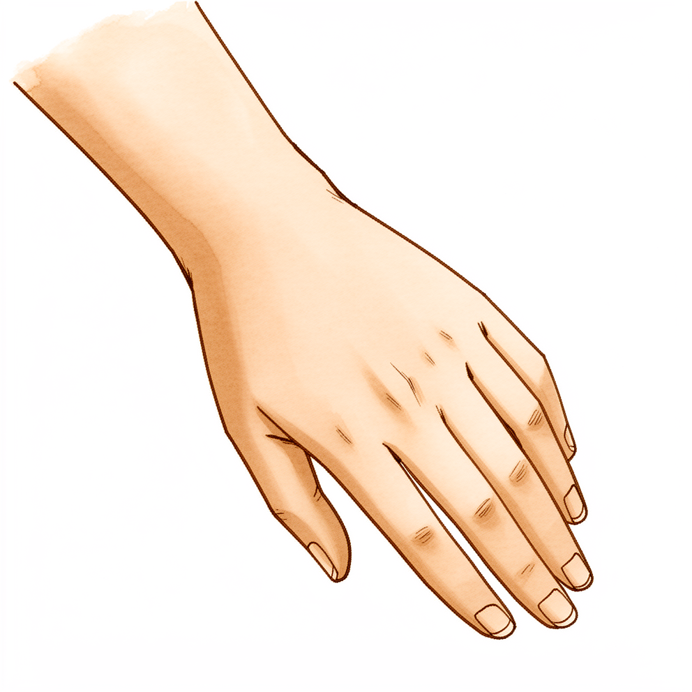
ہیمیٹ ہڈی کے ہک کا فریکچر (hook of hamate fracture)، یعنی کلائی کی ایک چھوٹی ہڈی کے ہک نما ابھار کا ٹوٹنا، ایک ہی لمحے میں ہوتا ہے۔ اس کی عام وجہ آپ کے ہاتھ کی ہتھیلی والی طرف، چھوٹی انگلی کی جڑ کے قریب، براہ راست ضرب لگنا ہے۔ یہ چوٹ بیس بال کے کھلاڑیوں اور گالف کھیلنے والوں میں عام ہے، جہاں بیٹ، کلب یا ریکٹ اس جگہ پر زور سے دباتا ہے۔ ہتھیلی کے لیگامنٹوں (ligaments)، یعنی وہ مضبوط بندھن جو ایک ہڈی کو دوسری ہڈی سے جوڑتے ہیں، اور ٹینڈنوں (tendons)، یعنی وہ مضبوط ڈوریاں جو پٹھوں کو ہڈی سے جوڑتی ہیں، پر اچانک پڑنے والی قوت سے بھی یہ ہو سکتا ہے۔
شروع ہی سے آپ کو ہاتھ کی ہتھیلی والی طرف، چھوٹی انگلی کے قریب جڑ میں درد محسوس ہوتا ہے۔ اس جگہ کو چھونے پر درد ہوتا ہے۔ زیادہ تر نئے (acute) فریکچروں میں چھونے پر یہ درد ہر بار موجود ہوتا ہے۔ ہاتھ سے دھکیلنے یا کھینچنے میں درد ہوتا ہے، اس لیے دروازے کا ہینڈل پکڑنا، کافی کا مگ تھامنا یا مرتبان کا ڈھکن گھمانا مشکل ہو جاتا ہے۔ ہو سکتا ہے آپ کو سوجن نظر آئے۔ درد شروع میں تیز ہوتا ہے، پھر کم ہو کر گہری دکھن میں بدل جاتا ہے۔
پہلے دن اور ہفتے پریشان کن ہو سکتے ہیں۔ ہاتھ استعمال کرنے پر درد ہوتا ہے، اور شروع میں دکھن آپ کی نیند میں خلل ڈال سکتی ہے۔ یہ چوٹ کم ہوتی ہے، اور اکثر کلائی کے عام ایکسرے میں نظر نہیں آتی۔ اس کا مطلب ہے کہ شروع میں یہ نظر انداز ہو سکتی ہے، خاص طور پر جب ایکسرے نارمل نظر آئیں۔ اگر آپ کی علامات اس انداز سے ملتی ہوں، تو ایک خاص زاویے سے لیا گیا ایکسرے یا سی ٹی اسکین فریکچر کو ڈھونڈ سکتا ہے۔ اس چوٹ کے لیے سی ٹی اسکین بہت درست ہوتا ہے۔
کچھ خطرے کی علامات میں فوری طبی امداد ضروری ہے۔ اگر آپ کا ہاتھ یا بازو گرم، سرخ، سوجا ہوا اور دردناک ہو جائے، خاص طور پر بخار کے ساتھ، تو اسی دن ہسپتال کی ایمرجنسی جائیں۔ اگر چوٹ کی جگہ پر جلد پھٹ گئی ہو، ہڈی نظر آ رہی ہو، یا شکل واضح طور پر بگڑی ہوئی ہو، تو اسی دن ایمرجنسی جائیں۔ اگر آپ کی انگلیاں یا ہاتھ سفید پڑنے لگیں، ٹھنڈے ہو جائیں، بالکل سفید یا نیلے ہو جائیں، یا اچانک احساس یا حرکت ختم ہو جائے، تو اسی دن ایمرجنسی جائیں۔
اگر آپ کی علامات کم نہ ہو رہی ہوں، ہفتوں کے دوران بڑھ رہی ہوں، آپ کو رات کو جگا دیتی ہوں، یا آپ کو کام کرنے یا اپنا ہاتھ استعمال کرنے سے روکتی ہوں، تو اپنے فیملی ڈاکٹر (GP) سے ملیں یا ماہر سے معائنہ کروانے کا کہیں۔ اگر آپ دفتری اوقات کے بعد یا ہفتے کے آخر میں کلینک سے رابطہ نہ کر سکیں، تو اپنے قریب ترین ہسپتال کی ایمرجنسی جائیں۔
دراصل کیا ہو رہا ہے
آپ کی کلائی میں گہرائی میں، ہتھیلی والی طرف، ایک چھوٹی خمدار ہڈی ہوتی ہے جسے ہیمیٹ (hamate) کہتے ہیں۔ اس میں ہڈی کا ایک چھوٹا سا ہک ہوتا ہے جس کا رخ آگے آپ کی ہتھیلی کی طرف ہوتا ہے۔ یہ ہک چھوٹی انگلی کو حرکت دینے والے کچھ لیگامنٹوں اور پٹھوں کے جڑنے کی جگہ ہے، اور جب آپ گرفت کرتے ہیں تو آپ کی انگلیوں کے ٹینڈن بالکل اس کے ساتھ لگ کر سرکتے ہیں۔
جب آپ ہاتھ کے بل گرتے ہیں یا بیٹ، کلب یا ریکٹ گھماتے ہیں، تو یہ ہک دباؤ برداشت کرتا ہے۔ ایک ہی زوردار ضرب اس میں دراڑ ڈال سکتی ہے۔ ایک ہی طرح سے ہزاروں بار گھمانا بھی اسے آہستہ آہستہ کمزور کر سکتا ہے یہاں تک کہ وہ ٹوٹ جائے، جیسے پیپر کلپ کو بار بار آگے پیچھے موڑا جائے تو وہ ٹوٹ جاتا ہے۔ یہ ہڈی چھوٹی ہے، اور کلائی کی ہڈیوں کے فریکچروں میں سے صرف تقریباً 2 فیصد یہاں ہوتے ہیں۔
ہک کو خون کی فراہمی کم ہوتی ہے، اور یہ بات اہم ہے۔ جسم کے کسی بھی دوسرے حصے کی طرح ہڈی کو دوبارہ جڑنے کے لیے خون کی ضرورت ہوتی ہے۔ گرفت کرتے وقت ہک ہلتا بھی ہے، کیونکہ اس پر دباؤ ڈالنے والے ٹینڈن ٹوٹے ہوئے سروں کو ہلاتے رہتے ہیں۔ جو فریکچر ہلتا رہے اور جسے خون کم ملے، وہ اکثر مشکل سے جڑتا ہے۔ ڈاکٹر اسے نان یونین (nonunion) کہتے ہیں، یعنی ہڈی کبھی نہیں جڑتی۔
اگر ٹوٹے ہوئے ٹکڑے اپنی معمول کی جگہ پر ہوں، تو شروع میں ہاتھ کو ساکن رکھنے سے ہڈی اب بھی جڑ سکتی ہے۔ اگر ٹکڑے ایک دوسرے سے ہٹ گئے ہوں، یا فریکچر کا دیر سے پتا چلے، تو سادہ طریقے سے جڑنے کا امکان کم ہو جاتا ہے۔ ایسی صورت میں علاج کا مطلب عموماً یا تو ہڈی کو ایک چھوٹے پیچ سے جوڑ کر رکھنا ہوتا ہے، یا ہک کو مکمل طور پر نکال دینا۔ اسے نکالنا سننے میں سخت قدم لگتا ہے، لیکن ہاتھ اس چھوٹے ہک کے بغیر اچھی طرح کام کر لیتا ہے، اور بہت سے لوگ اپنے پچھلے معیار پر کھیل میں واپس آ جاتے ہیں۔
ایک اور بات جاننے کے قابل ہے۔ ٹوٹے ہوئے ہک کا کھردرا، نہ جڑا ہوا کنارا بالکل آپ کی انگلیوں کے ٹینڈنوں کے ساتھ لگا ہوتا ہے۔ وقت کے ساتھ یہ انہیں گھسا سکتا ہے، جیسے کوئی رسی کسی تیز دھار چٹان پر رگڑ کھاتی ہے۔ اگر کوئی ٹینڈن ٹوٹ جائے، تو ٹوٹا ہوا ہک نکال دیا جاتا ہے اور ہڈی کو ہموار کر دیا جاتا ہے تاکہ ٹینڈن دوبارہ کام کر سکے۔
ہم اس کے لیے کیا کر سکتے ہیں
ڈاکٹر کیرن ہِرپرا، جو Mater Private Hospital Rockhampton میں بازو اور ہاتھ کے سرجن (upper-limb surgeon) ہیں، علاج کو آپ کی مخصوص چوٹ کے مطابق طے کرتے ہیں۔ ہیمیٹ ہڈی کے ہک کے کچھ فریکچر آپریشن کے بغیر ٹھیک ہو جاتے ہیں، اور کچھ کے لیے جلد سرجری کی ضرورت ہوتی ہے، اس لیے بروقت معائنہ اہم ہے۔ مریضوں کو عام طور پر ان کا فیملی ڈاکٹر (GP) ہمارے کلینک بھیجتا ہے؛ اگر کسی فزیوتھراپسٹ نے آپ کو ہم سے ملنے کا مشورہ دیا ہے، تب بھی میڈیکیئر (Medicare) کی رقم کی واپسی کا حق دار ہونے کے لیے آپ کو اپنے فیملی ڈاکٹر سے ریفرل (referral) کی ضرورت ہو گی۔ کلینک میں ہم آپ کی طبی تاریخ لیتے ہیں، آپ کے ہاتھ کا معائنہ کرتے ہیں، اور جہاں ضرورت ہو وہاں امیجنگ کا انتظام کرتے ہیں۔ چونکہ یہ فریکچر اکثر عام ایکسرے میں چھپا رہتا ہے، اس لیے اسے واضح طور پر دیکھنے کے لیے ہم ایک خاص زاویے سے لیا گیا ایکسرے یا سی ٹی اسکین استعمال کر سکتے ہیں۔
اگر ٹوٹے ہوئے ٹکڑے اپنی معمول کی جگہ پر ہوں اور فریکچر کا جلد پتا چل جائے، تو ہم آپ کے ہاتھ کو اسپلنٹ یا پلستر میں ساکن رکھ سکتے ہیں تاکہ ہڈی جڑ سکے۔ ہم بار بار امیجنگ کے ذریعے ہڈی کے جڑنے پر نظر رکھتے ہیں، اور آپ کی ہینڈ تھراپسٹ (ہاتھ کی بحالی کی ماہر)، Extend Rehabilitation کی Ruby Doolan، آپ کو جس اسپلنٹ کی ضرورت ہو وہ بناتی ہیں اور صحیح مرحلے پر حرکت کی طرف واپسی میں آپ کی رہنمائی کرتی ہیں۔ کھلاڑی بعض اوقات فریکچر کے ساتھ کھیلتے رہنے اور نہ جڑے ہوئے ٹکڑے کا علاج بعد میں کروانے کا انتخاب کرتے ہیں، اور ہم آپ سے بات کریں گے کہ آپ کے لیے اس کا کیا مطلب ہے۔
اگر ٹکڑے ایک دوسرے سے ہٹ گئے ہوں، یا فریکچر غیر مستحکم ہو، تو ہم شروع ہی سے سرجری تجویز کر سکتے ہیں۔ ایک راستہ ہڈی کے ہک کو نکال دینا ہے، جس سے آپ نسبتاً جلد اپنے کھیل میں واپس جا سکتے ہیں۔ دوسرا راستہ ہڈی کو ایک چھوٹے پیچ سے جوڑ کر رکھنا ہے، جو ہم کم عمر مریضوں کے لیے گرفت کی طاقت برقرار رکھنے کی خاطر تجویز کر سکتے ہیں۔ ان میں سے کسی بھی آپریشن کے دوران ہماری کوشش ہوتی ہے کہ قریبی عصب اور خون کی فراہمی محفوظ رہے۔ ان راستوں کے درمیان انتخاب ایک ایسا فیصلہ ہے جو ہم واقعی آپ کے ساتھ مل کر کرتے ہیں: کبھی اسپلنٹ کام کر سکتا ہے، لیکن ہو سکتا ہے درد یا ہڈی کی آخری پوزیشن آپ کے ہاتھ یا آپ کے کھیل کے لیے مناسب نہ ہو۔
آپ جو بھی راستہ اختیار کریں، ابتدائی ہفتے ملتے جلتے ہوتے ہیں۔ ہم درد پر قابو پانے میں آپ کی مدد کریں گے، اور ہڈی جڑنے کے دوران آپ اپنے ہاتھ کی حفاظت کریں گے۔ صحیح مرحلے پر Ruby کے ساتھ ہینڈ تھراپی شروع ہوتی ہے، تاکہ آپ کی گرفت اور حرکت محفوظ طریقے سے واپس آئے۔ اگر آپ کی علامات کم نہ ہو رہی ہوں، ہفتوں کے دوران بڑھ رہی ہوں، آپ کو رات کو جگا دیتی ہوں، یا آپ کو کام کرنے یا اپنا ہاتھ استعمال کرنے سے روکتی ہوں، تو اپنے فیملی ڈاکٹر سے ملیں یا ماہر سے معائنہ کروانے کا کہیں۔
کیا توقع رکھیں
صحت یابی اس بات پر منحصر ہے کہ آپ اس سفر میں کس مرحلے پر ہیں۔ اگر آپ کے فریکچر کا جلد پتا چل جائے اور ٹکڑے اپنی معمول کی جگہ پر ہوں، تو ہاتھ کو اسپلنٹ یا پلستر میں ساکن رکھنے سے ہڈی کو جڑنے کا موقع ملتا ہے۔ اگر فریکچر کا دیر سے پتا چلے، یا ٹکڑے ہٹ گئے ہوں، تو ہو سکتا ہے ہڈی کبھی خود سے نہ جڑے۔ ایسی صورت میں سرجری کا مطلب عموماً یا تو ہڈی کو جوڑ کر رکھنے کے لیے ایک چھوٹا پیچ ہوتا ہے یا ہک کو مکمل طور پر نکال دینا۔
اگر ہک نکالنے کے لیے آپ کی سرجری ہو، تو زیادہ تر لوگ نسبتاً جلد اپنے کھیل میں واپس آ جاتے ہیں۔ اعلیٰ سطح کے شوقیہ کھلاڑیوں میں سرجری کے بعد وہ چوٹ سے پہلے والی کارکردگی کی سطح پر کھیل میں واپس آ سکتے ہیں، ٹیسٹ کرنے پر ان کے ہاتھ کی کارکردگی نارمل ہوتی ہے، درد کم ہوتا ہے، اور وہ نتیجے سے مطمئن ہوتے ہیں۔ ایک پیشہ ور باؤلر (bowler) کے ہاتھ کی کارکردگی ہک نکالے جانے کے 2.5 ماہ بعد تسلی بخش تھی۔ جلد اور دیر سے کی گئی دونوں طرح کی سرجری کے بعد لوگ چوٹ سے پہلے والی سرگرمی کی سطح پر واپس آ جاتے ہیں۔ ہڈی کو پیچ سے جوڑ کر رکھنا بھی اچھا کام کر سکتا ہے اور اس کے بعد مسائل کم ہوتے ہیں، اور نہ جڑے ہوئے فریکچر کی مرمت گرفت کی طاقت برقرار رکھ سکتی ہے اور درد کو مکمل طور پر دور کر سکتی ہے۔
صحت یابی بتدریج واپسی ہے، کوئی بٹن دبانے سے ہونے والی چیز نہیں۔ شروع میں آپ کے ہاتھ میں دکھن ہوگی، اور یہ دکھن ہفتوں میں کم ہو جاتی ہے۔ ہینڈ تھراپسٹ کی رہنمائی میں ہاتھ کو دوبارہ استعمال کرنے سے گرفت کی طاقت واپس آتی ہے۔ مگ تھامنے یا چابی گھمانے جیسے روزمرہ کام اس وقت آسان ہو جاتے ہیں جب ٹوٹے ہوئے ٹکڑے کی وجہ سے ٹینڈنوں میں ہونے والی جلن (irritation) ختم ہو جاتی ہے۔
کچھ حقیقی خطرات ہیں جنہیں جاننا ضروری ہے۔ ہک کو خون کی فراہمی کم ہوتی ہے، اس لیے سب سے بڑی فکر ہڈی کا دیر سے جڑنا یا نہ جڑنا ہے۔ اگر ہڈی نہ جڑے تو اس کا کھردرا کنارا آپ کی انگلیوں کے ٹینڈنوں سے رگڑ کھا سکتا ہے۔ چھوٹی انگلی کا ٹینڈن اصل چوٹ کے 6 ماہ سے 25 سال بعد تک کسی بھی وقت ٹوٹا ہے، اور اس کے بعد گرفت کی طاقت اوسطاً دوسرے ہاتھ کی 83 فیصد تھی۔ اگر کوئی ٹینڈن واقعی ٹوٹ جائے، تو ٹوٹا ہوا ہک نکال دیا جاتا ہے اور ہڈی کو ہموار کر دیا جاتا ہے تاکہ ٹینڈن دوبارہ کام کر سکے۔ خود سرجری میں معمولی پیچیدگیوں کا خطرہ کم ہوتا ہے۔ اگر آپ کی علامات کم نہ ہو رہی ہوں، ہفتوں کے دوران بڑھ رہی ہوں، آپ کو رات کو جگا دیتی ہوں، یا آپ کو کام کرنے یا اپنا ہاتھ استعمال کرنے سے روکتی ہوں، تو اپنے فیملی ڈاکٹر سے ملیں یا ماہر سے معائنہ کروانے کا کہیں۔
ڈاکٹر کو کب دکھائیں
اگر چوٹ کی جگہ پر کھلا زخم ہو، ہڈی نظر آ رہی ہو، یا شکل واضح طور پر بگڑی ہوئی ہو، تو فوری طبی امداد حاصل کریں۔ اگر آپ کی انگلیاں یا ہاتھ سفید پڑنے لگیں، ٹھنڈے ہو جائیں، بالکل سفید یا نیلے ہو جائیں، یا اچانک احساس یا حرکت ختم ہو جائے، تو اسی دن ایمرجنسی جائیں۔ ان علامات کے لیے ہسپتال کی ایمرجنسی ضروری ہے، کسی کلینک کی انتظار گاہ میں انتظار کرنا نہیں۔
باقی ہر چیز کے لیے پہلے اپنے فیملی ڈاکٹر سے رجوع کریں۔ اگر درد کم نہ ہو رہا ہو، یا ہڈی جڑنے کے دوران سوجن، حرکت یا گرفت میں ہفتہ بہ ہفتہ بہتری نہ آ رہی ہو، تو ماہر سے معائنہ کروانے کا کہیں۔ یہ چوٹ عام ایکسرے میں چھپ جاتی ہے، اس لیے پہلا نتیجہ نارمل آنے سے اس کا امکان ختم نہیں ہو جاتا۔ سی ٹی اسکین اسے 95 فیصد معاملات میں ڈھونڈ لیتا ہے، اور جلد تشخیص بعد میں آپ کی گرفت اور ٹینڈنوں کی حفاظت کرتی ہے۔
Evidence & references
This is the clinical evidence summary written for health professionals. It is technical, and it lists the research this page was built from. You do not need to read it to understand your treatment or to make a decision about it.
Overview
- Hook of hamate fractures are rare injuries [7, 15].
- The most usual mechanism for hook of hamate fractures is a direct blow to the area of the hook [7, 15].
- Fractures of the hook of the hamate have been infrequently reported in the literature [2].
- Hook of hamate fractures with normal standard x-rays may be difficult to diagnose, especially for a nonhand surgeon [1].
- A specific radiographic view is recommended in addition to standard views in all cases of suspected fractures of the hook of the hamate [8].
- The incidence of acute hook of hamate fracture in association with high-energy distal fractures in patients undergoing CT evaluation of the wrist is 2.7% [18].
- Hamate hook fractures can be successfully treated acutely by immediate immobilization [3].
- Non-displaced acute fractures and delayed union of the hamate hook can be treated successfully by dorsal percutaneous cannulated mini-screw fixation with minimal morbidity and complications [5].
- The dorsal percutaneous approach is a reliable method that provides safe fixation of non-displaced fractures of the hook of hamate [23].
- Surgical excision as treatment for hook of hamate fractures is safe and allows a relatively rapid return to play in most cases [4].
- Surgical excision of the hook of the hamate resulted in good postoperative outcomes with a mean follow-up of 6 years in athletes [19].
- Early and late surgical excision of the hook of the hamate leads to excellent results with return to preinjury levels of activity [13].
- Surgical excision of hook of hamate fractures in high-level amateur athletes allows for successful return to sports participation at preinjury performance levels, achievement of normal function as measured by validated objective outcome measures, significant reduction in pain, and high overall patient satisfaction [27].
- Surgical excision is the treatment of choice for fractures of the hook of the hamate in any athlete, as the incidence of nonunion precludes the use of open reduction internal fixation [30].
- The treatment of choice for a symptomatic nonunion, and possibly for an acute fracture, appears to be surgical excision of the hook of the hamate [9].
- Symptomatic partial union of the hook of the hamate fracture in athletes should be managed with excision of the hook of the hamate and repair of the ligament attachments [10].
- The open carpal tunnel approach is a successful technique for open excision of symptomatic ununited hook of hamate fractures due to its familiarity, ease of performance, excellent visualization, and low morbidity [22].
- Both surgical procedures appear to yield acceptable outcomes in the treatment of hook of hamate fractures [11].
- Repair by open reduction and internal fixation for ununited fracture hook of the hamate is feasible and desirable for preservation of grip strength and complete relief of pain [14].
- In younger patients with nonunion of the hook of the hamate, repair of the nonunion rather than simple excision should be aimed for to preserve grip strength [20].
- Hook of hamate regeneration can occur after fracture fragment excision [12].
- The incidence of a hypoplastic hook of the hamate is common in patients with carpal tunnel syndrome [47].
Anatomy & Pathophysiology
Bony Anatomy
- The hamate consists of a body and a hook (hamulus) [59].
- The hook of the hamate serves as an attachment site for the transverse carpal ligament [59].
- The hook of the hamate serves as an origin for the flexor digiti minimi and opponens digiti minimi muscles [59].
- The distal end of the transverse carpal ligament is located 10 mm from the hook of the hamate [36].
- The boundaries of Guyon’s canal include the hook of the hamate radially and the pisiform ulnarly [56].
Vascular Supply
- The extraosseous blood supply to the carpus is provided by terminal branches of the radial, ulnar, and anterior interosseous arteries through dorsal and palmar transverse arterial arches [62].
- The dorsal intercarpal arch is the largest of the dorsal arches and supplies the distal carpal row [62].
- The deep palmar arch at the level of the metacarpal bases is consistent and communicates with the dorsal basal metacarpal arch and palmar metacarpal arteries [62].
- The hamate hook has a poor blood supply, which is a main reason for the increased risk of nonunion [48].
- The hamate hook is vulnerable to osteonecrosis and nonunion due to its vascularity [3].
Ligamentous Anatomy
- The capitohamate ligament is a thick ligament with a cross-section of 5 × 5 mm [57].
- The capitohamate ligament has extensions to the third or fourth metacarpals [57].
- The distal row of carpal bones is rigid with little motion between its bones due to stout intercarpal ligaments [57].
Biomechanics & Kinematics
- The hamate is part of the central column of the wrist, which controls flexion-extension of the wrist [60].
- The hamate is part of the metacolumn in Weber's three-column theory, which includes the triquetrum and hamate [60].
- The lunate, capitate, hamate, trapezium, and trapezoid function collectively as the "stable central column" in the Sandow et al. model [60].
- The hamate hook provides a biomechanical advantage for flexor tendon function [43].
- Cadaveric changes in tendon force after hamate hook excision suggest that power grip may be decreased [43].
- No significant change in the position of deep or superficial digital flexor tendons relative to the hook of hamate was noted with finger extension or flexion [49].
Pathophysiology & Injury Mechanisms
- Hook of hamate fractures are traditionally thought to be caused by direct trauma [69].
- Fracture of the hook of hamate is more likely to result from a fatigue response due to repetitive load applied by adjacent deep flexor tendons [69].
- Direct pressure of tendons on critical local vessels reduces blood flow, leading to mechanical and vascular effects that create pathological osseous change and weakening [69].
- These pathological changes predispose to stress fracture and nonunion in repetitive gripping activities [69].
- High-energy single-impact hamate fractures likely occur in otherwise healthy bone without pre-existing stress response [72].
- Fractures of the hook of the hamate can occur by direct impact applied to the hamate during a fall on the heel of the hand [48].
- Fractures of the hook of the hamate can occur by microtraumatism in sports requiring repeated use of a racquet or a bat [48].
- The growing popularity of golf and other racquet sports has led to an increase in stress fractures of the hamate hook [48].
- Mobility of the fracture site is a main reason for the increased risk of nonunion of the hamate hook [48].
- Delayed diagnosis is a main reason for the increased risk of nonunion of the hamate hook [48].
- Combined fracture of the hook of the hamate and palmar dislocation of the fifth carpometacarpal joint results from a sudden, violent contraction of the flexor carpi ulnaris against a fixed wrist [35].
- Chronic repetitive wear and fraying of flexor tendons against the rough surface of the hook is the proposed etiology for closed rupture of the flexor tendon [37].
- The misleading presentation of a hamate hook stress fracture in a professional bowler is related to a new drilling layout of the bowling ball and technical changes during ball release [29].
- Technical changes in bowling redistribute micro-constraints on the hook of the hamate using an indirect mechanism of injury through ligaments and flexor tendons [29].
Classification
Epidemiology and Mechanism
- Fractures of the hook of the hamate are rare injuries [7].
- Hamate hook fracture represents 2% of carpal bone fractures [48].
- The incidence of acute hook of hamate fracture in association with high-energy distal radius fractures is 2.7% [18].
- Combined intraarticular fracture of the body and hook of hamate is an extremely rare injury pattern [21].
Diagnostic Challenges
- Fractures of the hook of the hamate are not seen on standard two or three view wrist examination [26].
- The similarities between a fracture of the hook of the hamate and other conditions that cause ulnar wrist pain can lead to failure of clinical diagnosis [17].
- Combined intraarticular fracture of the body and hook of hamate requires a high index of suspicion for diagnosis [21].
Imaging and Physical Examination
- The carpal tunnel radiographic view should be considered in addition to routine radiographic views for patients with a possible hook of the hamate fracture [6].
- The hook of hamate pull test is an easy, cheap, sensitive, and specific test that can be readily performed in the clinic to diagnose hook of hamate fractures [25].
- A CT scan has 95% specificity and 95% accuracy in diagnosing hamulus fractures [33].
- Pain is provoked by resistive extension of the ring and middle finger PIP joints in the diagnosis of hamulus fracture or nonunion [33].
Clinical Presentation
Diagnostic Challenges and Rarity
- These fractures are not seen on standard two or three view wrist examination [26].
- Combined intraarticular fracture of the body and hook of hamate is an extremely rare injury pattern requiring a high index of suspicion for diagnosis [21].
- Hook of hamate fractures present with similarities to other conditions that cause ulnar wrist pain [17].
Physical Examination Findings
- Volar tenderness on palpation over the hook of hamate was present in 100% of cases in a study of acute hook of hamate fractures [67].
- The pull test was painful for all patients in a study of acute hook of hamate fractures [67].
- Pain over the ulnar-palmar base of the hand should prompt a presumptive diagnosis of fracture of the hook of the hamate until proven otherwise [31].
Imaging and Diagnostic Modalities
- Lateral trispiral tomography is clearly superior to other diagnostic methods for hamate hook fractures [41].
- An incidence of 2.7% of acute hook of hamate fracture was identified in association with high-energy distal fractures in patients undergoing CT evaluation of the wrist [18].
Mechanism of Injury
- Hamate hook stress fractures can result from an indirect mechanism of injury through the ligaments and flexor tendons due to redistributed micro-constraints [29].
Investigations
Clinical Examination
- Fractures of the hook of the hamate have clinical similarities to other conditions that cause ulnar wrist pain [17].
- In institutions where this protocol is followed, any patient with pain over the ulnar-palmar base of the hand should have a carpal tunnel view obtained with a presumptive diagnosis of hook of hamate fracture until proven otherwise [31].
Radiography
- Hook of hamate fractures may be difficult to diagnose on standard x-rays, particularly for non-hand surgeons [1].
- A specific radiographic view is recommended in addition to standard views for all cases of suspected fractures of the hook of the hamate [8].
- Fractures of the pisiform and hamulus are not seen on standard two or three view wrist examinations [26].
- Three variations of the hook of hamate were identified with radiographic evaluation using the carpal tunnel view [80].
Advanced Imaging
- MRI is the modality of choice for imaging radiographically occult fractures of the hand and wrist [58].
- Bone marrow edema on MRI may reveal fractures of the carpal bones that are radiographically occult [66].
- An incidence of 2.7% of acute hook of hamate fracture was identified in association with high-energy distal radius fractures in patients undergoing CT evaluation of the wrist [18].
Treatment
Non-Operative Management
- Hamate hook fractures diagnosed early may heal with nonoperative management [16].
- Acute hook of hamate fractures in athletes may be treated with casting [34].
- Ultrasound treatment might be useful for nonunion of the hook of the hamate caused by repeated stress [46].
- Low-intensity pulsed ultrasound may be useful in the treatment of ununited fractures of the hook of hamate [50].
Operative Management: Excision
- Surgical excision as treatment for hook of the hamate fractures is safe and allows a relatively rapid return to play [4].
- Surgical excision of the hook of the hamate resulted in good postoperative outcomes with a mean follow-up of 6 years [19].
- Surgical excision remains an effective method of management, with a low risk of minor complications for both acute and chronic hook of hamate fractures in professional baseball players [28].
- MLB players sustaining hook of hamate fractures can reasonably expect to return to their pre-injury performance levels following operative treatment [71].
- The open carpal tunnel approach is a successful technique for open excision of symptomatic ununited hook of hamate fractures because of its familiarity, ease of performance, excellent visualization and low morbidity [22].
- A modified surgical approach through Guyon’s Canal and the proximal ulnar border of the carpal tunnel allows for safe excision of the hook of the hamate [32].
- The modified approach through Guyon’s Canal and the proximal ulnar border of the carpal tunnel allows direct visualization of the vital structures most at risk, including the motor branch of the ulnar nerve, unlike blind or unilateral approaches [32].
- The modified approach through Guyon’s Canal and the proximal ulnar border of the carpal tunnel preserves the nerve supply and blood supply to the hypothenar muscles [32].
- A lateral approach to the hook of hamate for its fracture is believed to replace the conventional volar approach because of its benefits in terms of time, safety, and simplicity [42].
- Functional recovery of the hand was satisfactory 2.5 months after surgical resection of the hamate’s hook in a professional bowler with a stress fracture [29].
Operative Management: Fixation
- The cadaveric study confirms that the dorsal percutaneous approach is a reliable method and provides safe fixation of non-displaced fractures of the hook of hamate [23].
- In younger patients with nonunion of the hook of the hamate, one should aim for the repair of the nonunion rather than simple excision to preserve grip strength [20].
- A simplified dorsal approach to fracture of the hamate hook with percutaneous fixation with screws avoids damage to the blood supply of the hamate, surrounding soft tissues, and the ulnar nerve [45].
- A simplified dorsal approach to fracture of the hamate hook with percutaneous fixation with screws preserves important stabilising ligaments [45].
Outcomes and Biomechanics
- Patient outcomes were good to excellent regardless of the treatment chosen, with minimal pain and good hand functionality [44].
- The hamate hook provides some biomechanical advantage for flexor tendon function [43].
- Cadaveric changes in tendon force after hamate hook excision suggest that power grip may be decreased after hamate hook excision [43].
Diagnostic Challenges
- A specific radiographic view is recommended in addition to standard ones in all cases of suspected fractures of the hook of the hamate [8].
- Fractures of the hook of the hamate share similarities with other conditions that cause ulnar wrist pain, leading to failure of clinical diagnosis [17].
Nonunion and Healing Complications
- Hamate hook fractures that are diagnosed early may heal with nonoperative management [16].
- Symptomatic partial union of the hook of the hamate fracture in athletes should be managed no differently from a symptomatic nonunion [10].
- In younger patients with nonunion of the hook of the hamate, repair of the nonunion is preferred over simple excision to preserve grip strength [20].
- Range of motion and grip strengths were equivalent on the operated and unoperated sides following bone grafting for nonunion [24].
Surgical Complications and Outcomes
- Surgical excision as treatment for hook of the hamate fractures is safe and allows a relatively rapid return to play in most cases [4].
- Dorsal percutaneous cannulated mini-screw fixation for non-displaced acute fractures and delayed union of the hamate hook is associated with minimal morbidity and complications [5].
- Both surgical procedures for hook of hamate fractures appear to yield acceptable outcomes [11].
- The open carpal tunnel approach for excision of symptomatic ununited hook of hamate fractures is associated with low morbidity [22].
- The dorsal percutaneous approach provides safe fixation of non-displaced fractures of the hook of hamate [23].
- Surgical excision for acute and chronic hook of hamate fractures in professional baseball players carries a low risk of minor complications [28].
Associated Injuries and Anatomical Variations
- The incidence of acute hook of hamate fracture in association with high-energy distal radius fractures is 2.7% in patients undergoing CT evaluation of the wrist [18].
- Preoperative evaluation of the morphology of the hooks may help predict adverse events in cases of hypoplastic hooks undergoing endoscopic carpal tunnel release [47].
Recovery
Non-Operative Management
Operative Management
- Dorsal percutaneous cannulated mini-screw fixation treats non-displaced acute fractures and delayed union of the hamate hook with minimal morbidity and complications [5].
- In younger patients with nonunion of the hook of the hamate, repair of the nonunion rather than simple excision is recommended to preserve grip strength [20].
- Range of motion and grip strengths were equivalent on the operated and unoperated sides following bone grafting of a hook of hamate nonunion [24].
Key Evidence
- [Paper] Hook of hamate fractures, with normal standard x-rays, may be difficult to diagnose, especially for a nonhand surgeon. [1] (10.1016/j.hcl.2021.06.013)
- [L4] Fractures of the hook of the hamate have been infrequently reported in the literature. [2] (10.1016/0020-1383(89)90169-1)
- [L5] The data support the recommendation that hamate hook fractures can be successfully treated acutely by immediate immobilization. [3] (10.1016/0363-5023(93)90405-r)
- [L4] In most cases, surgical excision as treatment for hook of the hamate fractures is safe and allows a relatively rapid return to play. [4] (10.1016/j.jhsa.2017.06.108)
- [L4] This pilot study demonstrates that non-displaced acute fractures and delayed union of the hamate hook can be treated successfully by dorsal percutaneous cannulated mini-screw fixation with minimal morbidity and complications. [5] (10.1142/s0218810412970039)
- [L4] The carpal tunnel radiographic view should be considered in addition to routine radiographic views for patients with a possible hook of the hamate fracture. [6] (10.2519/jospt.2010.0408)
- [L4] Fractures of the hook of the hamate are rare injuries, with the most usual mechanism being a direct blow to the area of the hook. [7] (10.1016/0266-7681(85)90019-1)
- [L5] We recommend this view, in addition to the standard ones, in all cases of suspected fractures of the hook of the hamate. [8] (10.1016/s0363-5023(88)80026-1)
- [L4] The treatment of choice for a symptomatic nonunion, and possibly for an acute fracture, appears to be surgical excision of the hook of the hamate. [9] (10.1016/s0363-5023(88)80107-2)
- [L4] This condition should be managed no differently from a symptomatic nonunion, with excision of the hook of the hamate and repair of the ligament attachments. [10] (10.1177/03635465030310010201)
- [L4] Both surgical procedures appear to yield acceptable outcomes in the treatment of hook of hamate fractures. [11] (10.1016/j.jhsg.2023.11.011)
- [L4] Hook of hamate regeneration can occur after fracture fragment excision. [12] (10.1016/j.jhsg.2024.08.008)
- [L4] Early and late surgical excision of the hook of the hamate leads to excellent results with return to preinjury levels of activity. [13] (10.1053/j.otsm.2010.03.003)
- [L5] The author maintains that repair by open reduction and internal fixation for ununited fracture hook of the hamate is not only feasible but also desirable, both for preservation of grip strength and complete relief of pain. [14] (10.1016/0363-5023(89)90032-4)
- [L4] Fractures of the hook of the hamate are rare injuries, with the most usual mechanism being a direct blow to the area of the hook. [15] (10.1016/0266-7681_85_90019-1)
- [L4] Our results show that hamate hook fractures that are diagnosed early may heal with nonoperative management. [16] (10.1016/0363-5023(92)90363-t)
- [L4] These cases are reported to emphasize the similarities of a fracture of the hook of the hamate to other conditions that cause ulnar wrist pain. [17] (10.1016/s0363-5023(85)80258-6)
- [L4] The series identified an incidence of 2.7% of acute hook of hamate fracture in association with high-energy distal fractures in patients undergoing CT evaluation of the wrist. [18] (10.1177/1753193411436293)
- [L4] Surgical excision of the hook of the hamate resulted in good postoperative outcomes with a mean follow-up of 6 years. [19] (10.3109/17453679308993670)
- [L5] In younger patients with nonunion of the hook of the hamate, the author believes one should aim for the repair of the nonunion rather than simple excision to preserve grip strength. [20] (10.1016/s0363-5023(86)80127-7)
- [L5] Combined intraarticular fracture of the body and hook of hamate is an extremely rare injury pattern requiring a high index of suspicion for diagnosis. [21] (10.1007/s12593-012-0070-2)
- [Paper] The study highlights the open carpal tunnel approach as a successful technique for open excision of symptomatic ununited hook of hamate fractures, because of its familiarity, ease of performance, excellent visualization and low morbidity. [22] (10.1016/j.injury.2014.05.008)
- [L5] The cadaveric study confirms that the dorsal percutaneous approach is a reliable method and provides safe fixation of non-displaced fractures of the hook of hamate. [23] (10.1177/1753193421991761)
- [L4] The range of motion and grip strengths were equivalent on the operated and unoperated sides. [24] (10.1016/s0363-5023(89)80008-5)
- [L4] The hook of hamate pull test is an easy, cheap, sensitive, and specific test that can be readily performed in the clinic to diagnose hook of hamate fractures. [25] (10.1016/j.jhsa.2010.08.024)
- [L4] These fractures are not seen on standard two or three view wrist examination. [26] (10.1016/s0736-4679(98)00016-x)
- [L4] Surgical excision of hook of hamate fractures in high-level amateur athletes allows for successful return to sports participation at preinjury performance levels, achievement of normal function as measured by validated objective outcome measures, significant reduction in pain, and high overall patient satisfaction. [27] (10.1016/j.jhsa.2012.10.011)
- [L4] Surgical excision remains an effective method of management, with a low risk of minor complications for both acute and chronic hook of hamate fractures in professional baseball players. [28] (10.1016/j.jhsa.2021.03.015)
- [L5] [29] (10.1016/j.hansur.2016.06.007)
- [L5] Surgical excision is the treatment of choice for fractures of the hook of the hamate in any athlete, as the incidence of nonunion precludes the use of open reduction internal fixation. [30] (10.1016/j.hcl.2012.05.013)
- [L5] The author states that in their institution, anyone with pain over the ulnar-palmar base of the hand should have a carpal tunnel view with the presumptive diagnosis of fracture of the hook of the hamate until proven otherwise. [31] (10.1016/s0363-5023(86)80125-3)
- [L4] This modified approach allows direct visualization of the vital structures most at risk, including the motor branch of the ulnar nerve, unlike blind or unilateral approaches, and preserves the nerve supply and blood supply to the hypothenar muscles. [32] (10.1016/j.jhsa.2019.07.015)
- [L5] [33] (10.1016/j.jhsa.2013.06.004)
- [L5] Acute hook of hamate fractures in athletes may be treated with casting or allowed to return to play with nonunion addressed later via excision; definitive treatment for stress fractures is excision. [34] (10.1016/j.hcl.2012.05.012)
- [L5] These findings suggest that such an injury results from a sudden, violent contraction of the flexor carpi ulnaris against the fixed wrist. [35] (10.1016/s0266-7681(96)80043-x)
- [L5] The distal end of the transverse carpal ligament was located 10 mm from the hook of the hamate. [36] (10.1016/0363-5023(94)90073-6)
- [L4] The proposed etiology of the tendon ruptures is chronic repetitive wear and fraying against the rough surface of the hook. [37] (10.1142/s2424835516720164)
- [L4] Lateral trispiral tomography is clearly superior to other diagnostic methods for hamate hook fractures. [41] (10.1016/0363-5023(88)90217-1)
- [L5] The authors believe this lateral approach should replace the conventional volar approach because of its benefits in terms of time, safety, and simplicity. [42] (10.1016/0266-7681(86)90028-8)
- [L5] The hamate hook provides some biomechanical advantage for flexor tendon function and cadaveric changes in tendon force after its excision suggest that power grip may be decreased after hamate hook excision. [43] (10.1053/jhsu.2003.50005)
- [L4] Patient outcomes were good to excellent regardless of the treatment chosen, with minimal pain and good hand functionality. [44] (10.1177/1753193417729603)
- [L4] This minimally invasive technique avoids damage to the blood supply of the hamate, surrounding soft tissues, and the ulnar nerve, while preserving important stabilising ligaments. [45] (10.3109/02844310801956714)
- [L4] Ultrasound treatment might be useful for nonunion of the hook of the hamate caused by repeated stress, and we consider that the ultrasound treatment for nonunion of the hook of the hamate is one option in various treatment methods. [46] (10.1007/s00167-003-0425-0)
- [L4] The incidence of a hypoplastic hook of the hamate is common in patients with carpal tunnel syndrome, and preoperative evaluation of the morphology of the hooks and indications for endoscopic carpal tunnel release in cases of hypoplastic hooks may help predict adverse events. [47] (10.1016/j.jhsg.2023.12.010)
- [L5] [48] (10.1097/bth.0b013e31826577f8)
- [L4] No significant change with finger extension or flexion was noted in either the deep or superficial digital flexor tendons. [49] (10.1142/s2424835519500139)
- [L5] Based on the results of this case low-intensity pulsed ultrasound may be useful in the treatment of ununited fractures of the hook of hamate. [50] (10.1053/jhsu.2000.jhsu025a0077)
- [L4] [67] (10.1016/j.jhsa.2019.01.014)
- [L5] [69] (10.1177/17531934241235803)
- [L3] MLB players sustaining hook of hamate fractures can reasonably expect to return to their pre-injury performance levels following operative treatment. [71] (10.1123/jsr.2017-0071)
- [L5] [72] (10.1177/17531934241304249)
- [L4] Three variations of the hook of hamate were identified with radiographic evaluation using the carpal tunnel view. [80] (10.1016/j.jhsa.2005.05.018)
References
[1] Hook of Hamate Fractures. Hand Clinics. 2021. DOI: 10.1016/j.hcl.2021.06.013
[2] Fractures of the hook of the hamate. Injury. 1989. DOI: 10.1016/0020-1383(89)90169-1
[3] Hook of hamate vascularity: Vulnerability to osteonecrosis and nonunion. The Journal of Hand Surgery. 1993. DOI: 10.1016/0363-5023(93)90405-r
[4] Return to Play and Complications After Hook of the Hamate Fracture Surgery. The Journal of Hand Surgery. 2017. DOI: 10.1016/j.jhsa.2017.06.108
[5] DORSAL PERCUTANEOUS CANNULATED MINI-SCREW FIXATION FOR FRACTURES OF THE HAMATE HOOK. Hand Surgery. 2012. DOI: 10.1142/s0218810412970039
[6] Hook of the Hamate Fracture. Journal of Orthopaedic & Sports Physical Therapy. 2010. DOI: 10.2519/jospt.2010.0408
[7] Fractures of the hook of the hamate. The Journal of Hand Surgery: Journal of the British Society for Surgery of the Hand. 1985. DOI: 10.1016/0266-7681(85)90019-1
[8] Radiographic evaluation of the hook of the hamate: A new technique. The Journal of Hand Surgery. 1988. DOI: 10.1016/s0363-5023(88)80026-1
[9] Excision of the hook of the hamate: A retrospective survey and review of the literature. The Journal of Hand Surgery. 1988. DOI: 10.1016/s0363-5023(88)80107-2
[10] Symptomatic, Partial Union of the Hook of the Hamate Fracture in Athletes. The American Journal of Sports Medicine. 2003. DOI: 10.1177/03635465030310010201
[11] Surgical Management of Hook of Hamate Fractures: A Systematic Review of Outcomes. Journal of Hand Surgery Global Online. 2024. DOI: 10.1016/j.jhsg.2023.11.011
[12] Hook of Hamate Regrowth After Surgical Excision: A Report of Two Cases. Journal of Hand Surgery Global Online. 2024. DOI: 10.1016/j.jhsg.2024.08.008
[13] Hook of the Hamate Fractures. Operative Techniques in Sports Medicine. 2010. DOI: 10.1053/j.otsm.2010.03.003
[14] Fracture of hamate hook. The Journal of Hand Surgery. 1989. DOI: 10.1016/0363-5023(89)90032-4
[15] Fractures of the Hook of the Hamate. Journal of Hand Surgery. 1985. DOI: 10.1016/0266-7681_85_90019-1
[16] Nonoperative treatment of acute hamate hook fractures. The Journal of Hand Surgery. 1992. DOI: 10.1016/0363-5023(92)90363-t
[17] Fractures of the hook of the hamate—A failure of clinical diagnosis. The Journal of Hand Surgery. 1985. DOI: 10.1016/s0363-5023(85)80258-6
[18] Concomitant high-energy fractures of the distal radius and hook of hamate. Journal of Hand Surgery (European Volume). 2012. DOI: 10.1177/1753193411436293
[19] Fractures of the hook of the hamate in athletes: 8 cases followed for 6 years. Acta Orthopaedica Scandinavica. 1993. DOI: 10.3109/17453679308993670
[20] Fracture of the hook of the hamate. The Journal of Hand Surgery. 1986. DOI: 10.1016/s0363-5023(86)80127-7
[21] Combined Intraarticular Fracture of the Body and the Hook of Hamate: An Unusual Injury Pattern. Journal of Hand and Microsurgery. 2013. DOI: 10.1007/s12593-012-0070-2
[22] Surgical excision of ununited hook of hamate fractures via the carpal tunnel approach. Injury. 2014. DOI: 10.1016/j.injury.2014.05.008
[23] Safety and reliability of the dorsal percutaneous approach for non-displaced hook of hamate fracture: an anatomical study. Journal of Hand Surgery (European Volume). 2021. DOI: 10.1177/1753193421991761
[24] Nonunion of the hook of the hamate: An argument for bone grafting the nonunion. The Journal of Hand Surgery. 1989. DOI: 10.1016/s0363-5023(89)80008-5
[25] Hook of Hamate Pull Test. The Journal of Hand Surgery. 2010. DOI: 10.1016/j.jhsa.2010.08.024
[26] Pisiform and Hamulus Fractures: Easily Missed Wrist Fractures Diagnosed on a Reverse Oblique Radiograph. The Journal of Emergency Medicine. 1998. DOI: 10.1016/s0736-4679(98)00016-x
[27] Outcomes of Hook of Hamate Fracture Excision in High-Level Amateur Athletes. The Journal of Hand Surgery. 2013. DOI: 10.1016/j.jhsa.2012.10.011
[28] Hook of Hamate Fractures in Major and Minor League Baseball Players. The Journal of Hand Surgery. 2021. DOI: 10.1016/j.jhsa.2021.03.015
[29] Hamate hook stress fracture in a professional bowler: Case report of an unusual causal sport. Hand Surgery and Rehabilitation. 2017. DOI: 10.1016/j.hansur.2016.06.007
[30] Treatment of Fracture of Hook of the Hamate in Baseball Players. Hand Clinics. 2012. DOI: 10.1016/j.hcl.2012.05.013
[31] Fractures of the hook of the hamate. The Journal of Hand Surgery. 1986. DOI: 10.1016/s0363-5023(86)80125-3
[32] A Modified Surgical Approach Through Guyon’s Canal and the Proximal Ulnar Border of the Carpal Tunnel Allows for Safe Excision of the Hook of the Hamate. The Journal of Hand Surgery. 2019. DOI: 10.1016/j.jhsa.2019.07.015
[33] Hook of Hamate Fractures. The Journal of Hand Surgery. 2013. DOI: 10.1016/j.jhsa.2013.06.004
[34] Hook of Hamate and Pisiform Fractures in Basketball and Hockey Players. Hand Clinics. 2012. DOI: 10.1016/j.hcl.2012.05.012
[35] Combined Fracture of the Hook of the Hamate and Palmar Dislocation of the Fifth Carpometacarpal Joint. Journal of Hand Surgery. 1996. DOI: 10.1016/s0266-7681(96)80043-x
[36] Clinical location of hook of hamate: A technical note for endoscopic carpal tunnel release. The Journal of Hand Surgery. 1994. DOI: 10.1016/0363-5023(94)90073-6
[37] Closed Rupture of the Flexor Tendon Secondary to Sclerosis of the Hook of the Hamate: A Report of Two Cases. The Journal of Hand Surgery (Asian-Pacific Volume). 2016. DOI: 10.1142/s2424835516720164
[41] Fracture of the hamate hook. The Journal of Hand Surgery. 1988. DOI: 10.1016/0363-5023(88)90217-1
[42] Lateral approach to the hook of hamate for its fracture. The Journal of Hand Surgery: Journal of the British Society for Surgery of the Hand. 1986. DOI: 10.1016/0266-7681(86)90028-8
[43] Biomechanical evaluation of flexor tendon function after hamate hook excision. The Journal of Hand Surgery. 2003. DOI: 10.1053/jhsu.2003.50005
[44] Diagnosis and management of hook of hamate fractures. Journal of Hand Surgery (European Volume). 2017. DOI: 10.1177/1753193417729603
[45] Simplified dorsal approach to fracture of the hamate hook with percutaneous fixation with screws. Journal of Plastic Surgery and Hand Surgery. 2010. DOI: 10.3109/02844310801956714
[46] Ultrasound treatment of nonunion of the hook of the hamate in sports activities. Knee Surgery, Sports Traumatology, Arthroscopy. 2003. DOI: 10.1007/s00167-003-0425-0
[47] Anatomic Variation of the Hamate Hook as a Potential Risk in Endoscopic Carpal Tunnel Release. Journal of Hand Surgery Global Online. 2024. DOI: 10.1016/j.jhsg.2023.12.010
[48] Hamate Hook Nonunion Treated With a Hook Plate. Techniques in Hand & Upper Extremity Surgery. 2012. DOI: 10.1097/bth.0b013e31826577f8
[49] Sliding Position of the Flexor Tendons Relative to the Hook of Hamate in CT Scans. The Journal of Hand Surgery (Asian-Pacific Volume). 2019. DOI: 10.1142/s2424835519500139
[50] Treatment of ununited fracture of the hook of hamate by low-intensity pulsed ultrasound: A case report. The Journal of Hand Surgery. 2000. DOI: 10.1053/jhsu.2000.jhsu025a0077
[56] A Lange Medical Book Current Diagnosis Treatment In Orthopedics Fifth Edition. 2Musculoskeletal Trauma Surgery > FRACTURES AND DISLOCATIONS OF THE DISTAL AND MID-FOREARM.
[57] Aaos Comprehensive Orthopaedic Review 3. Carpal Instability* > II. Anatomy and Biomechanics (See Chapter 92).
[58] Orthopaedic Knowledge Update 13 Ebook Without Multimedia. Anatomy, Evaluation, Clinical Examination, and Imaging > Imaging: Advances in Imaging of the Hand and Upper Extremity > Magnetic Resonance Imaging.
[59] Aaos Comprehensive Orthopaedic Review 3. Anatomy of the Hand and Wrist > VII. The Wrist.
[60] Green S Operative Hand Surgery. WRIST BIOMECHANICS > Carpal Kinematics.
[62] Campbell S Operative Orthopaedics 4 Volume Set. NERVE INJURIES AT THE LEVEL OF THE HAND AND WRIST > CIRCULATION.
[66] Campbell S Operative Orthopaedics 4 Volume Set. WRIST AND ELBOW.
[67] Minimally Invasive Fixation With a Volar Approach Using a Cannulated Compression Screw for Acute Hook of Hamate Fractures. The Journal of Hand Surgery. 2019. DOI: 10.1016/j.jhsa.2019.01.014
[69] The aetiology of fracture and nonunion in the hook of the hamate. Journal of Hand Surgery (European Volume). 2024. DOI: 10.1177/17531934241235803
[71] Performance Outcomes After Hook of Hamate Fractures in Major League Baseball Players. Journal of Sport Rehabilitation. 2018. DOI: 10.1123/jsr.2017-0071
[72] Re: Campbell FC, Jones SW, Campbell DA. The aetiology of fracture and nonunion in the hook of the hamate. J Hand Surg Eur. 2024, 49: 1172-8. Journal of Hand Surgery (European Volume). 2024. DOI: 10.1177/17531934241304249
[80] Anatomic Variations of the Hook of Hamate and the Relationship to Carpal Tunnel Syndrome. The Journal of Hand Surgery. 2005. DOI: 10.1016/j.jhsa.2005.05.018