Patients › Hand
ہیمیٹ ہڈی کے فریکچر اور انگوٹھی والی اور چھوٹی انگلی کے سی ایم سی جوڑ کے فریکچر کے ساتھ جوڑ کا نکلنا
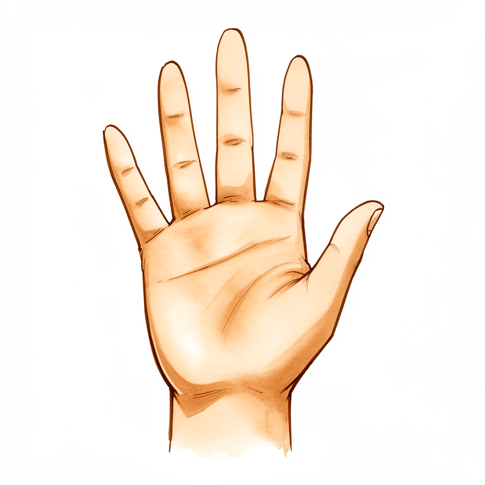
آپ کو کیا محسوس ہوتا ہے
یہ اچانک لگنے والی چوٹ ہے، آہستہ آہستہ بڑھنے والا مسئلہ نہیں۔ یہ عموماً اس وقت ہوتی ہے جب آپ کسی چیز پر زور سے مکا ماریں، اپنی مٹھی کے بل گریں، یا ہاتھ پر کوئی بھاری ضرب لگے۔ آپ کو انگوٹھی والی یا چھوٹی انگلی کی جڑ پر کچھ چٹخنے کا احساس ہو سکتا ہے یا اس کی آواز سنائی دے سکتی ہے۔ درد فوراً شروع ہو جاتا ہے، ہتھیلی کے باہری کنارے پر کلائی کے قریب۔
آپ کی چھوٹی انگلی کی جڑ اور وہ ہڈی جس پر یہ ٹکی ہوتی ہے، یعنی ہیمیٹ (hamate) جو کلائی کی چھوٹی ہڈیوں میں سے ایک ہے، یہ زور سہتی ہیں۔ وہاں کا جوڑ ٹوٹ سکتا ہے اور اپنی جگہ سے کھسک سکتا ہے۔ آپ کو ہاتھ کی پشت پر سوجن نظر آئے گی، اور نیل بھی پڑ سکتا ہے۔ آپ کی چھوٹی انگلی سیدھ سے ہٹی ہوئی نظر آ سکتی ہے یا پہلے سے مختلف انداز میں بیٹھی ہوئی لگ سکتی ہے۔ اس انگلی کو حرکت دینے، کوئی چیز پکڑنے، یا ہاتھ کے سہارے زور لگا کر اٹھنے سے درد ہوگا۔ روزمرہ کے کام، جیسے کافی کا مگ پکڑنا، دروازے کا دستہ گھمانا، یا ہاتھ ملانا، مشکل ہو جاتے ہیں۔
پہلے چند دنوں سے لے کر چند ہفتوں تک، درد اکثر رات کو اور انگلی کو حرکت دینے کی کوشش کرنے پر زیادہ ہوتا ہے۔ جیسے جیسے ہڈی اور جوڑ بھرنا شروع ہوتے ہیں، یہ آہستہ آہستہ کم ہو جاتا ہے۔ سوجن وقت کے ساتھ کم ہو جاتی ہے، لیکن اگر آپ ہاتھ پر ٹیک لگائیں یا زور سے پکڑیں تو ہاتھ دبنے پر دکھتا رہتا ہے۔
یہ چوٹیں عام نہیں ہیں، اور عام ایکسرے پر نقصان دیکھنا مشکل ہو سکتا ہے۔ اسی لیے احتیاط سے معائنہ، اور خاص زاویوں سے لی گئی تصاویر یا سی ٹی اسکین، اہم ہیں۔ اگر چوٹ کا پتا نہ چلے تو ہو سکتا ہے ہڈی نہ جڑے، جوڑ وقت سے پہلے گھس سکتا ہے، اور آپ کی گرفت کی طاقت اور انگلیوں کی حرکت کم ہو سکتی ہے۔ اگر جوڑ کا بروقت علاج نہ کیا جائے تو یہ غیر مستحکم بھی ہو سکتا ہے۔
ایک بات پر نظر رکھیں: جو عصب (nerve) آپ کی چھوٹی انگلی تک جاتا ہے وہ اس ہڈی کے بالکل قریب سے گزرتا ہے۔ اس انگلی میں کمزوری یا احساس میں تبدیلی چوٹ کے کچھ دن یا ہفتے بعد ظاہر ہو سکتی ہے، اس لیے شروع میں ہی آپ کے عصب کے کام کی جانچ کی جاتی ہے اور پھر اس پر قریب سے نظر رکھی جاتی ہے۔
اگر چوٹ کے اوپر جلد پھٹی ہوئی ہو، ہڈی باہر نظر آئے، واضح ٹیڑھا پن ہو، انگلیاں سفید پڑنے لگیں، ٹھنڈی ہو جائیں، بالکل سفید یا نیلی ہو جائیں، یا اچانک احساس یا حرکت ختم ہو جائے، تو اسی دن ہسپتال کی ایمرجنسی جائیں۔
دراصل کیا ہو رہا ہے
اپنی چھوٹی انگلی کی جڑ کو ایک چھوٹے بلاک کی طرح سمجھیں جو آپ کی کلائی کی ایک ہڈی (ہیمیٹ) پر بنی گھوڑے کی زین جیسی ایک ابھری ہوئی سطح پر ٹکا ہوتا ہے۔ اس سطح میں دو کم گہرے گڑھے ہوتے ہیں جن کے درمیان ایک ابھار ہوتا ہے، اور جب آپ کچھ پکڑتے ہیں اور ہاتھ کو پیالے کی شکل میں موڑتے ہیں تو بلاک اس پر ہلکا سا جھولتا ہے۔ آپ کی انگوٹھی والی انگلی بھی اسی ہڈی پر ساتھ ہی ٹکی ہوتی ہے، اور دونوں مل کر کام بانٹتی ہیں۔ دونوں مل کر اس جگہ تقریباً 15 سے 30 ڈگری حرکت ممکن بناتی ہیں جہاں انگلیاں کلائی سے ملتی ہیں۔
جب آپ مکا مارتے ہیں یا آپ کو زوردار ضرب لگتی ہے تو زور سیدھا چھوٹی انگلی سے ہوتا ہوا اوپر اس جوڑ میں پہنچتا ہے۔ یہ سطح چٹخ سکتی ہے، اور بلاک دھکا کھا کر اوپر اور پیچھے کی طرف اپنے گڑھے سے باہر نکل سکتا ہے۔ قریب ہی جڑے ہوئے کئی چھوٹے ٹینڈن (tendon)، یعنی وہ مضبوط ڈوریاں جو پٹھے کو ہڈی سے جوڑتی ہیں، ٹوٹے ہوئے ٹکڑوں کو کھینچتے ہیں، جس سے جوڑ مزید سیدھ سے باہر کھسک جاتا ہے۔ اسی لیے انگلی معمول کے مطابق نہ بیٹھتی ہے نہ حرکت کرتی ہے: جس سطح پر وہ ٹکی ہے وہ ٹوٹی ہوئی بھی ہے اور غیر مستحکم بھی۔
ہڈی دوبارہ آپس میں جڑ کر بھرتی ہے، جیسے ٹوٹی ہوئی ٹہنی کے اوپر نئی سخت تہہ بن کر وہ جڑ جاتی ہے۔ لیکن اگر ٹکڑے ایک دوسرے سے ہٹے ہوئے ہوں تو ہو سکتا ہے وہ غلط پوزیشن میں جڑ جائیں، یا بالکل نہ جڑیں۔ اگر جوڑ اپنی جگہ سے ہٹا رہے تو اس کی سطحیں غیر ہموار طریقے سے رگڑ کھاتی ہیں اور وقت سے پہلے گھس جاتی ہیں۔ اس لیے علاج کا مقصد ٹکڑوں کو سیدھ میں لانا اور جڑنے کے دوران انہیں ساکت رکھنا ہے۔
یہ چوٹیں عام نہیں ہیں، اور اکثر بہت زیادہ زور لگنے سے ہوتی ہیں، اس لیے ہاتھ کی دوسری چوٹیں بھی ساتھ ہو سکتی ہیں۔ یہ ایک وجہ ہے کہ عام ایکسرے پر پورا نقصان نظر انداز ہو سکتا ہے، اور اسی لیے خاص زاویوں سے لی گئی تصاویر یا سی ٹی اسکین استعمال کیے جاتے ہیں۔
ایک اور بات جاننا مفید ہے: ہیمیٹ کلائی کی چھوٹی ہڈیوں میں سے ایک ہے، اور کلائی کی ہڈیوں کے تمام فریکچر (ہڈی کا ٹوٹنا) میں سے 2 سے 4 فیصد اسی ہڈی کے ہوتے ہیں۔ چھوٹی انگلی اور کلائی کے درمیان کا جوڑ عام طور پر تقریباً 25 ڈگری تک مڑتا اور سیدھا ہوتا ہے، جو انگوٹھی والی انگلی کے جوڑ کی حرکت سے زیادہ ہے۔ یہی حرکت کم ہو جانے کی وجہ سے، ٹھیک ہونے تک، چیزیں پکڑنا اور ہاتھ کو پیالے کی شکل میں موڑنا مختلف محسوس ہوتا ہے۔
ہم اس کے لیے کیا کر سکتے ہیں
ڈاکٹر کیرن ہِرپرا، جو Mater Private Hospital Rockhampton میں بازو اور ہاتھ کے سرجن (upper-limb surgeon) ہیں، علاج کو آپ کی مخصوص چوٹ کے مطابق طے کرتے ہیں۔ ان میں سے کچھ چوٹیں آپریشن کے بغیر ٹھیک ہو جاتی ہیں، اور کچھ کو چوٹ لگنے کے کچھ ہی عرصے بعد سرجری کی ضرورت ہوتی ہے، اس لیے بروقت معائنہ اہم ہے۔ مریضوں کو عام طور پر ان کے فیملی ڈاکٹر (GP) ہمارے کلینک بھیجتے ہیں؛ اگر کسی فزیوتھراپسٹ نے آپ کو ہمیں دکھانے کا مشورہ دیا ہے، تب بھی میڈیکیئر (Medicare) کی رقم کی واپسی (rebate) کا اہل ہونے کے لیے آپ کو اپنے فیملی ڈاکٹر سے ریفرل (referral) لینا ہوگا۔ کلینک میں ہم آپ سے آپ کے مسئلے کی تفصیل پوچھتے ہیں، آپ کے ہاتھ کا معائنہ کرتے ہیں، اور جہاں ضرورت ہو ایکسرے یا اسکین کا انتظام کرتے ہیں۔ پھر ہم آپ کے ساتھ مل کر علاج کے مختلف راستوں پر بات کرتے ہیں۔
اگر جوڑ مستحکم ہو یا اپنی جگہ سے صرف تھوڑا سا ہٹا ہو، تو ہو سکتا ہے ہم سرجری کے بغیر علاج کریں۔ ہم جوڑ کو واپس سیدھ میں لاتے ہیں اور ہڈی جڑنے کے دوران اسے اسپلنٹ (splint) یا پلستر میں ساکت رکھتے ہیں۔ بھرنے کے دوران ہم دوبارہ ایکسرے یا اسکین کر کے اس کی پوزیشن جانچتے ہیں۔ جب حرکت دینا محفوظ ہو جائے تو آپ کے تھراپسٹ مرحلہ وار آپ کی حرکت اور گرفت واپس لاتے ہیں۔ کچھ لوگ یہ جانتے ہوئے یہ راستہ چنتے ہیں کہ ہو سکتا ہے جوڑ کی سطح بالکل ٹھیک نہ بیٹھے، اور کچھ درد یا انگلی کی پوزیشن میں تبدیلی باقی رہ جائے۔ فائدے اور نقصان کا یہ توازن اس فیصلے کا حصہ ہے جو ہم آپ کے ساتھ مل کر کرتے ہیں۔
جب جوڑ واضح طور پر اپنی جگہ سے ہٹا ہوا ہو، غیر مستحکم ہو، یا کئی ٹکڑوں میں ٹوٹا ہو، یا جب چوٹ کی نوعیت اس کا تقاضا کرے، تو شروع سے ہی سرجری کا مشورہ دیا جاتا ہے۔ یہ ان لوگوں کے لیے بھی مناسب ہو سکتی ہے جن کا کام یا کھیل ہاتھ پر بہت زیادہ بوجھ ڈالتا ہے۔ آپریشن میں ہڈی اور جوڑ کو سیدھ میں لا کر بھرنے تک وہیں تھامے رکھا جاتا ہے۔ اس میں کیا کیا جاتا ہے، اس پر ہم آپ سے الگ سے بات کریں گے۔
آپ جو بھی راستہ اختیار کریں، پہلے چند ہفتے ملتے جلتے ہوتے ہیں۔ سوجن کم ہونے تک ہم آپ کے درد کو قابو میں رکھتے ہیں۔ بھرنے کے دوران آپ ہاتھ کی حفاظت کرتے ہیں اور اسے اونچا رکھتے ہیں۔ ہینڈ تھراپی (ہاتھ کی بحالی کا علاج) صحیح مرحلے پر شروع ہوتی ہے، نہ بہت جلد اور نہ بہت دیر سے، تاکہ بھرتی ہوئی ہڈی کو چھیڑے بغیر آپ کی انگلیوں اور گرفت کی حرکت واپس آ جائے۔
کیا توقع رکھیں
صحت یابی کا آغاز ہڈی کے دوبارہ جڑنے سے ہوتا ہے، جبکہ اسے ساکت رکھا جاتا ہے۔ اگر آپ کی چوٹ کا علاج سرجری کے بغیر ہو تو پہلے چند ہفتے آپ اسپلنٹ یا پلستر پہنتے ہیں، اور اس دوران قریبی جانچ ہوتی رہتی ہے تاکہ تسلی ہو کہ جوڑ سیدھ میں رہے۔ اگر آپ کی سرجری ہو تو بھرنے تک ہڈی اور جوڑ کو اپنی جگہ پر تھامے رکھا جاتا ہے، اور Extend Rehabilitation میں Ruby Doolan کے ساتھ ہینڈ تھراپی صحیح مرحلے پر شروع ہوتی ہے تاکہ حرکت اور گرفت بحال ہو۔
صحت یابی مرحلہ وار ہوتی ہے۔ ہو سکتا ہے علاج کے بعد 3 ماہ سے زیادہ عرصے تک آپ کی انگلیاں سیدھی ہونے میں سست رہیں، اور اکڑاؤ بھرنے کے عمل کا ایک عام حصہ ہے، ناکامی کی علامت نہیں۔ گرفت کی طاقت واپس آنے میں انگلیوں کی حرکت سے زیادہ وقت لگ سکتا ہے۔ جیسے جیسے سوجن کم ہوتی ہے اور ہڈی جڑتی ہے، زیادہ تر لوگوں کے ہاتھ کا کارآمد کام واپس آ جاتا ہے، اور گرفت واپس آنے کے ساتھ ساتھ مگ پکڑنا یا دستہ گھمانا جیسے روزمرہ کام آسان ہو جاتے ہیں۔
دونوں راستوں کے نتائج حوصلہ افزا ہیں۔ جب جوڑ کو جلد سیدھ میں لایا جائے اور وہ مستحکم رہے تو سرجری کے بغیر علاج اچھا کام کر سکتا ہے۔ ان چوٹوں کی سرجری سے بھی ہاتھ کا کام اچھی طرح بحال ہوتا ہے، اور جن لوگوں کا اس قسم کی چوٹ کا علاج ہوا ہو ان کے ہاتھ کے کام میں بہت کم مستقل کمی رہتی ہے۔ سرجری کروانے والے کچھ لوگ کام پر اپنی پوری ذمہ داریوں پر واپس آ جاتے ہیں، اگرچہ آپ کا اپنا وقت اس بات پر منحصر ہے کہ آپ کا ہاتھ روزانہ کیا کام کرتا ہے۔
کچھ حقیقی خطرات ہیں جن کے بارے میں جاننا ضروری ہے۔ ہڈی غلط پوزیشن میں جڑ سکتی ہے، جس کا تعلق مسلسل درد اور جوڑ کے وقت سے پہلے گھسنے سے ہے۔ جب جوڑ ٹھیک سیدھ میں نہ ہو تو گرفت کی کمزوری سب سے بڑا دیرپا مسئلہ ہے۔ یہ بھی ہو سکتا ہے کہ ہڈی بالکل نہ جڑے۔ خود سرجری میں پیچیدگیوں کی ایک حقیقی شرح ہے، اگرچہ زیادہ تر لوگوں میں ہاتھ کا کام آخرکار اچھا ہو جاتا ہے۔ چوٹ کے بعد آپ کے عصب کے کام پر قریب سے نظر رکھی جاتی ہے، کیونکہ چھوٹی انگلی میں احساس کی تبدیلی یا کمزوری کچھ دن یا ہفتے بعد ظاہر ہو سکتی ہے۔
اگر آپ کی علامات کم نہ ہو رہی ہوں، ہفتوں کے دوران بگڑ رہی ہوں، آپ کو رات کو جگا دیتی ہوں، یا آپ کو کام کرنے یا ہاتھ استعمال کرنے سے روکتی ہوں، تو اپنے فیملی ڈاکٹر کو دکھائیں یا ماہر ڈاکٹر سے معائنہ کروانے کی درخواست کریں۔
ڈاکٹر کو کب دکھائیں
اگر آپ کی انگلی اپنی جگہ سے ہٹی ہوئی یا ٹیڑھی نظر آئے، چوٹ کے اوپر جلد پھٹی ہو، آپ کی انگلیاں سفید پڑنے لگیں، ٹھنڈی ہو جائیں، بالکل سفید یا نیلی ہو جائیں، یا ہاتھ میں اچانک احساس یا حرکت ختم ہو جائے، تو فوری طبی مدد حاصل کریں۔ ان صورتوں میں اسی دن ہسپتال کی ایمرجنسی جانا ضروری ہے۔
اگر آپ نے کسی سخت چیز پر مکا مارا ہو اور چھوٹی انگلی کی جڑ کا درد کم نہ ہو رہا ہو، یا بھرنے کے ساتھ ساتھ سوجن، حرکت یا گرفت ہر ہفتے بہتر نہ ہو رہی ہو، تو دیر کرنے کے بجائے جلد اپنے فیملی ڈاکٹر کو دکھائیں۔ عام ایکسرے پر ان چوٹوں کو دیکھنا مشکل ہو سکتا ہے، اس لیے احتیاط سے معائنہ اور خاص زاویوں سے لی گئی تصاویر یا سی ٹی اسکین اہم ہیں۔ اگر چوٹ کا پتا نہ چلے تو ہو سکتا ہے ہڈی نہ جڑے، جوڑ وقت سے پہلے گھس سکتا ہے، اور گرفت کی طاقت اور انگلیوں کی حرکت کم ہو سکتی ہے۔
Evidence & references
This is the clinical evidence summary written for health professionals. It is technical, and it lists the research this page was built from. You do not need to read it to understand your treatment or to make a decision about it.
Overview
Epidemiology and Recognition
- Injuries to the body of the hamate, especially involving the little finger metacarpal articulation, are likely more common than reported and should be recognized [2].
- CMC joint fracture dislocation from second to fifth finger is an extremely rare injury that needs thorough clinical examination and radiological assessment [4].
- Hamato-metacarpal injuries are uncommon and radiographic findings are often subtle, with the extent of injury often not appreciated [11].
- Carpometacarpal joint dislocations and fracture dislocations are uncommon injuries that can be very disabling if not recognized early [23].
- Fractures of the hook of the hamate often go unrecognized, although rare, they can lead to non-trivial tendon complications [9].
- Treating physicians need to maintain a high index of suspicion regarding lesser digit carpometacarpal joint fracture-dislocations as delay in diagnosis and treatment can lead to a suboptimal outcome [18].
Anatomy and Mechanism
- The hamate articulates with the ring and small metacarpals by two concave facets separated by a ridge [24].
- The base of the fifth metacarpal consists of a concave-convex facet that articulates with the hamate and a flat radial facet that articulates with the fourth metacarpal base [24].
- Dorsal and palmar intermetacarpal ligaments and an interosseous ligament stabilize the intermetacarpal joint [24].
- The injury results from a longitudinally directed force along the long axis of the fifth metacarpal [24].
- Usually a radial portion of the fifth metacarpal base remains articulated with the hamate and fourth CMC base, whereas the rest of the metacarpal is subluxated proximally and dorsally [24].
- The displacement is accentuated by the pull of the extensor carpi ulnaris [24].
- The pull of the abductor digiti minimi as well as the oblique slope of the hamate also contributed to the instability of CMC fracture-dislocations of the small finger [24].
- Coronal hamate body fractures with CMC instability frequently presented as dorsal oblique patterns following punching injuries and were commonly associated with fourth metacarpal base fractures [8].
Diagnosis
- Because the extent of the injury is frequently missed on routine x-rays, Bora and Didizian have recommended an anteroposterior (AP) view with the forearm pronated 30 degrees from the fully supinated position [24].
- The diagnosis can also be made by CT or a Brewerton view [24].
- Kim et al. recently reported three-dimensional CT reformatted views to have a higher interobserver and intraobserver reliability than standard two-dimensional CT [24].
Complications of Missed or Untreated Injury
- The risks to not detecting a hamate body fracture are nonunion of the bone, posttraumatic arthritis, decreased grip strength, and decreased range of motion of the hand [6].
- It is important to diagnose and treat the Carpometacarpal joint dislocation to avoid considerable morbidity associated with it [16].
- Delayed ulnar-nerve palsy can occur following a fracture of the hamate due to extraneural fibrosis, and it is important to assess ulnar-nerve function at the time of injury and follow patients closely for possible development of delayed symptoms [25].
Treatment Principles
- Successful treatment of coronal hamate fractures requires identification and treatment of associated disruption or instability of the ulnar 2 carpometacarpal joints [5].
- In cases of five CMC joint dislocations associated with a hamate body fracture, the authors recommend addressing the hamate fracture with open reduction and internal fixation prior to proceeding with reduction and pinning of the CMC joints [10].
- In patients with delayed presentation of CMC fracture dislocations, we recommend ORIF [41].
- If the diagnosis of an ulnar CMC joint dislocation or fracture–dislocation is early accomplished and a concentric and stable reduction is initially achieved, the nonoperative treatment may be a successful option to take into account but requiring a close follow-up for the first week [20].
- There is no consensus regarding optimal treatment of these fractures as evidenced by a recent systematic review [24].
- Options run the gamut from closed reduction and cast immobilization to spanning external fixation or open reduction and internal fixation [24].
- Reduction of these fractures may be unstable, and I agree with several authors that have advocated simple closed reduction and percutaneous pin fixation of the fifth metacarpal to the fourth metacarpal or carpus to maintain reduction [24].
- When the hamate shear fracture is a single fragment, screw fixation of the hamate fragment to the body serves to treat both the fracture and the dislocation [24].
- Fracture-dislocations of the fourth and fifth metacarpal joints, in association with comminuted dorsal hamate fractures or coronal fractures through the hamate, were particularly unstable and thus open reduction was uniformly necessary [24].
Operative Techniques
- The surgical treatment of coronal plane hamate fractures associated with the fourth and fifth metacarpal base fracture-dislocations can provide good functional recovery in these complex fractures [1].
- The study showed favorable outcomes after open reduction and internal fixation of the hamate body fracture with interfragmentary screws, when combined with stabilization of the CMC dislocation with percutaneous Kirschner wires [3].
- It was successfully treated by open reduction and fixation of the hook of the hamate fracture and stabilization of the carpometacarpal joint [7].
- Intra-articular fractures of the hamate are uncommon injuries and we would recommend open reduction and internal fixation with K-wires in an attempt to reconstruct the articular surface [13].
- Operative treatment with headless compression screws is a safe procedure with good clinical outcomes for acute dislocated hamate corpus fractures and nondisplaced fractures of the hook of the hamate bone [15].
- When fracture of the body of the hamate occurs with wide separation of the fracture surfaces, the most reliable form of treatment is open reduction and internal fixation [17].
- Open reduction and buttress plate application has been demonstrated in a case series of 11 patients to successfully reduce the fracture while maintaining the mobility of the ulnar-sided CMC joints by not securing the distal end of the plate to the metacarpal [24].
- Five patients (45%) required a second surgery for hardware removal [24].
- Dorsal buttress plating between the hamate and the capitate could be an alternative technique for the treatment of fracture-dislocation of the fifth CMC joint with avulsion fracture of the hamate [28].
- Despite relatively high surgical complication rates, operative management of CMC fracture dislocations results in good-to-excellent functional outcomes [12].
Outcomes
- Patients who sustain hamate fractures have minimal functional deficits whether managed operatively or nonsurgically [14].
- Weakness of grip was the major functional disability resulting from inadequate reduction [24].
- Petrie and Lamb treated 14 fracture-dislocations of the fifth metacarpal-hamate joint by immediate, unrestricted motion and reviewed them at 4.5 years [24].
- Despite persistent metacarpal shortening, incongruity in the articular surface, and widening of the joint, only 1 patient had pain significant enough to affect work [24].
- Kjaer-Petersen et al. reported that regardless of the method of treatment (closed, percutaneous, or open), 19 of 50 (38%) patients had some symptoms at a median follow-up of 4.3 years [24].
- Papaloizos et al. also advocated open reduction and noted that pain directly correlated with degenerative arthritis, which was attributed to nonanatomic reduction in several of their cases [24].
Anatomy & Pathophysiology
Bony Anatomy
- In the ring finger, there is 15 to 30 degrees of mobility between the ring and small finger metacarpal bases and the hamate at the CMC joint [31].
- The ring finger and small metacarpal bases share a common articulation with the hamate [31].
- The base of the fifth metacarpal can slide radially if the entire fourth metacarpal is excised [31].
- The possibility of radial sliding of the fifth metacarpal base is enhanced by the lack of tendinous attachments to the fourth metacarpal base [31].
- Fractures of the hamate, including hook fractures and those of the hamate body, are stated with 2–4% of all carpal fractures in the literature [22].
- Hamate-metacarpal fracture-dislocation constitutes 10–15% of carpometacarpal region fracture-dislocations [22].
- Body fractures of the hamate are less frequent than hook fractures [22].
- The first classification of hamate fractures was established by Milch et al. in 1934 and does not include coronal fractures [22].
- Cain’s classification concentrates on the dislocation of the fifth CMC and further co-fractures of the hamate [22].
- Ebraheim’s classification puts the course of the fracture line through the hamate’s body in the focus of classifying [22].
Ligamentous Anatomy
- Two distinct dorsal ligaments attach to the dorsal aspect of the fifth metacarpal: one extends from the ulnar base of the fifth metacarpal to the hamate, and the other from the radial base of the fifth metacarpal to the hamate and sometimes to the fourth metacarpal ulnar base [48].
- An intermetacarpal ligament attaches the radial base of the fifth metacarpal to the ulnar base of the fourth metacarpal [48].
- One volar ligament attaches to the fifth metacarpal base and extends either to the hook of the hamate or to the ulnar base of the fourth metacarpal [48].
- There are no intra-articular ligaments at the fifth CMC joint except for one located between the third/fourth metacarpal and the capitate/hamate [48].
- The alignment of the interosseous ligaments between the fourth and fifth metacarpals differs from the ligament alignment between the second-third and third-fourth metacarpals [48].
- The fifth CMC joint allows approximately 25° of flexion/extension compared with approximately 15° at the fourth CMC joint [48].
- When the metacarpals flex at the CMC joints, the dorsal interosseous ligament tightens and the anterior interosseous ligament relaxes [48].
- When the metacarpals extend at the CMC joints, the anterior ligament tightens and the posterior ligament relaxes [48].
- The extensor carpi ulnaris tendon attaches on the dorsal ulnar aspect of the fifth metacarpal base [74].
Pathomechanics
- Articular fractures of the hamate-metacarpal joint are typically the result of either a direct blow with a closed fist or high-energy trauma [24].
- These injuries are usually associated with proximal and dorsal subluxation of the metacarpal, though palmar dislocations are also reported [24].
- The pull of the abductor digiti minimi as well as the oblique slope of the hamate contribute to the instability of CMC fracture-dislocations of the small finger [24].
- A force acting along the longitudinal axis of the fifth metacarpal causes a fracture dislocation at the base of the bone in a dorsal-ulnar direction because of the anatomy of the joint and the mechanical properties of the bone and the intermetacarpal ligament [80].
- The most common mechanism of CMC dislocations with simultaneous hamate fracture is hyperflexion of the metacarpal heads [37].
- These injuries usually require a great force [37].
- The mechanism of injury for hamate dislocation associated with fracture of the trapezial ridge is believed to be traction force through the transverse carpal ligament caused by a flattening of the transverse carpal arch [92].
Clinical Presentation and Diagnosis
- Hamatometacarpal fracture-dislocations can easily go unnoticed, leading to pain and decreased grip strength [55].
- The extent of the injury is frequently missed on routine x-rays [24].
- Bora and Didizian have recommended an anteroposterior (AP) view with the forearm pronated 30 degrees from the fully supinated position for diagnosis [24].
- Three-dimensional CT reformatted views have a higher interobserver and intraobserver reliability than standard two-dimensional CT [24].
- The authors recommend obtaining radiographs of the hand in 60° of pronation from the lateral if there is suspicion for a fifth CMC subluxation or dislocation [29].
- 3D-CT allows for precise analysis of the injury pattern and better surgical planning [55].
- The relative metacarpal shortening (RMS) is a radiographic measurement with almost perfect agreement between reviewers and is increased in fourth and fifth carpometacarpal fracture dislocations [40].
- Carpometacarpal dislocations of the hand with a simultaneous fracture of the hamate have been infrequently cited in the medical bibliography occurring in less than 1% of osseous hand injuries [37].
- Diagnosis can be missed or delayed because of the high likelihood of other severe concomitant injuries [37].
- Applying a high index of suspicion and performing a good clinical examination play a prominent role in recognizing this rare condition [37].
- This is a rare hand injury that requires careful radiological assessment and meticulous, stable reduction [30].
Functional Consequences
- Carpometacarpal dislocations are uncommon and prone to wrist instability if not treated promptly [94].
- The overall mobility of the 2nd and 3rd CMC joints is limited compared with the 4th and 5th CMC joints [88].
- The study found more variability and often multiple distinct joint surfaces or facets forming the articulations between adjacent MCs and/or adjacent distal carpal bones [90].
Classification
Epidemiology and Recognition
- Hamate fractures, including hook fractures and those of the hamate body, constitute 2–4% of all carpal fractures [22].
- Hamate-metacarpal fracture-dislocations constitute 10–15% of carpometacarpal region fracture-dislocations [22].
- Injuries to the body of the hamate, especially involving the little finger metacarpal articulation, are likely more common than reported [2].
- Fracture of the body of the hamate is a more common injury presenting to accident and emergency departments than previously described, especially in young adults [32].
- CMC joint fracture dislocation from second to fifth finger is an extremely rare injury [4].
- Intra-articular fractures of the hamate are uncommon injuries [13].
Fracture Patterns and Mechanisms
- Coronal hamate body fractures with CMC instability frequently presented as dorsal oblique patterns following punching injuries [8].
- Coronal hamate body fractures with CMC instability were commonly associated with fourth metacarpal base fractures [8].
- A coronal fracture of the body of the hamate is described as an unusual entity [42].
Classification Systems
- The Kim and Shin (2012) classification system for ring and little finger CMC joint fracture subluxations is based on 2D CT images [36].
- A new classification system for ring and little finger CMC joint fracture subluxations based on 3D CT images has been proposed [36].
Clinical Presentation
Epidemiology and Prevalence
- Hamate fractures, including hook fractures and body fractures, account for 2–4% of all carpal fractures [22].
- Injuries to the body of the hamate, especially those involving the little finger metacarpal articulation, are likely more common than reported [2].
- CMC joint fracture dislocation from the second to fifth finger is an extremely rare injury [4].
- Carpometacarpal dislocations of the hand with a simultaneous fracture of the hamate occur in less than 1% of osseous hand injuries [37].
Mechanism of Injury
- Coronal hamate body fractures with CMC instability frequently present as dorsal oblique patterns following punching injuries [8].
- The most common mechanism of injury for CMC dislocations with simultaneous hamate fracture is hyperflexion of the metacarpal heads [37].
- An axial load to the fourth and fifth metacarpal heads secondary to a clenched-fist blow is often cited as a mechanism for these injuries [48].
Clinical Findings and Diagnostic Challenges
- CMC joint fracture dislocation from the second to fifth finger requires thorough clinical examination and radiological assessment [4].
- Isolated palmar ulnar dislocation of the fifth CMC joint is a rare injury with subtle radiographic findings that may be easily overlooked [54].
- Applying a high index of suspicion and performing a good clinical examination play a prominent role in recognizing CMC dislocations with associated hamate fractures [37].
- Fractures of the hook of the hamate often go unrecognized [9].
- The diagnosis of carpometacarpal dislocations must be considered even though they are rare [27].
Radiographic Assessment
- Bora and Didizian recommended an anteroposterior (AP) view with the forearm pronated 30 degrees from the fully supinated position for diagnosing fifth CMC injuries [24].
- The diagnosis of fifth CMC injuries can also be made by CT or a Brewerton view [24].
- Kim et al. reported that three-dimensional CT reformatted views have higher interobserver and intraobserver reliability than standard two-dimensional CT [24].
- Standard radiographs should be supplemented with three-quarter and strict profile views for diagnosing carpometacarpal dislocations [27].
- Where a hamate fracture is suspected, an oblique x-ray view should be considered as part of the initial diagnostic investigations [26].
- Both the 2-5 IMA and the 3-5 IMA are useful screening measurements on lateral hand radiographs for detection of ulnar-sided carpometacarpal fracture-dislocations [56].
Associated Injuries and Complications
- Fractures of the hook of the hamate can lead to non-trivial tendon complications, such as flexor digitorum profundus rupture [9].
- Delayed ulnar-nerve palsy can occur following a fracture of the hamate due to extraneural fibrosis [25].
- It is important to assess ulnar-nerve function at the time of injury and follow patients closely for possible development of delayed symptoms [25].
Investigations
Radiographic Assessment
- Standard radiographs for carpometacarpal dislocations should be supplemented with three-quarter and strict profile views [27].
- A true lateral radiograph is required for accurate diagnosis of carpometacarpal fracture-dislocations because swelling can obscure the deformity [82].
- Loss of parallel joint surfaces at the carpometacarpal articulations on a posteroanterior radiograph is indicative of carpometacarpal fracture-dislocation [82].
- An oblique x-ray view should be considered as part of the initial diagnostic investigations when a hamate fracture is suspected [26].
- Radiographs of the hand in 60° of pronation from the lateral are recommended if there is suspicion for a fifth carpometacarpal subluxation or dislocation [29].
- The relative metacarpal shortening (RMS) is a radiographic measurement with almost perfect agreement between reviewers that is increased in fourth and fifth carpometacarpal fracture dislocations [40].
- Both the 2-5 intermetacarpal angle (IMA) and the 3-5 IMA are useful screening measurements on lateral hand radiographs for detection of ulnar-sided carpometacarpal fracture-dislocations [56].
- Isolated palmar ulnar dislocation of the fifth carpometacarpal joint has subtle radiographic findings that may be easily overlooked [54].
- Hamato-metacarpal injuries often present with subtle radiographic findings, and the extent of injury is often not appreciated [11].
- Fracture-dislocation of the metacarpal bases is often not recognized due to swelling and metacarpal overlap on lateral plain films [82].
Advanced Imaging
- A CT scan is beneficial to determine the extent of joint surface involvement and to guide appropriate intervention for carpometacarpal fracture-dislocations [82].
- 3D-CT allows for precise analysis of the injury pattern and better surgical planning for hamatometacarpal fracture-dislocations [55].
- Careful evaluation of intraoperative and postoperative imaging, particularly CT, is important to detect rare persistent palmar trapezoid dislocations that may be missed on standard radiographs [99].
Clinical Examination and Diagnostic Considerations
- CMC joint fracture dislocation from second to fifth finger requires thorough clinical examination and radiological assessment [4].
- Delayed diagnosis makes closed reduction difficult and was associated with less favorable radiographic outcome for carpometacarpal fracture-dislocations [60].
Treatment
Operative Management: Hamate Fractures
- Surgical treatment of coronal plane hamate fractures associated with fourth and fifth metacarpal base fracture-dislocations can provide good functional recovery [1].
- Open reduction and internal fixation of the hamate body fracture with interfragmentary screws, combined with stabilization of the CMC dislocation with percutaneous Kirschner wires, showed favorable outcomes [3].
- In cases of five CMC joint dislocations associated with a hamate body fracture, the hamate fracture should be addressed with open reduction and internal fixation prior to proceeding with reduction and pinning of the CMC joints [10].
- Open reduction and internal fixation is the most reliable form of treatment when fracture of the body of the hamate occurs with wide separation of the fracture surfaces [17].
- Open reduction and internal fixation with K-wires is recommended for intra-articular fractures of the hamate in an attempt to reconstruct the articular surface [13].
- Undisplaced coronal hamate fractures may be treated conservatively with good results, while displaced fractures or those with associated metacarpal subluxation are better treated with open reduction and internal fixation to minimize residual deformity and dysfunction [63].
- Dorsal drilling of coronal hamate fractures appears to be safe, as volar drill tips are well away from ulnar nerve motor and sensory branches [58].
- When a hamate shear fracture is a single fragment, screw fixation of the hamate fragment to the body serves to treat both the fracture and the dislocation [24].
Operative Management: Carpometacarpal Fracture-Dislocations
- Operative management of CMC fracture dislocations results in good-to-excellent functional outcomes despite relatively high surgical complication rates [12].
- Simple closed reduction and percutaneous pin fixation of the fifth metacarpal to the fourth metacarpal or carpus is advocated to maintain reduction when closed reduction may be unstable [24].
- Open reduction and buttress plate application has been demonstrated to successfully reduce the fracture while maintaining the mobility of the ulnar-sided CMC joints by not securing the distal end of the plate to the metacarpal [24].
- Fracture-dislocations of the fourth and fifth metacarpal joints associated with comminuted dorsal hamate fractures or coronal fractures through the hamate are particularly unstable and thus open reduction is uniformly necessary [24].
- Surgical treatment using a suture button implant for delayed ulnar carpometacarpal fracture-dislocations allowed early motion from 2 weeks after surgery, resulting in good motion of the ulnar CMC joints and increased hand grip [43].
- Stabilized arthroplasty for old fracture dislocations of the fifth carpometacarpal joint provides better mobility than arthrodesis and restores metacarpal length better than nonstabilized resectional arthroplasty [57].
Non-Operative Management
- If the diagnosis of an ulnar CMC joint dislocation or fracture–dislocation is early accomplished and a concentric and stable reduction is initially achieved, nonoperative treatment may be a successful option requiring close follow-up for the first week [20].
- Conservative treatment through immediate reduction and splint immobilization can be sufficient for acute, uncomplicated ulnar CMC dislocations [102].
- A patient with an ulnopalmar fifth carpometacarpal joint dislocation was successfully treated by nonoperative means [100].
Diagnostic Considerations for Treatment Planning
- The diagnosis of carpometacarpal dislocations must be considered even though they are rare, and standard radiographs should be supplemented with three-quarter and strict profile views [27].
- Radiographs of the hand in 60° of pronation from the lateral are recommended if there is suspicion for a fifth CMC subluxation or dislocation [29].
- Bora and Didizian have recommended an anteroposterior (AP) view with the forearm pronated 30 degrees from the fully supinated position to diagnose fifth CMC injuries [24].
- Three-dimensional CT reformatted views have a higher interobserver and intraobserver reliability than standard two-dimensional CT for diagnosing these injuries [24].
Complications
Missed Diagnosis and Diagnostic Challenges
- The authors believe that where a hamate fracture is suspected, an oblique x-ray view should be considered as part of the initial diagnostic investigations [26].
Bony Complications
- Fractures of the hook of the hamate, although rare, can lead to non-trivial tendon complications [9].
- The complication of tendon rupture not uncommonly follows basilar hook of the hamate fractures [89].
- Tendon rupture of the flexor digitorum profundus of the little finger can occur secondary to hamate non-union [9].
- Postoperatively, extension of the fingers remained poor for over 3 months in a case of unusual carpometacarpal fracture-dislocation [34].
Neurological Complications
- It is important to assess ulnar-nerve function at the time of injury and follow patients closely for possible development of delayed symptoms of ulnar nerve palsy [25].
Surgical Complications
- In a case series of 11 patients treated with open reduction and buttress plate application for fifth metacarpal-hamate fracture-dislocations, five patients (45%) required a second surgery for hardware removal [24].
Recovery
Functional Outcomes
- Six months after injury, finger and wrist motions were equal bilaterally, and measured grip strength was equal in both hands [51].
- Five years and two months postoperatively, radiographs showed anatomical positioning of the metacarpals and degenerative arthritis of the carpometacarpal joints was not observed [39].
Complications and Risks
- Fractures of the hook of the hamate can lead to non-trivial tendon complications, such as flexor digitorum profundus rupture of the little finger secondary to non-union [9].
- Postoperatively, extension of the fingers remained poor for over 3 months in a reported case of carpometacarpal fracture-dislocation [34].
Recovery Timeline and Monitoring
- Nonoperative treatment of ulnar CMC joint dislocations or fracture-dislocations requires close follow-up for the first week if a concentric and stable reduction is initially achieved [20].
Key Evidence
- [L4] The surgical treatment of coronal plane hamate fractures associated with the fourth and fifth metacarpal base fracture-dislocations can provide good functional recovery in these complex fractures. [1] (10.52312/jdrs.2025.1997)
- [L5] Injuries to the body of the hamate, especially involving the little finger metacarpal articulation, are likely more common than reported and should be recognized. [2] (10.1016/j.jhsa.2014.08.026)
- [Paper] The study showed favorable outcomes after open reduction and internal fixation of the hamate body fracture with interfragmentary screws, when combined with stabilization of the CMC dislocation with percutaneous Kirschner wires. [3] (10.1055/s-0039-1692326)
- [L4] CMC joint fracture dislocation from second to fifth finger is an extremely rare injury that needs thorough clinical examination and radiological assessment. [4] (10.4055/cios.2015.7.4.430)
- [L5] Successful treatment of coronal hamate fractures requires identification and treatment of associated disruption or instability of the ulnar 2 carpometacarpal joints. [5] (10.1016/j.hcl.2012.05.010)
- [L5] The risks to not detecting a hamate body fracture are nonunion of the bone, posttraumatic arthritis, decreased grip strength, and decreased range of motion of the hand. [6] (10.1016/j.ajem.2014.03.050)
- [L5] It was successfully treated by open reduction and fixation of the hook of the hamate fracture and stabilization of the carpometacarpal joint. [7] (10.1016/s0266-7681(96)80043-x)
- [L4] Coronal hamate body fractures with CMC instability frequently presented as dorsal oblique patterns following punching injuries and were commonly associated with fourth metacarpal base fractures. [8] (10.1007/s00402-026-06366-5)
- [L5] Fractures of the hook of the hamate often go unrecognized, although rare, they can lead to non-trivial tendon complications. [9] (10.1016/j.main.2014.10.147)
- [Case_report] In cases of five CMC joint dislocations associated with a hamate body fracture, the authors recommend addressing the hamate fracture with open reduction and internal fixation prior to proceeding with reduction and pinning of the CMC joints. [10] (10.1177/1558944716668860)
- [L4] Hamato-metacarpal injuries are uncommon and radiographic findings are often subtle, with the extent of injury often not appreciated. [11] (10.1016/s0266-7681(05)80063-4)
- [L4] Despite relatively high surgical complication rates, operative management of CMC fracture dislocations results in good-to-excellent functional outcomes. [12] (10.1016/j.jhsg.2024.11.003)
- [L5] Intra-articular fractures of the hamate are uncommon injuries and we would recommend open reduction and internal fixation with K-wires in an attempt to reconstruct the articular surface. [13] (10.1016/0020-1383(93)90187-b)
- [L4] Patients who sustain hamate fractures have minimal functional deficits whether managed operatively or nonsurgically. [14] (10.1016/j.jhsg.2024.02.002)
- [L4] Operative treatment with headless compression screws is a safe procedure with good clinical outcomes for acute dislocated hamate corpus fractures and nondisplaced fractures of the hook of the hamate bone. [15] (10.1055/s-0039-1695765)
- [Paper] It is important to diagnose and treat the Carpometacarpal joint dislocation to avoid considerable morbidity associated with it. [16] (10.1007/s12593-010-0018-3)
- [L5] The author believes that when fracture of the body of the hamate occurs with wide separation of the fracture surfaces, the most reliable form of treatment is open reduction and internal fixation. [17] (10.1016/s0363-5023(83)80181-6)
- [L5] Treating physicians need to maintain a high index of suspicion regarding lesser digit carpometacarpal joint fracture-dislocations as delay in diagnosis and treatment can lead to a suboptimal outcome. [18] (10.1016/j.jhsa.2026.02.016)
- [L4] If the diagnosis of an ulnar CMC joint dislocation or fracture–dislocation is early accomplished and a concentric and stable reduction is initially achieved, the nonoperative treatment may be a successful option to take into account but requiring a close follow-up for the first week. [20] (10.1055/s-0039-1688468)
- [L4] [22] (10.3205/iprs000131)
- [L5] Carpometacarpal joint dislocations and fracture dislocations are uncommon injuries that can be very disabling if not recognized early. [23] (10.5435/jaaos-d-25-00583)
- [L4] Delayed ulnar-nerve palsy can occur following a fracture of the hamate due to extraneural fibrosis, and it is important to assess ulnar-nerve function at the time of injury and follow patients closely for possible development of delayed symptoms. [25] (10.2106/00004623-196850030-00017)
- [Case_report] The authors believe that where a hamate fracture is suspected, an oblique x-ray view should be considered as part of the initial diagnostic investigations. [26] (10.1186/1749-799x-5-64)
- [L5] The diagnosis of carpometacarpal dislocations must be considered even though they are rare, and standard radiographs should be supplemented with three-quarter and strict profile views. [27] (10.1016/s1297-3203(01)00065-8)
- [Case_report] Dorsal buttress plating between the hamate and the capitate could be an alternative technique for the treatment of fracture-dislocation of the fifth CMC joint with avulsion fracture of the hamate. [28] (10.1007/s00402-018-3072-0)
- [L4] The authors recommend obtaining radiographs of the hand in 60° of pronation from the lateral if there is suspicion for a fifth CMC subluxation or dislocation. [29] (10.1016/j.jhsa.2018.04.012)
- [L5] This is a rare hand injury that requires careful radiological assessment and meticulous, stable reduction. [30] (10.1016/j.ijscr.2023.108417)
- [L4] Fracture of the body of the hamate is a more common injury presenting to accident and emergency departments than previously described, especially in young adults. [32] (10.1016/0020-1383(91)90129-3)
- [L5] Postoperatively recovery of the wrist was rapid, though extension of the fingers remained poor for over 3 months. [34] (10.1016/0020-1383(94)90161-9)
- [L4] [36] (10.1177/1753193415602589)
- [L5] [37] (10.1155/2020/2861604)
- [L5] Five years and two months postoperatively, radiographs showed anatomical positioning of the metacarpals and degenerative arthritis of the carpometacarpal joints was not observed. [39] (10.1016/s0020-1383(97)82145-6)
- [L4] The relative metacarpal shortening (RMS) is a radiographic measurement with almost perfect agreement between reviewers and is increased in fourth and fifth carpometacarpal fracture dislocations. [40] (10.1016/j.jhsa.2025.08.011)
- [L4] In patients with delayed presentation of CMC fracture dislocations, we recommend ORIF. [41] (10.1016/j.jhsa.2015.07.017)
- [L5] A coronal fracture of the body of the hamate is described. [42] (10.1016/s0363-5023(88)80139-4)
- [L4] The surgical treatment using a suture button implant allowed early motion from 2 weeks after surgery, which resulted in good motion of the ulnar CMC joints and increased hand grip. [43] (10.1055/s-0039-1693051)
- [Paper] [48] (10.1016/j.hcl.2011.05.010)
- [L5] Six months after injury, finger and wrist motions were equal bilaterally, and measured grip strength was equal in both hands. [51] (10.1016/s0363-5023(84)80020-9)
- [L4] Isolated palmar ulnar dislocation of the fifth CMC joint is a rare injury with subtle radiographic findings that may be easily overlooked. [54] (10.1007/s004029900111)
- [L5] Hamatometacarpal fracture-dislocations can easily go unnoticed, leading to pain and decreased grip strength. 3D-CT allows for precise analysis of the injury pattern and better surgical planning. [55] (10.1016/j.main.2006.12.001)
- [L4] Both the 2-5 IMA and the 3-5 IMA are useful screening measurements on lateral hand radiographs for detection of ulnar-sided carpometacarpal fracture-dislocations. [56] (10.1016/s0363-5023(11)60044-0)
- [L5] Compared with the more commonly used operative procedures, stabilized arthroplasty provides a better mobility than arthrodesis and restores metacarpal length better than nonstabilized resectional arthroplasty. [57] (10.1097/bth.0b013e3181aa25c4)
- [L5] Dorsal drilling of coronal hamate fractures appears to be safe, as volar drill tips are well away from ulnar nerve motor and sensory branches. [58] (10.1016/j.jhsa.2022.04.023)
- [L4] Delayed diagnosis makes closed reduction difficult and was associated with less favorable radiographic outcome. [60] (10.1177/1558944719852743)
- [L4] Undisplaced fractures may be treated conservatively with good results, while displaced fractures or those with associated metacarpal subluxation are better treated with open reduction and internal fixation to minimize residual deformity and dysfunction. [63] (10.1177/1753193408098907)
- [L5] A force acting along the longitudinal axis of the fifth metacarpal causes a fracture dislocation at the base of the bone in a dorsal-ulnar direction, because of the anatomy of the joint and the mechanical properties of the bone and the intermetacarpal ligament. [80] (10.1016/s0363-5023(79)80070-2)
- [Paper] The overall mobility of the 2nd and 3rd CMC joints is limited compared with the 4th and 5th CMC joints. [88] (10.1053/jhsu.2001.28761)
- [L4] The complication of tendon rupture not uncommonly follows basilar hook of the hamate fractures. [89] (10.1016/0363-5023(90)90147-j)
- [L5] The study found more variability and often multiple distinct joint surfaces or facets forming the articulations between adjacent MCs and/or adjacent distal carpal bones. [90] (10.1053/jhsu.2001.26329)
- [L5] The mechanism of injury is believed to be traction force through the transverse carpal ligament caused by a flattening of the transverse carpal arch. [92] (10.1016/s0363-5023(86)80007-7)
- [L4] Carpometacarpal dislocations are uncommon and prone to wrist instability if not treated promptly. [94] (10.1016/j.otsr.2016.04.003)
- [L4] This report highlights the importance of careful evaluation of intraoperative and postoperative imaging, particularly CT, to detect rare persistent palmar trapezoid dislocations that may be missed on standard radiographs. [99] (10.1016/j.jhsg.2025.100769)
- [L5] A patient with an ulnopalmar fifth carpometacarpal joint dislocation was successfully treated by nonoperative means. [100] (10.1016/s0363-5023(86)80189-7)
- [L4] Although operative treatment is recommended in the literature, these cases show that conservative treatment through immediate reduction and splint immobilization can be sufficient for acute, uncomplicated ulnar CMC dislocations. [102] (10.1007/s11552-011-9347-3)
References
[1] Surgical treatment of coronal plane hamate fractures: Clinical and radiological outcomes. Joint Diseases and Related Surgery. 2024. DOI: 10.52312/jdrs.2025.1997
[2] Are Hamate Fractures Common? Letter Regarding “Carpal Fractures” Article. The Journal of Hand Surgery. 2014. DOI: 10.1016/j.jhsa.2014.08.026
[3] Carpometacarpal 4/5 Fracture Dislocations: Fracture Morphology and Surgical Treatment. Journal of Hand and Microsurgery. 2020. DOI: 10.1055/s-0039-1692326
[4] Carpometacarpal Joint Fracture Dislocation of Second to Fifth Finger. Clinics in Orthopedic Surgery. 2015. DOI: 10.4055/cios.2015.7.4.430
[5] Fractures of the Hamate and Pisiform Bones. Hand Clinics. 2012. DOI: 10.1016/j.hcl.2012.05.010
[6] Hamate body and capitate fracture in punch injury. The American Journal of Emergency Medicine. 2014. DOI: 10.1016/j.ajem.2014.03.050
[7] Combined Fracture of the Hook of the Hamate and Palmar Dislocation of the Fifth Carpometacarpal Joint. Journal of Hand Surgery. 1996. DOI: 10.1016/s0266-7681(96)80043-x
[8] Coronal hamate body fractures with dorsal fourth and fifth carpometacarpal joint instability: fracture patterns, injury mechanisms, surgical strategies, and outcomes. Archives of Orthopaedic and Trauma Surgery. 2026. DOI: 10.1007/s00402-026-06366-5
[9] Tendon rupture of the flexor digitorum profundus of the little finger secondary to hamate non-union. Chirurgie de la Main. 2015. DOI: 10.1016/j.main.2014.10.147
[10] Coronal Hamate Fracture Associated With Carpometacarpal Dislocations of All of the Fingers: Review of the Literature and Case Report. HAND. 2016. DOI: 10.1177/1558944716668860
[11] Combined Fourth and Fifth Metacarpal Fracture and Fifth Carpometacarpal Joint Dislocation. Journal of Hand Surgery. 1995. DOI: 10.1016/s0266-7681(05)80063-4
[12] Operative Treatment of Non-Thumb Carpometacarpal Joint Fracture Dislocations. Journal of Hand Surgery Global Online. 2025. DOI: 10.1016/j.jhsg.2024.11.003
[13] Intra-articular fractures of the hamate. Injury. 1993. DOI: 10.1016/0020-1383(93)90187-b
[14] Study of Hamate Fractures in a Single Tertiary Hands Unit: A Retrospective Cohort Study and Literature Review. Journal of Hand Surgery Global Online. 2024. DOI: 10.1016/j.jhsg.2024.02.002
[15] Functional Outcome following Headless Compression Screw Fixation for Hamate Fractures. Journal of Wrist Surgery. 2019. DOI: 10.1055/s-0039-1695765
[16] Dislocation of Second and Third Carpometacarpal Joints Along with Fracture of First Metacarpal–Possible Mechanisms. Journal of Hand and Microsurgery. 2010. DOI: 10.1007/s12593-010-0018-3
[17] Fracture of the body of the hamate bone. The Journal of Hand Surgery. 1983. DOI: 10.1016/s0363-5023(83)80181-6
[18] Current Concepts: Lesser Digit Carpometacarpal Joint Fracture-Dislocations. The Journal of Hand Surgery. 2026. DOI: 10.1016/j.jhsa.2026.02.016
[20] Nonoperative Treatment of Ulnar Carpometacarpal Fracture–Dislocations. Journal of Wrist Surgery. 2019. DOI: 10.1055/s-0039-1688468
[22] Hamate's coronal fracture: diagnostic and therapeutic approaches based on a long-term follow-up. GMS Interdiscip Plast Reconstr Surg DGPW. 2019. DOI: 10.3205/iprs000131
[23] Carpometacarpal Joint Dislocations and Fracture Dislocations of the Index Through Small Digits. Journal of the American Academy of Orthopaedic Surgeons. 2025. DOI: 10.5435/jaaos-d-25-00583
[24] Green S Operative Hand Surgery. Metacarpal Base Fractures and Carpometacarpal Fracture-Dislocations > Fracture-Dislocations of the Small Finger Carpometacarpal Joint.
[25] Delayed Ulnar-Nerve Palsy Following a Fracture of the Hamate. The Journal of Bone & Joint Surgery. 1968. DOI: 10.2106/00004623-196850030-00017
[26] Lessons to be learned from a missed case of Hamate fracture: a case report. Journal of Orthopaedic Surgery and Research. 2010. DOI: 10.1186/1749-799x-5-64
[27] Fracture-luxation carpométacarpienne du deuxième rayon à propos d'un cas et revue de la littérature. Chirurgie de la Main. 2001. DOI: 10.1016/s1297-3203(01)00065-8
[28] Dorsal buttress plate fixation for the treatment of fracture–dislocation of the fifth carpometacarpal joint with avulsion fracture of the hamate: a case report. Archives of Orthopaedic and Trauma Surgery. 2018. DOI: 10.1007/s00402-018-3072-0
[29] Optimal Oblique Radiographs to Identify Fifth Carpometacarpal Dorsal Subluxations: A Cadaveric Study. The Journal of Hand Surgery. 2018. DOI: 10.1016/j.jhsa.2018.04.012
[30] Carpometacarpal dislocation second to fifth with associated hamate fracture: A case report of rare injury. International Journal of Surgery Case Reports. 2023. DOI: 10.1016/j.ijscr.2023.108417
[31] Green S Operative Hand Surgery. Ring Finger Ray Amputation Without Transposition.
[32] Fractures of the body of the hamate. Injury. 1991. DOI: 10.1016/0020-1383(91)90129-3
[34] An unusual carpometacarpal fracture—dislocation. Injury. 1994. DOI: 10.1016/0020-1383(94)90161-9
[36] Reliability of classification of ring and little finger carpometacarpal joint fracture subluxations: a comparison between two-dimensional computed tomography and three-dimensional computed tomography classifications. Journal of Hand Surgery (European Volume). 2015. DOI: 10.1177/1753193415602589
[37] Carpometacarpal Dislocation of the Third to Fifth Fingers and an Associated Fracture of the Hamate in a Military Paratrooper. Case Reports in Orthopedics. 2020. DOI: 10.1155/2020/2861604
[39] Open reduction and internal fixation of an unusual multiple carpometacarpal dislocation using one plate: a case report. Injury. 1997. DOI: 10.1016/s0020-1383(97)82145-6
[40] Relative Metacarpal Shortening as a Radiographic Measure of Fourth and Fifth Carpometacarpal Fracture Dislocation. The Journal of Hand Surgery. 2026. DOI: 10.1016/j.jhsa.2025.08.011
[41] The Effect of Timing on the Treatment and Outcome of Combined Fourth and Fifth Carpometacarpal Fracture Dislocations. The Journal of Hand Surgery. 2015. DOI: 10.1016/j.jhsa.2015.07.017
[42] An unusual coronal fracture of the body of the hamate bone. The Journal of Hand Surgery. 1988. DOI: 10.1016/s0363-5023(88)80139-4
[43] Surgical Treatment for Delayed Ulnar Carpometacarpal Fracture–Dislocations. Journal of Wrist Surgery. 2019. DOI: 10.1055/s-0039-1693051
[48] Arthroscopic Reduction and Percutaneous Fixation of Fifth Carpometacarpal Fracture Dislocations. Hand Clinics. 2011. DOI: 10.1016/j.hcl.2011.05.010
[51] Dorsal dislocation of the fourth and fifth carpometacarpal joints and simultaneous dislocation of the metacarpophalangeal joint of the small finger: A case report. The Journal of Hand Surgery. 1984. DOI: 10.1016/s0363-5023(84)80020-9
[54] Isolated palmar dislocation of the fifth carpometacarpal joint diagnosed by stress X-rays. Archives of Orthopaedic and Trauma Surgery. 2000. DOI: 10.1007/s004029900111
[55] Stabilisation intramédullaire de la fracture–luxation hamatométacarpienne. À propos d'un cas. Chirurgie de la Main. 2007. DOI: 10.1016/j.main.2006.12.001
[56] The Interpetacarpal Angle: A Useful Screening Test for Ulnar-Sided Carpometacarpal Fracture-Dislocations. The Journal of Hand Surgery. 2011. DOI: 10.1016/s0363-5023(11)60044-0
[57] “Stabilized Arthroplasty” for Old Fracture Dislocations of the Fifth Carpometacarpal Joint. Techniques in Hand & Upper Extremity Surgery. 2009. DOI: 10.1097/bth.0b013e3181aa25c4
[58] Dorsal Fixation of Coronal Hamate and Fifth Metacarpal Base Fractures: An Anatomic Evaluation of the Ulnar Nerve. The Journal of Hand Surgery. 2024. DOI: 10.1016/j.jhsa.2022.04.023
[60] Carpometacarpal Fracture-Dislocations: A Retrospective Review of Injury Characteristics and Radiographic Outcomes. HAND. 2019. DOI: 10.1177/1558944719852743
[63] Outcome following coronal fractures of the hamate. Journal of Hand Surgery (European Volume). 2009. DOI: 10.1177/1753193408098907
[74] Rockwood And Green S Fractures In Adults. 42: Fractures of the Distal Radius and Ulna > Open Reduction Surgical Fixation of CMC Joint of the Fingers: Key Surgical Steps.
[80] The mechanism of dorsal fracture dislocation of the fifth carpometacarpal joint. The Journal of Hand Surgery. 1979. DOI: 10.1016/s0363-5023(79)80070-2
[82] Campbell S Operative Orthopaedics 4 Volume Set. FRACTURES, DISLOCATIONS, AND LIGAMENTOUS INJURIES OF THE HAND AND WRIST > FINGER METACARPALS.
[88] Three-dimensional kinematic analysis of the second through fifth carpometacarpal joints. The Journal of Hand Surgery. 2001. DOI: 10.1053/jhsu.2001.28761
[89] Flexor tendon ruptures secondary to hamate hook fractures. The Journal of Hand Surgery. 1990. DOI: 10.1016/0363-5023(90)90147-j
[90] The ligament and skeletal anatomy of the second through fifth carpometacarpal joints and adjacent structures. The Journal of Hand Surgery. 2001. DOI: 10.1053/jhsu.2001.26329
[92] Dislocation of the hamate associated with fracture of the trapezial ridge. The Journal of Hand Surgery. 1986. DOI: 10.1016/s0363-5023(86)80007-7
[94] Rare carpometacarpal dislocations. Orthopaedics & Traumatology: Surgery & Research. 2016. DOI: 10.1016/j.otsr.2016.04.003
[99] Diagnosis and Management of Persistent Trapezoid Dislocation Following Surgical Treatment of Second through Fifth Carpometacarpal Fracture Dislocation. Journal of Hand Surgery Global Online. 2025. DOI: 10.1016/j.jhsg.2025.100769
[100] Ulnopalmar dislocation of the fifth carpometacarpal joint—Successful closed reduction: Review of the literature and anatomic reevaluation. The Journal of Hand Surgery. 1986. DOI: 10.1016/s0363-5023(86)80189-7
[102] Acute Ulnar Carpometacarpal Dislocations. Can it be Treated Conservatively? A Review of Four Cases. HAND. 2011. DOI: 10.1007/s11552-011-9347-3