Patients › Elbow
کہنی کے لیگامنٹ کی دوبارہ تعمیر (جوڑ کو مستحکم کرنا)
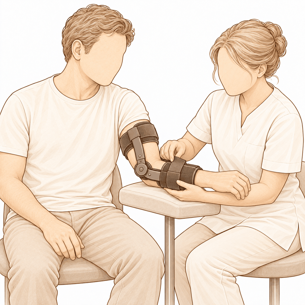
یہ آپریشن کیوں تجویز کیا گیا ہے
ڈاکٹر کیرن ہِرپرا، جو Mater Private Hospital Rockhampton میں بازو اور ہاتھ کے سرجن (upper-limb surgeon) ہیں، علاج کو آپ کی مخصوص چوٹ کے مطابق طے کرتے ہیں۔ مریضوں کو عام طور پر ان کے فیملی ڈاکٹر (GP) ہمارے کلینک میں بھیجتے ہیں؛ اگر کسی فزیوتھراپسٹ نے آپ کو ہمیں دکھانے کا مشورہ دیا ہے، تب بھی میڈیکیئر (Medicare) کی طرف سے رقم کی واپسی (rebate) کا حق دار ہونے کے لیے آپ کو اپنے فیملی ڈاکٹر سے ریفرل (referral) لینا ہوگا۔ ملاقات کے دوران ہم آپ سے تفصیل پوچھتے ہیں، آپ کی کہنی کا معائنہ کرتے ہیں، اور یہ جاننے کے لیے کہ مسئلہ کیا ہے، ضرورت ہو تو اسکین کا انتظام کرتے ہیں۔
کہنی کے لیگامنٹ کی دوبارہ تعمیر ایک ایسا آپریشن ہے جس میں گھسے ہوئے یا پھٹے ہوئے لیگامنٹ (ligament)، یعنی وہ مضبوط بندھن جو ایک ہڈی کو دوسری ہڈی سے جوڑتا ہے، کو عطیہ کیے گئے ٹینڈن (tendon)، یعنی وہ مضبوط ڈوری جو پٹھے کو ہڈی سے جوڑتی ہے، کے ایک ٹکڑے سے دوبارہ بنایا جاتا ہے تاکہ جوڑ مستحکم رہے۔ ہم عام طور پر یہ آپریشن اس وقت تجویز کرتے ہیں جب دوسرے علاج سے کافی بہتری نہ آنے کے بعد بھی آپ کی کہنی ایسا محسوس ہو کہ جیسے جواب دے رہی ہے، یا اپنی جگہ پر نہ ٹکتی ہو۔ یہ اس وقت بھی پیش کیا جاتا ہے جب کہنی کو صرف تقریباً سیدھا رکھ کر ہی جوڑ میں ٹکایا جا سکتا ہو، یا جب جوڑ کے ارد گرد کسی فریکچر (ہڈی کا ٹوٹنا) نے اسے غیر مستحکم کر دیا ہو۔ مقصد ایک مستحکم کہنی ہے جسے آپ حرکت دے سکیں، استعمال کر سکیں اور درد کے بغیر اس پر بھروسہ کر سکیں۔ کہنی کے پیچھے اور باہر کی طرف گھوم کر نکلنے والے ڈھیلے پن (posterolateral rotatory instability) والی تقریباً 85 فیصد کہنیوں میں عطیہ کیے گئے ٹشو (allograft) سے دوبارہ تعمیر کہنی کا استحکام بحال کر دیتی ہے۔ ہم اس پر آپ سے تفصیل سے بات کریں گے اور آپ کے ساتھ مل کر فیصلہ کریں گے کہ کیا یہ آپ کی کہنی اور آپ کے مقاصد کے لیے موزوں ہے۔
آپریشن سے پہلے
سرجری سے پہلے کے ہفتوں میں ہم ایکسرے، ایم آر آئی (ایک اسکین جو لیگامنٹ جیسے نرم ٹشو دکھاتا ہے) یا الٹراساؤنڈ جیسے اسکین کے ذریعے منصوبہ بندی مکمل کرتے ہیں۔ آپریشن کے دن، آپریشن سے سات گھنٹے پہلے کھانا اور پینا بند کر دیں۔ ہم سات گھنٹے اس لیے کہتے ہیں تاکہ اگر آپریشن کی فہرست وقت سے پہلے چل رہی ہو تو آپ کا آپریشن پہلے کیا جا سکے؛ آپ کے سرجن آپ کے فاقے کا صحیح وقت بتائیں گے۔ ہو سکتا ہے آپ کو سرجری سے پہلے کچھ دوائیں روکنی پڑیں، اور ہم آپ کو واضح ہدایات دیں گے کہ کون سی دوائیں اور کتنے عرصے کے لیے۔ آپ جو کچھ بھی لیتے ہیں، بشمول گولیاں، قطرے اور کریمیں، ان سب کی لکھی ہوئی فہرست ساتھ لائیں۔ آپریشن کے بعد آپ کو گھر لے جانے کے لیے کسی کا انتظام کریں۔ ڈھیلے، آرام دہ کپڑے پہنیں جن کی آستینیں آسانی سے کہنی پر سے گزر جائیں۔ اگر آپ کو دوسری طبی بیماریاں ہیں تو ہو سکتا ہے آپ کو خون کے ٹیسٹ یا بے ہوشی کے ڈاکٹر (anaesthetist)، یعنی وہ ڈاکٹر جو بے ہوشی کی دوا دیتے ہیں، سے معائنے کی بھی ضرورت ہو۔
آپریشن کے دن
آپ ہسپتال کے سرجری کے داخلے کے شعبے میں پہنچتے ہیں، جہاں آپ کا اندراج کیا جاتا ہے اور آپ کو آپریشن تھیٹر کے لیے تیار کیا جاتا ہے۔ وہاں آپ کی ملاقات بے ہوشی کے ڈاکٹر سے ہوگی۔ یہ آپریشن جنرل اینستھیزیا (مکمل بے ہوشی) میں کیا جاتا ہے۔ آپریشن کے بعد درد میں آرام کے لیے بعض اوقات اعصابی بلاک (nerve block)، یعنی ایک انجکشن جو جسم کے ایک حصے کو سُن کر دیتا ہے، بھی شامل کیا جاتا ہے؛ بے ہوشی کے ڈاکٹر اسی دن آپ سے اس پر بات کریں گے۔ پھر آپ کو آپریشن تھیٹر میں لے جایا جاتا ہے، جہاں آپریشن کیا جاتا ہے۔ آپریشن مکمل ہونے پر آپ ریکوری روم میں جاگتے ہیں، جہاں بے ہوشی کی دوا کا اثر ختم ہونے تک نرسیں آپ کی نگرانی کرتی ہیں۔ جب آپ کی حالت مستحکم ہو جائے تو آپریشن کی نوعیت اور آپ کی صحت یابی کے مطابق آپ یا تو وارڈ میں جاتے ہیں یا گھر چلے جاتے ہیں۔
اس دن کا مقصد سادہ ہے: آپ کی کہنی میں اتنا استحکام بحال کرنا کہ سرجری کے جلد بعد اس کی حرکت شروع ہو سکے۔ آپریشن کے بعد کہنی کو زیادہ دیر تک بے حرکت رکھنے سے یہ اکڑ جاتی ہے، اس لیے جلد حرکت شروع کرنا منصوبے کا حصہ ہے۔
آپریشن میں کیا کیا جاتا ہے
صحیح مراحل اس بات پر منحصر ہیں کہ آپ کی کہنی کے ارد گرد کون سے حصے زخمی ہیں، اور ہم آپریشن کے دن سے پہلے آپ کے اسکین کی مدد سے اس کی منصوبہ بندی کرتے ہیں۔ اگر مسئلے میں کوئی فریکچر بھی شامل ہے تو آپ کے سرجن پہلے پیچ یا چھوٹی پلیٹ کے ذریعے ہڈی کے ٹوٹے ہوئے ٹکڑوں کو جوڑتے ہیں۔ اگر بازو کے نچلے حصے (forearm) کے اوپری سرے پر ہڈی کا کوئی ٹوٹا ہوا ٹکڑا جوڑا نہ جا سکے تو اس کی جگہ دھات کا امپلانٹ (جسم میں لگایا گیا مصنوعی پرزہ) لگایا جا سکتا ہے۔ ہڈی کے مضبوطی سے جڑ جانے کے بعد پھٹے ہوئے لیگامنٹ کی مرمت کی جاتی ہے یا انہیں دوبارہ بنایا جاتا ہے۔
لیگامنٹ کو دوبارہ بنانے کا مطلب ٹینڈن کے ایک ٹکڑے سے نیا لیگامنٹ بنانا ہے۔ یہ ٹکڑا عطیہ کیے گئے ٹشو سے، یا آپ کے اپنے جسم سے لیا جا سکتا ہے۔ چھوٹے اینکر (anchors)، یعنی ہڈی میں لگائے جانے والے چھوٹے پرزے جو ٹانکوں کے دھاگے کو تھامتے ہیں، نئے لیگامنٹ کو صحیح جگہوں پر ہڈی سے جوڑے رکھتے ہیں، اور آپریشن ختم کرنے سے پہلے آپ کے سرجن چیک کرتے ہیں کہ کہنی اپنی پوری حرکت کی حد میں جوڑ کے اندر ٹکی رہتی ہے۔ بعض اوقات مرمت کے ساتھ ایک مضبوط ٹانکوں والی پٹی (suture tape) بھی لگائی جاتی ہے جو اندرونی بریس کی طرح کام کرتی ہے اور لیگامنٹ کے ٹھیک ہونے تک اسے سہارا دیتی ہے۔ اگر کہنی پھر بھی خود کو مستحکم نہ رکھ سکے تو حالات سنبھلنے تک اسے اپنی جگہ پر رکھنے کے لیے ایک عارضی قبضہ (hinge) لگایا جا سکتا ہے۔
یہ آپریشن اکثر ایک لمبے چیرے کے بجائے چھوٹے چیروں کے ذریعے کیا جا سکتا ہے۔ لیگامنٹ کو دوبارہ بنانے کے کچھ آپریشنوں میں تقریباً 2 سے 3 سینٹی میٹر کا چیرا کافی ہوتا ہے۔ چھوٹے چیروں کے ذریعے کام کرنے سے جوڑ کے ارد گرد صحت مند ٹینڈن اور پٹھے محفوظ رہتے ہیں، اور جوڑ کی اندرونی جھلی کو چھیڑنے کی ضرورت نہیں پڑتی۔
مرمت مکمل ہونے پر چیروں کو ٹانکوں سے بند کر کے زخم کی پٹی سے ڈھانپ دیا جاتا ہے۔ پورے آپریشن کا مقصد ایک مستحکم کہنی ہے جس کی حرکت جلد شروع ہو سکے، کیونکہ اسے زیادہ دیر تک بے حرکت رکھنے سے یہ اکثر اکڑ جاتی ہے۔
آپریشن کے بعد
آپ ریکوری روم میں جاگیں گے، جہاں بے ہوشی کی دوا کا اثر ختم ہونے تک نرسیں آپ پر کڑی نظر رکھتی ہیں۔ آپ کا بازو سلنگ (sling)، یعنی گلے سے لٹکی وہ پٹی جو بازو کو سہارا دیتی ہے، میں ہوگا یا تکیوں پر ٹکا ہوگا، اور زخموں پر پٹی ہوگی۔ ہم آپ کو درد کش دوا دیں گے اور آپ کے چلنے پھرنے سے پہلے چیک کریں گے کہ یہ اثر کر رہی ہے۔ گھر جانے کے بعد پہلے 24 گھنٹے کسی کو آپ کے ساتھ رہنا چاہیے۔ آپ کی ٹیم آپ کو بتائے گی کہ آپ اسی دن گھر جائیں گے یا ایک رات ہسپتال میں رہیں گے۔ ہم زخم کی پٹی تقریباً 10 دن تک لگی رہنے دیتے ہیں؛ براہ کرم اس سے پہلے اسے نہ اتاریں، جب تک ہم آپ کو ایسا کرنے کا نہ کہیں۔ جب ہم آپ کو دیکھتے ہیں تو اسے بدل دیتے ہیں یا اتار دیتے ہیں۔ زیادہ تر لوگ فوراً چل پھر سکتے ہیں اور ہلکے کام کر سکتے ہیں، لیکن جب آپ چل پھر رہے ہوں تو اپنی کہنی کو سلنگ میں آرام سے رکھیں۔
صحت یابی
پہلے چند دن آپ کی کہنی میں درد اور سوجن رہے گی۔ یہ آہستہ آہستہ کم ہو جاتی ہے۔ آرام، بازو کو اونچا رکھنا، اور ہماری دی ہوئی درد کش دوا تکلیف کو کم کریں گے۔ جوڑ کو پہلی بار حرکت دینا شروع کرنے پر کچھ دکھن ہونا معمول کی بات ہے اور ہفتوں کے ساتھ اس میں بہتری آتی ہے۔
آپ سلنگ میں بازو کے ساتھ گھر جائیں گے۔ آپ فوراً چل پھر سکتے ہیں اور ہلکے کام کر سکتے ہیں، لیکن جب آپ چل پھر رہے ہوں تو اپنی کہنی کو سلنگ میں آرام سے رکھیں۔ آپ کا ہاتھ اور کلائی جلد حرکت کر سکتے ہیں، اور ان کے لیے ہلکی ورزشیں عام طور پر چند دنوں کے اندر شروع ہو جاتی ہیں۔ سرجری کے بعد ہینڈ تھراپی Extend Rehabilitation میں Ruby Doolan کے ساتھ ہوتی ہے۔ وہ ہینڈ تھراپسٹ (ہاتھ کی بحالی کی ماہر) ہیں: وہ آپ کی ورزشوں میں رہنمائی کریں گی اور جس اسپلنٹ کی آپ کو ضرورت ہو وہ بنائیں گی۔ تھراپی کا مقصد مسلسل اور جلد حرکت ہے، کیونکہ کہنی کو زیادہ دیر تک بے حرکت رکھنے سے یہ اکثر اکڑ جاتی ہے۔
جیسے جیسے سوجن کم ہوتی ہے، آپ اپنا بازو زیادہ استعمال کریں گے: کھانا کھانا، لکھنا اور گھر کے ہلکے کام۔ جب آپ کے سرجن مطمئن ہو جائیں کہ مرمت مضبوط ہے تو سلنگ اتر جاتی ہے اور آپ خود کہنی کو موڑنا اور سیدھا کرنا شروع کرتے ہیں۔ حرکت اکثر شروع میں تیزی سے بہتر ہوتی ہے، پھر آہستہ۔ جب آپ درد کے بغیر چیزوں کو پکڑ اور تھام سکیں تو روزمرہ کام آسان محسوس ہونے لگتے ہیں۔ جب تک آپ کا بازو سلنگ میں ہے، گاڑی چلانا محفوظ نہیں ہے، اور گاڑی چلانے کے لیے ضروری ہے کہ آپ دونوں ہاتھوں سے اسٹیئرنگ پکڑ سکیں، اچانک بریک لگانے کی صورت میں ردعمل دے سکیں، اور تیز درد کش دوائیں نہ لے رہے ہوں۔ ہماری رہنمائی بازو اور ہاتھ کی سرجری کے بعد گاڑی چلانا بتاتی ہے کہ آپ کب دوبارہ گاڑی چلا سکتے ہیں۔
ہر شخص اپنی رفتار سے ٹھیک ہوتا ہے۔ آپ کا وقت مختلف ہو سکتا ہے، اور آپ کے سرجن اور تھراپسٹ پورے راستے میں آپ کی رہنمائی کریں گے۔
کیا مسائل پیدا ہو سکتے ہیں
زیادہ تر مریض ٹھیک رہتے ہیں، لیکن کبھی کبھار مسائل پیدا ہو سکتے ہیں۔ آپ کے سرجن اور ٹیم کسی بھی مسئلے کو جلد پکڑنے کے لیے آپ کی کڑی نگرانی کرتے ہیں۔
آپ کی کہنی کے اندر کی طرف چلنے والے عصب میں سرجری کے بعد جلن (irritation) پیدا ہو سکتی ہے۔ آپ کو انگوٹھی والی انگلی اور چھوٹی انگلی میں جھنجھناہٹ، سوئیاں چبھنے جیسا احساس یا سُن پن محسوس ہو سکتا ہے۔ اکثر یہ خود بخود ٹھیک ہو جاتا ہے، لیکن اگر یہ کم نہ ہو، یا انگلیاں کمزور محسوس ہوں تو ہمیں بتائیں۔
بعض اوقات کہنی ڈھیلی رہ سکتی ہے یا دوبارہ ایسا محسوس ہو سکتا ہے کہ جیسے جواب دے رہی ہے۔ اگر آپ کی کہنی غیر مستحکم محسوس ہونے لگے، یا اپنی جگہ سے کھسک جائے تو فوراً کلینک سے رابطہ کریں۔
کہنی اکڑ بھی سکتی ہے۔ ہو سکتا ہے آپ کو بازو کو پوری طرح سیدھا کرنے یا موڑنے میں دشواری ہو، اور حرکت دردناک ہونے کے بجائے رکی ہوئی محسوس ہو۔ اپنے معائنے پر اس کا ذکر کریں، کیونکہ اضافی تھراپی یا مزید علاج مدد کر سکتا ہے۔
انفیکشن غیر معمولی ہے لیکن سنگین ہے۔ ایسے گہرے، دھڑکتے ہوئے درد پر نظر رکھیں جو سادہ درد کش دواؤں سے کم نہ ہو، زخم سے باہر کی طرف پھیلتی سرخی، یا زخم سے رطوبت کا اخراج۔ اگر آپ کو یہ علامات نظر آئیں تو اسی دن کلینک کو فون کریں، یا اگر آپ کو بخار محسوس ہو یا طبیعت خراب لگے تو ہسپتال کی ایمرجنسی جائیں۔
مرمت کو تھامنے کے لیے استعمال ہونے والے دھات کے چھوٹے اینکر یا پیچ بعض اوقات جلن (irritation) کا سبب بن سکتے ہیں، یا عارضی قبضہ ڈھیلا ہو سکتا ہے۔ آپ کو کٹ کٹ کی نئی آواز، اٹکنا، یا جلد کے نیچے گانٹھ محسوس ہو سکتی ہے۔ اپنے اگلے معائنے پر اس کا ذکر کریں۔
جہاں لیگامنٹ کو دوبارہ بنانے کے لیے آپ کے اپنے ٹینڈن کا ٹکڑا استعمال کیا جاتا ہے، وہ جگہ کچھ عرصے تک چھونے پر درد والی رہ سکتی ہے یا اس میں دکھن رہ سکتی ہے۔ اگر یہ بہتر ہونے کے بجائے خراب ہو تو ہمیں بتائیں۔
بعض اوقات ہڈی ایسی جگہ بن سکتی ہے جہاں اسے نہیں بننا چاہیے، جوڑ کے ارد گرد یا اس کے اندر۔ اس سے حرکت محدود ہو سکتی ہے یا اٹکنا اور رگڑ محسوس ہو سکتی ہے۔ اگر آپ کی کہنی میں بہتری رک جائے تو اپنے معائنے پر اس کا ذکر کریں۔
زخم کا نشان خود بھی کبھی کبھار کھل سکتا ہے یا اس سے رطوبت رس سکتی ہے۔ اگر زخم کھل جائے، زیادہ سرخ ہو جائے، یا اس سے رطوبت کا اخراج شروع ہو جائے تو انتظار کرنے کے بجائے ہم سے رابطہ کریں۔
اگر آپ تفصیل جاننا چاہتے ہیں تو اس صفحے پر پیچیدگیوں کی جدول میں ان کی عام شرحیں درج ہیں۔
ہمیں کب فون کریں
زیادہ تر مسائل جلد ظاہر ہو جاتے ہیں، اور ہم ان کے بارے میں دیر سے سننے کے بجائے جلد سننا پسند کریں گے۔ اگر آپ کو بخار ہو، زخم زیادہ سرخ ہو جائے یا اس سے رطوبت رسنے لگے، یا آپ کا درد کم ہونے کے بجائے بڑھتا رہے تو ہمیں فون کریں۔ اگر آپ کی پنڈلی کے پچھلے حصے میں سوجن یا درد ہو، یا اچانک سانس پھولنے لگے تو ہسپتال کی ایمرجنسی جائیں۔ اگر آپ کی انگوٹھی والی انگلی اور چھوٹی انگلی سُن ہو جائیں، آپ کا ہاتھ کمزور محسوس ہو، یا آپ اپنا بازو بالکل حرکت نہ دے سکیں تو فوراً ہمیں فون کریں۔ اگر آپ کی کہنی اپنی جگہ سے کھسک جائے یا دوبارہ ایسا محسوس ہو کہ جیسے جواب دے رہی ہے تو اسی دن کلینک سے رابطہ کریں۔
Evidence & references
This is the clinical evidence summary written for health professionals. It is technical, and it lists the research this page was built from. You do not need to read it to understand your treatment or to make a decision about it.
Overview
- Surgical ligament reconstruction or repair is the most favorable treatment option for restoration of normal elbow function in posterolateral rotatory instability [4].
- Repair and reconstruction of the lateral ulnar collateral ligament offers practical and often successful solutions for patients with posterolateral rotatory instability of the elbow [10].
- Improving arthroscopic techniques provide a satisfactory alternative in stabilizing the elbow [7].
- The gravity varus test has biomechanical plausibility and confirms that pronation stabilizes the lateral elbow [1].
- An all-arthroscopic reconstruction technique for the lateral ulnar collateral ligament is reproducible and avoids residual instability [2].
- Open posterolateral ligament plication and lateral ulnar collateral ligament repair using an all-suture construct allows for complete posterolateral stabilization of the elbow with a single implant and bone preservation [3].
- Suture-augmented lateral ulnar collateral ligament and radial collateral ligament reconstruction provides a reproducible, anatomically based construct that restores posterolateral elbow stability and addresses the complex spectrum of lateral-sided injuries observed in posterolateral rotatory instability [5].
- Further follow-up and wider experience of the Surgilig/LockDown technique is required to assess its long-term functional outcome in comparison with more established surgical techniques for stabilization of posterolateral elbow instability [6].
- Arthroscopic lateral ulnar collateral ligament plication/reconstruction with augmented lateral collateral ligament imbrication allows effective management of elbow instability while promoting quicker patient recovery and long-term functional restoration [8].
- Acute repair of the lateral ulnar collateral ligament with a cortical button resulted in satisfactory clinical outcomes and successful restoration of elbow stability [9].
- The use of suture button fixation for repair of the lateral ulnar collateral ligament in terrible triad injuries has not been previously described [11].
- Corrective open-wedge osteotomy with structural bone allograft restored radial head alignment, eliminated dynamic subluxation, and achieved stable elbow function without the need for ligament reconstruction in complex posterolateral rotatory instability due to malunited radius fracture [12].
- Reconstruction of the lateral ulnar collateral ligament with a tendon graft offers an alternative that restores stability through a dynamic “sling effect” rather than rigid constraint [13].
Anatomy & Pathophysiology
Bony Anatomy
- The elbow is a trocho-ginglymoid joint consisting of medial and lateral articulations that provide bony stability [22].
- The ulnohumeral joint is formed by the articulation of the trochlea with the ulna within the greater sigmoid notch [22].
- The ulnohumeral joint provides highly congruent anatomy through almost 180° of articular contact, except for a bare area of the greater sigmoid notch devoid of cartilage [22].
- The radiocapitellar joint is formed by the articulation of the capitellum and the radial head [22].
- The radial head is a concave elliptical structure covered with articular cartilage along the radiocapitellar joint and approximately 270° of the articular margin [22].
- The radius is held in close approximation to the ulna at the proximal radioulnar joint by the annular ligament [22].
- The distal humeral articulation is angled 30° from the longitudinal axis of the humerus [22].
- The axis of rotation is angulated 5° to 7° in the coronal plane relative to the epicondylar axis, with the medial side more distal than the lateral side [22].
- The articular surface of the distal humerus is angled 30 degrees anterior to the humeral shaft axis [28].
- In full extension, 60% of axial load is transmitted through the radiocapitellar joint [28].
- The medial column of the distal humerus diverges from the humeral shaft at a 45-degree angle in the coronal plane [30].
- The lateral column of the distal humerus diverges from the humeral shaft at a 20-degree angle in the coronal plane [30].
- The trochlea is covered by articular cartilage anteriorly, inferiorly, and posteriorly, creating an arc of almost 270 degrees [33].
- The olecranon fossa and coronoid fossa are separated by a thin bony septum which is occasionally absent [33].
- The ulna medially bends approximately 8° at 8 cm from the tip of the olecranon [22].
- The articulation to the tip of the coronoid is approximately 30° from the long axis of the ulna in the sagittal plane [22].
Ligamentous Anatomy
- The lateral collateral ligament complex consists of the radial collateral ligament, the lateral ulnar collateral ligament (LUCL), and the annular ligament [33].
- The radial collateral ligament originates from an isometric point on the lateral epicondyle and fans out to attach to the annular ligament [33].
- The LUCL arises from an isometric point on the lateral epicondyle and attaches to the crista supinatoris of the proximal ulna [33].
- The annular ligament attaches to the anterior and posterior margins of the lesser sigmoid notch [33].
- The LUCL origin center is located 10.7 mm from the lateral epicondyle [21].
- The LUCL insertion is located 3.3 mm from the apex of the supinator crest [21].
- The LUCL acts as a posterior buttress for the radial head and the ulna to prevent subluxation [17].
- The LUCL is considered the principal constraint of the elbow joint against posterolateral rotatory instability (PLRI) [17].
- The lateral collateral ligament contributes 14% of varus stability of the elbow with the joint in full extension [37].
- The lateral collateral ligament contributes 9% of varus stability of the elbow with the joint in 90 degrees of flexion [37].
- The medial ulnar collateral ligament (MUCL) is the primary restraint to valgus stress within functional elbow range of motion [28].
- The MUCL originates on the posterior medial epicondyle and inserts on the sublime tubercle of the medial coronoid process [28].
- The anterior bundle of the MUCL is the primary restraint to valgus stress within functional elbow range of motion [28].
- The posterior bundle of the MUCL is the primary restraint to valgus stress with the elbow in maximal flexion [28].
- The common extensor tendon is a secondary stabilizer of the lateral elbow [16].
- The joint capsule is a secondary stabilizer of the elbow [19].
- The radiocapitellar articulation is a secondary stabilizer of the elbow [19].
Biomechanics & Pathophysiology
- Elbow stability is determined by primary and secondary stabilizers, with injury to these structures causing instability [19].
- The three primary stabilizers of the elbow are the ulnohumeral articulation, the MUCL, and the LUCL complex [19].
- Secondary stabilizers of the elbow include the radiocapitellar articulation, common flexor tendon, common extensor tendon, and joint capsule [19].
- Tensile forces are present at the medial elbow while compressive forces are present at the lateral elbow [23].
- The capsule allows maximum distension at approximately 70 to 80 degrees of flexion [23].
- The anterior capsule attaches at a point approximately 6 mm distal to the tip of the coronoid [23].
- Pronation stabilizes the lateral elbow [1].
- The LUCL is taut in both flexion and extension [37].
- Disruption of the LUCL results in posterolateral rotatory instability [37].
- Failure of primary stabilizers results in increased stress on secondary stabilizers, leading to capsular contractures, chondromalacia, osteophytes, and loose bodies [37].
- Ulnar nerve symptoms may develop from traction, scarring, or osteophyte impingement following primary stabilizer failure [37].
- The typical injury pattern for traumatic elbow dislocation involves a valgus internal rotation force of the forearm on a slightly flexed extremity [37].
- In traumatic elbow dislocation, structures are disrupted on the lateral side first, progressing medially as more force is applied [37].
- Posterolateral structures are most commonly affected when recurrence or persistence of instability results from traumatic dislocation [37].
- A coronoid fracture in association with disruption of the posterior band of the ulnar collateral ligament can result in symptomatic posteromedial instability [37].
- The normal elbow has a range of motion from 0° to 140° from extension to flexion [19].
- The normal elbow has a range of motion of 75° and 85° in pronation and supination respectively [19].
- A functional arc for elbow flexion and extension is 100° [19].
- A functional arc for forearm rotation is 100° [19].
- The normal valgus carrying angle of the elbow is 5 to 10 degrees for men and 10 to 15 degrees for women [28].
- The overall valgus angle of the elbow in extension, including the ulna, is 10 to 17 degrees [33].
- The distal humerus articular surface is internally rotated 3 to 8 degrees axially [33].
- As the elbow flexes, it internally rotates resulting in slight varus alignment [33].
Classification
- The lateral ulnar collateral ligament (LUCL) acts as a posterior buttress for the radial head and the ulna to prevent subluxation [17].
- Pronation stabilizes the lateral elbow in a cadaveric model of posterolateral rotatory instability [1].
Clinical Presentation
- The gravity varus test has biomechanical plausibility for evaluating posterolateral rotatory instability of the elbow [1].
- Complex posterolateral rotatory instability can be caused by a malunited radius fracture [12].
- Four-dimensional computed tomography scan can be used to diagnose complex posterolateral rotatory instability due to malunited radius fracture [12].
- Dynamic subluxation is a feature of complex posterolateral rotatory instability associated with malunited radius fracture [12].
Investigations
Physical Examination
- Pronation stabilizes the lateral elbow during the gravity varus test [1].
- Elbow stability is determined by primary stabilizers, which include the ulnohumeral articulation, the medial ulnar collateral ligament (MUCL), and the lateral ulnar collateral ligament (LUCL) complex [19].
- Secondary stabilizers of the elbow include the radiocapitellar articulation, the common flexor tendon, the common extensor tendon, and the joint capsule [19].
- The physical examination for elbow pathology is directed by the history and the location of patient pain in the anterior, posterior, medial, or lateral aspect of the elbow [19].
- The LUCL is considered the principal constraint of the elbow joint against posterolateral rotatory instability [17].
- The lateral ulnar collateral ligament injury is the cause of recurrent instability following elbow dislocation [41].
Imaging
- Plain radiographs remain the hallmark and best screening test for elbow evaluation [19].
- Standard radiographic views for elbow evaluation include AP, lateral, and oblique views [36].
- CT is helpful when assessing for malunion architecture and the location and pattern of osteophytes or loose bodies [36].
- Three-dimensional CT is used to check for heterotopic ossification [36].
- CT is not necessary when elbow stiffness is entirely soft-tissue related [36].
- CT is beneficial if any joint incongruity or abnormal bony anatomy is present [36].
- MRI can be used to evaluate ligaments and tendons, but it is rarely indicated for elbow stiffness [36].
- Magnetic resonance evaluation of the elbow includes imaging of ligament complexes [20].
- MRI findings in acute elbow dislocation provide insight into the mechanism of injury [20].
- MR evaluation of instability patterns emphasizes the soft-tissue lesions that result from dislocation [20].
- Four-dimensional computed tomography scan can be used for diagnosis in complex posterolateral rotatory instability due to malunited radius fracture [12].
Treatment
Operative Techniques and Constructs
- Surgical ligament reconstruction or repair is the most favorable treatment option for restoration of normal elbow function [4].
- Repair and reconstruction of the lateral ulnar collateral ligament (LUCL) offers practical and often successful solutions for patients with posterolateral rotatory instability (PLRI) of the elbow [10].
- An all-arthroscopic reconstruction technique for the LUCL is reproducible and achieves reconstruction while avoiding residual instability [2].
- Open posterolateral ligament plication and LUCL repair using an all-suture construct allows for complete posterolateral stabilization of the elbow with a single implant and bone preservation [3].
- A suture-augmented LUCL and radial collateral ligament reconstruction method provides a reproducible, anatomically based construct that restores posterolateral elbow stability and addresses the complex spectrum of lateral-sided injuries observed in PLRI [5].
- Acute repair of the LUCL with a cortical button resulted in satisfactory clinical outcomes and successful restoration of elbow stability [9].
- Arthroscopic LUCL plication/reconstruction with augmented lateral collateral ligament imbrication is a minimally invasive method that allows effective management of elbow instability while promoting quicker patient recovery and long-term functional restoration [8].
- Arthroscopic-assisted LUCL reconstruction provides less insult and dissection to the soft tissue at the lateral side of the elbow while being an excellent tool to diagnose any concomitant intraarticular pathologies [15].
- A knotless, onlay technique for LUCL reconstruction performs reconstruction with a tendon graft without violation of the extensor origin and soft tissue envelop [16].
- The use of knotless anchors and an onlay technique shortens operative time, reduces the required surgical exposure, and removes the risk of tunnel osteolysis or fracture and resultant graft failure while maintaining a broad bone surface for graft incorporation [16].
- A minimally-invasive dissection prevents iatrogenic injury to the common extensor origin, an important secondary stabilizer of the lateral elbow, and the articular capsule [16].
- Remaining extracapsular with a minimally-invasive technique avoids plication of the capsular structures or risk of formation of intra-articular adhesions, theoretically reducing the risk of any postoperative loss of range of motion [16].
Outcomes and Considerations
- The docking technique originally described by Jones et al. in 2012 is the most common method in use in contemporary practice for LUCL reconstruction [16].
- The docking technique has shown recurrent instability rates as high as 25% [16].
- A minimally-invasive approach allows for earlier rehabilitation and return of range of motion, reduced postoperative pain, and reduced operative time as a repair of the common extensor origin is not required following LUCL reconstruction [16].
- Fluoroscopic guidance can help to reduce injuries to unintended structures that could be foreseen due to a limited exposure [16].
- Postoperative stiffness is a known complication of LUCL reconstruction and occurs not uncommonly [16].
Non-Ligamentous Interventions
- Corrective open-wedge osteotomy with structural bone allograft restored radial head alignment, eliminated dynamic subluxation, and achieved stable elbow function without the need for ligament reconstruction in cases of complex PLRI due to malunited radius fracture [12].
Instability and Recurrence
- The docking technique for LUCL reconstruction has shown recurrent instability rates as high as 25% [16].
- The knotless, onlay technique for LUCL reconstruction removes the risk of tunnel osteolysis or fracture and resultant graft failure [16].
- The arthroscopic technique for LUCL reconstruction achieves reconstruction and avoids residual instability [2].
- The all-suture construct for posterolateral ligament plication and LUCL repair allows for complete posterolateral stabilization of the elbow [3].
- The suture-augmented LUCL and radial collateral ligament reconstruction method restores posterolateral elbow stability [5].
- Acute repair of the LUCL with a cortical button resulted in successful restoration of elbow stability [9].
- Repair and reconstruction of the LUCL offers practical and often successful solutions for patients with PLRI of the elbow [10].
- Reconstruction of the lateral ulnar collateral ligament with a tendon graft restores stability through a dynamic “sling effect” rather than rigid constraint [13].
Stiffness and Range of Motion
- The minimally-invasive, extracapsular technique for LUCL reconstruction theoretically reduces the risk of postoperative loss of range of motion by avoiding plication of capsular structures or risk of formation of intra-articular adhesions [16].
- The minimally-invasive method for managing elbow instability promotes quicker patient recovery and long-term functional restoration [8].
Iatrogenic Injury and Soft Tissue
- The minimally-invasive dissection for LUCL reconstruction prevents iatrogenic injury to the common extensor origin, an important secondary stabilizer of the lateral elbow [16].
- The minimally-invasive dissection for LUCL reconstruction prevents iatrogenic injury to the articular capsule [16].
- The arthroscopic-assisted LUCL reconstruction provides less insult and dissection to the soft tissue at the lateral side of the elbow [15].
Long-Term Outcomes
Recovery
- The minimally invasive arthroscopic method allows effective management of elbow instability while promoting quicker patient recovery and long-term functional restoration [8].
- The all-arthroscopic reconstruction technique is reproducible and achieves the reconstruction of the lateral ulnar collateral ligament as well as avoids residual instability [2].
- The open posterolateral ligament plication and lateral ulnar collateral ligament repair technique allows for complete posterolateral stabilization of the elbow with a single implant and bone preservation [3].
- The suture-augmented lateral ulnar collateral ligament and radial collateral ligament reconstruction method provides a reproducible, anatomically based construct that restores posterolateral elbow stability and addresses the complex spectrum of lateral-sided injuries observed in posterolateral rotatory instability [5].
- Reconstruction of the lateral ulnar collateral ligament with a tendon graft offers an alternative, restoring stability through a dynamic “sling effect” rather than rigid constraint [13].
- Clinical practice supports formal reconstruction of the lateral ulnar collateral ligament for chronic late presenting injury with posterolateral rotatory instability in the patient with open physes, without concern for causing significant growth arrest [14].
Key Evidence
- [L5] These findings support the biomechanical plausibility of the test and confirm that pronation stabilizes the lateral elbow. [1] (10.1016/j.jseint.2026.101792)
- [L5] The presented arthroscopic technique is reproducible and achieves the reconstruction of the LUCL of the elbow as well as avoids residual instability. [2] (10.1016/j.eats.2024.103096)
- [L5] The technique allows for complete posterolateral stabilization of the elbow with a single implant and bone preservation. [3] (10.1016/j.eats.2024.103172)
- [L4] Surgical ligament reconstruction or repair is the most favorable treatment option for restoration of normal elbow function. [4] (10.1016/s1058-2746(03)00091-0)
- [L5] The described method provides a reproducible, anatomically based construct that restores posterolateral elbow stability and addresses the complex spectrum of lateral-sided injuries observed in PLRI. [5] (10.1016/j.eats.2025.103797)
- [L4] Further follow-up and wider experience of the technique is required to assess its long-term functional outcome in comparison with more established surgical techniques for stabilization of posterolateral elbow instability. [6] (10.1097/bte.0000000000000025)
- [L5] Improving arthroscopic techniques provide a satisfactory alternative in stabilizing the elbow. [7] (10.1016/s0278-5919(05)70246-5)
- [L5] This minimally invasive method allows effective management of elbow instability while promoting quicker patient recovery and long-term functional restoration. [8] (10.1016/j.eats.2025.103529)
- [L4] The study found that acute repair of the LUCL with a cortical button resulted in satisfactory clinical outcomes and successful restoration of elbow stability. [9] (10.1097/bte.0000000000000178)
- [L5] Repair and reconstruction of the LUCL now offers practical and often successful solutions for patients with PLRI of the elbow. [10] (10.1016/j.csm.2004.06.010)
- [L4] The use of suture button fixation for repair of lateral ulnar collateral ligament has not been previously described. [11] (10.1016/j.eats.2023.10.004)
- [L5] Corrective open-wedge osteotomy with structural bone allograft restored radial head alignment, eliminated dynamic subluxation, and achieved stable elbow function without the need for ligament reconstruction. [12] (10.1016/j.xrrt.2026.100840)
- [L5] Reconstruction of the lateral ulnar collateral ligament with a tendon graft offers an alternative, restoring stability through a dynamic “sling effect” rather than rigid constraint. [13] (10.1002/atn2.70037)
- [L5] Clinical practice supports formal reconstruction of the LUCL for chronic late presenting injury with PLRI in the patient with open physes, without concern for causing significant growth arrest. [14] (10.1097/bth.0b013e3181dd88f3)
- [L5] It provides less insult and dissection to the soft tissue at the lateral side of the elbow while being an excellent tool to diagnose any concomitant intraarticular pathologies. [15] (10.1016/j.eats.2024.103101)
- [L5] [16] (10.1002/atn2.70135)
- [L5] On the basis of these morphological features, the LUCL might act a posterior buttress for the radial head and the ulna to prevent its subluxation, and therefore it is considered to be the principal constraint of the elbow joint against PLRI. [17] (10.1177/1558944716660555mm)
References
[1] Biomechanical Evaluation of the Gravity Varus Test in a Cadaveric Model of Posterolateral Rotatory Instability of the Elbow. JSES International. 2026. DOI: 10.1016/j.jseint.2026.101792
[2] Posterolateral Elbow Dislocation: An All‐Arthroscopic Reconstruction of the Lateral Ulnar Collateral Ligament. Arthroscopy Techniques. 2024. DOI: 10.1016/j.eats.2024.103096
[3] Open Posterolateral Ligament Plication and Lateral Ulnar Collateral Ligament Repair in Posterolateral Rotatory Instability of the Elbow Using an All‐Suture Construct. Arthroscopy Techniques. 2024. DOI: 10.1016/j.eats.2024.103172
[4] Surgical reconstruction for posterolateral rotatory instability of the elbow. Journal of Shoulder and Elbow Surgery. 2003. DOI: 10.1016/s1058-2746(03)00091-0
[5] Suture‐Augmented Lateral Ulnar Collateral Ligament and Radial Collateral Ligament Reconstruction for Subacute and Chronic Posterolateral Rotatory Instability. Arthroscopy Techniques. 2025. DOI: 10.1016/j.eats.2025.103797
[6] Surgical Repair of Posterolateral Rotatory Instability of the Elbow Using Surgilig/LockDown. Techniques in Shoulder & Elbow Surgery. 2014. DOI: 10.1097/bte.0000000000000025
[7] Posterolateral Rotatory Instability of the Elbow. Clinics in Sports Medicine. 2001. DOI: 10.1016/s0278-5919(05)70246-5
[8] Arthroscopic Lateral Ulnar Collateral Ligament Plication/Reconstruction With Augmented Lateral Collateral Ligament Imbrication. Arthroscopy Techniques. 2025. DOI: 10.1016/j.eats.2025.103529
[9] Clinical Outcomes of Lateral Ulnar Collateral Ligament Repair of the Elbow Using a Cortical Button. Techniques in Shoulder & Elbow Surgery. 2019. DOI: 10.1097/bte.0000000000000178
[10] PLRI: posterolateral rotatory instability of the elbow. Clinics in Sports Medicine. 2004. DOI: 10.1016/j.csm.2004.06.010
[11] Suture Button Repair for Lateral Ulnar Collateral Ligament in Terrible Triad Injuries: Surgical Technique. Arthroscopy Techniques. 2024. DOI: 10.1016/j.eats.2023.10.004
[12] Complex posterolateral rotatory instability due to malunited radius fracture: diagnosis with four-dimensional computed tomography scan and corrective osteotomy. JSES Reviews, Reports, and Techniques. 2026. DOI: 10.1016/j.xrrt.2026.100840
[13] Arthroscopic Lateral Collateral Ligament Reconstruction With Tendon Graft in High‐Grade Atraumatic Posterolateral Rotatory Instability in Elbows. Arthroscopy Techniques. 2026. DOI: 10.1002/atn2.70037
[14] Surgical Treatment of Posterolateral Rotatory Instability of the Elbow in Children. Techniques in Hand & Upper Extremity Surgery. 2010. DOI: 10.1097/bth.0b013e3181dd88f3
[15] Arthroscopic‐Assisted Lateral Ulnar Collateral Ligament Reconstruction for Posterolateral Rotatory Instability of the Elbow: A Technical Note. Arthroscopy Techniques. 2024. DOI: 10.1016/j.eats.2024.103101
[16] Lateral Ulnar Collateral Ligament Reconstruction Through a Mini‐Invasive Approach. Arthroscopy Techniques. 2026. DOI: 10.1002/atn2.70135
[17] Morphology of the Lateral Ulnar Collateral Ligament of the Elbow. HAND. 2016. DOI: 10.1177/1558944716660555mm
[19] Orthopaedic Knowledge Update 13 Ebook Without Multimedia. Anatomy, Biomechanics, Physical Examination, and Imaging of the Elbow > Summary and Conclusions.
[20] Orthopaedic Knowledge Update Sports Medicine 6. Magnetic Resonance Imaging of the Elbow > Annotated References.
[21] Orthopaedic Knowledge Update 13 Ebook Without Multimedia. Anatomy, Biomechanics, Physical Examination, and Imaging of the Elbow > Annotated References.
[22] Orthopaedic Knowledge Update 13 Ebook Without Multimedia. Anatomy, Biomechanics, Physical Examination, and Imaging of the Elbow > Anatomy > Bony Anatomy.
[23] Miller S Review Of Orthopaedics. SECTION 16 PATELLAR TRACKING IN TOTAL KNEE ARTHROPLASTY > TABLE 2.3 Shoulder Spaces.
[28] Miller S Review Of Orthopaedics. ELBOW.
[30] A Lange Medical Book Current Diagnosis Treatment In Orthopedics Fifth Edition. 2Musculoskeletal Trauma Surgery > INJURIES AROUND THE ELBOW.
[33] Rockwood And Green S Fractures In Adults. 29: Principles of Nonunion and Bone Defect Treatment > Pathoanatomy and Applied Anatomy Related to Distal Humerus Fractures.
[36] Aaos Comprehensive Orthopaedic Review 3. Elbow Stiffness* > IV. Evaluation.
[37] Campbell S Operative Orthopaedics 4 Volume Set. POSTERIOR SURGICAL APPROACH FOR QUADRILATERAL SPACE SYNDROME > MCLAUGHLIN PROCEDURE > ARTHROSCOPIC SURGERY.
[41] A Lange Medical Book Current Diagnosis Treatment In Orthopedics Fifth Edition. 2Musculoskeletal Trauma Surgery > ELBOW DISLOCATION.