Patients › Hand
انگلی کے درمیانی جوڑ (PIP joint) کا فیوژن
PIP joint fusion stabilizes a severely damaged middle finger joint, relieving pain when other treatments fail.
یہ آپریشن کیوں تجویز کیا گیا ہے
ڈاکٹر کیرن ہِرپرا، جو Mater Private Hospital Rockhampton میں بازو اور ہاتھ کے سرجن (upper-limb surgeon) ہیں، سب سے کم مداخلت والے ان طریقوں سے آغاز کرتے ہیں جو آپ کی حالت کے لیے مناسب ہوں۔ مریض عام طور پر اپنے فیملی ڈاکٹر (GP) کے بھیجنے پر ہمارے کلینک آتے ہیں؛ اگر کسی فزیوتھراپسٹ نے آپ کو ہمیں دکھانے کا مشورہ دیا ہے، تب بھی میڈیکیئر (Medicare) کی رقم واپسی کا حق دار ہونے کے لیے آپ کو اپنے فیملی ڈاکٹر سے ریفرل (referral) لینا ہوگا۔ ملاقات کے وقت ہم آپ سے تکلیف کی تفصیل لیتے ہیں، آپ کے ہاتھ کا معائنہ کرتے ہیں، اور اگر مسئلہ سمجھنے کے لیے ضرورت ہو تو ایکسرے یا اسکین کرواتے ہیں۔
یہ آپریشن آپ کی انگلی کے درمیانی جوڑ کا فیوژن (دو ہڈیوں کو جوڑ کر ایک کر دینا) ہے۔ اس جوڑ کو بنانے والی دو ہڈیوں کو آپس میں جوڑ دیا جاتا ہے تاکہ وہ ایک ہو کر جڑ جائیں، اور پھر جوڑ میں حرکت نہیں ہوتی۔ ہم عموماً اس کا مشورہ اس وقت دیتے ہیں جب جوڑ دردناک، اکڑا ہوا یا ٹیڑھا ہو، اور سادہ علاج، جیسے سرگرمیوں میں تبدیلی، فزیوتھراپی یا ہینڈ تھراپی، اور اسپلنٹ، سے آپ کو کافی بہتری نہ ملی ہو۔ کچھ چوٹوں یا ساخت کے مسائل میں شروع میں ہی سرجری کا مشورہ دیا جا سکتا ہے۔ مقصد درد ختم کر کے اور جوڑ کو مستحکم رکھ کر ہاتھ کا کام بحال کرنا ہے۔
آپریشن سے پہلے
سرجری سے پہلے کے ہفتوں میں ہم آپ کی انگلی کی نئی تصاویر، جیسے ایکسرے، اور کبھی کبھار ایم آر آئی یا الٹراساؤنڈ اسکین کے ذریعے منصوبے کی تصدیق کرتے ہیں۔ یہ سرجن کو دکھاتی ہیں کہ جوڑ کیسا نظر آتا ہے اور فیوژن کے زاویے کی منصوبہ بندی میں مدد دیتی ہیں۔ زیادہ تر لوگوں کو اس سے زیادہ کسی چیز کی ضرورت نہیں ہوتی۔ اگر آپ کو دوسری طبی بیماریاں ہیں تو آپ کو خون کے ٹیسٹ یا بے ہوشی کے ڈاکٹر (anaesthetist) سے معائنے کی بھی ضرورت ہو سکتی ہے، لیکن یہ معمول کا حصہ نہیں ہے۔ آپریشن سے پہلے کے دنوں میں ہم آپ کو بتائیں گے کہ اپنی معمول کی دواؤں میں سے کون سی اور کب بند کرنی ہے۔ سرجری سے پہلے سات گھنٹے تک کچھ نہ کھائیں اور نہ پئیں؛ ہم تھوڑا اضافی وقت اس لیے مانگتے ہیں تاکہ اگر آپریشن کی فہرست وقت سے پہلے چل رہی ہو تو آپ کی باری پہلے لائی جا سکے۔ گھر واپسی کے لیے کسی سواری کا انتظام کریں، کیونکہ آپ خود گاڑی نہیں چلا سکیں گے۔ اپنی موجودہ دواؤں کی لکھی ہوئی فہرست ساتھ لائیں اور ڈھیلے، آرام دہ کپڑے پہنیں۔
آپریشن کے دن
آپ ہسپتال کے سرجیکل داخلے کے شعبے میں پہنچتے ہیں، جہاں آپ کا اندراج ہوتا ہے اور آپ کو آپریشن تھیٹر کے لیے تیار کیا جاتا ہے۔ وہیں آپ کی ملاقات بے ہوشی کے ڈاکٹر سے ہوگی۔ یہ آپریشن جنرل اینستھیزیا (مکمل بے ہوشی) میں کیا جاتا ہے۔ آپریشن کے دوران آپ مکمل طور پر بے ہوش ہوں گے۔ کچھ مریضوں کو آپریشن کے بعد درد میں آرام کے لیے اعصابی بلاک (nerve block)، یعنی ایک انجکشن جو جسم کے ایک حصے کو سُن کر دیتا ہے، بھی لگایا جا سکتا ہے؛ بے ہوشی کے ڈاکٹر اسی دن آپ کے انفرادی حالات کی بنیاد پر اس کا فیصلہ کرتے ہیں۔ پھر آپ کو آپریشن تھیٹر میں لے جایا جاتا ہے، جہاں آپریشن کیا جاتا ہے۔
آپ ریکوری روم میں جاگتے ہیں، جہاں نرسیں بے ہوشی کی دوا کا اثر ختم ہونے تک آپ کی نگرانی کرتی ہیں۔ جب آپ کی حالت مستحکم ہو جائے تو طریقہ کار اور آپ کی صحت یابی کے لحاظ سے آپ یا تو وارڈ میں جاتے ہیں یا گھر۔
آپریشن میں کیا کیا جاتا ہے
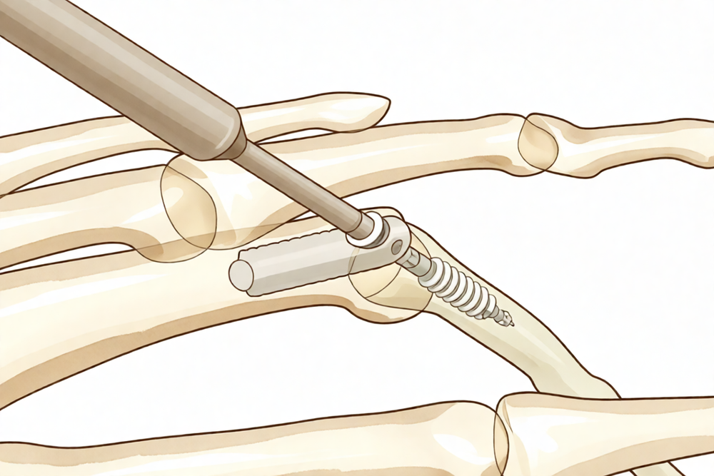
آپ کے سرجن آپ کی انگلی کے درمیانی جوڑ کے اوپر ایک چیرا لگاتے ہیں۔ اس چیرے کے ذریعے جوڑ کو کھولا جاتا ہے اور دونوں ہڈیوں کی گھسی ہوئی، خراب سطحوں کو تیار کیا جاتا ہے تاکہ وہ آپس میں جڑ سکیں۔ ہڈیوں کے سروں کو ایک دوسرے کے ساتھ فٹ ہونے کی شکل دی جاتی ہے، اور انگلی کو ایک ایسے مڑے ہوئے زاویے پر رکھا جاتا ہے جس سے آپ کا ہاتھ پکڑنے اور چٹکی کی گرفت کے لیے اچھی طرح کام کر سکے۔
پھر ہڈیوں کے جڑنے تک انہیں آپس میں تھام کر رکھا جاتا ہے۔ آپ کے سرجن دھات کے چھوٹے پرزے، جیسے تار یا پیچ، استعمال کرتے ہیں تاکہ دونوں ہڈیاں ساکن اور صحیح حالت میں رہیں۔ یہی تھامنا جوڑ کو جڑ کر ایک ٹھوس ہڈی بننے دیتا ہے۔ ہڈیاں سیدھ میں آ کر جکڑ دیے جانے کے بعد زخم کو ٹانکوں سے بند کر دیا جاتا ہے اور اس پر زخم کی پٹی رکھ دی جاتی ہے۔
اس سب کا مقصد سادہ ہے: دردناک، حرکت کرنے والا جوڑ ختم کر کے اس کی جگہ دونوں ہڈیوں کے درمیان ایک مستحکم، درد سے پاک جڑاؤ بنانا۔ چونکہ جوڑ اب حرکت نہیں کرتا، انگلی پہلے سے زیادہ اکڑی ہوئی ہوگی، لیکن استعمال کرنے پر اب اسے اٹکنا یا دکھنا نہیں چاہیے۔
آپریشن کے بعد
اس آپریشن کے بعد زیادہ تر مریض ایک رات ہسپتال میں رہتے ہیں، اگرچہ کچھ اسی دن گھر جا سکتے ہیں۔ آپ ریکوری روم میں جاگتے ہیں، جہاں بے ہوشی کی دوا کا اثر ختم ہونے تک نرسیں آپ کا خیال رکھتی ہیں۔ آپ کے ہاتھ پر زخم کی پٹی ہوگی اور اسے اسپلنٹ سے سہارا دیا جا سکتا ہے، اور ہم آپ کو آرام میں رکھنے کے لیے درد کش دوا دیں گے۔ جیسے ہی آپ خود کو سنبھلا ہوا محسوس کریں، آپ اٹھ کر چل پھر سکتے ہیں۔ براہِ کرم پہلے 24 گھنٹوں کے لیے کسی کا انتظام کریں جو آپ کے ساتھ رہے۔ ہم زخم کی پٹی تقریباً 10 دن تک لگی رہنے دیتے ہیں؛ جب تک ہم نہ کہیں، براہِ کرم اس سے پہلے اسے نہ اتاریں۔ جب ہم آپ کو دیکھتے ہیں تو ہم اسے بدل دیتے ہیں یا اتار دیتے ہیں۔
صحت یابی
پہلے چند دنوں اور ہفتوں میں آپ کی انگلی دکھے گی اور سوجی رہے گی۔ یہ معمول کی بات ہے اور آہستہ آہستہ ٹھیک ہو جاتی ہے۔ اپنا ہاتھ تکیے پر اونچا رکھنا، سوتے وقت بھی، سوجن کم کرنے میں مدد دیتا ہے۔ جو درد کش دوا ہم آپ کو دیتے ہیں وہ اس دوران آپ کو آرام میں رکھتی ہے۔
ہڈیوں کے جڑنے کے دوران انگلی کی حفاظت کے لیے آپ اس پر اسپلنٹ پہنیں گے۔ سرجری کے بعد ہینڈ تھراپی Extend Rehabilitation میں Ruby Doolan کے ساتھ ہوتی ہے۔ آپ کی ہینڈ تھراپسٹ (ہاتھ کی بحالی کی ماہر) Ruby ہیں: وہ آپ کی ورزشوں کی رہنمائی کرتی ہیں اور جس اسپلنٹ کی بھی ضرورت ہو وہ بناتی ہیں۔ آپ کی تھراپی کا زور ہاتھ اور باقی انگلیوں کو حرکت میں رکھنے پر ہوگا، تاکہ جڑا ہوا جوڑ پُرسکون ہونے تک آپ کی گرفت اور چٹکی کی گرفت مضبوط رہے۔
آپ گھر پر روزمرہ کی زیادہ تر ہلکی سرگرمیاں فوراً شروع کر سکتے ہیں، اور مدد کے لیے دوسرا ہاتھ استعمال کر سکتے ہیں۔ جب تک ہم اجازت نہ دیں، بھاری وزن اٹھانے، زور سے پکڑنے، یا کسی بھی ایسے کام سے پرہیز کریں جس سے جڑتی ہوئی انگلی پر زور پڑے۔ پٹی اترنے اور زخم بھرنے کے بعد آپ روزمرہ کاموں کے لیے ہاتھ زیادہ آزادی سے استعمال کر سکتے ہیں۔ جب تک اسپلنٹ کی وجہ سے آپ اسٹیئرنگ کو صحیح طرح پکڑ نہ سکیں، گاڑی چلانا محفوظ نہیں ہے۔ اسپلنٹ اترنے اور سرجن کی اجازت کے بعد ہمارا صفحہ بازو اور ہاتھ کی سرجری کے بعد گاڑی چلانا دیکھیں۔
صحت یابی ہر شخص میں مختلف ہوتی ہے۔ آپ کی صحت یابی کا دورانیہ مختلف ہو سکتا ہے، اور ہر معائنے پر آپ کے سرجن اور تھراپسٹ آپ کی رہنمائی کریں گے۔
کیا مسائل پیدا ہو سکتے ہیں
زیادہ تر مریض ٹھیک رہتے ہیں، لیکن کبھی کبھار مسائل پیدا ہو سکتے ہیں۔ آپ کے سرجن اور ٹیم کسی بھی مسئلے کو جلد پکڑنے کے لیے آپ کی قریب سے نگرانی کرتے ہیں۔
فیوژن میں سب سے بڑا خطرہ یہ ہے کہ دونوں ہڈیاں آپس میں نہ جڑیں۔ اگر ایسا ہو تو انگلی دردناک رہ سکتی ہے، اور آپ کو وہاں کلک یا حرکت محسوس ہو سکتی ہے جہاں کوئی حرکت نہیں ہونی چاہیے۔ اگر درد توقع کے مطابق ٹھیک نہ ہو تو اگلے معائنے پر ہمیں بتائیں۔
ہڈیوں کو تھامنے والے دھات کے پرزے بھی مسئلہ پیدا کر سکتے ہیں۔ کوئی تار یا پیچ ڈھیلا ہو سکتا ہے، ٹوٹ سکتا ہے، یا جلد پر دباؤ ڈال سکتا ہے۔ آپ کو جلد کے نیچے کوئی تیز کنارہ محسوس ہو سکتا ہے، یا انگلی سیدھ سے ہٹی ہوئی نظر آ سکتی ہے۔ معائنے کے وقت اس کا ذکر کریں تاکہ ہم اسے جانچ سکیں۔
انفیکشن غیر معمولی ہے لیکن اس پر جلد توجہ ضروری ہے۔ ایسے گہرے، ٹیس والے درد پر نظر رکھیں جو سادہ درد کش دوا سے کم نہ ہو، زخم سے باہر کی طرف پھیلتی سرخی، یا زخم سے رطوبت کا رسنا۔ آپ کو بخار جیسی گرمی اور طبیعت کی عمومی خرابی محسوس ہو سکتی ہے۔ اگر آپ کو ان میں سے کوئی بھی علامت نظر آئے تو فوراً کلینک کو فون کریں۔ اگر کلینک کا وقت ختم ہو چکا ہو یا سرخی تیزی سے پھیل رہی ہو، تو ہسپتال کی ایمرجنسی جائیں۔
انفیکشن انگلی کے کسی جوڑ تک انگلی پر لگے کٹ یا کسی چیز کے چبھنے سے بنے زخم سے، قریب کی جلد کے انفیکشن سے، یا جسم میں کسی اور جگہ سے خون کے راستے بھی پہنچ سکتا ہے۔ ہاتھ کے چھوٹے جوڑ میں انفیکشن کی علامات اکثر چوٹ کے چند دنوں کے اندر شروع ہو جاتی ہیں۔ اگر کسی ضرب یا زخم کے بعد انگلی گرم، سوجی ہوئی اور دردناک ہو جائے تو بغیر دیر کیے اپنے فیملی ڈاکٹر کو دکھائیں، تاکہ اس کا جلد علاج ہو سکے۔
فیوژن اچھی طرح جڑ جانے کے باوجود بھی اکڑاؤ اور جاری رہنے والا درد ہو سکتا ہے۔ اگر صحت یابی کی متوقع مدت کے بعد بھی آپ کی انگلی بہت اکڑی ہوئی یا دردناک رہے، تو ہم اس کا جائزہ لیں گے اور بات کریں گے کہ کیا کیا جا سکتا ہے۔
اگر آپ تفصیل جاننا چاہتے ہیں تو اس صفحے پر پیچیدگیوں کے جدول میں عام شرحیں دی گئی ہیں۔
ہمیں کب فون کریں
اگر آپ کو بخار ہو، اگر زخم کے آس پاس کی جلد زیادہ سرخ ہو جائے یا اس سے رطوبت رسنے لگے، یا اگر اچانک ایسا شدید درد ہو جو سادہ درد کش دوا سے کم نہ ہو، تو فوراً ہمیں فون کریں۔ اگر آپ کی پنڈلی میں سوجن یا درد ہو، یا سانس پھولے، تو ہسپتال کی ایمرجنسی جائیں، کیونکہ یہ خون کے لوتھڑے کی علامت ہو سکتی ہیں۔ اگر آپ کی انگلیاں سُن ہو جائیں، ان کا رنگ اُڑ جائے، یا آپ انہیں بالکل حرکت نہ دے سکیں، تو بھی ایمرجنسی جائیں۔ اگر کلینک کا وقت ختم ہو چکا ہو اور آپ پریشان ہوں، تو کلینک کھلنے کا انتظار کرنے کے بجائے ایمرجنسی جائیں۔
اس مسئلے کے بارے میں مزید کہاں پڑھیں
یہ صفحہ خود آپریشن کے بارے میں ہے۔ جس مسئلے کا یہ علاج کرتا ہے، بشمول اس کے کہ شواہد کیا بتاتے ہیں کہ سرجری کب مدد کرتی ہے اور کب نہیں، اس کی مزید تفصیل انگلی کے درمیانی جوڑ کا آرتھرائٹس کے صفحے پر دی گئی ہے۔
Evidence & references
This is the clinical evidence summary written for health professionals. It is technical, and it lists the research this page was built from. You do not need to read it to understand your treatment or to make a decision about it.
Anatomy & Pathophysiology
General Hand Architecture
- The hand is composed of 19 bones, 17 articulations, and 19 muscles situated entirely within the hand [2].
- The hand contains approximately the same number of tendons activated by forearm muscles as it has intrinsic muscles [2].
- The hand functions as an organ designed to obtain information and an organ of execution [2].
- The dorsal aspect of the hand is convex, while the palmar or volar aspect is concave [2].
- The dorsal surface of the hand is usually visible and aesthetically important, whereas the palmar surface is usually hidden and functional [2].
- The hand moves within a large volume of space with the shoulder as the apex, allowing it to reach any part of the body fairly easily due to the mobility of the shoulder, elbow, and wrist [2].
- Distal to the elbow, the wrist and forearm function as a single physiological unit that places the hand in a position for grasping [2].
- The hand is capable of conforming to the shape of objects to be grasped or studied [2].
Digits and Phalanges
- The digits are divided into the thumb and four fingers [2].
- The thumb has a more proximal and lateral position, allowing movement inward and outward from the palm [2].
- The four fingers are the distal extension of the carpometacarpal part of the hand [2].
- Each finger ray is a polyarticular chain composed of a metacarpal and three phalanges, except the thumb which has only two phalanges [2].
- When fingers are extended and separated, their tips lie on the circumference of a circle whose center is the head of the third metacarpal [2].
- The web space of the thumb is the largest and deepest among the digital web spaces [2].
Cutaneous Units
- The dorsal covering of the interphalangeal articulations of the digits forms a unique cutaneous unit characterized by a considerable excess of skin when the digits are in extension [3].
- The fine tight skin of the dorsal aspect of the middle phalanx forms a distinct cutaneous unit [3].
- The dorsal integument of the distal phalanx is characterized by the nail bed with its matrix [3].
- The palm forms a cutaneous unit extending from the distal transverse crease of the wrist up to the transverse crease at the base of the digits [3].
- The skin of the radial portion of the palm covers the thenar eminence and is relatively well vascularized and mobile [3].
- The skin of the ulnar and distal portion of the palm covers the hypothenar eminence and has poor mobility [3].
- The central triangular part of the palm has fixed, poorly vascularized skin that covers almost directly the superficial palmar aponeurosis [3].
- The integument of the palmar face of the digits is subdivided into phalangeal units separated by digital flexion folds [3].
- When a digit is completely flexed, the integument of adjacent phalanges comes into contact in the zones of the flexion creases, establishing areas of cutaneous contact in the form of a diamond [3].
- The sides of the diamond-shaped cutaneous contact zones do not undergo variations in length during movements of flexion and extension [3].
- Incisions made along the lines of the diamond-shaped cutaneous contact zones present a minimal chance of retraction [3].
- The dorsal slope of the web spaces has a gradual incline and its supple skin is not adherent to the subjacent region [3].
- The palmar surface of the web spaces is flat and precipitously interrupted, with skin densely adherent to the commissural skeleton [3].
Intrinsic Muscles
- There are seven interosseous muscles in the hand, consisting of four dorsal and three volar muscles [4].
- The dorsal interossei are abductors [4].
- The volar interossei are adductors [4].
- The middle finger has two dorsal interossei and no volar interossei because the central axis of the hand lies within it [4].
- Each dorsal interosseous muscle, with the exception of the third, has two muscle heads [4].
- The superficial head of the dorsal interosseous muscles abducts and weakly flexes the proximal phalanx [4].
- The deep head of the dorsal interosseous muscles flexes and weakly abducts the proximal phalanx while extending the middle and distal phalanges [4].
- Transverse fibers arch dorsally from each lateral band to join over the dorsum of the finger, flexing the proximal phalanx [4].
- Oblique fibers from the lateral bands insert onto the lateral tubercles at the base of the middle phalanx to extend the middle phalanx at the PIP joint [4].
- The lateral bands are joined by the lateral slips of the extensor tendon to form the conjoined lateral band [4].
- The two conjoined lateral bands to each finger unite at the distal third of the middle phalanx to form the terminal tendon [4].
- The terminal tendon inserts at the base of the distal phalanx to extend it [4].
- Each volar interosseous muscle has only one muscle head and none insert onto the proximal phalanx [4].
- The volar interossei form the ulnar lateral band of the index finger and the radial lateral band of the ring and little fingers [4].
- The abductor digiti quinti and flexor digiti quinti brevis are similar in structure and function to the superficial and deep heads of the dorsal interossei, respectively [4].
- The opponens digiti quinti arises from the pisohamate ligament and the hook of the hamate and inserts onto the ulnar side of the diaphysis of the fifth metacarpal [4].
Metacarpal Arch and Stability
- The metacarpal arch is endowed with a great deal of adaptability because of the mobility of the peripheral metacarpals [7].
- The index metacarpal is the most firmly fixed [7].
- The ring metacarpal has about 10 degrees of mobility in flexion and extension [7].
- The fifth metacarpal has a range of flexion–extension of approximately 20 degrees [7].
- The second to fifth metacarpals are bound together by various fibrous structures, the most distal of which is the deep transverse intermetacarpal ligament [7].
- The deep transverse intermetacarpal ligament is also known as the interglenoid ligament because it ties together the anterior glenoid ligaments of the metacarpophalangeal articulations [7].
- The metacarpophalangeal articulations are the keystones of the longitudinal arches of the hand [7].
- The thick anterior glenoid capsules, known as volar plates, prevent hyperextension at the metacarpophalangeal joints [7].
- The stability of the metacarpophalangeal joints is essential to the support of the longitudinal arch as well as of the transverse metacarpal arch [7].
Vascular Anatomy
- The arteries of the thumb vary in both size and number, making surgical reconstruction delicate [8].
- The layout of the thumb arteries is the result of innumerable variations regarding origin, transit, connections, and size [8].
- In anatomical studies, only 15% of dissections of the palmar arteries of the thumb fall into the classical "typical" category [8].
- The princeps pollicis artery is the terminal branch of the radial artery that crosses the first intermetacarpal space [8].
- The princeps pollicis artery runs along the ulnar side of the first metacarpal bone and along the volar surface of the adductor muscle [8].
- The princeps pollicis artery emerges onto the subcutaneous palmar tissue at the level of the cutaneous flexion crease of the metacarpophalangeal joint [8].
- At the metacarpophalangeal joint level, the princeps pollicis divides into two terminal rami known as the collateral palmar arteries of the thumb [8].
- The collateral palmar arteries run along the digital tunnel symmetrically and are of equal caliber [8].
- The collateral palmar arteries head distally to finally unite in the pulp arcade [8].
- An arcade located deep in the flexor tendon joins together the two arteries at the level of the distal metaphysis of the first phalanx [8].
- Vessels originating from the subtendinous arcade enter the vincula and irrigate the flexor tendon [8].
- In the second segment of the thumb, the main artery is the ulnar collateral artery [8].
- The subtendinous anastomosis situated at the level of the neck of the first phalanx acts as a moderator between the two collateral arteries [8].
- In cases where the palmar ulnar collateral artery is absent, the dorsal artery takes its place by means of a branch through the subtendinous arcade [8].
- In the pulp segment, the two arteries are of similar size and run through the thick fatty subcutaneous padding [8].
- The dorsal arteries of the thumb originate from the palmar arteries at the level of the first metacarpal [8].
- The dorsal arteries run laterally along the metacarpophalangeal joint and continue obliquely from volar to dorsal [8].
- At the level of the neck of the first phalanx, an anastomosis can be found which originates from the palmar arteries [8].
- The dorsal arteries are joined by three arcades: one inconstant arcade under the extensor tendon at the neck of the first phalanx, the arcade of the nail matrix, and the arcade of the nailbed [8].
Surgical Approaches and Incisions
- Distal palmar incisions are transverse, while proximal palmar incisions tend to be more longitudinal with the distal end curving radially [9].
- Proximal palmar incisions should parallel the thenar crease [9].
- Incisions extended proximal to the wrist should not cross the flexor wrist creases at a right angle [9].
- The most important structure in the thenar area is the recurrent branch (motor) of the median nerve [9].
- Anatomic studies have shown that there is no single longitudinal incision in the proximal palm that completely avoids the palmar cutaneous branches of the median and ulnar nerves [9].
- Midlateral incisions described for the fingers are also suitable for the thumb [9].
- The radial side of the thumb is more accessible for midlateral incisions [9].
- Care should be taken to avoid the dorsal branch of the superficial radial nerve to the radial side of the thumb during midlateral incisions [9].
- The volar midoblique incision crosses the flexion creases obliquely in the midline of the finger between the neurovascular bundles [9].
Investigations
- Clinical evaluation of the hand and wrist requires combining patient history with a careful physical examination to pinpoint or narrow the scope of possible pathologic processes [1].
- Diagnostic tests such as imaging and serum laboratory studies are useful in determining pathology but can be expensive, time consuming, and often nonspecific [1].
- A careful physical examination is essential to direct care and future testing if indicated [1].
- A systematic method for approaching the physical examination of the hand is essential due to the number of structures in a small space [1].
- Clinicians may organize the hand examination by anatomic location or region, or by organ system or pathology [1].
- The skeleton of the hand and wrist consists of 27 bones, of which 19 are long bones [12].
- The skeleton is divided into five rays, each comprising a polyarticulated chain of metacarpals and phalanges [12].
- The base of each metacarpal articulates with the distal row of the carpus [12].
- The wrist has three axes of movement, permitting the hand to be positioned in any spatial configuration for grasping [12].
- The radial ray (first ray) is the shortest, made up of a metacarpal and two phalanges [12].
- The other four digital rays are formed by four skeletal segments: a metacarpal and three phalanges [12].
- The thumb metacarpal is the shortest, while the index metacarpal is the longest [12].
- The proximal and middle phalanges of the middle and ring fingers are longer than those of the index finger [12].
- The transverse axis of the palm forms an acute angle of approximately 75 degrees with the longitudinal axis [12].
- Epiphyseal plates are located at the proximal ends of the phalanges and the first metacarpal [12].
- Epiphyseal plates are located at the distal ends of the other metacarpals [12].
References
[1] Orthopaedic Knowledge Update 13 Ebook Without Multimedia. Anatomy, Evaluation, Clinical Examination, and Imaging > Evaluation and Clinical Examination: Current Concepts.
[2] Exam Of The Hand Wrist 2Ed. INTRODUCTION.
[3] Exam Of The Hand Wrist 2Ed. Functional cutaneous units.
[4] Green S Operative Hand Surgery. Interosseous and Hypothenar Muscles.
[7] Exam Of The Hand Wrist 2Ed. The arches of the hand > The metacarpal arch.
[8] Exam Of The Hand Wrist 2Ed. Techniques of investigation of the arterial supply by J P Melki > Vascularization of the thumb > Palmar aspect.
[9] Campbell S Operative Orthopaedics 4 Volume Set. RESULTS OF SUTURE OF THE SCIATIC NERVE > PALMAR INCISIONS.
[12] Exam Of The Hand Wrist 2Ed. 1.1 SKELETON OF THE HAND > The osseous skeleton.