Patients › Wrist
کلائی کے گینگلیئن سسٹ کو نکالنے کا آپریشن (Wrist ganglion excision)
Ganglion excision — when to consider surgery, arthroscopic vs open approaches, and expected recovery.
یہ آپریشن کیوں تجویز کیا گیا ہے
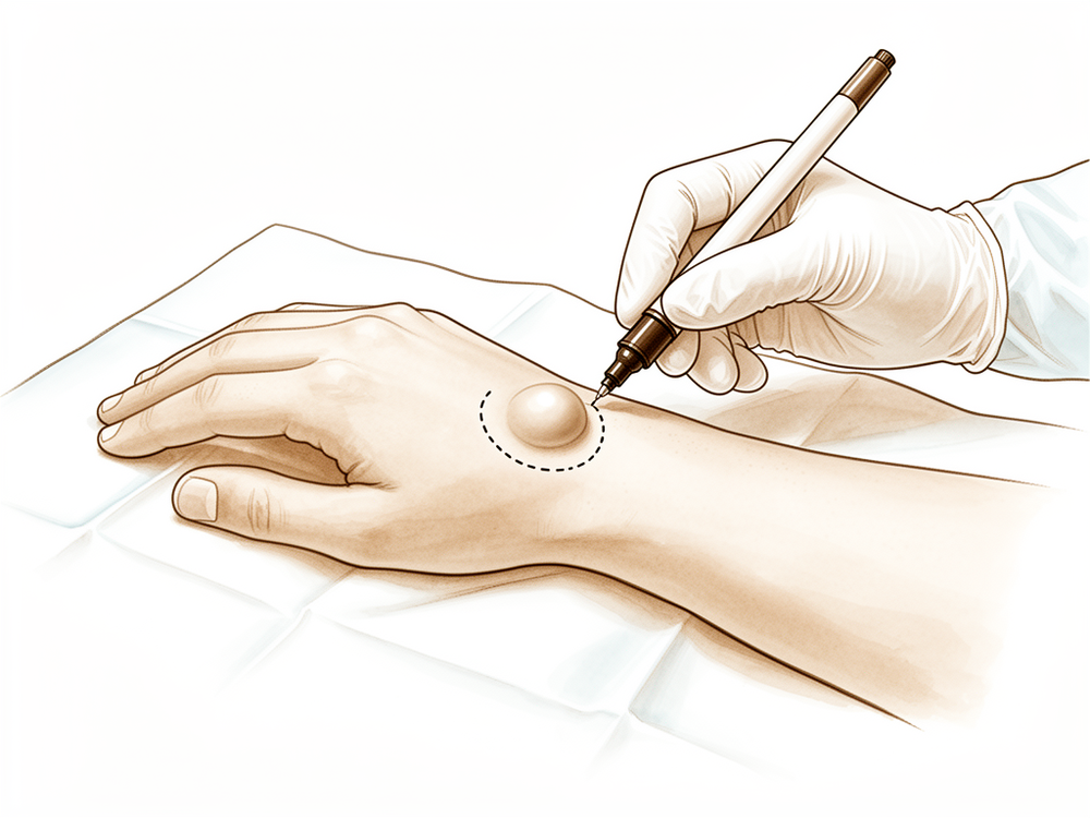
ڈاکٹر کیرن ہِرپرا، جو Mater Private Hospital Rockhampton میں بازو اور ہاتھ کے سرجن (upper-limb surgeon) ہیں، آپ کی حالت کے مطابق سب سے کم چیر پھاڑ والے طریقوں سے آغاز کرتے ہیں۔ کلائی کا گینگلیئن سسٹ (ganglion) کلائی کے جوڑ کے قریب سیال سے بھری ایک گانٹھ ہے۔ اسے نکالنے کو ایکسیژن (excision) کہتے ہیں۔ مریضوں کو عام طور پر ان کے فیملی ڈاکٹر (GP) ریفرل (referral) دے کر ہمارے کلینک بھیجتے ہیں؛ اگر کسی فزیوتھراپسٹ نے آپ کو ہمیں دکھانے کا مشورہ دیا ہے، تب بھی میڈیکیئر (Medicare) سے رقم کی واپسی (rebate) کا اہل ہونے کے لیے آپ کو اپنے فیملی ڈاکٹر سے ریفرل لینا ہوگا۔ ملاقات پر ہم آپ سے آپ کی تکلیف کی تفصیل پوچھتے ہیں، آپ کی کلائی کا معائنہ کرتے ہیں، اور ضرورت ہو تو اسکین کرواتے ہیں۔
کلائی کے زیادہ تر گینگلیئن کا پہلا علاج سرجری کے بغیر کیا جاتا ہے۔ ہم عموماً سرگرمیوں میں تبدیلی، ہینڈ تھراپی (ہاتھ کی بحالی کے ماہر سے علاج)، اسپلنٹ (splint) یا گانٹھ کو سوئی سے خالی کرنا آزماتے ہیں۔ جب ان اقدامات سے آپ کو کافی بہتری نہ ملے تو اگلا قدم سرجری ہوتا ہے۔ کلائی کے تقریباً 40 فیصد گینگلیئن پہلے 6 سال میں خود ہی سکڑ جاتے ہیں، اس لیے شروع میں انتظار کرنا اکثر مناسب ہوتا ہے۔ گانٹھ کو سوئی سے خالی کرنے سے عموماً اس کا واپس آنا نہیں رکتا، جبکہ سرجری سے نکالنے کے بعد گانٹھ کے دوبارہ ہونے کی شرح تقریباً 10 فیصد ہے۔ آپریشن کا مقصد گانٹھ کو نکالنا اور آپ کا درد کم کرنا ہے تاکہ آپ کی کلائی دوبارہ اچھی طرح کام کرے۔
آپریشن سے پہلے
زیادہ تر لوگوں کو بہت کم تیاری کی ضرورت ہوتی ہے۔ آپ کو بتایا جائے گا کہ سرجری سے 7 گھنٹے پہلے سے کچھ نہ کھائیں اور نہ پئیں۔ ہم عام 6 کے بجائے 7 گھنٹے اس لیے کہتے ہیں تاکہ اگر آپریشن کی فہرست وقت سے پہلے چل رہی ہو تو آپ کو پہلے بلایا جا سکے۔ آپ کے سرجن آپ کو بتائیں گے کہ کون سی دوائیں کب بند کرنی ہیں، اور آپ جو کچھ بھی لیتے ہیں اس کی فہرست ساتھ لائیں۔ بعد میں آپ کو گھر لے جانے کے لیے کسی کا بندوبست کریں جو گاڑی چلائے، اور ڈھیلے، آرام دہ کپڑے پہنیں۔ ہو سکتا ہے آپریشن کی منصوبہ بندی کے لیے ایکسرے، الٹراساؤنڈ یا ایم آر آئی اسکین پہلے ہی ہو چکا ہو۔ اگر آپ کو دوسری طبی بیماریاں ہیں تو آپ کو خون کے ٹیسٹ یا بے ہوشی کے ڈاکٹر (anaesthetist) سے معائنے کی ضرورت پڑ سکتی ہے۔
آپریشن کے دن
آپ ہسپتال کے سرجری کے داخلے کے شعبے میں پہنچتے ہیں، جہاں آپ کا اندراج ہوتا ہے اور آپ کو آپریشن تھیٹر کے لیے تیار کیا جاتا ہے۔ وہیں آپ کی ملاقات بے ہوشی کے ڈاکٹر سے ہوتی ہے۔ یہ آپریشن جنرل اینستھیزیا (مکمل بے ہوشی) میں کیا جاتا ہے۔ آپریشن کے دوران آپ پوری طرح بے ہوش ہوں گے۔ کچھ مریضوں کو آپریشن کے بعد درد میں آرام کے لیے اعصابی بلاک (nerve block)، یعنی ایک انجکشن جو جسم کے ایک حصے کو سُن کر دیتا ہے، بھی دیا جا سکتا ہے؛ بے ہوشی کے ڈاکٹر آپ کے انفرادی حالات کو دیکھ کر اسی دن اس کا فیصلہ کرتے ہیں۔ پھر آپ کو آپریشن تھیٹر میں لے جایا جاتا ہے، جہاں آپریشن کیا جاتا ہے۔
آپ ریکوری روم میں جاگتے ہیں، جہاں بے ہوشی کا اثر ختم ہونے تک نرسیں آپ کی نگرانی کرتی ہیں۔ جب آپ کی حالت مستحکم ہو جائے تو آپریشن اور آپ کی صحت یابی کے لحاظ سے آپ یا تو وارڈ میں جاتے ہیں یا گھر۔ زیادہ تر لوگ اسی دن گھر چلے جاتے ہیں۔ اگر آپ گھر جائیں تو، جیسا کہ سرجری سے پہلے طے کیا گیا تھا، کسی اور کو ہی گاڑی چلا کر آپ کو لے جانا ہوگا۔ جانے سے پہلے آپ کو کلائی کی دیکھ بھال اور اگلے معائنے کے بارے میں ہدایات دی جائیں گی۔
آپریشن میں کیا کیا جاتا ہے
آپریشن میں گینگلیئن، یعنی سیال سے بھری گانٹھ، کو اس چھوٹی ڈنڈی سمیت نکالا جاتا ہے جو اسے کلائی کے جوڑ سے ملاتی ہے۔ اس ڈنڈی کو نکالنا اہم ہے، کیونکہ اگر یہ پیچھے رہ جائے تو گانٹھ کے دوبارہ بننے کا امکان بڑھ جاتا ہے۔
سرجن کہاں کام کریں گے، یہ اس پر منحصر ہے کہ گانٹھ کہاں ہے۔ کلائی کی پشت کی گانٹھ کے لیے آپریشن دو یا تین چھوٹے سوراخ نما چیروں (keyhole cuts) سے کیا جاتا ہے، جن میں سے ہر ایک تقریباً 1 سینٹی میٹر لمبا ہوتا ہے۔ ایک پتلا کیمرہ کلائی کے جوڑ میں ڈالا جاتا ہے تاکہ سرجن اندر دیکھ سکیں اور گانٹھ اور اس کی ڈنڈی کو نکال سکیں۔ کلائی کی ہتھیلی والی طرف کی گانٹھ کے لیے آپریشن گانٹھ کے اوپر ایک کھلے چیرے سے کیا جاتا ہے۔ سرجن احتیاط سے گانٹھ کو آس پاس کے حصوں سے الگ کرتے ہیں۔ ہتھیلی والی طرف گانٹھ اکثر اس شریان کے قریب ہوتی ہے جو ہاتھ کو خون پہنچاتی ہے، اور کبھی کبھی اس کے گرد لپٹی ہوتی ہے، اس لیے آپریشن کا یہ حصہ آہستہ اور نرمی سے کیا جاتا ہے۔ آپ کے سرجن پہلے سے یہ بھی جانچ لیں گے کہ ہاتھ کی دونوں بڑی شریانوں کے ذریعے ہاتھ میں خون کا بہاؤ ٹھیک ہے۔
گانٹھ نکالنے کے بعد چیروں کو ٹانکوں سے بند کر دیا جاتا ہے۔ اوپر زخم کی پٹی لگائی جاتی ہے، اور آپ یہ پٹی تقریباً 10 دن تک لگی رہنے دیتے ہیں۔ آپ کے سرجن آپ کو کلائی کو جلد حرکت دینا شروع کرنے کی ترغیب دیں گے، سرجری کے بعد پہلے 2 ہفتوں کے اندر، کیونکہ ہلکی حرکت اکڑاؤ سے بچانے میں مدد دیتی ہے۔
آپریشن کے بعد
یہ عموماً ایک دن کا آپریشن ہوتا ہے، اس لیے آپ اسی دن گھر جانے کی توقع رکھ سکتے ہیں، اگرچہ کبھی کبھار مریض رات بھر ٹھہرتے ہیں۔ پہلے 24 گھنٹے کسی کو آپ کے ساتھ رہنا چاہیے۔ چیروں کے آس پاس آپ کو کچھ درد محسوس ہو سکتا ہے؛ عام درد کش دوا سے عموماً یہ ٹھیک ہو جاتا ہے۔ آرام کرتے وقت سوجن کم کرنے کے لیے اپنا ہاتھ تکیے پر اونچا رکھیں۔ جیسے ہی آپ خود کو سنبھلا ہوا محسوس کریں، گھر میں چل پھر سکتے ہیں، اور کلائی کو جلد ہلکی حرکت دینے کی ترغیب دی جاتی ہے۔ ہم پٹی تقریباً 10 دن تک لگی رہنے دیتے ہیں؛ جب تک ہم نہ کہیں، اس سے پہلے اسے نہ اتاریں۔ جب ہم آپ کو دیکھتے ہیں تو اسے بدل دیتے ہیں یا اتار دیتے ہیں۔ زیادہ تر لوگ دو سے تین ہفتوں کے اندر، جب وہ اسٹیئرنگ آرام سے پکڑ سکیں، دوبارہ گاڑی چلانے لگتے ہیں۔
صحت یابی
پہلے چند دن آپ کی کلائی میں درد ہو گا اور چیروں کے آس پاس تھوڑی سوجن ہو گی۔ عام درد کش دوا سے عموماً یہ ٹھیک ہو جاتا ہے، اور ہاتھ کو تکیے پر اونچا رکھنے سے سوجن کم ہونے میں مدد ملتی ہے۔ پہلے دو ایک ہفتوں میں تکلیف مسلسل کم ہوتی جاتی ہے۔
آپ پٹی تقریباً 10 دن تک لگی رہنے دیں گے، اور جب ہم آپ کو دیکھتے ہیں تو اسے بدل دیتے ہیں یا اتار دیتے ہیں۔ کلائی کی ہلکی حرکت جلد، پہلے 2 ہفتوں کے اندر، شروع ہو جاتی ہے، کیونکہ جلد حرکت دینے سے اکڑاؤ سے بچنے میں مدد ملتی ہے۔ سرجری کے بعد ہینڈ تھراپی Extend Rehabilitation میں Ruby Doolan کے ساتھ ہوتی ہے۔ آپ کے ہینڈ تھراپسٹ آپ کی ورزشوں میں رہنمائی کریں گے اور حرکت اور گرفت کی واپسی کے ساتھ ساتھ انہیں جانچتے رہیں گے۔ جیسے ہی آپ خود کو سنبھلا ہوا محسوس کریں، گھر میں چل پھر سکتے ہیں، اور جتنا آرام سے ہو سکے ہلکے کاموں کے لیے ہاتھ استعمال کریں گے۔
جیسے جیسے سوجن کم ہوتی ہے، روزمرہ کے کام آسان ہوتے جاتے ہیں۔ جب آپ بغیر درد کے پکڑ سکیں گے تو لکھنا، کھانا اور کپڑے پہننا دوبارہ معمول کے مطابق لگنے لگے گا۔ گاڑی چلانا عموماً دو سے تین ہفتوں کے اندر دوبارہ شروع ہو جاتا ہے، جب زخم میں تکلیف نہ رہے اور آپ آرام سے اسٹیئرنگ پکڑ سکیں؛ ہمارا صفحہ "بازو اور ہاتھ کی سرجری کے بعد گاڑی چلانا" دیکھیں۔ حرکت اور طاقت واپس آنے کے ساتھ زیادہ تر لوگ اپنے معمول کے کام اور سرگرمیوں پر واپس آ جاتے ہیں۔
صحت یابی ہر شخص میں مختلف ہوتی ہے، اس لیے آپ کا وقت مختلف ہو سکتا ہے۔ آپ کے سرجن اور ہینڈ تھراپسٹ ہر ملاقات پر آپ کی رہنمائی کریں گے، اور جیسے جیسے آپ کی کلائی ٹھیک ہوتی ہے، آپ مسلسل بہتری کی توقع رکھ سکتے ہیں۔
کیا مسائل پیدا ہو سکتے ہیں
زیادہ تر مریض ٹھیک رہتے ہیں، لیکن کبھی کبھار مسائل پیدا ہو سکتے ہیں۔ آپ کے سرجن اور ٹیم آپ کی قریب سے نگرانی کرتے ہیں تاکہ کسی بھی مسئلے کو جلد پہچان سکیں۔
سرجری کے بعد گانٹھ دوبارہ بن سکتی ہے۔ ہو سکتا ہے آپ دیکھیں کہ ویسا ہی نرم ابھار کلائی کے قریب واپس آ رہا ہے، کبھی کبھی ہفتوں یا مہینوں بعد۔ اگر آپ اسے واپس آتا دیکھیں یا محسوس کریں تو اپنے اگلے معائنے پر اس کا ذکر کریں۔ اگر اسے مزید علاج کی ضرورت ہو تو آپ کے سرجن آپ کو مختلف راستوں کے بارے میں بتا سکتے ہیں۔
کچھ لوگوں کو آپریشن کے بعد کلائی میں درد رہ جاتا ہے۔ اس کا امکان زیادہ ہے اگر سرجری سے پہلے آپ کی کلائی میں درد تھا، یا اگر آپ کے کام یا کھیل میں کلائی کو زور کے ساتھ پیچھے کی طرف موڑنا پڑتا ہے۔ یہ درد گہری دکھن جیسا محسوس ہو سکتا ہے جو کام کاج سے بڑھ جاتی ہے اور عام درد کش دواؤں سے پوری طرح ٹھیک نہیں ہوتی۔ کچھ لوگوں کو یہ بھی لگتا ہے کہ ان کا ہاتھ اتنی آسانی سے کام نہیں کرتا جتنی انہیں امید تھی، اور گرفت یا روزمرہ کے کام پہلے سے مشکل محسوس ہوتے ہیں۔ اگر ابتدائی بحالی کے عرصے کے بعد بھی درد یا کمزوری باقی رہے تو معائنے پر اپنے سرجن کو بتائیں تاکہ ہینڈ تھراپی یا دوسرے اقدامات پر غور کیا جا سکے۔
کلائی کی پشت پر دوربین کے ذریعے آپریشن کے دوران انگلیاں سیدھی کرنے والے ٹینڈنوں کو نقصان ایک نایاب مسئلہ ہے۔ آپ اسے ایک یا زیادہ انگلیاں سیدھی کرنے میں دشواری، یا کسی ایسی انگلی سے پہچانیں گے جو باقی انگلیوں سے نیچے لٹکی ہو۔ اگر آپ کوئی انگلی ٹھیک سے سیدھی نہ کر سکیں تو فوراً کلینک سے رابطہ کریں۔
کلائی کی ہتھیلی والی طرف کی گانٹھ کے لیے کچھ نایاب مسائل ایسے ہیں جو اس بات سے جڑے ہیں کہ گانٹھ ہاتھ کو خون پہنچانے والی شریان کے قریب ہوتی ہے۔ خطرے کی علامات میں ہاتھ کا سفید پڑ جانا، نیلا پڑ جانا یا معمول سے ٹھنڈا لگنا، یا ایسا سُن پن اور جھنجھناہٹ شامل ہیں جو ٹھیک نہ ہو۔ اگر آپ ان میں سے کچھ بھی دیکھیں تو انتظار کرنے کے بجائے ہسپتال کی ایمرجنسی جائیں۔
اگر آپ تفصیلات جاننا چاہتے ہیں تو اس صفحے پر پیچیدگیوں کا جدول عام شرحیں دکھاتا ہے۔
ہمیں کب فون کریں
اگر آپ کو بخار ہو، یا چیروں کے آس پاس کی جلد زیادہ سرخ یا سوجی ہوئی ہو جائے یا اس سے رطوبت رسنے لگے تو ہمیں فون کریں۔ اگر آپ کا درد کم ہونے کے بجائے بڑھتا ہی جائے، یا آپ کوئی انگلی سیدھی نہ کر سکیں تو ہمیں فون کریں۔ اگر آپ کا ہاتھ سفید پڑ جائے، نیلا ہو جائے یا ٹھنڈا محسوس ہو، اگر ہاتھ میں احساس ختم ہو جائے، یا آپ اسے حرکت نہ دے سکیں تو ہسپتال کی ایمرجنسی جائیں۔ اگر آپ کی پنڈلی میں سوجن ہو یا سانس پھولے تو بھی ہسپتال کی ایمرجنسی جائیں۔
اس مسئلے کے بارے میں مزید کہاں پڑھیں
یہ صفحہ خود آپریشن کے بارے میں ہے۔ جس بیماری کا یہ علاج کرتا ہے، بشمول اس کے کہ شواہد کیا بتاتے ہیں کہ سرجری کب مدد کرتی ہے اور کب نہیں، اس کی مزید تفصیل کلائی کا گینگلیئن سسٹ کے صفحے پر ہے۔
Evidence & references
This is the clinical evidence summary written for health professionals. It is technical, and it lists the research this page was built from. You do not need to read it to understand your treatment or to make a decision about it.
Anatomy & Pathophysiology
Bony Anatomy
- The wrist includes the distal radioulnar, radiocarpal, and ulnocarpal joints and the eight carpal bones and their proximal and distal articulations [4].
- The proximal row of carpal bones consists of the scaphoid, lunate, triquetrum, and pisiform [4].
- The distal row of carpal bones consists of the trapezium, trapezoid, capitate, and hamate [4].
- The pisiform and trapezoid are the smallest carpal bones, while the capitate is the largest [4].
- The radiocarpal joints are formed by the articulation of the distal radius with the scaphoid and lunate through their respective concave facets on the distal radius [4].
- The triquetrum articulates with the triangular fibrocartilage at the radiocarpal joint [4].
- The distal concave articular surfaces of the proximal carpal row form the midcarpal articulations with the distal row [4].
- The distal row articulates with the metacarpals, allowing mobility in the thumb, stability in the index and long finger metacarpals, and increased mobility in the ring and little finger metacarpals [4].
- The distal ulnar convexity articulates at the lesser sigmoid notch of the distal radius [4].
- The sigmoid notch articular surface accommodates the ulnar head through two thirds of its arc [4].
- There is about a 20-degree inclination of the distal ulna at its articulation with the radius [4].
- The ulnar styloid lies dorsal to the ulnar head and extends distally [4].
- The distal radius has three articular components: the scaphoid fossa, the lunate fossa, and the sigmoid notch [8].
- A ridge corresponding with the scapholunate interval is located between the scaphoid and lunate fossae on the distal radius [8].
- The radial styloid allows attachment of the brachioradialis tendon [8].
- The radial styloid is the origin of the radial scapholunate and radial lunocapitate ligaments [8].
- The concave elliptical distal radius is oriented in the sagittal plane with an average of 11 degrees of volar tilt [8].
- In the frontal plane, the average radial inclination of the distal radius is 23 degrees [8].
- Radial length is measured from the tip of the radial styloid to the ulnar articular surface and averages 13 mm [8].
- The ulnar shaft remains fixed in its rotation at the ulnohumeral joint, while the radius rotates around the ulna in pronation and supination [8].
- The radius has a lateral bow that is crucial to the maintenance of full pronation and supination [8].
Ligamentous Anatomy
- The triangular fibrocartilage complex (TFCC) attaches to the base of the ulnar styloid and separates the hyaline cartilage–covered ulnar head from the styloid [4].
- The TFCC includes the ulnar collateral ligament, dorsal and volar radioulnar ligaments, articular disc, meniscal homologue, extensor carpi ulnaris sheath, and ulnolunate and ulnotriquetral ligaments [4].
- Interosseous intrinsic ligaments connect the carpal bones in the proximal and distal carpal rows [4].
- The scapholunate and lunotriquetral interosseous ligaments connect the proximal carpal row [4].
- Ligaments connecting the trapezium to the trapezoid, trapezoid to the capitate, and capitate to the hamate connect the distal carpal row [4].
- Extrinsic ligaments extend from the radius and ulna distally across the carpal rows [4].
- The radial collateral ligament extends from the radial styloid to the scaphoid waist [4].
- The ulnar collateral ligament extends from the base of the ulnar styloid to the pisiform [4].
- The transverse carpal ligament is an extrinsic ligament [4].
- Volar extrinsic ligaments include the radioscapocapitate, radiolunotriquetral, and radioscapolunate ligaments on the radial side [4].
- The ulnolunate and ulnotriquetral components of the TFCC are volar extrinsic ligaments on the ulnar side [4].
- The space of Poirier is a relatively thin area on the palmar side of the carpus between the radiolunotriquetral and radioscapocapitate ligaments [4].
- The space of Poirier overlies the palmar surface of the lunate [4].
- Dorsal extrinsic ligaments include the dorsal radiocarpal and dorsal intercarpal ligaments [4].
- The trapezoidal dorsal radiocarpal ligament attaches along the dorsal radial articular margin of the lunate fossa, from the Lister tubercle to the lesser sigmoid notch [4].
- The trapezoidal dorsal radiocarpal ligament spans the lunotriquetral joint and inserts on the dorsal surface of the triquetrum [4].
- The dorsal intercarpal ligament is attached to the distal, dorsal surface of the triquetrum and passes across the midcarpal joint to attach to the dorsal surfaces of the scaphoid waist and the trapezoid [4].
- Extrinsic carpal ligaments connect the radius or the ulna to the carpus [9].
- Volar ligaments are generally stronger than dorsal ligaments [9].
- The radioscaphocapitate ligament connects to the waist of the scaphoid and limits ulnar translation of the carpus [9].
- The long radiolunate ligament helps limit ulnar translocation of the carpus [9].
- The short radiolunate ligament helps control lunate position [9].
- The radioscapholunate ligament is a vascular conduit and not a true ligament, also known as the ligament of Testut [9].
- The ulnolunate ligament attaches to the palmar radioulnar ligament and the lunate [9].
- The ulnocapitate ligament attaches to the ulnar head and is the most superficial or palmar palmar ulnocarpal ligament [9].
- The ulnotriquetral ligament attaches to the palmar radioulnar ligament and the triquetrum [9].
- The dorsal radiocarpal ligament has a trapezoidal shape and passes from the dorsal rim of the distal radius to the lunate and triquetrum [9].
- Fibers of the dorsal radiocarpal ligament insert onto the dorsal lunotriquetral interosseous ligament [9].
- The scapholunate interosseous ligament is a major stabilizer of the wrist and the most commonly injured wrist ligament [9].
- The scapholunate interosseous ligament is C-shaped, consisting of dorsal, palmar, and interosseous portions, with the dorsal portion being the strongest and thickest [9].
- The scapholunate interosseous ligament provides a flexion force on the lunate given its attachment to the scaphoid [9].
- The lunotriquetral interosseous ligament is C-shaped, with the volar portion being the thickest and strongest [9].
- The lunotriquetral interosseous ligament provides an extension moment on the lunate given its attachment to the triquetrum [9].
- The capitohamate ligament is a thick ligament with a 5 × 5 mm cross section and extensions to the third or fourth metacarpals [9].
- The dorsal intercarpal ligament passes from the dorsal tubercle of the triquetrum to the distal pole of the scaphoid [9].
- The dorsal intercarpal ligament reinforces the elastic dorsal wrist capsule and helps stabilize the scapholunate articulation with a contribution to the dorsal scapholunate interosseous ligament from its deep fibers [9].
- The space of Poirier is an area adjacent to the proximal capitate without ligamentous attachment, situated ulnar to the radioscaphocapitate ligament and radial to the long radiolunate in the floor of the carpal tunnel [9].
- The space of Poirier is a weak area vulnerable to instability, through which the distal carpal row separates from the lunate during a perilunate dislocation [9].
Soft Tissue Anatomy
- Six dorsal compartments contain wrist and digital extensor tendons on the dorsal surface of the wrist [8].
- The contents of the carpal tunnel reside on the volar surface, including nine flexor tendons and the median nerve [8].
- The flexor carpi ulnaris tendon can be palpated near its insertion on the pisiform on the ulnar surface [8].
- The boundaries of Guyon’s canal are the volar carpal ligament and transverse carpal ligament, the hook of the hamate radially, and the pisiform ulnarly [8].
- Guyon’s canal contains the ulnar artery and nerve [8].
- The interosseous membrane connects the shafts of the radius and ulna in the interosseous space [8].
- The central portion of the interosseous membrane is thickened and important in force transmission between the radius and ulna [8].
Ganglion Pathophysiology
- The dorsal wrist ganglion accounts for 60% to 70% of all hand and wrist ganglions [3].
- The main cyst of a dorsal wrist ganglion is usually located directly over the scapholunate ligament [3].
- Dorsal wrist ganglions may occur anywhere else between the extensor tendons [3].
- Failure to identify and excise the pedicle attachment to the scapholunate ligament increases the likelihood of recurrence for dorsal wrist ganglions [3].
- A small, mucin-filled duct is invariably seen piercing the transverse fibers of the scapholunate ligament, connecting the underlying scapholunate joint with the main cyst [3].
- The volar wrist ganglion is the second most common ganglion of the hand and wrist, accounting for 18% to 20% [1].
- The majority of volar ganglions occur either directly over the distal edge of the radius or slightly more distally over the scaphoid tubercle [1].
- Volar ganglions arising over the distal edge of the radius arise from the capsular and ligamentous fibers of the radiocarpal joint [1].
- Volar ganglions arising over the distal edge of the radius occur under the volar wrist crease between the flexor carpi radialis and abductor pollicis longus tendons [1].
- The main cyst of a volar wrist ganglion arising from the radiocarpal joint may be intertwined with bifurcating branches of the radial artery [1].
- Another type of volar ganglion arises from the capsule of the scaphotrapezial joint [1].
- Volar ganglions may appear small clinically but can be surprisingly extensive at surgery [1].
- Multiloculated volar ganglion cysts may extend under the thenar muscles, along the flexor carpi radialis tendon, into the carpal canal, and under the first extensor compartment adjacent to the dorsal branch of the radial artery [1].
- Extensions of volar ganglions may reach as far dorsally as the first web space [1].
- The radial artery is frequently intimately attached to the wall of a volar ganglion and may be completely encircled by it [1].
Biomechanics and Kinematics
- The eight carpal bones represent the most complex articular system in the human body [5].
- The human wrist allows precise positioning of the hand and optimization of power and prehensile tasks throughout a nearly hemispherical arc of wrist motion [5].
- The wrist joint’s motion planes include flexion, extension, radial deviation, ulnar deviation, and circumduction [9].
- There is minimal carpal motion with pronosupination [9].
- Approximately 62° of wrist extension occurs through the radiocarpal joint [9].
- 62% of wrist flexion occurs through the midcarpal joint [9].
- The midcarpal joint is mostly responsible for 20° of radial deviation and 40° of ulnar deviation [9].
- The midcarpal joint is responsible for the “dart thrower’s motion,” which involves moving from radial extension into ulnar flexion positioning of the wrist [9].
- The radius bears 80% of the axial load transmitted through the radiocarpal joint in neutral ulnar variance [9].
- The ulna bears 20% of the axial load transmitted through the radiocarpal joint in neutral ulnar variance [9].
- The proximal row of carpal bones forms an intercalated segment between the distal carpal row and the distal radius, bound into a functional unit by the scapholunate and lunotriquetral interosseous ligaments [9].
- The distal row is rigid with little motion between its bones due to stout intercarpal ligaments, acting as a functional unit with the scaphoid bridging both rows [9].
- During wrist flexion from neutral, the distal row flexes and ulnarly deviates slightly while the scaphoid pronates [9].
- During wrist flexion from neutral, the proximal row flexes differentially, with more rotation through the scaphoid, followed by the triquetrum and the lunate [9].
- The proximal row translates dorsally during wrist flexion [9].
- During wrist extension from neutral, the distal row extends and radially deviates slightly while the scaphoid supinates [9].
- During wrist extension from neutral, the proximal row extends differentially, with more motion in the scaphoid, followed by the triquetrum and then the lunate [9].
- The proximal row translates palmarly during wrist extension [9].
Clinical Presentation
- Volar wrist ganglions are the second most common ganglion of the hand and wrist, accounting for 18% to 20% of cases [1].
- The majority of volar ganglions occur directly over the distal edge of the radius or slightly more distally over the scaphoid tubercle [1].
- Ganglions arising from the distal edge of the radius originate from capsular and ligamentous fibers of the radiocarpal joint [1].
- Radiocarpal volar ganglions occur under the volar wrist crease between the flexor carpi radialis and abductor pollicis longus tendons [1].
- The main cyst of a radiocarpal volar ganglion may be intertwined with bifurcating branches of the radial artery [1].
- A second type of volar ganglion arises from the capsule of the scaphotrapezial joint [1].
- Multiloculated cysts may extend under the thenar muscles, along the flexor carpi radialis tendon, into the carpal canal, and under the first extensor compartment adjacent to the dorsal branch of the radial artery [1].
- Extensions of volar ganglions can reach as far dorsally as the first web space [1].
- Extensions of volar ganglions can often be appreciated preoperatively by careful palpation and digital compression [1].
- The patency of the radial and ulnar arteries must be assessed prior to surgery [1].
- The Allen test should be performed routinely to exclude ulnar artery occlusion [1].
- Preservation of the radial artery is particularly important in patients with a radial-dominant circulation [1].
Investigations
Magnetic Resonance Imaging
- MRI for hand and wrist imaging was first reported in 1986 [10].
- The primary advantages of MRI compared with CT and radiography are improved tissue characterization, especially of soft tissues such as ligamentous structures in the wrist and synovium in the hand, and the lack of ionizing radiation [10].
- MRI is the modality of choice for imaging radiographically occult fractures of the hand and wrist [10].
- Early MRI was limited by low magnetic field strength (0.15 T) and limited image options and image processing [10].
- Modern MRI is generally at 1.5T or 3T with a wide variety of imaging options and powerful image processing and postprocessing available [10].
- 3T is much preferred for hand and wrist imaging, especially for imaging small fields of view [10].
- Dedicated extremity magnets have been marketed, but image quality is poor compared with conventional MR imaging [10].
- 7T MRI has recently become approved for clinical use [10].
- 7T MRI has the potential to become a powerful tool for hand and wrist imaging as applications are developed [10].
- MRI with contrast enhancement is most commonly used to determine whether soft-tissue lesions are solid or cystic [10].
- In the case of rheumatologic imaging, MRI with contrast enhancement is used to better visualize erosions and synovial burden [10].
- Dynamic contrast enhancement has been used with inconsistent results to assess for the presence of avascular necrosis in the lunate or scaphoid after injury [10].
- MR angiography of the hand and wrist can be helpful in situations such as diagnosis of the hypothenar hammer syndrome [10].
- MR arthrography can be performed for evaluation of the triangular fibrocartilage and intercarpal ligament tears [10].
- MR arthrography is generally unnecessary with the increasing availability of high field MRI [10].
Computed Tomography and Other Imaging Modalities
- Hybrid SPECT/CT has been used for the diagnosis of radiographic occult fractures of the wrist [6].
- Cone-beam CT has been used in the diagnosis of scaphoid fractures [6].
- CT and MRI have a diagnostic impact on wrist injuries in young adults [6].
- Cine MRI is a new approach to the diagnosis of scapholunate dissociation [6].
- Cone-beam computed tomography arthrography is an innovative modality for the evaluation of wrist ligament and cartilage injuries [6].
- 3D analysis of the wrist has been described [6].
- Normal and variant anatomy of the wrist and hand can be evaluated on MR imaging [6].
- A practical guide to diagnostic imaging of the upper extremity has been published [7].
- 3-Tesla imaging of the wrist and hand has specific techniques and applications [7].
- MR imaging of the human hand and wrist at 7 T has been described [7].
- Magnetic resonance imaging can be used for the anterior radiocarpal ligaments [7].
- The carpal contents/canal ratio can be established by means of magnetic resonance imaging [7].
- MR imaging can be used for trauma of the elbow and wrist [7].
- Magnetic resonance imaging of the wrist has been described [7].
- MR imaging can be used for ulnar impaction [7].
- Rupture of the distal biceps tendon can be evaluated with MR imaging [7].
- MR imaging can be used for the wrist and elbow in clinical sports medicine [7].
- Magnetic resonance imaging of the musculoskeletal system, including the elbow, has been described [7].
- The efficacy of magnetic resonance imaging of the elbow has been evaluated [7].
- Sports and occupational injuries of the elbow can be evaluated with MR imaging findings [7].
- Magnetic resonance imaging of the elbow has been described [7].
- Magnetic resonance imaging can be used in Kienbock disease [7].
- Magnetic resonance imaging of the elbow, including normal anatomy, imaging technique, and osseous abnormalities, has been described [7].
- Magnetic resonance imaging of the elbow, including abnormalities of the ligaments, tendons, and nerves, has been described [7].
- Low-field MRI can be used for scaphoid fracture [7].
- MR imaging can be used for traumatic and overuse injuries of the wrist and hand in athletes [7].
- MR imaging diagnosis of TFC pathology can be correlated with arthroscopy [7].
- MR imaging of the elbow in baseball pitchers has been described [7].
- Overuse syndromes and injuries involving the elbow can be evaluated with MR imaging findings [7].
- Occult wrist fractures can be detected by magnetic resonance imaging [7].
- Ulnar collateral ligament injury in the throwing athlete can be evaluated with saline-enhanced MR arthrography [7].
- Magnetic resonance imaging of the triangular fibrocartilage complex has been described [7].
- Preoperative evaluation of the ulnar collateral ligament by magnetic resonance imaging and computed tomography arthrography has been evaluated in baseball players with surgical confirmation [7].
- Histologic and magnetic resonance imaging correlations in Kienbock’s disease have been described [7].
- MRI can be used for the diagnosis of occult dorsal wrist ganglion [7].
Radiography and Stress Views
- Distal radioulnar joint stress radiography can be used for detecting radioulnar ligament injury [6].
- Radiographic stress views can be compared for scapholunate dynamic instability in a cadaver model [6].
- Imaging of radial wrist pain involves various imaging modalities and anatomy [6].
- Intrinsic ligament and triangular fibrocartilage complex tears of the wrist can be compared using MDCT arthrography, conventional 3-T MRI, and MR arthrography [6].
- Simple plain radiographic signs and measures can be used to diagnose acute scapholunate ligament injuries of the wrist [11].
- Radiographic clues can be used for determining carpal instability and treatment protocol for scaphoid fractures [11].
- The role of imaging in diagnosing diseases of the distal radioulnar joint, triangular fibrocartilage complex, and distal ulna has been described [11].
Diagnostic Accuracy and Systematic Reviews
- The efficacy of magnetic resonance imaging and clinical tests in diagnostics of wrist ligament injuries has been evaluated in a systematic review [11].
- The role of magnetic resonance imaging in scaphoid fractures has been described [11].
Treatment
Dorsal Wrist Ganglion
- Failure to identify and excise the pedicle attachment to the scapholunate ligament increases the likelihood of recurrence [3].
- Careful preoperative palpation of the cyst with digital compression often reveals its extent and the direction of the pedicle [3].
- Transillumination or aspiration confirms the diagnosis preoperatively [3].
- Review of preoperative radiographs to rule out an interosseous component is recommended before dissection [3].
- Most dorsal ganglions can be approached through a transverse incision over the proximal carpal row [3].
- A modified incision or second transverse incision may be necessary for ganglions not directly over the scapholunate ligament [3].
- The diagnosis of ganglion cyst should be made before commitment to a transverse incision because this incision is not readily incorporated into a limb-sparing incision if a subsequent diagnosis of a malignant soft tissue tumor is made [3].
- The extensor pollicis longus and extensor digitorum communis tendons are retracted radially and ulnarly, respectively, to expose the dorsal ganglion [3].
- The joint capsule is opened along the border of the radius and scaphoid's proximal pole with the wrist in volar flexion [3].
- The capsule is elevated and retracted distally to expose the capsular attachments to the scapholunate ligament [3].
- Smaller intraarticular cysts are often seen attached to the scapholunate ligament [3].
- The capsular incision is continued around the ganglion, leaving all capsular attachments to the ligament intact [3].
- The capsular incision is extended laterally if capsular ducts are encountered, which can be identified by small amounts of mucin drainage [3].
- The ganglion and its capsular attachments are tangentially excised off the scapholunate ligament [3].
- A small, mucin-filled duct is invariably seen piercing the transverse fibers of the scapholunate ligament [3].
- This duct appears to connect the underlying scapholunate joint with the main cyst [3].
- Synovial and capsular attachments along the distal margin of the scapholunate ligament are excised to give an unobstructed view of the head and neck of the capitate [3].
Volar Wrist Ganglion
- Volar ganglions arising from the distal edge of the radius occur under the volar wrist crease between the flexor carpi radialis and abductor pollicis longus tendons [1].
- The main cyst of a volar ganglion may be intertwined with bifurcating branches of the radial artery [1].
- Extensions of volar ganglions can be appreciated preoperatively by careful palpation and digital compression [1].
- The Allen test should be performed routinely to assess the patency of the radial and ulnar arteries [1].
- Ulnar artery occlusion should be excluded preoperatively [1].
- The radial artery must be preserved, particularly in patients with a radial-dominant circulation [1].
- Exposure and precise identification of the capsular attachments of a volar ganglion are more difficult than for a dorsal ganglion [1].
- The incision for volar ganglion excision must be planned to allow for extension into the carpal tunnel or base of the thenar muscles [1].
- Longitudinal incisions allow for optimal visualization during volar ganglion excision [1].
- The forearm fascia is incised longitudinally to identify and mobilize the dome of the cyst [1].
- The radial artery is frequently intimately attached to the wall of the ganglion and may be completely encircled by it [1].
- Loupe magnification aids in the dissection to protect the radial artery [1].
- The pedicle is traced to the volar joint capsule, usually the scaphotrapezial or radiocarpal ligament [1].
- The joint is opened and explored, and the ganglion attachments are excised approximately 3 ± 4 mm [1].
- Surrounding tissues are compressed digitally after excision to rule out further mucin-filled pockets [1].
- Unidentified extensions of the ganglion must be excised if present [1].
- Capsular closure is unnecessary and only delays early mobilization [1].
- A bulky bandage and elevation of the hand ensure early postoperative comfort [1].
- Motion of the wrist should begin within the first 2 weeks after surgery [1].
- Unexpected branches of the radial sensory or lateral antebrachial cutaneous nerves may be injured, leading to troublesome neuromas [1].
- Extensions of the routine incision into the carpal canal must avoid injury to the palmar cutaneous branch of the median nerve [1].
- Injuries to the radial artery can be repaired microscopically [1].
- Some authors recommend leaving a portion of the cyst wall attached to the artery to avoid arterial injury [1].
- Stiffness of the wrist is less common than with dorsal ganglions but can occur if early motion is not encouraged [1].
- Curved incisions appear to consistently provide more attractive scars, especially near the volar wrist creases [1].
References
[1] Green S Operative Hand Surgery. BOX 59.1 Ganglions of the Hand and Wrist > Operative Treatment > Volar Wrist Ganglion.
[3] Green S Operative Hand Surgery. BOX 59.1 Ganglions of the Hand and Wrist > Operative Treatment > Dorsal Wrist Ganglion.
[4] Campbell S Operative Orthopaedics 4 Volume Set. NERVE INJURIES AT THE LEVEL OF THE HAND AND WRIST > ANATOMY.
[5] Green S Operative Hand Surgery. INTRODUCTION.
[6] Campbell S Operative Orthopaedics 4 Volume Set. ANATOMIC RECONSTRUCTION OF THE DISTAL RADIOULNAR LIGAMENTS > RADIOGRAPHIC TECHNIQUES.
[7] Campbell S Operative Orthopaedics 4 Volume Set. ELBOW, WRIST, AND HAND.
[8] A Lange Medical Book Current Diagnosis Treatment In Orthopedics Fifth Edition. 2Musculoskeletal Trauma Surgery > FRACTURES AND DISLOCATIONS OF THE DISTAL AND MID-FOREARM.
[9] Aaos Comprehensive Orthopaedic Review 3. Carpal Instability* > II. Anatomy and Biomechanics (See Chapter 92).
[10] Orthopaedic Knowledge Update 13 Ebook Without Multimedia. Anatomy, Evaluation, Clinical Examination, and Imaging > Imaging: Advances in Imaging of the Hand and Upper Extremity > Magnetic Resonance Imaging.
[11] Campbell S Operative Orthopaedics 4 Volume Set. ANATOMIC RECONSTRUCTION OF THE DISTAL RADIOULNAR LIGAMENTS > DIAGNOSIS AND EVALUATION.