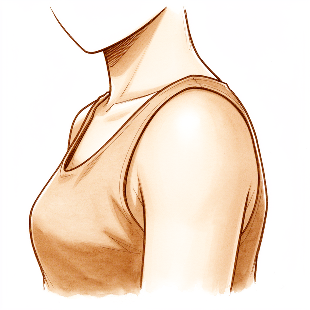
آپ کو کیا محسوس ہوتا ہے
سنیپنگ اسکیپولا (snapping scapula) کا مطلب ہے کہ جب آپ کے کندھے کی پچھلی چپٹی ہڈی (shoulder blade) سینے کی دیوار پر حرکت کرتی ہے تو آپ کو اس میں سے رگڑ، کلک یا کھٹ کی آواز سنائی دیتی ہے یا محسوس ہوتی ہے۔ ڈاکٹر کبھی کبھی اسے "واش بورڈ سنڈروم" (washboard syndrome) کہتے ہیں، کیونکہ یہ آواز ایسی لگ سکتی ہے جیسے کوئی پہیہ کپڑے دھونے کے لہریے دار تختے پر چل رہا ہو۔ یہ آواز بذاتِ خود عام ہے۔ جن لوگوں کو کوئی بھی علامت نہیں ہوتی، ان میں سے تقریباً ایک تہائی کے کندھے کی پچھلی چپٹی ہڈی سے کچھ نہ کچھ رگڑ کی آواز آتی ہے۔ اگر درد نہ ہو تو صرف اس آواز کو کسی علاج کی ضرورت نہیں۔
لوگ عام طور پر درد کی وجہ سے ڈاکٹر کے پاس آتے ہیں۔ یہ درد کندھے کی پچھلی چپٹی ہڈی کے ارد گرد یا اس کے نیچے ہوتا ہے، اکثر اس کے اندرونی کنارے کے قریب جو آپ کی ریڑھ کی ہڈی کے سب سے قریب ہے۔ یہ سر سے اوپر کی سرگرمیوں سے، اوپر کی طرف یا جسم کے آر پار ہاتھ بڑھانے سے بڑھ جاتا ہے، اور کھیل میں کارکردگی کم ہونے کی صورت میں بھی سامنے آ سکتا ہے۔ یہ درد سوزش زدہ برسا (bursa) سے ہوتا ہے، یعنی سیال سے بھری وہ پتلی گدی جو کندھے کی پچھلی چپٹی ہڈی کو پسلیوں پر آسانی سے پھسلنے دیتی ہے، یا خود ہڈی کی شکل سے ہوتا ہے۔ سینے کی دیوار ایک ساؤنڈ باکس کی طرح کام کرتی ہے، اس لیے جو آواز آپ سنتے ہیں وہ اسے پیدا کرنے والی حرکت سے زیادہ اونچی ہوتی ہے۔
یہ درد روزمرہ کے کاموں کو مشکل بنا سکتا ہے۔ کوئی بھاری دیگچی اونچی شیلف پر رکھنا، تار پر کپڑے لٹکانا، یا سیٹ بیلٹ کی طرف ہاتھ بڑھانا، یہ سب اسے شروع کر سکتے ہیں۔ کچھ لوگوں کو یہ سرگرمی کے دوران کے بجائے سرگرمی کے بعد زیادہ محسوس ہوتا ہے۔ یہ اتنا شدید ہو سکتا ہے کہ نیند میں خلل ڈالے یا آپ کو اپنا بازو معمول کے مطابق استعمال کرنے سے روک دے۔
اگر آپ کی علامات ٹھیک نہیں ہو رہیں، ہفتوں کے دوران بڑھ رہی ہیں، آپ کو رات کو جگا دیتی ہیں، یا آپ کو کام کرنے یا اپنا بازو استعمال کرنے سے روکتی ہیں، تو اپنے فیملی ڈاکٹر (GP) کو دکھائیں یا کسی ماہر ڈاکٹر سے معائنے کی درخواست کریں۔
دراصل کیا ہو رہا ہے
آپ کے کندھے کی پچھلی چپٹی ہڈی پھسلنے کے لیے بنی ہے۔ یہ آپ کی سینے کی دیوار پر ٹکی ہوتی ہے، اور اس سے ایک پتلی، چکنی، چربی والی تہہ کے ذریعے الگ رہتی ہے جو دو حرکت کرنے والی سطحوں کے درمیان گسکیٹ (gasket) کی طرح کام کرتی ہے۔ آپ کے کندھے کو حرکت دینے کے لیے چار جوڑ مل کر کام کرتے ہیں، اور تقریباً دو تہائی حرکت خود گیند اور پیالے والے جوڑ سے آتی ہے، جبکہ باقی ایک تہائی کندھے کی پچھلی چپٹی ہڈی کے پسلیوں پر پھسلنے سے آتی ہے۔ جب یہ پھسلن ہموار ہو تو آپ کو اس کا کبھی پتہ نہیں چلتا۔
سنیپنگ اسکیپولا میں مسئلہ یہ ہے کہ کوئی چیز اس پھسلن میں خلل ڈالتی ہے۔ کبھی یہ ہڈی کی شکل ہوتی ہے: کندھے کی پچھلی چپٹی ہڈی کا اندرونی کنارہ معمول سے زیادہ پسلیوں کی طرف مڑا یا خم دار ہو سکتا ہے، اس لیے یہ سینے کی دیوار کے اس سے زیادہ قریب ہوتا ہے جتنا ہونا چاہیے۔ کبھی یہ ہڈی پر اضافی ہڈی کا ایک چھوٹا سا ابھار ہوتا ہے۔ اکثر یہ ہڈی کے ارد گرد کے پٹھوں کا غیر متوازن کام ہوتا ہے، جس کی وجہ سے حرکت کرتے وقت ہڈی ہموار راستے پر نہیں چلتی۔ ان میں سے کوئی بھی چیز ہڈی کو پھسلنے کے بجائے رگڑ کھانے یا اٹکنے پر مجبور کر سکتی ہے۔
یہ رگڑ ہڈی اور پسلیوں کے درمیان کی گدی میں جلن (irritation) پیدا کرتی ہے۔ یہ گدی، جسے برسا کہتے ہیں، سیال سے بھری ایک پتلی تھیلی ہے جو ہڈی اور نرم بافتوں کے درمیان جھٹکا جذب کرنے والی چیز کی طرح کام کرتی ہے۔ جب اس میں سوزش ہو جاتی ہے تو یہ موٹی ہو جاتی ہے اور چھونے پر درد کرتی ہے، اور یہی وہ درد ہے جو آپ کو کندھے کی پچھلی چپٹی ہڈی کے نیچے یا ارد گرد محسوس ہوتا ہے۔ رگڑ سے خود بافت بھی ردِعمل کے طور پر موٹی ہو سکتی ہے، جو بار بار کی رگڑ پر جسم کا جواب ہے۔
اس سے اوپر بیان کی گئی علامات کی وضاحت ہوتی ہے۔ آواز کھردری سطحوں کے ایک دوسرے پر حرکت کرنے سے آتی ہے، اور سینے کی دیوار اسے ساؤنڈ باکس کی طرح اونچا کر دیتی ہے۔ درد سوزش زدہ گدی سے آتا ہے، خود آواز سے نہیں۔ اسی لیے دونوں مسائل کا علاج الگ الگ ہوتا ہے، اور اسی لیے علاج کا مرکز سوزش کو کم کرنا اور ہڈی کو قابو میں رکھنے والے پٹھوں کو دوبارہ تربیت دینا ہوتا ہے۔
ہم اس کے لیے کیا کر سکتے ہیں
ڈاکٹر کیرن ہِرپرا، جو Mater Private Hospital Rockhampton میں بازو اور ہاتھ کے سرجن (upper-limb surgeon) ہیں، آپ کے مسئلے کے مطابق سب سے کم چیر پھاڑ والے طریقوں سے آغاز کرتے ہیں۔ مریضوں کو عام طور پر ان کے فیملی ڈاکٹر ہمارے کلینک ریفر کرتے ہیں؛ اگر کسی فزیوتھراپسٹ نے آپ کو ہمارے پاس آنے کا مشورہ دیا ہے، تب بھی میڈیکیئر (Medicare) کی رقم کی واپسی کا حق دار ہونے کے لیے آپ کو اپنے فیملی ڈاکٹر سے ریفرل (referral) کی ضرورت ہوگی۔ کلینک میں ہم آپ سے تفصیل پوچھتے ہیں، آپ کے کندھے کا معائنہ کرتے ہیں اور جہاں ضرورت ہو تصاویر (اسکین) کا انتظام کرتے ہیں، تاکہ معلوم ہو سکے کہ آواز اور درد کی وجہ کیا ہے۔
زیادہ تر لوگ آپریشن کے بغیر ٹھیک ہو جاتے ہیں، اس لیے علاج سرجری کے بغیر شروع ہوتا ہے۔ پہلا قدم فزیوتھراپی کا ایک پروگرام ہے جو آپ کے کندھے کی پچھلی چپٹی ہڈی کو قابو میں رکھنے والے پٹھوں کو مضبوط کرتا ہے، اور ساتھ ہی ان پٹھوں کو اسٹریچ کیا جاتا ہے جو اکڑ گئے ہوں۔ جن سرگرمیوں سے علامات شروع ہوتی ہیں، ان میں تبدیلی بھی مدد دیتی ہے۔ مزید کسی چیز کے بارے میں سوچنے سے پہلے اسے 3 سے 6 ماہ تک پوری طرح آزمائیں۔
اگر درد برقرار رہے تو دوا سوزش کو کم کرنے میں مدد دے سکتی ہے۔ سوزش کم کرنے والی گولیاں، اسی قسم کی جو بہت سے لوگ پٹھوں کے درد کے لیے لیتے ہیں، اس مرحلے کا حصہ ہیں۔ ہم تصویری رہنمائی میں آپ کے کندھے کی پچھلی چپٹی ہڈی کے نیچے والے برسا میں اسٹیرائیڈ اور لوکل اینستھیزیا (صرف اس حصے کو سُن کرنا) کی دوا کا انجکشن بھی پیش کر سکتے ہیں۔ یہ دو کام کرتا ہے: یہ سوزش زدہ گدی کو پرسکون کرتا ہے، اور اس بات کی تصدیق کرتا ہے کہ آپ کا درد یہیں سے آ رہا ہے۔
سرجری پر صرف اس وقت غور کیا جاتا ہے جب ان علاجوں کے کئی ماہ بعد بھی درد جاری رہے اور ان سے آپ کو کافی بہتری نہ ملی ہو۔ یہ آپریشن دوربین کے ذریعے آپریشن (keyhole) ہے، جو کیمرے کی مدد سے چھوٹے چیروں کے ذریعے کیا جاتا ہے۔ اس میں آپ کے کندھے کی پچھلی چپٹی ہڈی اور پسلیوں کے درمیان کا سوزش زدہ برسا نکال دیا جاتا ہے۔ آپ کے کندھے کی پچھلی چپٹی ہڈی کا اوپری اندرونی کونا صرف اس صورت میں تراشا جاتا ہے جب آپریشن کے دوران یہ ابھرا ہوا پایا جائے۔ زیادہ تر لوگوں کا درد سرجری کے بعد کافی حد تک بہتر ہو جاتا ہے، لیکن اکثر کچھ کلک کی آواز باقی رہ جاتی ہے۔ ہم آپ سے بات کریں گے کہ سرجری آپ کے لیے کیا معنی رکھ سکتی ہے، اور آپ کے ساتھ مل کر فیصلہ کریں گے کہ کیا یہ صحیح قدم ہے۔
کیا توقع رکھیں
زیادہ تر لوگ آپریشن کے بغیر ٹھیک ہو جاتے ہیں۔ علاج کا آغاز فزیوتھراپی سے ہوتا ہے، جو آپ کے کندھے کی پچھلی چپٹی ہڈی کو قابو میں رکھنے والے پٹھوں کو مضبوط کرتی ہے، ساتھ میں سوزش کم کرنے والی دوا اور ان سرگرمیوں میں تبدیلی جن سے علامات شروع ہوتی ہیں۔ اسے 3 سے 6 ماہ تک پوری طرح آزمائیں۔ بہت سے لوگوں کا درد اس عرصے میں کم ہو جاتا ہے اور انہیں کبھی سرجری کی ضرورت نہیں پڑتی۔
اگر ان علاجوں کے کئی ماہ بعد بھی درد جاری رہے، تو سرجری پر غور کیا جا سکتا ہے۔ دوربین کے ذریعے آپریشن میں آپ کے کندھے کی پچھلی چپٹی ہڈی اور پسلیوں کے درمیان کا سوزش زدہ برسا نکال دیا جاتا ہے، اور ہڈی کا اوپری اندرونی کونا صرف اس صورت میں تراشا جاتا ہے جب یہ ابھرا ہوا پایا جائے۔ زیادہ تر لوگوں کا درد سرجری کے بعد کافی حد تک بہتر ہو جاتا ہے۔ اکثر کچھ کلک کی آواز باقی رہ جاتی ہے، کیونکہ جو علاج درد میں مدد دیتے ہیں وہ زیادہ تر درد کو ٹھیک کرتے ہیں، آواز کو نہیں۔ ہر علامت سے مکمل نجات عام نتیجہ نہیں ہے۔
علاج کے بغیر علامات برقرار رہ سکتی ہیں اور اتنی شدید ہو سکتی ہیں کہ نیند میں خلل ڈالیں یا آپ کو اپنا بازو معمول کے مطابق استعمال کرنے سے روک دیں۔ اس مسئلے کو پہچاننا اور اس کا علاج کرنا مشکل ہو سکتا ہے، اس لیے اس کے خود ٹھیک ہونے کا انتظار کرنے کے بجائے جلد واضح تشخیص کروا لینا بہتر ہے۔
اگر آپ کی علامات ٹھیک نہیں ہو رہیں، ہفتوں کے دوران بڑھ رہی ہیں، آپ کو رات کو جگا دیتی ہیں، یا آپ کو کام کرنے یا اپنا بازو استعمال کرنے سے روکتی ہیں، تو اپنے فیملی ڈاکٹر کو دکھائیں یا کسی ماہر ڈاکٹر سے معائنے کی درخواست کریں۔
ڈاکٹر کو کب دکھائیں
صرف آواز پریشانی کی وجہ نہیں ہے۔ جن لوگوں کو کوئی بھی علامت نہیں ہوتی، ان میں سے تقریباً ایک تہائی کے کندھے کی پچھلی چپٹی ہڈی سے کچھ نہ کچھ رگڑ کی آواز آتی ہے، اور صرف اس آواز کو کسی علاج کی ضرورت نہیں۔ اصل اہمیت درد کی ہے۔ اگر آپ کو کندھے کی پچھلی چپٹی ہڈی کے ارد گرد یا نیچے ایسا درد ہو جو ٹھیک نہیں ہو رہا، ہفتوں کے دوران بڑھ رہا ہے، آپ کو رات کو جگا دیتا ہے، یا آپ کو کام کرنے یا اپنا بازو استعمال کرنے سے روکتا ہے، تو اپنے فیملی ڈاکٹر کو دکھائیں یا کسی ماہر ڈاکٹر سے معائنے کی درخواست کریں۔ اگر درد اتنا شدید ہو کہ نیند میں خلل ڈالے، اگر کھیل میں آپ کی کارکردگی کم ہو گئی ہو، یا اگر سر سے اوپر کی سرگرمی، جیسے اوپر کی طرف یا جسم کے آر پار ہاتھ بڑھانا، درد شروع کر دے، تو بھی معائنے کی درخواست کریں۔ سنیپنگ اسکیپولا کو پہچاننا مشکل ہو سکتا ہے، اس لیے جلد واضح تشخیص کروا لینا فائدہ مند ہے۔
مزید تفصیل
Advanced reading: the deeper science (optional)
یہ حصہ اس سے آگے جاتا ہے جتنا آپ کو اپنے علاج کے فیصلوں کے لیے جاننے کی ضرورت ہے۔ سنیپنگ اسکیپولا کے بارے میں یہ اضافی مطالعہ اس لیے فائدہ مند ہے کہ آواز اور درد دو الگ مسائل ہیں، اور جو علاج مدد دیتے ہیں وہ زیادہ تر دوسرے مسئلے کو ٹھیک کرتے ہیں، پہلے کو نہیں۔
آواز عام ہے؛ اصل مسئلہ درد ہے
کندھے کی پچھلی چپٹی ہڈی سے رگڑ کی آواز بذاتِ خود کسی بیماری کی علامت نہیں ہے۔ اس مسئلے پر ایک مشہور جائزہ ایک ابتدائی تحقیق کا حوالہ دیتا ہے جس میں ایسے 100 لوگوں میں سے 31 فیصد میں، جنہیں کوئی بھی علامت نہیں تھی، کندھے کی پچھلی ہڈی میں رگڑ کی آواز (scapular crepitus) پائی گئی [1]۔ آواز کو ایک بیماری میں بدلنے والی چیز درد ہے، جو عام طور پر سوزش زدہ برسا سے ہوتا ہے: یعنی بافت کی وہ پتلی، سیال والی تہہ جو کندھے کی پچھلی چپٹی ہڈی کو پسلیوں پر پھسلنے دیتی ہے۔
ہڈی کی شکل کا بھی کردار ہے۔ تصویری اسکینوں کی ایک تحقیق میں، جن لوگوں کی کندھے کی پچھلی چپٹی ہڈی اپنے اندرونی کنارے کے ساتھ آگے کی طرف مڑی ہوئی تھی (ایک مقعر، یعنی اندر کو دبی ہوئی شکل، جس کا "میڈیل اسکیپولا کارپس اینگل" (medial scapula corpus angle) مثبت ہو)، ان میں سنیپنگ اسکیپولا کا 12 گنا زیادہ خطرہ تھا [2]۔ یہ پیمائش اتنی مفید ثابت نہیں ہوئی جتنی شروع میں لگی تھی: بعد کی ایک تحقیق میں پتہ چلا کہ یہی زاویہ اس سمت کے لحاظ سے کافی بدل جاتا ہے جس سے اسے دیکھا جائے، اس لیے یہ اکیلے اس مسئلے کی تشخیص کے لیے کافی قابلِ اعتماد نہیں ہے [3]۔ عطیہ کی گئی لاشوں پر کیے گئے ایک تجرباتی ماڈل (cadaver model) میں، ہڈی کی سب سے اہم خصوصیت یہ تھی کہ کندھے کی پچھلی چپٹی ہڈی کا اندرونی حصہ پسلیوں کی طرف کتنا آگے کو تھا [4]۔
زیادہ تر لوگ آپریشن کے بغیر ٹھیک ہو جاتے ہیں
ایک سیریز میں 28 مریضوں کی 30 کندھے کی پچھلی چپٹی ہڈیوں کا علاج پٹھوں کو مضبوط کرنے کے ایک خاص پروگرام سے کیا گیا؛ ان میں سے 14 (50 فیصد) صرف ورزشوں سے ٹھیک رہے اور 6 (21 فیصد) کو ورزشوں کے ساتھ صرف سوزش کم کرنے والی دوا کی ضرورت پڑی، اس طرح 71 فیصد نے تحقیق کے اپنے درجہ بندی کے پیمانے پر سرجری کے بغیر اچھا یا اس سے بہتر نتیجہ حاصل کیا [5]۔
سب سے لمبے عرصے تک مریضوں کو دیکھنے والی تحقیق فن لینڈ سے ہے۔ بارہ لوگ جن کا کھلا آپریشن ہوا تھا اور 9 جن کا علاج آپریشن کے بغیر کیا گیا تھا، اوسطاً 22 سال بعد دوبارہ جانچے گئے۔ محنت کے دوران درد دونوں گروپوں میں کم تھا: سرجری کے بعد 10 میں سے 0.8 اور سرجری کے بغیر 10 میں سے 1.5، یہ فرق اتنا کم تھا کہ یقین سے نہیں کہا جا سکتا کہ یہ حقیقی تھا (p = 0.357) [6]۔ سرجری کے بعد 12 میں سے 7 میں درد جلدی بہتر ہوا، لیکن لمبے عرصے میں دونوں راستے تقریباً ایک ہی جگہ پہنچے۔
برسا میں لوکل اینستھیزیا کی دوا اور اسٹیرائیڈ کا انجکشن دو کام کرتا ہے: یہ سوزش کا علاج کرتا ہے اور یہ جانچتا ہے کہ کیا واقعی برسا ہی درد کی جڑ ہے۔ برطانیہ کے ایک علاج کے نظام میں، 94 مریضوں میں سے 74 کو فزیوتھراپی کے بعد بھی تکلیف تھی اور انہیں الٹراساؤنڈ کی رہنمائی میں انجکشن لگایا گیا؛ 55 کو شروع میں اچھا فائدہ ہوا، جو 17 میں برقرار رہا [7]۔ جن 42 مریضوں کا آپریشن ہوا اور بعد میں ان کا معائنہ کیا گیا، ان میں سے جن پر انجکشن نے اچھا اثر کیا تھا وہ آپریشن کے بعد ان لوگوں سے زیادہ بہتر ہوئے جن پر اثر نہیں ہوا تھا [7]۔ امریکہ کی ایک بڑی سیریز، جس میں مریضوں کو کم از کم پانچ سال تک دیکھا گیا، میں یہی تعلق نہیں ملا [8]، اس لیے انجکشن کا کم اثر ہونا سرجری کو خارج از امکان نہیں کرتا۔
سرجری کیا حاصل کرتی ہے، اور کیا نہیں
لمبے عرصے کی سب سے بڑی سیریز میں 66 لوگوں کو برسا کو دوربین کے ذریعے نکالنے اور کندھے کی پچھلی چپٹی ہڈی کا کونا تراشنے کے بعد 5 سے 15 سال تک دیکھا گیا۔ کندھے کا اوسط اسکور 100 میں سے 56.7 سے بڑھ کر 87.2 ہو گیا، درد 10 میں سے 5.2 سے کم ہو کر 1.4 رہ گیا، اور 77.6 فیصد میں اتنی بہتری آئی جو محسوس کی جا سکے۔ کھیل کھیلنے والے 42 لوگوں میں سے 38 (90.5 فیصد) کھیل میں واپس گئے اور 31 (73.8 فیصد) اپنی پہلے والی سطح پر یا اس کے قریب واپس آئے۔ آٹھ (12.1 فیصد) کو دوبارہ آپریشن کی ضرورت پڑی [8]۔ کندھے کی پچھلی چپٹی ہڈیوں کی 74 کی ایک اور سیریز میں دوبارہ سرجری کی شرح ملتی جلتی تھی (74 میں سے 8)، اور اس میں پایا گیا کہ زیادہ عمر کے مریض، وہ لوگ جن کی علامات زیادہ عرصے سے تھیں، اور وہ لوگ جن کے سرجری سے پہلے ذہنی صحت کے اسکور کم تھے، ان کے نتائج کم اچھے رہنے کا رجحان تھا [9]۔
مکمل طور پر ٹھیک ہو جانا عام نہیں ہے۔ 203 آپریشن شدہ کندھوں کے ایک منظم جائزے (systematic review) میں پایا گیا کہ 42 (20.7 فیصد) مکمل طور پر ٹھیک ہو گئے، 139 (68.5 فیصد) بہتر ہوئے لیکن ان میں ابھی بھی کچھ علامات تھیں، اور 22 (10.8 فیصد) کا نتیجہ خراب رہا [10]۔ 464 مریضوں کے 2026 کے ایک جائزے میں مجموعی طور پر یہی نتیجہ نکلا: آپریشن کے بغیر علاج اور سرجری دونوں سے درد میں آرام آتا ہے، لیکن میکانیکی علامات، یعنی کلک اور رگڑ کی آواز، اکثر مکمل طور پر ختم نہیں ہوتیں [11]۔
کیا ہڈی نکالنا فائدہ مند ہے؟
کندھے کی پچھلی چپٹی ہڈی کا اوپری اندرونی کونا تراشنا اس آپریشن کا روایتی حصہ ہے، اس لیے یہ حیرت کی بات ہے کہ اس سے شاید بہت کم فائدہ ہوتا ہے۔ ایک مرکز نے 24 ایسے لوگوں کا موازنہ، جن کا صرف برسا نکالا گیا، 32 ایسے لوگوں سے کیا جن کا کونا بھی تراشا گیا۔ آخری درد کے اسکور (10 میں سے 3.4 اور 2.8، p = 0.351) اور کندھے کے اسکور (75.8 اور 76.5، p = 0.895) ایک جیسے تھے، اور کلک کی آواز 58 فیصد اور 59 فیصد میں ابھی بھی موجود تھی [12]۔ ہڈی کے کام کے بغیر آپریشن کم وقت میں ہوا: 37.5 منٹ بمقابلہ 61.2 منٹ [12]۔ گروپوں کو مختلف عرصے تک دیکھا گیا اور انہیں بے ترتیب (randomised) طریقے سے تقسیم نہیں کیا گیا تھا، اس لیے اس معاملے میں یہ آخری بات نہیں ہے، لیکن اسی وجہ سے ہڈی تراشنے کا فیصلہ پہلے سے نہیں بلکہ آپریشن کے دوران کیا جاتا ہے۔
حوالہ جات
[1] Kuhn JE, Plancher KD, Hawkins RJ. Symptomatic Scapulothoracic Crepitus and Bursitis. J Am Acad Orthop Surg. 1998;6(5):267-73. https://doi.org/10.5435/00124635-199809000-00001 [2] Spiegl UJ, Petri M, Smith SW, Ho CP, Millett PJ. Association between scapula bony morphology and snapping scapula syndrome. J Shoulder Elbow Surg. 2015;24(8):1289-95. https://doi.org/10.1016/j.jse.2014.12.034 [3] Percin B, Featherall J, Tashjian RZ, Chalmers PN, Joyce CD, Mortensen AJ, et al. The influence of scapular orientation on the medial scapula corpus angle in snapping scapula syndrome. JSES Int. 2025;9(1):70-8. https://doi.org/10.1016/j.jseint.2024.08.179 [4] Tahal DS, Katthagen JC, Marchetti DC, Mikula JD, Montgomery SR, Brady A, et al. A Cadaveric Model Evaluating the Influence of Bony Anatomy and the Effectiveness of Partial Scapulectomy on Decompression of the Scapulothoracic Space in Snapping Scapula Syndrome. Am J Sports Med. 2017;45(6):1276-82. https://doi.org/10.1177/0363546516687755 [5] Groh G, Simoni M, Allen T, Dwyer T, Heckman M, Rockwood C. Treatment of snapping scapula with a periscapular muscle strengthening program. J Shoulder Elbow Surg. 1996;5(2):S6. https://doi.org/10.1016/s1058-2746(96)80089-9 [6] Vastamäki M, Vastamäki H. Open Surgical Treatment for Snapping Scapula Provides Durable Pain Relief, but so Does Nonsurgical Treatment. Clin Orthop Relat Res. 2016;474(3):799-805. https://doi.org/10.1007/s11999-015-4614-1 [7] Tytherleigh-Strong G, Gill J, Griffiths E, Al-Hadithy N. Prognosis After Arthroscopic Superior Medial Scapuloplasty for Snapping Scapula Syndrome Improves After a Transient Beneficial Response With an Ultrasound-Guided Subscapular Cortisone Injection. Arthroscopy. 2020;36(12):2965-72. https://doi.org/10.1016/j.arthro.2020.07.024 [8] Rupp M, Geissbuhler AR, Rutledge JC, Amendola R, Hanson JA, Doan KC, et al. Minimum 5-Year Clinical and Return-to-Sport Outcomes After Primary Arthroscopic Scapulothoracic Bursectomy and Partial Scapulectomy for Snapping Scapula Syndrome. Am J Sports Med. 2024;52(6):1449-56. https://doi.org/10.1177/03635465241243072 [9] Menge TJ, Horan MP, Tahal DS, Mitchell JJ, Katthagen JC, Millett PJ. Arthroscopic Treatment of Snapping Scapula Syndrome: Outcomes at Minimum of 2 Years. Arthroscopy. 2016;33(4):726-32. https://doi.org/10.1016/j.arthro.2016.08.029 [10] Memon M, Kay J, Simunovic N, Ayeni OR. Arthroscopic management of snapping scapula syndrome improves pain and functional outcomes, although a high rate of residual symptoms has been reported. Knee Surg Sports Traumatol Arthrosc. 2017;26(1):221-39. https://doi.org/10.1007/s00167-017-4693-5 [11] Wallace K, Mitchell JK, Lauck BJ, Reed J, Conlon S, Kamath GV. Snapping Scapula Syndrome: A Systematic Review of Treatment Options and Outcomes. J Shoulder Elbow Surg. 2026. https://doi.org/10.1016/j.jse.2026.05.023 [12] Featherall J, Christensen GV, Mortensen AJ, Wheelwright JC, Chalmers PN, Tashjian RZ. Arthroscopic scapulothoracic bursectomy with and without superomedial angle scapuloplasty: a comparison of patient-reported outcomes. J Shoulder Elbow Surg. 2023;32(9):1945-52. https://doi.org/10.1016/j.jse.2023.03.020
Evidence & references
This is the clinical evidence summary written for health professionals. It is technical, and it lists the research this page was built from. You do not need to read it to understand your treatment or to make a decision about it.
Overview
- Snapping scapula syndrome is caused by either osseous lesions or scapulothoracic bursitis [3].
- The condition is also referred to as “washboard syndrome” and is attributed to bony and soft tissue abnormalities [9].
- Snapping scapula represents the clinical expression of scapulothoracic impingement due to mechanical factors and/or muscle imbalance [7].
- Osteochondroma of the scapula is a rare entity that may present with snapping scapula syndrome when it alters scapulothoracic mechanics [6].
- Snapping scapula syndrome can be a debilitating disorder [2].
- Diagnosis and management of snapping scapula syndrome remains challenging despite recent advances [1].
- The condition can be difficult to recognize and treat [3].
- Four-dimensional computed tomography scans facilitate preoperative planning by defining pathology and improving assessment of the amount and location of scapular bone and soft tissue causing symptoms [18].
Treatment
- Treatment of patients with snapping scapula syndrome begins with nonoperative methods [5].
- When nonoperative treatment fails, several surgical options exist [5].
- Arthroscopic management of snapping scapula syndrome yields improvement in pain, crepitus, and range of motion in a majority of patients [4].
- Most patients experience residual symptoms following arthroscopic management [4].
- Arthroscopic scapulothoracic treatment provides significant pain relief and functional improvement for painful snapping scapula symptoms not responding to non-surgical treatment modalities [8].
- Arthroscopic treatment for snapping scapula syndrome resulted in significant improvement in pain and function with high patient satisfaction at a mean follow-up of 3.4 years [14].
- Arthroscopic scapuloplasty leads to significant improvement in pain and function in patients with snapping scapula syndrome refractory to conservative treatment [15].
- Arthroscopic surgery is an effective treatment for snapping scapula syndrome in both primary and revision cases, showing significant improvements in all postoperative outcome scores at a mean of 3.4 years [17].
- Arthroscopic resection of the superomedial corner of the scapula is a useful technique in patients whose pain and clicking can be clinically localized and who have failed to respond to conservative measures [19].
- Arthroscopic resection of the medial superior hook formation in combination with partial bursectomy provided good results with significant gains with respect to the WORC score in 90 % of the patients troubled by painful snapping scapula for a long time and without relief by exercise-based rehabilitation [35].
- At the end of 1 year, all patients in a case series had a constant score above 90 and were completely relieved of snapping around the scapula having full pain-free movements at the shoulder and scapulothoracic joints [11].
- A patient remained asymptomatic with full range of motion and no further complaints of pain or snapping at twenty-six months postoperatively [12].
- At 2 years 6 months after the operation, snapping and pain were completely gone and the patient was satisfied with the results [13].
Anatomy & Pathophysiology
Etiology and Mechanism
- Snapping scapula is the clinical expression of scapulothoracic impingement due to mechanical factors and/or muscle imbalance [7].
- The condition is attributed to bony and soft tissue abnormalities [9].
- Osteochondroma of the scapula may present with snapping scapula syndrome when it alters scapulothoracic mechanics [6].
- Subscapular elastofibroma is a reactive pseudotumor resulting from mechanical friction of the scapula on the chest wall [86].
Bony Anatomy and Morphology
- The scapula is separated from the chest wall by thin gliding fibro-fatty tissue, allowing its smooth excursion over the chest wall [53].
- Anterior angulation of the medial scapula in the axial plane was associated with snapping scapula syndrome [34].
- Patients with a concave-shaped scapula and a positive medial scapula corpus angle may be at risk for snapping scapula syndrome [47].
- The anterior offset of the scapula appeared to be the most important bony parameter to consider during preoperative planning and the evaluation of scapulothoracic space decompression with partial scapulectomy [46].
- The weakest area of the circumference of the biomechanical body of the scapula is the connection of the scapular spine and the medial border of the scapula, known as the spinomedial angle [53].
- The superior angle and the adjacent part of the supraspinous fossa form an appendage that serves as a surface of insertion or origin of muscles but does not transmit compressive forces from the glenoid [53].
Soft Tissue and Neurovascular Anatomy
- Neurologic structures around the scapula vary significantly between the neutral arm position and the chicken-wing position commonly used in the arthroscopic treatment of snapping scapula syndrome [85].
- The scapula is attached to the axial skeleton by the clavicle, specifically by the acromioclavicular and sternoclavicular joints [53].
- The scapula serves for attachment of a number of muscles originating from the axial skeleton that control its motion [53].
Classification
- Mauclaire classified scapulothoracic crepitus into three types: froissement (a gentle friction sound thought to be physiologic), frottement (a louder sound with grating, which is usually pathologic), and craquement (a loud snapping sound, which is always pathologic) [26].
- Frottement may suggest a soft-tissue lesion or bursitis, while craquement may suggest an osseous lesion as the source of symptomatic scapulothoracic crepitus [26].
- The snapping scapula syndrome is caused by either osseous lesions or scapulothoracic bursitis [3].
- Snapping scapula is the clinical expression of the scapulothoracic impingement due to mechanical factors and/or muscle imbalance [7].
- The snapping scapula, also called “washboard syndrome,” is a controversial condition attributed to bony and soft tissue abnormalities [9].
- Rib fractures at the posterior angle could cause snapping scapula syndrome [20].
Clinical Presentation
- Snapping scapula syndrome is an uncommon condition that is likely underdiagnosed [42].
- The condition can produce severe shoulder pain and dysfunction [24].
- Patients often present with decreased athletic performance and increased pain with overhead activities [42].
- Crepitus is a reported symptom in patients with snapping scapula syndrome [42].
- High clinical suspicion is necessary in patients presenting with medial scapular border tenderness, palpable crepitus, and audible snapping [40].
- Potential factors causing snapping scapula syndrome include bony prominence at the superomedial scapular angle, imbalance of the periscapular muscles, and scapulothoracic bursitis [42].
- Scapulothoracic crepitus is not necessarily a pathologic condition, as it was found in 31% of 100 normal asymptomatic persons [26].
- Scapulothoracic crepitus is considered pathologic if it is associated with pain, winging, or other disorders of the scapulothoracic articulation [26].
- The condition has been referred to by various names, including snapping scapula, washboard syndrome, scapulothoracic syndrome, rolling scapula, grating scapula, and scapulocostal syndrome [26].
- Mauclaire classified scapulothoracic crepitus into three types: froissement (gentle friction sound), frottement (louder sound with grating), and craquement (loud snapping sound) [26].
- Scapular noises arise from anatomic changes in the tissue interposed between the scapula and the chest wall and/or an incongruent scapulothoracic articulation [26].
- The air-filled thoracic cavity acts as a resonating chamber that amplifies scapulothoracic crepitus [26].
- A mass composed of alternating strands of fatty and fibrous tissue located at the inferior pole of the scapula is pathognomonic for elastofibroma dorsi [36].
- Scapular osteochondroma is a rarely reported cause of scapular pseudo-winging and should be considered in the differential diagnosis when winging and a mass are present [43].
- The differential diagnosis for causes of snapping scapula includes idiopathic causes, scapulothoracic bursitis, osteochondromas, and elastofibromas [42].
- Four-dimensional computed tomography scans facilitate preoperative planning by defining pathology and assessing the amount and location of scapular bone and soft tissue causing symptoms [18].
- Plain radiographs and CT scans provide detailed information about osseous abnormalities in snapping scapula syndrome [42].
- MRI characterizes bursal and other soft-tissue pathologies in snapping scapula syndrome [42].
Investigations
- Symptomatic scapulothoracic crepitus is considered pathologic when associated with pain, winging, or other disorders of the scapulothoracic articulation [26].
- Scapulothoracic crepitus is attributed to anatomic changes in the tissue interposed between the scapula and the chest wall or an incongruent scapulothoracic articulation [26].
- The air-filled thoracic cavity acts as a resonating chamber that amplifies scapulothoracic crepitus noises [26].
- Frottement (a louder sound with grating) usually suggests a soft-tissue lesion or bursitis, while craquement (a loud snapping sound) always suggests an osseous lesion [26].
- Four-dimensional computed tomography (4D CT) scans define pathology well and improve the assessment of the amount and location of scapular bone and soft tissue causing symptoms in snapping scapula syndrome [18].
- Fluoroscopy provides a real-time method for evaluating and documenting abnormal osseous mechanics of the scapulothoracic bursal region [31].
- Anterior angulation of the medial scapula in the axial plane is associated with snapping scapula syndrome [34].
- The anterior offset of the scapula is the most important bony parameter to consider during preoperative planning and evaluation of scapulothoracic space decompression with partial scapulectomy [46].
- Imaging-guided injections provide an alternative means of diagnosis in cases where direct imaging is usually not successful [48].
- Rib fractures at the posterior angle can cause snapping scapula syndrome [20].
Treatment
Non-Operative Management
- In most cases, nonoperative treatment is curative and includes physical therapy for scapular muscle strengthening and nonsteroidal anti-inflammatory medications [29].
- Nonsurgical management remains the first treatment option, with surgery recommended after 6 months of nonsurgical treatment with no improvement [42].
- Nonoperative management of snapping scapula syndrome in the form of analgesia, physiotherapy, local corticosteroid injection, and/or extracorporeal shockwave therapy should be initiated for 3 to 6 months before considering surgical management [40].
- Strengthening weak periscapular muscle groups combined with simultaneous stretching of contracted muscles and training of antagonistic muscle groups can yield good clinical results [42].
- Physical therapy, injections with local anesthetics and/or steroids, and NSAIDs can be useful in nonsurgical management [42].
- Patients treated nonsurgically achieved results that were not distinguishable from the surgically treated group in a small series with a minimum of 16 years follow-up [24].
- At a minimum of 16 years follow-up, patients treated nonsurgically reported little pain, some crepitus (usually painless), and a consistent ability to return to work [24].
Operative Management
- When nonoperative treatment fails, several surgical options exist for snapping scapula syndrome [5].
- Most patients experience residual symptoms after arthroscopic management of snapping scapula syndrome [4].
- For patients who fail conservative management, arthroscopic bursectomy and partial scapulectomy is a reasonable option [23].
- Arthroscopic bursectomy and partial scapulectomy is technically demanding and requires an in-depth understanding of the complex anatomy of the scapulothoracic region [23].
- Improvement in functional outcomes and pain relief are reliably achieved after surgical treatment of snapping scapula syndrome [28].
- Poorer outcomes can be seen in workers' compensation patients as well as those with residual symptomatic shoulder pathology following surgical treatment [28].
- Operative time is shorter without scapuloplasty when performing arthroscopic scapulothoracic bursectomy [30].
- Open surgical treatment for snapping scapula provides durable pain relief [24].
- Patients treated surgically for snapping scapula report little pain, some crepitus (usually painless), and a consistent ability to return to work at a minimum of 16 years follow-up [24].
- At the end of 1 year, all patients in a case series of arthroscopic management for recalcitrant scapulothoracic bursitis had a constant score above 90 and were completely relieved of snapping around the scapula [11].
- Patients in a case series of arthroscopic management for recalcitrant scapulothoracic bursitis had full pain-free movements at the shoulder and scapulothoracic joints at 1 year [11].
- A patient with snapping scapula syndrome remained asymptomatic with full range of motion and no further complaints of pain or snapping at twenty-six months postoperatively [12].
- Snapping and pain were completely gone and the patient was satisfied with the results at 2 years 6 months after operation for abnormal angulation of the superior angle of the scapula [13].
- Arthroscopic access to the scapulothoracic space is simple and reproducible, with bursal tissue clearance optimizing visualization [25].
- Arthroscopic treatment of snapping scapula follows 8 key steps to ensure a safe and effective procedure [10].
- A technique for arthroscopic management of snapping scapula syndrome includes scapulothoracic bursectomy and superomedial partial scapulectomy, which has been refined over nearly two decades of experience [16].
- Most authors have recommended partial scapulectomy or resection of the superomedial angle of the scapula in patients with scapular crepitus and pain caused by bony incongruity [81].
- The decision for and timing of operative management should be individualized for each patient [81].
- Operative options include partial scapulectomy and open versus arthroscopic bursectomy [81].
Complications
- Arthroscopic management of snapping scapula syndrome results in most patients experiencing residual symptoms [4].
- Snapping scapula syndrome is difficult to recognize and treat [3].
Recovery
Non-Operative
- Nonoperative treatment for snapping scapula syndrome includes physical therapy for scapular muscle strengthening and nonsteroidal anti-inflammatory medications [29].
- In most cases, nonoperative treatment is curative [29].
Operative Outcomes
- Improvement in functional outcomes and pain relief are reliably achieved after surgical treatment [28].
- Poorer outcomes can be seen in workers' compensation patients as well as those with residual symptomatic shoulder pathology [28].
- At the end of 1 year, all the patients had a constant score above 90 and were completely relieved of snapping around the scapula having full pain-free movements at the shoulder and scapulothoracic joints [11].
- The patient remained asymptomatic with full range of motion and no further complaints of pain or snapping at twenty-six months postoperatively [12].
- A year after excision, the patient has not suffered recurrent pain in the left shoulder [90].
Surgical Technique and Planning
- Operative time is shorter without scapuloplasty [30].
- The 4D CT scan images defined pathology well in patients with snapping scapula syndrome and improved assessment of the amount and location of the scapular bone and soft tissue causing symptoms [18].
Key Evidence
- [L5] Diagnosis and management of snapping scapula syndrome remains challenging despite recent advances. [1] (10.5435/jaaos-21-04-214)
- [L4] Snapping scapula syndrome can be a debilitating disorder. [2] (10.1016/j.arthro.2012.05.889)
- [L5] The snapping scapula syndrome is caused by either osseous lesions or scapulothoracic bursitis and can be difficult to recognize and treat. [3] (10.1155/2013/635628)
- [L1] Arthroscopic management of snapping scapula syndrome yields improvement in pain, crepitus, and range of motion in a majority of patients; however, most patients experience residual symptoms. [4] (10.1007/s00167-017-4693-5)
- [L5] Treatment of patients with snapping scapula syndrome begins with nonoperative methods; when nonoperative treatment fails, several surgical options exist. [5] (10.1177/0363546504268790)
- [Case_report] Osteochondroma of the scapula is a rare entity that may present with snapping scapula syndrome when it alters scapulothoracic mechanics. [6] (10.1016/j.otsr.2017.01.019)
- [L4] Snapping scapula is the clinical expression of the scapulothoracic impingement due to mechanical factors and/or muscle imbalance. [7] (10.1016/s1058-2746(96)80466-6)
- [L4] Arthroscopic scapulothoracic treatment provides significant pain relief and functional improvement for painful snapping scapula symptoms not responding to non-surgical treatment modalities. [8] (10.2174/1874325001711010785)
- [L5] The snapping scapula, also called “washboard syndrome” is a controversial condition attributed to bony and soft tissue abnormalities. [9] (10.11138/mltj/2013.3.2.080)
- [L5] Arthroscopic treatment of snapping scapula follows 8 key steps to ensure a safe and effective procedure. [10] (10.1016/j.eats.2025.103671)
- [L4] At the end of 1 year, all the patients had a constant score above 90 and were completely relieved of snapping around the scapula having full pain-free movements at the shoulder and scapulothoracic joints. [11] (10.13107/jocr.2025.v15.i02.5290)
- [Case_report] The patient remained asymptomatic with full range of motion and no further complaints of pain or snapping at twenty-six months postoperatively. [12] (10.2106/00004623-198264060-00020)
- [L5] At 2 years 6 months after the operation, snapping and pain were completely gone and the patient was satisfied with the results. [13] (10.1016/s1058-2746(03)00178-2)
- [L3] Arthroscopic treatment for snapping scapula syndrome resulted in significant improvement in pain and function with high patient satisfaction at a mean follow-up of 3.4 years. [14] (10.1177/2325967116s00097)
- [L3] Arthroscopic scapuloplasty leads to significant improvement in pain and function in patients with snapping scapula syndrome refractory to conservative treatment. [15] (10.1016/j.arthro.2020.07.024)
- [L5] This Technical Note presents a technique for arthroscopic management of snapping scapula syndrome that includes scapulothoracic bursectomy and superomedial partial scapulectomy, which has been refined over nearly two decades of experience. [16] (10.1016/j.eats.2022.02.028)
- [L5] The editorial commentary notes that the referenced study by Menge et al. concludes that arthroscopic surgery is an effective treatment for snapping scapula syndrome in both primary and revision cases, showing significant improvements in all postoperative outcome scores at a mean of 3.4 years. [17] (10.1016/j.arthro.2016.11.017)
- [L5] The 4D CT scan images defined pathology well in patients with snapping scapula syndrome and improved assessment of the amount and location of the scapular bone and soft tissue causing symptoms. [18] (10.1016/j.jse.2014.09.020)
- [L4] Arthroscopic resection of the superomedial corner of the scapula is a useful technique in patients whose pain and clicking can be clinically localized and who have failed to respond to conservative measures. [19] (10.1016/s1058-2746(99)90056-3)
- [L5] It should be noted that rib fractures at the posterior angle could cause snapping scapula syndrome. [20] (10.1016/s1058-2746(03)00055-7)
- [Paper] For those patients who fail conservative management, arthroscopic bursectomy and partial scapulectomy is a reasonable option, but is technically demanding and requires an in-depth understanding of the complex anatomy of the scapulothoracic region. [23] (10.1016/j.csm.2014.06.003)
- [L3] [24] (10.1007/s11999-015-4614-1)
- [Paper] Arthroscopic access to the scapulothoracic space is simple and reproducible, with bursal tissue clearance optimizing visualization. [25] (10.1016/j.eats.2015.07.002)
- [L4] [26] (10.5435/00124635-199809000-00001)
- [L4] Improvement in functional outcomes and pain relief are reliably achieved after surgical treatment, although poorer outcomes can be seen in workers' compensation patients as well as those with residual symptomatic shoulder pathology. [28] (10.1177/2325967113505739)
- [L5] In most cases, nonoperative treatment is curative and includes physical therapy for scapular muscle strengthening and nonsteroidal anti-inflammatory medications. [29] (10.1016/j.arthro.2008.12.022)
- [L3] Operative time is shorter without scapuloplasty. [30] (10.1016/j.jse.2023.03.020)
- [L5] Fluoroscopy provides a realtime method of evaluating and documenting abnormal osseous mechanics of the scapulothoracic bursal region. [31] (10.1007/s11420-007-9047-2)
- [L3] Anterior angulation of the medial scapula in the axial plane was associated with SSS. [34] (10.1016/j.jse.2014.12.034)
- [L4] In this study, it was found that arthroscopic resection of the medial superior hook formation in combination with partial bursectomy provided good results with significant gains with respect to the WORC score in 90 % of the patients troubled by painful snapping scapula for a long time and without relief by exercise-based rehabilitation. [35] (10.1007/s00590-012-1154-1)
- [L4] A mass composed of alternating strands of fatty and fibrous tissue is pathognomonic if it is located at the inferior pole of the scapula. [36] (10.1016/j.jbspin.2004.04.006)
- [L4] [40] (10.1177/19417381211029211)
- [Case_report] Scapular osteochondroma is a rarely reported cause of scapular pseudo-winging, and should be considered in the differential diagnosis when winging and a mass are present. [43] (10.1007/s11552-014-9659-1)
- [L5] The anterior offset of the scapula appeared to be the most important bony parameter to consider during preoperative planning and the evaluation of scapulothoracic space decompression with partial scapulectomy. [46] (10.1177/0363546516687755)
- [L3] Those patients with a concave-shaped scapula and a positive MSCA may be at risk for SSS. [47] (10.1177/2325967114s00063)
- [L4] [48] (10.2214/ajr.181.5.1811232)
- [L5] [81] (10.1177/1941738109338359)
- [L5] Neurologic structures around the scapula vary significantly between the neutral arm position and the chicken-wing position commonly used in the arthroscopic treatment of SSS. [85] (10.1016/j.jse.2022.03.029)
- [L4] Subscapular elastofibroma is a reactive pseudotumor resulting from mechanical friction of the scapula on the chest wall. [86] (10.1016/s1058-2746(05)80008-4)
- [L5] A year after excision, the patient has not suffered recurrent pain in the left shoulder. [90] (10.1016/1058-2746(93)90078-u)
References
[1] Snapping Scapula Syndrome: Diagnosis and Management. Journal of the American Academy of Orthopaedic Surgeons. 2013. DOI: 10.5435/jaaos-21-04-214
[2] Technique and Outcomes of Arthroscopic Scapulothoracic Bursectomy and Partial Scapulectomy. Arthroscopy. 2012. DOI: 10.1016/j.arthro.2012.05.889
[3] Scapulothoracic Anatomy and Snapping Scapula Syndrome. Anatomy Research International. 2013. DOI: 10.1155/2013/635628
[4] Arthroscopic management of snapping scapula syndrome improves pain and functional outcomes, although a high rate of residual symptoms has been reported. Knee Surgery, Sports Traumatology, Arthroscopy. 2017. DOI: 10.1007/s00167-017-4693-5
[5] Nonoperative and Operative Management of Snapping Scapula. The American Journal of Sports Medicine. 2004. DOI: 10.1177/0363546504268790
[6] The unusual osteochondroma: A case of snapping scapula syndrome and review of the literature. Orthopaedics & Traumatology: Surgery & Research. 2017. DOI: 10.1016/j.otsr.2017.01.019
[7] Surgical management of snapping scapula. Journal of Shoulder and Elbow Surgery. 1996. DOI: 10.1016/s1058-2746(96)80466-6
[8] Outcome of Scapulothoracic Arthroscopy for Painful Snapping Scapula. The Open Orthopaedics Journal. 2017. DOI: 10.2174/1874325001711010785
[9] Snapping scapula syndrome: current concepts review in conservative and surgical treatment. Muscles, Ligaments and Tendons Journal. 2013. DOI: 10.11138/mltj/2013.3.2.080
[10] Full‐House Arthroscopic Treatment of Snapping Scapula Syndrome: Bursectomy, Levator Scapulae Release, and Partial Scapulectomy. Arthroscopy Techniques. 2025. DOI: 10.1016/j.eats.2025.103671
[11] Arthroscopic Management of Snapping Scapular Syndrome due to Recalcitrant Scapulothoracic Bursitis – A Case Series. Journal of Orthopaedic Case Reports. 2025. DOI: 10.13107/jocr.2025.v15.i02.5290
[12] The snapping scapula syndrome. A case report.. The Journal of Bone & Joint Surgery. 1982. DOI: 10.2106/00004623-198264060-00020
[13] Snapping scapula caused by abnormal angulation of the superior angle of the scapula. Journal of Shoulder and Elbow Surgery. 2004. DOI: 10.1016/s1058-2746(03)00178-2
[14] Two-Year Outcomes following Arthroscopic Treatment for Snapping Scapula Syndrome. Orthopaedic Journal of Sports Medicine. 2016. DOI: 10.1177/2325967116s00097
[15] Prognosis After Arthroscopic Superior Medial Scapuloplasty for Snapping Scapula Syndrome Improves After a Transient Beneficial Response With an Ultrasound‐Guided Subscapular Cortisone Injection. Arthroscopy. 2020. DOI: 10.1016/j.arthro.2020.07.024
[16] Arthroscopic Treatment of Snapping Scapula Syndrome With Scapulothoracic Bursectomy and Partial Scapulectomy. Arthroscopy Techniques. 2022. DOI: 10.1016/j.eats.2022.02.028
[17] Editorial Commentary: Arthroscopy of the Scapulothoracic Joint for Treatment of Snapping Scapula Syndrome—Minimum 2‐Year Results in a Large Series, a Long‐Awaited Publication. Arthroscopy. 2017. DOI: 10.1016/j.arthro.2016.11.017
[18] Four-dimensional computed tomography scans facilitate preoperative planning in snapping scapula syndrome. Journal of Shoulder and Elbow Surgery. 2015. DOI: 10.1016/j.jse.2014.09.020
[19] Arthroscopic partial resection of the scapula for snapping scapula: A new technique. Journal of Shoulder and Elbow Surgery. 1999. DOI: 10.1016/s1058-2746(99)90056-3
[20] Snapping scapula syndrome due to malunion of rib fractures: a case report. Journal of Shoulder and Elbow Surgery. 2004. DOI: 10.1016/s1058-2746(03)00055-7
[23] Snapping Scapula Syndrome in the Military. Clinics in Sports Medicine. 2014. DOI: 10.1016/j.csm.2014.06.003
[24] Open Surgical Treatment for Snapping Scapula Provides Durable Pain Relief, but so Does Nonsurgical Treatment. Clinical Orthopaedics & Related Research. 2016. DOI: 10.1007/s11999-015-4614-1
[25] Arthroscopic Scapulothoracic Decompression for Snapping Scapula Syndrome. Arthroscopy Techniques. 2015. DOI: 10.1016/j.eats.2015.07.002
[26] Symptomatic Scapulothoracic Crepitus and Bursitis. Journal of the American Academy of Orthopaedic Surgeons. 1998. DOI: 10.5435/00124635-199809000-00001
[28] Functional Outcomes After Arthroscopic Scapulothoracic Bursectomy and Partial Superomedial Angle Scapulectomy. Orthopaedic Journal of Sports Medicine. 2013. DOI: 10.1177/2325967113505739
[29] The Snapping Scapula: Diagnosis and Treatment. Arthroscopy. 2009. DOI: 10.1016/j.arthro.2008.12.022
[30] Arthroscopic scapulothoracic bursectomy with and without superomedial angle scapuloplasty: a comparison of patient-reported outcomes. Journal of Shoulder and Elbow Surgery. 2023. DOI: 10.1016/j.jse.2023.03.020
[31] Imaging-Guided Treatment of Scapulothoracic Bursitis. HSS Journal®: The Musculoskeletal Journal of Hospital for Special Surgery. 2007. DOI: 10.1007/s11420-007-9047-2
[34] Association between scapula bony morphology and snapping scapula syndrome. Journal of Shoulder and Elbow Surgery. 2015. DOI: 10.1016/j.jse.2014.12.034
[35] Arthroscopic treatment for snapping scapula: a prospective case series. European Journal of Orthopaedic Surgery & Traumatology. 2013. DOI: 10.1007/s00590-012-1154-1
[36] Imaging study findings in elastofibroma dorsi. Joint Bone Spine. 2004. DOI: 10.1016/j.jbspin.2004.04.006
[40] Diagnosis and Treatment of Snapping Scapula Syndrome: A Scoping Review. Sports Health: A Multidisciplinary Approach. 2021. DOI: 10.1177/19417381211029211
[42] Chapter 5 Adhesive Capsulitis, Cartilage Lesions, Nerve Compression Disorders, and Snapping Scapula. 2019.
[43] Pseudo-Winging of the Scapula Caused by Scapular Osteochondroma: Review of Literature and Case Report. HAND. 2014. DOI: 10.1007/s11552-014-9659-1
[46] A Cadaveric Model Evaluating the Influence of Bony Anatomy and the Effectiveness of Partial Scapulectomy on Decompression of the Scapulothoracic Space in Snapping Scapula Syndrome. The American Journal of Sports Medicine. 2017. DOI: 10.1177/0363546516687755
[47] Significant Association Between Snapping Scapula Syndrome and Anterior Angulation of the Superomedial Scapular Angle. Orthopaedic Journal of Sports Medicine. 2014. DOI: 10.1177/2325967114s00063
[48] Fluoroscopically Guided Scapulothoracic Injections. American Journal of Roentgenology. 2003. DOI: 10.2214/ajr.181.5.1811232
[53] Rockwood And Green S Fractures In Adults. 29: Principles of Nonunion and Bone Defect Treatment > Applied Anatomy Related to Scapular Fractures.
[81] Clinical Management of Scapulothoracic Bursitis and the Snapping Scapula. Sports Health: A Multidisciplinary Approach. 2010. DOI: 10.1177/1941738109338359
[85] Anatomic safe zones for arthroscopic snapping scapula surgery: quantitative anatomy of the superomedial scapula and associated neurovascular structures and the effects of arm positioning on safety. Journal of Shoulder and Elbow Surgery. 2022. DOI: 10.1016/j.jse.2022.03.029
[86] Subscapular elastofibroma: A reactive pseudotumor. Journal of Shoulder and Elbow Surgery. 1996. DOI: 10.1016/s1058-2746(05)80008-4
[90] Snapping scapula: Formation of an erosive, subscapular bursal cyst after partial scapulectomy. Journal of Shoulder and Elbow Surgery. 1993. DOI: 10.1016/1058-2746(93)90078-u