Patients › Hand
ٹینڈن کے غلاف کے جائنٹ سیل ٹیومر کو نکالنے کا آپریشن (Excision of Giant Cell Tumour of Tendon Sheath)
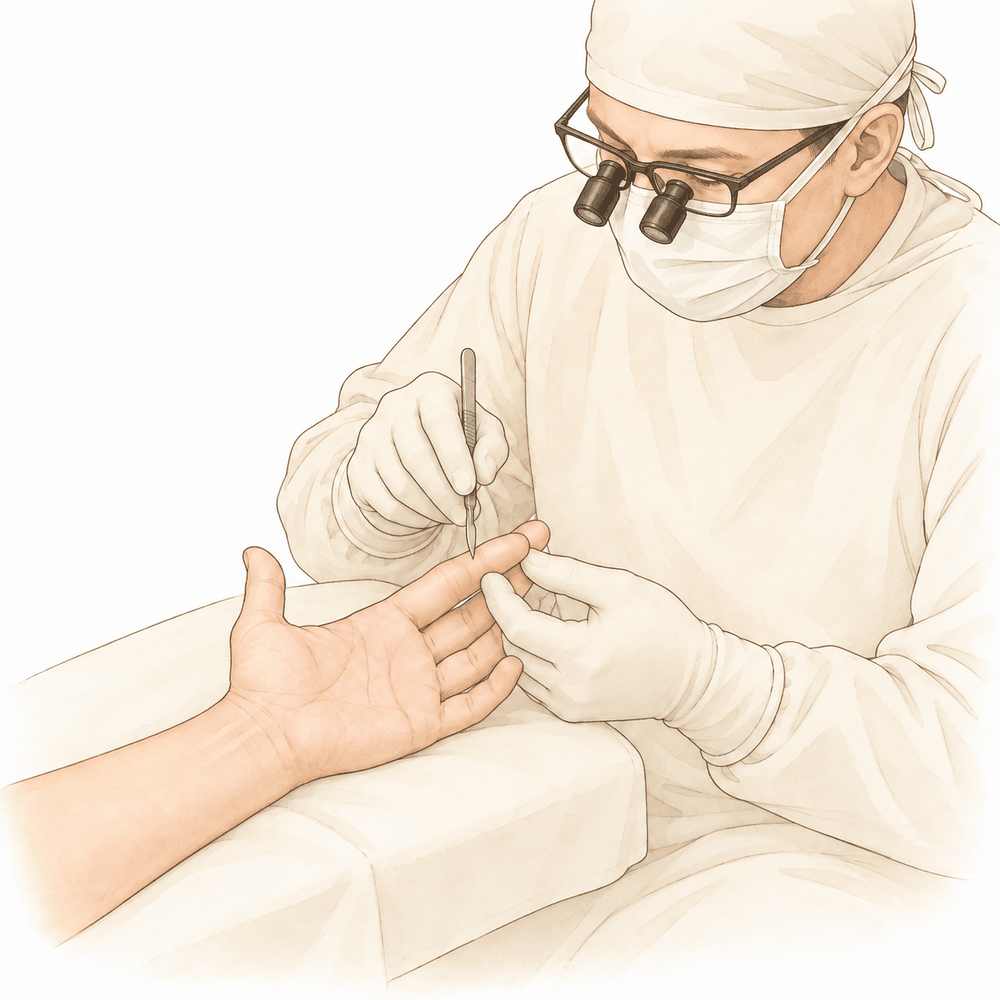
یہ آپریشن کیوں تجویز کیا گیا ہے
ڈاکٹر کیرن ہِرپرا، جو Mater Private Hospital Rockhampton میں بازو اور ہاتھ کے سرجن (upper-limb surgeon) ہیں، سب سے کم چیر پھاڑ والے ان طریقوں سے آغاز کرتے ہیں جو آپ کے مسئلے کے لیے مناسب ہوں۔ مریض عام طور پر اپنے فیملی ڈاکٹر (GP) کے ذریعے ہمارے کلینک بھیجے جاتے ہیں؛ اگر کسی فزیوتھراپسٹ نے آپ کو ہم سے ملنے کا مشورہ دیا ہے، تب بھی میڈیکیئر (Medicare) کی رقم کی واپسی کا حق دار ہونے کے لیے آپ کو اپنے فیملی ڈاکٹر سے ریفرل (referral) لینا ہوگا۔ آپ کی ملاقات پر ہم آپ سے مسئلے کی تفصیل پوچھتے ہیں، آپ کے ہاتھ کا معائنہ کرتے ہیں، اور جہاں ضرورت ہو تشخیص کی تصدیق کے لیے ایکسرے یا اسکین کا انتظام کرتے ہیں۔
ٹینڈن کے غلاف کا جائنٹ سیل ٹیومر (giant cell tumour of tendon sheath) ایک غیر کینسر والی گانٹھ ہے جو کسی ٹینڈن (tendon)، یعنی وہ مضبوط ڈوری جو پٹھے کو ہڈی سے جوڑتی ہے، پر یا اس کے قریب آہستہ آہستہ بڑھتی ہے۔ یہ جسم کے دوسرے حصوں میں نہیں پھیلتی۔ یہ گانٹھیں ہاتھ اور انگلیوں میں عام ہیں، اور عام طور پر ان میں درد نہیں ہوتا، اگرچہ گانٹھ بڑھنے کے ساتھ کچھ لوگوں کو سوجن، درد یا اکڑاؤ محسوس ہوتا ہے۔ چونکہ یہ گانٹھ خود بخود ختم نہیں ہوتی، اس لیے عام طور پر علاج اسے نکال دینا ہی ہوتا ہے۔
ہم جو آپریشن تجویز کرتے ہیں وہ سرجری سے گانٹھ کو کاٹ کر نکالنا (excision) ہے، یعنی گانٹھ کو پوری طرح نکال دینا۔ اس مسئلے کے لیے یہی سب سے زیادہ تسلیم شدہ علاج ہے۔ اصل مقصد گانٹھ کو پوری طرح نکالنا اور اس کی وجہ سے ہونے والی سوجن، درد یا اکڑاؤ کو کم کرنا ہے۔ چونکہ یہ گانٹھیں دوبارہ بن سکتی ہیں، اس لیے ہم سرجری کے بعد اس جگہ کو جانچنے کے لیے باقاعدہ ملاقاتوں کا انتظام کرتے ہیں۔
آپریشن سے پہلے
سرجری سے پہلے کے ہفتوں میں ہم ایکسرے یا اسکین کے ذریعے منصوبے کی تصدیق کرتے ہیں۔ اس میں ایکسرے، الٹراساؤنڈ یا ایم آر آئی اسکین شامل ہو سکتا ہے، جو مقناطیس کی مدد سے گانٹھ اور اس کے آس پاس کے ٹشو کی تفصیلی تصویریں لیتا ہے۔ یہ تصویریں ہمیں ٹھیک ٹھیک دیکھنے میں مدد دیتی ہیں کہ گانٹھ کہاں ہے اور اسے نکالنے کی منصوبہ بندی کیسے کرنی ہے۔
آپریشن والے دن، اپنے آپریشن کے وقت سے سات گھنٹے پہلے کھانا پینا بند کر دیں۔ ہم چھ کے بجائے سات گھنٹے کا کہتے ہیں تاکہ اگر آپریشن کی فہرست وقت سے پہلے چل رہی ہو تو آپ کی سرجری پہلے کی جا سکے۔ آپ کے سرجن آپ کو بتائیں گے کہ آپ کی معمول کی دواؤں میں سے کون سی لینی ہیں اور کون سی روکنی ہیں۔ جو کچھ بھی آپ لیتے ہیں اس کی لکھی ہوئی فہرست ساتھ لائیں، ڈھیلے آرام دہ کپڑے پہنیں، اور بعد میں آپ کو گاڑی میں گھر لے جانے کے لیے کسی کا انتظام کریں۔ اگر آپ کو دوسری طبی بیماریاں ہیں تو ہو سکتا ہے آپ کو خون کے ٹیسٹ یا بے ہوشی کے ڈاکٹر (anaesthetist)، یعنی وہ ڈاکٹر جو بے ہوشی کی دوا دیتے ہیں، سے معائنے کی ضرورت ہو۔
آپریشن کے دن
آپ ہسپتال کے سرجری کے داخلے کے شعبے میں پہنچتے ہیں، جہاں آپ کا اندراج کیا جاتا ہے اور آپ کو آپریشن تھیٹر کے لیے تیار کیا جاتا ہے۔ پھر آپ کی ملاقات بے ہوشی کے ڈاکٹر سے ہوگی، یعنی وہ ڈاکٹر جو بے ہوشی کی دوا دیتے ہیں۔ یہ آپریشن جنرل اینستھیزیا (مکمل بے ہوشی) میں کیا جاتا ہے۔ بعض اوقات آپریشن کے بعد درد میں آرام کے لیے اس کے ساتھ اعصابی بلاک (nerve block)، یعنی ایک انجکشن جو جسم کے ایک حصے کو سُن کر دیتا ہے، بھی لگایا جاتا ہے؛ بے ہوشی کے ڈاکٹر اسی دن آپ سے اس بارے میں بات کریں گے۔ پھر آپ کو آپریشن تھیٹر لے جایا جاتا ہے، جہاں آپریشن کیا جاتا ہے۔
آپ ریکوری روم میں جاگتے ہیں، جہاں بے ہوشی کا اثر ختم ہونے تک نرسیں آپ کی نگرانی کرتی ہیں۔ جب آپ کی حالت مستحکم ہو جائے تو آپریشن اور آپ کی صحت یابی کے لحاظ سے آپ یا تو وارڈ میں جاتے ہیں یا گھر چلے جاتے ہیں۔
آپریشن میں کیا کیا جاتا ہے
آپریشن کا مقصد گانٹھ کو پوری طرح نکالنا ہے، اس کے ان چھوٹے ٹکڑوں سمیت جو شاید آس پاس پھیل گئے ہوں۔ آپ کے سرجن گانٹھ کے اوپر ایک چیرا لگاتے ہیں، جس کا سائز اس بات کے مطابق ہوتا ہے کہ گانٹھ کہاں ہے اور کتنی دور تک پھیلی ہوئی ہے۔ گانٹھ اکثر ٹینڈن سے، یا اس کے ساتھ ساتھ چلنے والے اعصاب اور خون کی نالیوں سے، مضبوطی سے چپکی ہوتی ہے، اس لیے اسے احتیاط سے نکالا جاتا ہے، بعض اوقات ان حصوں کی حفاظت کے لیے عدسے (magnification) کی مدد سے۔
گانٹھ نکالنے کے بعد آپ کے سرجن اس جگہ کو چھوٹے سیٹلائٹ نوڈیولز (satellite nodules)، یعنی گانٹھ کے ننھے الگ ٹکڑے جو پیچھے رہ سکتے ہیں، کے لیے جانچتے ہیں۔ انہیں نکالنا اہم ہے، کیونکہ پیچھے رہ جانے والے ٹکڑے ہی بعد میں دوبارہ بڑھ سکتے ہیں۔ پھر چیرے کو ٹانکوں سے بند کر کے زخم کی پٹی سے ڈھانپ دیا جاتا ہے۔
سرجری سے پہلے، یہ تصدیق کرنے کے لیے کہ گانٹھ کیا ہے، باریک سوئی سے اس کا ایک چھوٹا نمونہ لیا جا سکتا ہے۔ اس سے منصوبہ بندی میں مدد ملتی ہے، تاکہ سرجن کو ٹھیک ٹھیک معلوم ہو کہ آپریشن کے دوران کیا توقع رکھنی ہے۔
آپریشن کے بعد
آپ ریکوری وارڈ میں جاگتے ہیں، جہاں بے ہوشی کا اثر ختم ہونے تک نرسیں آپ پر نظر رکھتی ہیں۔ آپ کے ہاتھ پر زخم کی پٹی ہوگی، اور آپ کو آرام دہ رکھنے کے لیے ہم آپ کو درد کی دوا دیں گے۔ جاگنے کے کچھ ہی دیر بعد آپ چل پھر سکتے ہیں، البتہ پہلے 24 گھنٹے کسی کو آپ کے ساتھ رہنا چاہیے۔ آپ کی ٹیم آپ کو بتائے گی کہ آپ اسی دن گھر جائیں گے یا ایک رات ہسپتال میں رہیں گے۔ ہم زخم کی پٹی تقریباً 10 دن تک لگی رہنے دیتے ہیں؛ براہ کرم اس سے پہلے اسے نہ اتاریں، جب تک ہم آپ کو ایسا کرنے کو نہ کہیں۔ جب ہم آپ کو دیکھتے ہیں تو اسے بدلتے یا اتار دیتے ہیں۔
صحت یابی
پہلے چند دن آپ کے ہاتھ میں درد اور سوجن رہے گی۔ یہ آہستہ آہستہ کم ہو جاتی ہے۔ ہاتھ کو تکیے پر اونچا رکھنا، بیٹھتے یا سوتے وقت بھی، سوجن اور تکلیف کو کم کرنے میں مدد دیتا ہے۔ آپ کی ٹیم کی ہدایت کے مطابق سادہ درد کش دوا آپ کو آرام دہ رکھے گی۔
آپ ہاتھ پر زخم کی پٹی کے ساتھ گھر جائیں گے، جسے ہم تقریباً 10 دن تک لگا رہنے دیتے ہیں۔ براہ کرم اسے صاف اور خشک رکھیں، اور اسے خود نہ اتاریں۔ جب ہم آپ کو دیکھتے ہیں تو زخم کا معائنہ کرتے ہیں، پٹی اتارتے ہیں اور ضرورت ہو تو ٹانکے نکال دیتے ہیں۔
زخم بھر جانے کے بعد آپ Extend Rehabilitation میں روبی ڈولن (Ruby Doolan) کے ساتھ ہینڈ تھراپی شروع کریں گے۔ روبی ایک ہینڈ تھراپسٹ (ہاتھ کی بحالی کی ماہر) ہیں جو آپ کی ورزشوں کی رہنمائی کرتی ہیں اور جو بھی اسپلنٹ آپ کو درکار ہو وہ بناتی ہیں۔ سوجن کم ہونے کے ساتھ یہ ورزشیں آپ کی انگلیوں اور ہاتھ کو دوبارہ حرکت اور طاقت حاصل کرنے میں مدد دیتی ہیں۔ آپ گھر پر روزمرہ کے ہلکے کاموں کے لیے ہاتھ استعمال کر سکتے ہیں، لیکن جب تک آپ کا تھراپسٹ اجازت نہ دے، بھاری وزن اٹھانے، زور سے پکڑنے یا کوئی بھی ایسا کام کرنے سے پرہیز کریں جس سے اس جگہ پر زور پڑے۔
زیادہ تر لوگوں کو محسوس ہوتا ہے کہ حرکت واپس آنے کے ساتھ اکڑاؤ مسلسل بہتر ہوتا جاتا ہے۔ جب آپ بغیر درد کے چیزیں پکڑ سکیں اور ہاتھ استعمال کر سکیں تو روزمرہ کے کام آپ کو بہت آسان لگیں گے۔ آپ کے سرجن آپ کو بتائیں گے کہ آپ کب کام، کھیل یا گاڑی چلانے پر واپس جا سکتے ہیں، اور ہماری ویب سائٹ پر بازو اور ہاتھ کی سرجری کے بعد گاڑی چلانے کے بارے میں ایک الگ رہنمائی موجود ہے۔
ہر شخص اپنی رفتار سے ٹھیک ہوتا ہے، اس لیے آپ کا وقت مختلف ہو سکتا ہے۔ آپ کے سرجن اور آپ کا ہینڈ تھراپسٹ پورے راستے میں آپ کی رہنمائی کریں گے۔
کیا مسائل پیدا ہو سکتے ہیں
زیادہ تر مریض ٹھیک ہو جاتے ہیں، لیکن کبھی کبھار مسائل پیدا ہو سکتے ہیں۔ آپ کے سرجن اور ٹیم آپ کی قریب سے نگرانی کرتے ہیں تاکہ کسی بھی مسئلے کو جلد پکڑا جا سکے۔
جس بات پر ہم سب سے زیادہ نظر رکھتے ہیں وہ گانٹھ کا دوبارہ بن جانا ہے۔ یہ سرجری کے کئی مہینوں بلکہ چند سال بعد بھی ہو سکتا ہے، اس لیے ہم اس جگہ کو جانچنے کے لیے باقاعدہ ملاقاتوں کا انتظام کرتے ہیں۔ اگر آپ کو اپنے زخم کے نشان کے قریب کوئی نئی گانٹھ یا سوجن نظر آئے تو اپنے اگلے معائنے میں اس کا ذکر کریں یا کلینک کو فون کریں۔ بعض اوقات زخم میں رہ جانے والا گانٹھ کا چھوٹا سا ٹکڑا وہیں بڑھ سکتا ہے، اس لیے خود زخم کے نشان کے اندر موجود گانٹھ ہمیں دکھانا ضروری ہے۔
شاذ و نادر ہی یہ گانٹھیں زیادہ سنگین انداز میں برتاؤ کر سکتی ہیں۔ اگر آپ کو ایسی سوجن ہو جو بڑھتی ہی جائے یا غیر معمولی لگے، تو ہم آپ کو جلد دیکھنا چاہتے ہیں تاکہ اس کی جانچ کر سکیں۔
گانٹھ بعض اوقات اعصاب پر دباؤ ڈال سکتی ہے یا ان کے قریب ہو سکتی ہے۔ اگر آپ کو اپنے ہاتھ یا بازو کے کسی حصے میں سُن پن، جھنجھناہٹ یا کمزوری محسوس ہو جو ٹھیک نہ ہو تو اپنے معائنے پر ہمیں بتائیں۔
ان گانٹھوں کو نکالنا تکنیکی طور پر مشکل ہو سکتا ہے کیونکہ یہ ٹینڈن اور اس کے ساتھ والے اعصاب اور خون کی نالیوں کو مضبوطی سے جکڑ لیتی ہیں۔ آپ کے سرجن ان حصوں کی حفاظت کرتے ہوئے پوری گانٹھ نکالنے کے لیے احتیاط سے کام کرتے ہیں۔ اگر گانٹھ کا کچھ حصہ اس لیے چھوڑ دیا جائے کہ اسے نکالنے سے کسی اہم چیز کو نقصان پہنچتا، تو ہم آپ کو یہ بات سمجھائیں گے اور بعد میں اس جگہ پر گہری نظر رکھیں گے۔
جو گانٹھیں ٹشو میں زیادہ وسیع طور پر پھیلی ہوتی ہیں، ان پر صرف سرجری سے پوری طرح قابو نہیں پایا جا سکتا۔ ایسی صورت میں ہاتھ کے کام کی حفاظت میں مدد کے لیے ریڈی ایشن تھراپی (radiation therapy)، یعنی نشانہ بنا کر دی جانے والی ایکسرے شعاعیں جو گانٹھ کی بڑھوتری کو سست کرتی ہیں، تجویز کی جا سکتی ہے۔ اگر کبھی یہ آپ کے معاملے پر لاگو ہوا تو آپ کے سرجن آپ سے اس بارے میں بات کریں گے۔
چونکہ یہ گانٹھیں خاموشی سے دوبارہ بن سکتی ہیں، اس لیے ہم آپ سے کہتے ہیں کہ ہاتھ ٹھیک محسوس ہونے پر بھی بعد کی ملاقاتوں پر ضرور آئیں۔
اگر آپ تفصیل جاننا چاہتے ہیں تو اس صفحے پر پیچیدگیوں کے جدول میں عام شرحیں درج ہیں۔
ہمیں کب فون کریں
اگر آپ کو بخار ہو، زخم کے گرد سرخی بڑھ رہی ہو، یا زخم سے رطوبت کا اخراج ہو تو ہمیں فون کریں۔ اگر آپ کا درد اچانک بڑھ جائے، یا ہاتھ میں سُن پن یا جھنجھناہٹ ٹھیک نہ ہو تو ہمیں فون کریں۔ اگر آپ اپنی انگلیوں یا ہاتھ کو پہلے کی طرح حرکت نہ دے سکیں تو ہمیں فون کریں۔ اگر آپ کو پنڈلی میں سوجن یا درد ہو، یا سانس پھولے تو ہسپتال کی ایمرجنسی جائیں۔ یہ خون کے لوتھڑے کی علامات ہو سکتی ہیں، اور انہیں فوراً چیک کروانا ضروری ہے۔
Evidence & references
This is the clinical evidence summary written for health professionals. It is technical, and it lists the research this page was built from. You do not need to read it to understand your treatment or to make a decision about it.
Overview
- Multicentric calcifying aponeurotic fibroma (CAF) with associated nerve compression and bony dysplasia is exceedingly rare [1].
- Pseudo-Volkmann contracture most commonly involves the ring finger flexor digitorum superficialis but can occur in all fingers [2].
- A series of 75 consecutive joint operations described finger-joints arthrodesed by making the opposing joint surfaces concave distally and convex proximally [3].
- Distal web creep occurred exclusively in grafted cases, reinforcing the biological limitations of grafted tissue in growing patients and supporting the preferential use of local flap reconstruction whenever feasible [4].
- The absence of a control group in a prospective clinical series of reverse dorsoradial digital artery flaps precludes establishing superiority over alternative reconstructive options [5].
- Performing digital mucous cyst excision in-office resulted in an 8.5-fold decrease in health care–related costs [6].
- A two-stage thumb reconstruction procedure shortened the time for recovery and the results recorded were extremely good [7].
- The outcome of the modified neurovascular Tranquilli-Leali flap was cosmetically acceptable to the patient and detailed follow-up showed the flaps to be functional with normal sensitivity and no bony tip pain [11].
- Soft tissue distraction surgery does not downgrade existing function, uses expendable donor nerve, and has minimal perioperative down time for patients, which might make it a more viable option than traditional tendon transfers for tetraplegia [18].
- A preliminary report described the use of 19 Hunter sliding implants in 14 children with flexor tendon injuries [38].
Anatomy & Pathophysiology
- The hand is composed of 19 bones, 17 articulations, and 19 muscles situated entirely within the hand [21].
- The hand contains approximately the same number of tendons activated by the forearm muscles as it has intrinsic muscles [21].
- The dorsal aspect of the hand is convex, while the anterior, palmar, or volar aspect is concave [21].
- The palmar surface of the hand is the functional surface, whereas the dorsal surface is usually visible and aesthetically important [21].
- The digits are divided into the thumb and four fingers [21].
- The thumb has a more proximal and lateral position, allowing movement inward and outward from the palm [21].
- The four fingers are the distal extension of the carpometacarpal part of the hand [21].
- The hinges of finger flexion and extension movements are located at the thenar crease and at the transverse distal palmar crease, not at the bases of the digits [21].
- When fingers are extended and separated, the tips lie on the circumference of a circle whose center is the head of the third metacarpal [21].
- The web spaces are formed from the union of two nonsymmetrical cutaneous surfaces [22].
- The dorsal slope of the web space has a gradual incline and its supple skin is not adherent to the subjacent region [22].
- The palmar surface of the web space is flat and precipitously interrupted, with skin densely adherent to the commissural skeleton [22].
- The commissural skeleton is formed by the interdigital palmar (natatory) ligament between the fingers and by the distal transverse ligament at the level of the thumb web [22].
- The distal transverse ligament at the level of the thumb web is the deepest and most mobile of the commissural ligaments [22].
- There are seven interosseous muscles in the hand, consisting of four dorsal and three volar muscles [23].
- The dorsal interossei are abductors [23].
- The volar interossei are adductors [23].
- The middle finger has two dorsal interossei and no volar interossei because the central axis of the hand lies within it [23].
- Each dorsal interosseous muscle, with the exception of the third, has two muscle heads: a superficial head and a deep head [23].
- The superficial head of the dorsal interosseous muscles arises most dorsally from the shaft of the contiguous metacarpals and is inserted by a medial tendon onto the lateral tubercle of the base of the proximal phalanx [23].
- The deep head of each dorsal interosseous muscle forms a lateral tendon, or lateral band, at the level of the metacarpophalangeal joint [23].
- The flexor digiti quinti brevis is structurally and functionally similar to the deep head of the dorsal interossei and forms the ulnar lateral band of the little finger [23].
- The three volar interossei arise from adjacent surfaces of contiguous metacarpal shafts [23].
- Each volar interosseous muscle has only one muscle head and none of them insert onto the proximal phalanx [23].
- The volar interossei form the ulnar lateral band of the index finger and the radial lateral band of the ring and little fingers [23].
- The abductor digiti quinti and flexor digiti quinti brevis are similar in both structure and function to the superficial and deep heads of the dorsal interossei, respectively, and arise from the fifth metacarpal [23].
- The opponens digiti quinti lies deepest among the hypothenar muscles, arising from the pisohamate ligament and the hook of the hamate [23].
- The opponens digiti quinti inserts onto the ulnar side of the diaphysis of the fifth metacarpal, which it flexes and supinates [23].
- The metacarpal arch is endowed with a great deal of adaptability because of the mobility of the peripheral metacarpals [26].
- The index metacarpal is the most firmly fixed [26].
- The ring metacarpal has about 10 degrees of mobility in flexion and extension [26].
- The fifth metacarpal has a range of flexion–extension of approximately 20 degrees [26].
- The second to fifth metacarpals are bound together by various fibrous structures, the most distal of which is the deep transverse intermetacarpal ligament [26].
- The deep transverse intermetacarpal ligament is also known as the interglenoid ligament because it ties together the anterior glenoid ligaments of the metacarpophalangeal articulations [26].
- The anterior glenoid ligaments of the metacarpophalangeal articulations are known as the volar plates [26].
- The longitudinal arches of the hand are composed of a fixed portion, the carpometacarpal, and a mobile portion, the digits [26].
- The keystones of the longitudinal arches are the metacarpophalangeal articulations [26].
- The thick anterior glenoid capsules, or volar plates, of the metacarpophalangeal joints prevent hyperextension [26].
- The volar plates are interconnected by the transverse interglenoid ligament [26].
- The stability of the metacarpophalangeal joints is essential to the support of the longitudinal arch as well as of the transverse metacarpal arch [26].
- The palmar cutaneous branch of the median nerve supplies the skin over the thenar eminence [9].
- The palmar cutaneous branch of the median nerve arises from the radial border of the median nerve [9].
- The palmar cutaneous branch of the median nerve arises 8.5 cm proximal to the wrist crease [9].
- The flexor retinaculum maintains and restrains the tendons of the extrinsic flexors of the digits within the carpal canal [26].
- The palmar tendons, especially the profundus, are kept close to the axis of flexion–extension of the wrist by the flexor retinaculum [26].
- The extensors of the wrist are more distant from the axis of flexion–extension than the flexors digitorum [26].
- The distance of the wrist extensors from the axis of flexion–extension provides a mechanical advantage that compensates for their difference in power compared to the flexors [26].
- The metacarpophalangeal joint is stabilized by collateral ligaments and by the thick volar articular capsule, the volar plate [26].
- The lateral accessory ligaments, the sagittal bands of the extensor apparatus, and the first annular segment of the pulley of the flexor tendons insert on the volar plate [26].
- The thumb metacarpal is independent and articulates with the trapezium [26].
- The middle metacarpals are united to the carpus by the intrinsic interlocking encasement of the bones themselves [26].
- The fifth metacarpal is semi-independent and articulates with the hamate [26].
- The fifth metacarpal is restrained on its radial side by its articulation with the base of the fourth metacarpal [26].
- The "princeps pollicis" artery is the terminal branch of the radial artery [27].
- The "princeps pollicis" artery crosses the first intermetacarpal space and runs along the ulnar side of the first metacarpal bone [27].
- The "princeps pollicis" artery runs along the volar surface of the adductor muscle [27].
- The "princeps pollicis" artery emerges onto the subcutaneous palmar tissue at the level of the cutaneous flexion crease of the metacarpophalangeal joint [27].
- At the metacarpophalangeal flexion crease, the "princeps pollicis" artery divides into two terminal rami, the collateral palmar arteries of the thumb [27].
- The collateral palmar arteries of the thumb run along the digital tunnel symmetrically and are of equal caliber [27].
- The collateral palmar arteries of the thumb head distally to finally unite in the pulp arcade [27].
- During their transit in the digital tunnel, the collateral palmar arteries break off into numerous collateral branches, which are cutaneous, articular, or osseous [27].
- An arcade located deep in the flexor tendon joins together the two arteries at the level of the distal metaphysis of the first phalanx [27].
- Vessels originating from the arcade at the distal metaphysis of the first phalanx enter the "vincula" and irrigate the flexor tendon [27].
- In anatomical studies, only 15% of dissections of the palmar arteries of the thumb fall into the classical category [27].
- In the second segment of the thumb, the two arteries run alongside the flexor tendon and behind the collateral nerves [27].
- In the second segment of the thumb, the main artery is the ulnar collateral artery [27].
- The subtendinous anastomosis situated at the level of the neck of the first phalanx acts as a "moderator" between the two collateral arteries [27].
- In cases where the palmar ulnar collateral artery is absent, the dorsal artery takes its place by means of a branch through the subtendinous arcade [27].
- In the pulp segment of the thumb, the two arteries are of similar size and run through the thick fatty subcutaneous padding [27].
- In the pulp segment of the thumb, the arteries cross over and convert into the ends of the digital nerves at the level of the median axis [27].
- The dorsal arteries of the thumb originate from the palmar arteries (princeps, commissural, or anastomoses of the superficial arcade) at the level of the first metacarpal [27].
- The dorsal arteries of the thumb run laterally along the metacarpophalangeal joint and continue obliquely from volar to dorsal [27].
- At the level of the neck of the first phalanx, an anastomosis can be found which originates from the palmar arteries for the dorsal aspect [27].
- The dorsal arteries of the thumb are joined by three arcades: one inconstant arcade located under the extensor tendon at the level of the neck of the first phalanx, the arcade of the nail matrix, and the arcade of the nailbed [27].
- The ulnar dorso-collateral artery generally stems from the "princeps pollicis" onto the medial border of the neck of the first metacarpal [27].
- The ulnar dorso-collateral artery heads distally remaining on the lateral surface of the finger [27].
- Distal palmar incisions are transverse as a rule [28].
- In the proximal palm, incisions tend to be more longitudinal, with the distal end curving radially and paralleling the closest major skin crease [28].
- After the skin and underlying fat have been incised in the palm, the fat is dissected from the palmar fascia and is carried with the skin flaps [28].
- Most vital structures in the palm are deep to the palmar fascia [28].
- In the distal palm, structures lying between the metacarpal heads are not protected by the palmar fascia [28].
- The superficial volar neurovascular arch should be protected when deeper exposure is required in the palm [28].
- Incisions in the more proximal palm should parallel the thenar crease [28].
- Incisions extended proximal to the wrist should not cross the flexor wrist creases at a right angle [28].
- The most important structure in the thenar area is the recurrent branch (motor) of the median nerve [28].
- Anatomic studies have shown that there is no single longitudinal incision in the proximal palm that completely avoids the palmar cutaneous branches of the median and ulnar nerves [28].
- The volar midoblique incision crosses the flexion creases obliquely in the midline of the finger between the neurovascular bundles [28].
- The volar midoblique incision is useful for a variety of procedures and often can be used instead of a volar zigzag incision [28].
- The volar zigzag finger incision does not require mobilizing either neurovascular bundle and directly exposes the volar surface of the flexor tendon sheath [28].
- When used on a contracted skin surface, the volar zigzag finger incision tends to straighten out and result in a more linear scar than is desirable [28].
- Midlateral incisions described for the fingers are also suitable for the thumb [28].
- The radial side of the thumb is more accessible for midlateral incisions [28].
- A midlateral incision on the radial side of the thumb can be extended by curving its proximal end at the midmetacarpal area and creating a flap on the palmar surface of the thumb [28].
- Care should be taken to avoid the dorsal branch of the superficial radial nerve to the radial side of the thumb during midlateral incisions [28].
- The necessary skin to cover the thumb distal to the metacarpophalangeal joint is about 9 cm wide and 8 cm long [25].
- Skin loss of the thumb and first metacarpal is 13 cm wide and 12 cm long [25].
- The skin cover of both the palmar and dorsal surfaces of the hand is 12 cm by 10 cm [25].
- The skin covering each finger is 7 cm by 10 cm on both the palmar and dorsal aspects [25].
- Skin grafting or flaps for both sides of the hand and digits requires a skin of 20 cm by 20 cm [25].
- Skin grafting or flaps for one aspect of the forearm from wrist to elbow requires skin of 30 cm by 15 cm [25].
- Skin grafting or flaps for both aspects of the forearm requires skin of 30 cm by 30 cm [25].
- The palm forms a cutaneous unit extending from the distal transverse crease of the wrist up to the transverse crease at the base of the digits [22].
- The palmar integument may be subdivided into two separate zones by the oppositional crease of the thumb [22].
- The oppositional crease of the thumb constitutes the oblique axis of the hand [22].
- The skin of the radial portion of the palm covers the thenar eminence and the external part of the palm and is the mobile portion [22].
- The skin of the ulnar and distal portion covers the hypothenar eminence where the skin has poor mobility [22].
- The distal part of the palm beyond the transverse distal palmar crease is a true hinge just at the level of the metacarpophalangeal articulations [22].
- The central triangular part of the palm has skin that is fixed and poorly vascularized, covering almost directly the superficial palmar aponeurosis [22].
- The superficial palmar aponeurosis inserts into the skin of the central triangular part of the palm [22].
- The integument of the palmar face of the digits may be subdivided into phalangeal units separated by the digital flexion folds [22].
- There are three digital flexion folds for the digits and two for the thumb [22].
- When a digit is completely flexed, the integument of the adjacent phalanges comes into contact in the zones of the flexion creases [22].
- The areas of cutaneous contact in the flexed digits are in the form of a diamond [22].
- The sides of the diamond-shaped cutaneous contact areas do not undergo variations in length during the movements of flexion and extension [22].
- Incisions made along the lines of the diamond-shaped cutaneous contact areas present a minimal chance of retraction [22].
- One cutaneous unit on the dorsum of the hand extends from the wrist to the proximal interphalangeal joints of the fingers and the interphalangeal joint of the thumb [22].
- The dorsal covering of the interphalangeal articulations of the digits forms a unique cutaneous unit characterized by a considerable excess of skin when the digits are in extension [22].
- The fine tight skin of the dorsal aspect of the middle phalanx forms another cutaneous unit [22].
- The dorsal integument of the distal phalanx is very special because of the nail bed with its matrix [22].
Clinical Presentation
- Multicentric calcifying aponeurotic fibroma with associated nerve compression and bony dysplasia is exceedingly rare [1].
- Pseudo-Volkmann contracture most commonly involves the ring finger flexor digitorum superficialis [2].
- Pseudo-Volkmann contracture can occur in all fingers [2].
- In a case of flexor tenosynovitis of the hand caused by Mycobacterium tuberculosis, the patient presented with diffuse erythema and swelling in the fifth finger extending into the palm [10].
- In a case of flexor tenosynovitis of the hand caused by Mycobacterium tuberculosis, a small cyst was noted on the ulnar side of the dorsal hand [10].
- In a case of flexor tenosynovitis of the hand caused by Mycobacterium tuberculosis, the patient had a fusiform digit and pain over the flexor sheath [10].
- In a case of flexor tenosynovitis of the hand caused by Mycobacterium tuberculosis, the patient did not have pain with passive extension or a flexed digit at rest [10].
Investigations
- Clinical evaluation of the hand and wrist requires a systematic physical examination to pinpoint or narrow the scope of possible pathologic processes, as patients often have difficulty accurately describing symptoms [15].
- Diagnostic tests such as imaging and serum laboratory studies are useful in determining pathology but can be expensive, time-consuming, and often nonspecific [15].
- A careful physical examination is essential to direct care and future testing if indicated [15].
- An 8-MHz Doppler tone assessment may be used to identify superficially displaced neurovascular bundles when Dupuytren cords lie beneath soft fleshy prominences, but false-negatives are possible [30].
- Doppler imaging is a promising improvement for identifying structures, but higher resolution imaging technology is needed [30].
- MR assessment of Dupuytren’s disease is hindered by the resolution of current equipment, orientation issues due to multiplanar deformities of the fingers, and lack of intraoperative availability [30].
- MRI is probably most useful in identifying additional pathology such as flexor tendon bowstringing [30].
- MRI may be helpful in providing a quantitative noninvasive measure of cellularity of affected areas, which is an index of biologic activity [30].
Treatment
- Successful treatment of finger flexor tenosynovitis from stonefish envenomation injury was obtained with serial debridement coupled with a prolonged antibiotic regimen for coverage of the marine base pathogens [13].
Complications
- The absence of a control group in the reverse dorsoradial digital artery flap series precludes establishing superiority over alternative reconstructive options [5].
- In closing wedge osteotomy for clinodactyly, the volar neurovascular bundles and extensor mechanism lie in close proximity and must be carefully preserved [8].
- In closing wedge osteotomy for clinodactyly, accurate localization of the proximal interphalangeal and distal interphalangeal joints is critical to avoid iatrogenic joint violation [8].
- In closing wedge osteotomy for clinodactyly, the periosteum should be preserved to maintain vascularity and enhance healing [8].
Operative Technique
- Finger-joints were arthrodesed by making the opposing joint surfaces concave distally and convex proximally in a series of 75 consecutive operations [3].
- The volar neurovascular bundles and extensor mechanism lie in close proximity and must be carefully preserved during surgical exposure [8].
- Surgical exposure is typically performed from the concave side of the deformity, which is the ulnar side in cases of radial deviation [8].
- The extensor mechanism, including the conjoined lateral bands, central slip, and terminal tendon, must be identified and protected throughout the procedure [8].
- Accurate localization of the proximal interphalangeal and distal interphalangeal (DIP) joints is critical to avoid iatrogenic joint violation [8].
- The periosteum should be preserved to maintain vascularity and enhance healing [8].
Outcomes and Complications
- A two-stage procedure shortened the time for recovery with extremely good results recorded [7].
- Distal web creep occurred exclusively in grafted cases, reinforcing the biological limitations of grafted tissue in growing patients [4].
- The absence of a control group precludes establishing superiority over alternative reconstructive options for reverse dorsoradial digital artery flaps [5].
- Modified neurovascular Tranquilli-Leali flaps resulted in outcomes that were cosmetically acceptable to the patient [11].
- Detailed follow-up of modified neurovascular Tranquilli-Leali flaps showed them to be functional with normal sensitivity and no bony tip pain [11].
Key Evidence
- [L5] Multicentric calcifying aponeurotic fibroma (CAF) with associated nerve compression and bony dysplasia is exceedingly rare. [1] (10.1016/j.jhsg.2026.101079)
- [L4] It most commonly involves the ring finger flexor digitorum superficialis but can occur in all fingers. [2] (10.5435/jaaosglobal-d-18-00031)
- [L4] The authors describe a series of 75 consecutive joint operations in which finger-joints were arthrodesed by making the opposing joint surfaces concave distally and convex proximally. [3] (10.1016/s0020-1383(71)80177-8)
- [L4] Distal web creep occurred exclusively in grafted cases, reinforcing the biological limitations of grafted tissue in growing patients and supporting the preferential use of local flap reconstruction whenever feasible. [4] (10.1016/j.jhsg.2026.101055)
- [L4] The absence of a control group precludes establishing superiority over alternative reconstructive options. [5] (10.1016/j.jhsg.2026.101107)
- [Paper] Performing the cyst excision in-office resulted in an 8.5-fold decrease in health care–related costs. [6] (10.1177/15589447251350168)
- [L4] The two-stage procedure shortened the time for recovery and the results recorded are extremely good. [7] (10.1016/s0020-1383(70)80031-6)
- [L5] [8] (10.1016/j.jhsg.2026.101048)
- [L5] [9] (10.1177/17531934231212064)
- [L5] [10] (10.5435/jaaosglobal-d-17-00083)
- [L5] The outcome was cosmetically acceptable to the patient and detailed follow-up showed the flaps to be functional with normal sensitivity and no bony tip pain. [11] (10.1016/s0020-1383(98)00126-0)
- [L5] Successful treatment was obtained with serial debridement coupled with a prolonged antibiotic regimen for coverage of the marine base pathogens. [13] (10.5435/jaaosglobal-d-19-00024)
- [L4] This surgery does not downgrade existing function, uses expendable donor nerve, and has minimal perioperative down time for patients, which might make it a more viable option than traditional tendon transfers for tetraplegia. [18] (10.1016/j.jhsa.2013.08.033)
- [L4] This paper is a preliminary report on the use of 19 Hunter sliding implants in 14 children with flexor tendon injuries. [38] (10.1016/s0020-1383(71)80175-4)
References
[1] Multicentric Calcifying Aponeurotic Fibroma of the Upper Extremity With Associated Nerve Compression and Bony Dysplasia. Journal of Hand Surgery Global Online. 2026. DOI: 10.1016/j.jhsg.2026.101079
[2] Pseudo-Volkmann Contracture: A Case Report and Review of the Current Literature. JAAOS: Global Research and Reviews. 2018. DOI: 10.5435/jaaosglobal-d-18-00031
[3] Concave-convex arthrodeses. Injury. 1972. DOI: 10.1016/s0020-1383(71)80177-8
[4] Dorsal Island Flap for Syndactyly Reconstruction: A Skin-Sparing Technique Associated With Reduced Web Creep. Journal of Hand Surgery Global Online. 2026. DOI: 10.1016/j.jhsg.2026.101055
[5] Reverse Dorsoradial Digital Artery Flap for Reconstruction of Thumb Soft Tissue Defects: A Prospective Clinical Series. Journal of Hand Surgery Global Online. 2026. DOI: 10.1016/j.jhsg.2026.101107
[6] Safety and Cost-Effectiveness of In-Office Digital Mucous Cyst Excisions. HAND. 2025. DOI: 10.1177/15589447251350168
[7] Thumb reconstruction. Injury. 1970. DOI: 10.1016/s0020-1383(70)80031-6
[8] Closing Wedge Osteotomy With Cannulated Screw Fixation in Clinodactyly: A Surgical Technique Guide. Journal of Hand Surgery Global Online. 2026. DOI: 10.1016/j.jhsg.2026.101048
[9] European Board of Hand Surgery (EBHS) Examination Questions. Journal of Hand Surgery (European Volume). 2023. DOI: 10.1177/17531934231212064
[10] Flexor Tenosynovitis of the Hand Caused by Mycobacterium tuberculosis. JAAOS: Global Research and Reviews. 2018. DOI: 10.5435/jaaosglobal-d-17-00083
[11] Finger flaps: using the modified neurovascular Tranquilli-Leali flap. Injury. 1998. DOI: 10.1016/s0020-1383(98)00126-0
[13] Finger Flexor Tenosynovitis From Stonefish Envenomation Injury. JAAOS: Global Research and Reviews. 2019. DOI: 10.5435/jaaosglobal-d-19-00024
[15] Orthopaedic Knowledge Update 13 Ebook Without Multimedia. Anatomy, Evaluation, Clinical Examination, and Imaging > Evaluation and Clinical Examination: Current Concepts.
[18] The Effect of Soft Tissue Distraction on the Outcomes of Centralization for Radial Longitudinal Deficiency. The Journal of Hand Surgery. 2013. DOI: 10.1016/j.jhsa.2013.08.033
[21] Exam Of The Hand Wrist 2Ed. INTRODUCTION.
[22] Exam Of The Hand Wrist 2Ed. Functional cutaneous units.
[23] Green S Operative Hand Surgery. Interosseous and Hypothenar Muscles.
[25] Exam Of The Hand Wrist 2Ed. Planning skin cover of the hand and forearm.
[26] Exam Of The Hand Wrist 2Ed. The arches of the hand > The metacarpal arch.
[27] Exam Of The Hand Wrist 2Ed. Techniques of investigation of the arterial supply by J P Melki > Vascularization of the thumb > Palmar aspect.
[28] Campbell S Operative Orthopaedics 4 Volume Set. RESULTS OF SUTURE OF THE SCIATIC NERVE > PALMAR INCISIONS.
[30] Dupuytren S Disease And Related Hyperproliferative Disorders. 54. The Future of Dupuytren’s Research and Treatment > 54.4 Mechanical Measurements and Procedures > 54.4.4 Imaging.
[38] Tendon-sheaths from sliding artificial implants. Injury. 1972. DOI: 10.1016/s0020-1383(71)80175-4