Patients › Elbow
ریڈیل ٹنل سنڈروم (Radial Tunnel Syndrome)
Radial tunnel syndrome — causes forearm pain, weakness straightening fingers, and is distinct from tennis elbow.
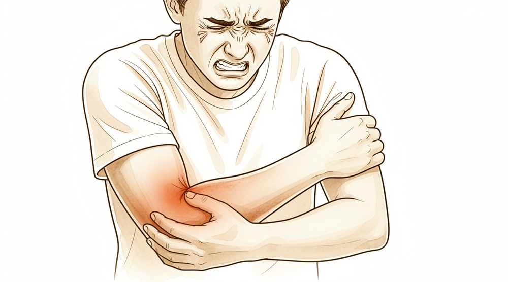
آپ کو کیا محسوس ہوتا ہے
سب سے بڑی علامت آپ کے بازو کے نچلے حصے (forearm) کی باہر والی طرف، کہنی کے قریب، درد ہے۔ یہ درد عموماً اس جگہ سب سے زیادہ ہوتا ہے جو کہنی کی باہر والی طرف کے ہڈی کے ابھار سے چند سینٹی میٹر نیچے ہے اور جسے چھونے پر درد ہوتا ہے۔ اس جگہ کو دبانے سے اکثر درد ہوتا ہے۔ درد بازو کے نچلے حصے کی پشت سے ہوتا ہوا کلائی کی طرف پھیل سکتا ہے۔
کچھ خاص حرکتیں اسے بڑھا دیتی ہیں۔ رکاوٹ کے خلاف ہتھیلی کو اوپر کی طرف گھمانا، یا رکاوٹ کے خلاف درمیانی انگلی کو سیدھا کرنا، درد شروع کر سکتا ہے۔ ایسی سرگرمی سے بھی درد شروع ہو سکتا ہے جو بازو کے نچلے حصے پر بار بار بوجھ ڈالتی رہے۔ بہت سے لوگ محسوس کرتے ہیں کہ درد آرام کے وقت کے بجائے کام یا کھیل کے بعد بھڑکتا ہے۔
یہ بیماری ایک ایسے عصب (nerve) کو متاثر کرتی ہے جس کا کام جلد کے بجائے پٹھوں تک پیغام پہنچانا ہے۔ اس کا مطلب ہے کہ آپ کو سُن پن یا جھنجھناہٹ ہونے کا امکان کم ہے، اور خود عصب عموماً معمول کے مطابق کام کرتا ہے۔ مسئلہ درد ہے، کمزوری یا حس کا ختم ہونا نہیں۔
روزمرہ زندگی میں درد عموماً ان کاموں میں سامنے آتا ہے جن میں پکڑنا اور مروڑنا پڑتا ہے۔ کیتلی اٹھانا، دروازے کا ہینڈل گھمانا، پیچ کس استعمال کرنا یا کپڑا نچوڑنا، یہ سب تکلیف دہ ہو سکتے ہیں۔ چونکہ درد والی جگہ کہنی کی باہر والی طرف کے قریب ہوتی ہے، اس لیے اس بیماری کو اکثر ٹینس ایلبو (کہنی کے باہر کی طرف درد) سمجھ لیا جاتا ہے، اور دونوں ایک ساتھ بھی ہو سکتے ہیں۔
کوئی ایک ٹیسٹ ایسا نہیں جو اس بیماری کی تصدیق کر دے۔ آپ کے سرجن آپ کی بتائی ہوئی تفصیل اور معائنے سے تشخیص تک پہنچتے ہیں، اور اس مخصوص جگہ پر چھونے سے ہونے والے درد اور آپ کے درد کے انداز کو جانچتے ہیں۔
ان علامات پر نظر رکھیں۔ اگر آپ کی علامات ٹھیک نہیں ہو رہیں، ہفتوں کے دوران بگڑتی جا رہی ہیں، رات کو آپ کو جگا دیتی ہیں، یا آپ کو کام کرنے یا بازو استعمال کرنے سے روکتی ہیں، تو اپنے فیملی ڈاکٹر (GP) سے ملیں یا کسی ماہر ڈاکٹر (specialist) سے معائنے کی درخواست کریں۔ اگر آپ کا بازو گرم، سرخ، سوجا ہوا اور دردناک ہو جائے، خاص طور پر بخار کے ساتھ، یا اگر آپ کا ہاتھ سفید پڑ جائے، ٹھنڈا ہو جائے، یا اس کا رنگ سفید یا نیلا ہو جائے، تو اسی دن ہسپتال کی ایمرجنسی جائیں۔ اگر آپ دفتری اوقات کے بعد یا ہفتے کے آخر میں کلینک سے رابطہ نہ کر سکیں، تو اپنے قریب ترین ہسپتال کی ایمرجنسی جائیں۔
دراصل کیا ہو رہا ہے
درد عصب سے آتا ہے، خود کہنی کے جوڑ سے نہیں۔ عصب ایک تار کی طرح ہے جو آپ کے دماغ اور پٹھوں کے درمیان پیغامات لے جاتا ہے۔ ریڈیل عصب (radial nerve) کی ایک شاخ آپ کے بازو کے نچلے حصے کی باہر والی طرف، کہنی کے پاس سے گزرتی ہوئی نیچے جاتی ہے، اور ان پٹھوں تک پیغام پہنچاتی ہے جو آپ کی کلائی اور انگلیوں کو سیدھا کرتے ہیں۔
اس راستے میں کئی الگ الگ ساختیں عصب کے قریب ہوتی ہیں۔ کسی پٹھے کا کنارہ، ریشے دار ٹشو کی ایک پٹی، یا اوپر سے گزرنے والی خون کی چھوٹی نالیوں کا ایک گچھا، ان میں سے ہر ایک اس پر دباؤ ڈال سکتا ہے۔ یہ کوئی ایک تنگ راستہ نہیں ہے۔ یہ زیادہ تر ایک پیدل راستے جیسا ہے جس میں کئی جگہیں ایسی ہیں جہاں دباؤ پڑ سکتا ہے، اور ان میں سے کوئی بھی جگہ ساتھ ساتھ چلنے والی تار کو دبا سکتی ہے۔
جب ان میں سے کوئی ساخت عصب پر دباؤ ڈالتی ہے تو عصب میں جلن (irritation) پیدا ہو جاتی ہے اور اس میں درد ہونے لگتا ہے۔ اسی لیے درد کہنی کی باہر والی طرف کے ہڈی کے ابھار کے نیچے اس جگہ پر رہتا ہے جسے چھونے پر درد ہوتا ہے، اور اسی لیے رکاوٹ کے خلاف ہتھیلی کو اوپر گھمانے یا درمیانی انگلی کو سیدھا کرنے سے درد بڑھ جاتا ہے۔ یہ حرکتیں ان پٹھوں کو کھینچتی ہیں جن تک یہ عصب پیغام پہنچاتا ہے، اور جلن والا عصب شکایت کرنے لگتا ہے۔
عصب اب بھی کام کرتا ہے، اس لیے آپ کے پٹھے کام کرتے رہتے ہیں اور آپ کو کوئی سُن پن یا جھنجھناہٹ محسوس نہیں ہوتی۔ مسئلہ جلن اور درد ہے، پیغام کا رک جانا نہیں۔ چونکہ درد والی جگہ کہنی کی باہر والی طرف کے بہت قریب ہوتی ہے، اس لیے اس درد کو آسانی سے ٹینس ایلبو سمجھ لیا جاتا ہے، جو اسی علاقے میں ایک ٹینڈن (tendon)، یعنی وہ مضبوط ڈوری جو پٹھے کو ہڈی سے جوڑتی ہے، کو متاثر کرتا ہے۔ دونوں ایک ساتھ بھی ہو سکتے ہیں۔
ہم اس کے لیے کیا کر سکتے ہیں
ڈاکٹر کیرن ہِرپرا، جو Mater Private Hospital Rockhampton میں بازو اور ہاتھ کے سرجن (upper-limb surgeon) ہیں، علاج کا آغاز ان کم سے کم تکلیف دہ طریقوں سے کرتے ہیں جو آپ کی حالت کے لیے مناسب ہوں۔ مریضوں کو عام طور پر ان کا فیملی ڈاکٹر (GP) ہمارے کلینک میں بھیجتا ہے؛ اگر کسی فزیوتھراپسٹ نے آپ کو ہمیں دکھانے کا مشورہ دیا ہے، تب بھی میڈیکیئر (Medicare) کی رقم واپسی کا حق دار ہونے کے لیے آپ کو اپنے فیملی ڈاکٹر سے ریفرل (referral) لینا ہوگا۔ کلینک میں ہم آپ کی بیماری کی تاریخ لیتے ہیں، آپ کے بازو کا معائنہ کرتے ہیں، اور اگر مددگار ہو تو ایکسرے یا اسکین کا انتظام کرتے ہیں۔ اس بیماری میں ہم عام طور پر پہلے آپریشن کے بغیر علاج آزماتے ہیں، اور اگر اس سے کافی بہتری نہ آئے تو سرجری پر غور کرتے ہیں۔
پہلا قدم یہ بدلنا ہے کہ آپ اپنا بازو کیسے استعمال کرتے ہیں۔ پکڑنے اور مروڑنے والے جو کام درد کو بھڑکاتے ہیں انہیں کم کرنے سے جلن والے عصب کو پرسکون ہونے کا موقع ملتا ہے۔ ہینڈ تھراپی (ہاتھ کی بحالی کے ماہر کا علاج) کا مقصد درد کو کم کرنا اور بازو کے نچلے حصے پر بوجھ برداشت کرنے کی آپ کی صلاحیت کو دوبارہ بڑھانا ہے، تاکہ کام اور روزمرہ کے کام پھر سے آرام دہ ہو جائیں۔ ہم آپ سے کہتے ہیں کہ کسی مزید قدم کے بارے میں سوچنے سے پہلے اسے مناسب موقع دیں۔
درد کش دوائیں اور سوزش کم کرنے والی دوائیں درد کے بھڑکنے کے دوران مدد دے سکتی ہیں۔ جب تک دوسرے اقدامات اپنا کام کرتے ہیں، یہ تکلیف کو کم کرتی ہیں۔ یہ خود عصب پر پڑنے والے دباؤ کو ٹھیک نہیں کرتیں۔
اگر آپریشن کے بغیر علاج سے کافی بہتری نہ آئی ہو تو سرجری ایک راستہ ہے۔ اس آپریشن کو ریڈیل ٹنل ریلیز (radial tunnel release، عصب پر دباؤ ختم کرنے کا آپریشن) کہتے ہیں۔ یہ عصب کو ان تمام جگہوں پر آزاد کر کے کام کرتا ہے جہاں قریب کی کوئی ساخت اس پر دباؤ ڈال رہی ہو۔ اس کی سفارش کرنے سے پہلے ہم احتیاط سے جانچتے ہیں کہ عصب کہاں دب رہا ہے، تاکہ آپریشن صحیح جگہ کو نشانہ بنائے اور آس پاس کے ٹشو کو کم سے کم چھیڑا جائے۔ چونکہ تشخیص کسی ایک واضح ٹیسٹ کے بجائے آپ کی بتائی ہوئی تفصیل اور معائنے پر منحصر ہے، اس لیے ہم آپریشن کے فیصلے کو مشترکہ فیصلہ سمجھتے ہیں، جو ہم آپ کے ساتھ مل کر اس وقت کرتے ہیں جب آپ سمجھ لیں کہ سرجری کیا ٹھیک کر سکتی ہے اور کیا نہیں۔
کیا توقع رکھیں
زیادہ تر لوگوں میں یہ بیماری عصب کو نقصان نہیں پہنچاتی۔ عصب کام کرتا رہتا ہے، اس لیے آپ کے پٹھے کام کرتے رہتے ہیں اور آپ کی حس ختم نہیں ہوتی۔ مسئلہ درد ہے، اور جلن والے عصب کا درد ٹھیک ہونے میں کافی وقت لے سکتا ہے۔ یہ شاذ و نادر ہی راتوں رات ختم ہوتا ہے، اور کم ہونے سے پہلے اکثر پکڑنے اور مروڑنے والے کاموں سے بھڑکتا رہتا ہے۔
سچی تصویر یہ ہے کہ اس بیماری کے بارے میں پیش گوئی کرنا مشکل ہے۔ کچھ لوگ آرام، سرگرمیوں میں تبدیلی اور ہینڈ تھراپی سے ٹھیک ہو جاتے ہیں۔ دوسروں کو آپریشن کے بغیر اچھے علاج کے باوجود درد رہتا ہے۔ تشخیص کی تصدیق کے لیے کوئی قابلِ اعتماد ٹیسٹ نہیں ہے، جس کی وجہ سے پہلے سے یہ کہنا مشکل ہوتا ہے کہ آپ کس گروپ میں ہوں گے۔ ہم یہ ضرور کہہ سکتے ہیں کہ درد مسلسل بڑھنے کے بجائے اس بات کے ساتھ آتا جاتا رہتا ہے کہ آپ بازو کے نچلے حصے پر کتنا بوجھ ڈالتے ہیں۔
اگر آپریشن کے بغیر علاج کام کر جائے، تو جیسے جیسے عصب کی جلن کم ہوتی ہے، درد ہفتوں سے مہینوں میں آہستہ آہستہ کم ہوتا ہے۔ آپ توقع رکھ سکتے ہیں کہ پہلے کیتلی اٹھانے یا دروازے کا ہینڈل گھمانے جیسے روزمرہ کام کم تکلیف دہ ہو جائیں گے، اور اس کے بعد بھاری کام دوبارہ معمول کے مطابق محسوس ہوگا۔ اگر آپریشن کے بغیر علاج سے کافی مدد نہ ملنے کے بعد سرجری کی جائے، تو صحت یابی بھی یک دم نہیں بلکہ آہستہ آہستہ ہوتی ہے، اور کچھ لوگوں کو اس کے بعد بھی کچھ درد رہتا ہے۔
اگر بیماری کو یوں ہی چھوڑ دیا جائے تو ہو سکتا ہے یہ خود ہی ٹھیک ہو جائے، لیکن یہ برقرار بھی رہ سکتی ہے، یا جب بھی آپ ان کاموں پر واپس جائیں جو اسے بڑھاتے ہیں، بار بار بھڑک سکتی ہے۔ پہلے سے یہ جاننے کا کوئی طریقہ نہیں کہ کیا ہوگا۔ جو بات عموماً اہم ہوتی ہے وہ یہ ہے کہ علاج کو مناسب موقع دیا جائے اور ایسے درد کے باوجود زور نہ لگایا جائے جو ٹھیک نہیں ہو رہا۔
اگر آپ کی علامات ٹھیک نہیں ہو رہیں، ہفتوں کے دوران بگڑتی جا رہی ہیں، رات کو آپ کو جگا دیتی ہیں، یا آپ کو کام کرنے یا بازو استعمال کرنے سے روکتی ہیں، تو اپنے فیملی ڈاکٹر سے ملیں یا کسی ماہر ڈاکٹر سے معائنے کی درخواست کریں۔ اگر آپ کا بازو گرم، سرخ، سوجا ہوا اور دردناک ہو جائے، خاص طور پر بخار کے ساتھ، یا اگر آپ کا ہاتھ سفید پڑ جائے، ٹھنڈا ہو جائے، یا اس کا رنگ سفید یا نیلا ہو جائے، تو اسی دن ہسپتال کی ایمرجنسی جائیں۔ اگر آپ دفتری اوقات کے بعد یا ہفتے کے آخر میں کلینک سے رابطہ نہ کر سکیں، تو اپنے قریب ترین ہسپتال کی ایمرجنسی جائیں۔
ڈاکٹر کو کب دکھائیں
اس بیماری میں انتباہی علامات کا تعلق ایسے درد سے ہے جو ٹھیک نہ ہو رہا ہو، کسی خطرے سے نہیں۔ اگر آرام اور سرگرمیوں میں تبدیلی کو مناسب موقع دینے کے بعد بھی آپ کے بازو کے نچلے حصے کا درد ٹھیک نہ ہوا ہو، یا یہ بار بار آپ کو کام کرنے یا بازو استعمال کرنے سے روکتا ہو، تو اپنے فیملی ڈاکٹر سے ملیں یا کسی ماہر ڈاکٹر سے معائنے کی درخواست کریں۔ اگر کہنی کی باہر والی طرف کے نیچے چھونے پر درد والی جگہ ایک نمایاں علامت ہو، یا اگر رکاوٹ کے خلاف ہتھیلی کو اوپر گھمانے یا درمیانی انگلی کو سیدھا کرنے سے ہر بار درد شروع ہو جاتا ہو، تو دیر کرنے کے بجائے جلد معائنہ کروائیں۔ یہ انداز ٹینس ایلبو کے بجائے اسی بیماری کی طرف اشارہ کرتے ہیں، اور انہیں اچھی طرح جانچ کر حل کرنا فائدہ مند ہے۔
یہ بیماری عموماً عصب کو نقصان نہیں پہنچاتی، اس لیے کلائی یا انگلیوں کی اچانک کمزوری اس کا عام حصہ نہیں ہے۔ اگر آپ کو کلائی، انگلیوں یا انگوٹھے کو سیدھا کرنے میں کوئی نئی کمزوری محسوس ہو، تو انتظار کرنے کے بجائے جلد از جلد کسی ماہر ڈاکٹر سے معائنے کی درخواست کریں، کیونکہ یہ انداز عصب کے کسی دوسرے مسئلے کی طرف اشارہ کرتا ہے جس کا معائنہ اس کے اپنے الگ وقت کے مطابق کیا جاتا ہے۔
اگر آپ کا بازو گرم، سرخ، سوجا ہوا اور دردناک ہو جائے، خاص طور پر بخار کے ساتھ، یا اگر آپ کا ہاتھ سفید پڑ جائے، ٹھنڈا ہو جائے، یا اس کا رنگ سفید یا نیلا ہو جائے، تو اسی دن ہسپتال کی ایمرجنسی جائیں۔ اگر آپ دفتری اوقات کے بعد یا ہفتے کے آخر میں کلینک سے رابطہ نہ کر سکیں، تو اپنے قریب ترین ہسپتال کی ایمرجنسی جائیں۔
مزید تفصیل
Advanced reading: the deeper science (optional)
یہ حصہ اس سے آگے جاتا ہے جتنا آپ کو اپنے علاج کے فیصلوں کے لیے ضرورت ہے۔ ریڈیل ٹنل سنڈروم کے بارے میں اضافی مطالعہ ایک ناخوشگوار وجہ سے فائدہ مند ہے: اس ویب سائٹ پر یہی وہ بیماری ہے جس کے حق میں شواہد سب سے کمزور ہیں، کوئی ایسا ٹیسٹ نہیں جو اس کی تصدیق کرے، اور آپریشن کے کسی بھی فیصلے میں یہ بات شامل ہونی چاہیے کہ آپ کو صاف صاف بتایا جائے کہ یہ کتنی غیر یقینی ہے۔
تصدیق کرنے والا کوئی ٹیسٹ نہیں ہے
عصب پر دباؤ کی زیادہ تر صورتیں ٹیسٹ سے ثابت کی جا سکتی ہیں۔ کارپل ٹنل سنڈروم (کلائی میں عصب کا دبنا) میں کلائی کے آر پار عصب میں پیغام کی رفتار سست ہو جاتی ہے؛ کیوبیٹل ٹنل (cubital tunnel) میں کہنی کے آر پار ایسا ہوتا ہے۔ ریڈیل ٹنل سنڈروم میں عام طور پر ایسا نہیں ہوتا، کیونکہ اس میں شامل عصب کی شاخ جلد کے بجائے پٹھوں کو اعصابی پیغام پہنچاتی ہے، اس لیے سُن پن کا کوئی حصہ نہیں ہوتا جس کا نقشہ بنایا جا سکے، اور عصب کی رفتار کے ٹیسٹ (nerve conduction studies) اکثر نارمل ہوتے ہیں۔
اس لیے تشخیص کا دارومدار درد کے انداز، کہنی کی ہڈی کے ابھار (lateral epicondyle) کے اوپر کے بجائے اس سے چند سینٹی میٹر نیچے دبانے پر درد، اور لوکل اینستھیزیا (صرف اس حصے کو سُن کرنا) کے انجکشن پر ردعمل پر ہوتا ہے۔ ان میں سے ہر ایک اشارہ دیتا ہے؛ کوئی بھی حتمی ثبوت نہیں ہے۔ حالیہ جائزے تشخیص اور نتائج دونوں کے بارے میں جاری اختلاف بیان کرتے ہیں [1]۔
اس کا عملی نتیجہ یہ ہے کہ اسے اکثر ٹینس ایلبو سمجھ لیا جاتا ہے، جو بالکل ساتھ والی جگہ پر ہوتا ہے اور اس کے ساتھ موجود بھی ہو سکتا ہے۔ ایسا مستقل "ٹینس ایلبو" جو اچھے علاج سے ٹھیک نہ ہوا ہو، ان سب سے عام حالات میں سے ایک ہے جن میں آخرکار ریڈیل ٹنل سنڈروم پر غور کیا جاتا ہے۔
اس کے علاج کے حق میں شواہد واقعی بہت کم ہیں
مختلف علاجوں کے ایک منظم جائزے (systematic review) میں یہ پایا گیا کہ ایسا رجحان ہے کہ سرجری کے ذریعے دباؤ ختم کرنا مؤثر ہو سکتا ہے، اور، جو زیادہ حیران کن بات تھی، یہ کہ آپریشن کے بغیر علاجوں کی افادیت معلوم نہیں ہے، کیونکہ ان میں سے زیادہ تر کے بارے میں کوئی تحقیق موجود ہی نہیں تھی [2]۔
یہ عام "شواہد محدود ہیں" والا بیان نہیں ہے۔ اس بیماری کے لیے پیش کیے جانے والے آپریشن کے بغیر زیادہ تر علاجوں پر تحقیق کی ہی نہیں گئی۔ موجودہ جائزے اب بھی آپریشن کے بغیر علاج کو پہلے رکھتے ہیں، اور ضدی کیسوں کے لیے دباؤ ختم کرنے کی سرجری کو ایک قابل عمل راستہ سمجھتے ہیں [1]، اور یہ ترتیب کسی ثابت شدہ برتری کی بجائے معقول احتیاط کی عکاسی کرتی ہے۔
فیصلے کے لیے اس کا کیا مطلب ہے
اس سے دو باتیں نکلتی ہیں، اور وہ مخالف سمتوں میں کھینچتی ہیں۔
جلد سرجری کے خلاف: تشخیص کی تصدیق نہیں ہو سکتی، اس لیے ہو سکتا ہے آپریشن ایک ایسے عصب سے دباؤ ہٹا رہا ہو جو درد کا ذریعہ ہی نہیں ہے۔ جہاں دباؤ ختم کرنے کا آپریشن ناکام ہو جائے، وہاں اکثر یہ طے کرنا ناممکن ہوتا ہے کہ آپریشن ناکافی تھا یا تشخیص غلط تھی۔
صحیح مریض میں اس پر غور کرنے کے حق میں: متبادل علاجوں کی حمایت میں بھی کوئی شواہد نہیں ہیں، اس لیے انتظار کرنا وہ شواہد پر مبنی راستہ نہیں ہے جو لوگ سمجھتے ہیں۔ یہ صرف کم خطرے والا راستہ ہے۔
معقول موقف یہ ہے کہ اس ویب سائٹ پر کسی بھی دوسری جگہ کے مقابلے میں یہاں پُراعتماد تشخیص زیادہ اہم ہے، یعنی ایک ہی طرح کی بیماری کی تاریخ، ایسا معائنہ جو درد کو کہنی کی ہڈی کے ابھار (epicondyle) کے بجائے ریڈیل ٹنل میں ظاہر کرے، ایسی تصویری جانچ جو جگہ گھیرنے والی کسی گانٹھ یا رسولی کو خارج کر دے، اور ایسا ناقابل واپسی قدم اٹھانے سے پہلے مثالی طور پر تشخیصی انجکشن (diagnostic block) پر ایک قائل کرنے والا ردعمل۔
ایک متعلقہ لیکن مختلف مسئلہ
پوسٹیریئر انٹراوسیئس عصب کا فالج (palsy)، یعنی درد کے بجائے انگلیوں اور انگوٹھے کو سیدھا کرنے میں کمزوری، ایک الگ بیماری ہے جس کا علاج زیادہ واضح ہے۔ جہاں تصویری ٹیسٹ میں دباؤ ڈالنے والی کوئی گانٹھ نظر نہ آئے، وہاں پہلے آپریشن کے بغیر علاج آزمانا چاہیے، اور سرجری کو ان صورتوں کے لیے رکھنا چاہیے جہاں دباؤ ڈالنے والی گانٹھ ثابت ہو یا آپریشن کے بغیر علاج ناکام ہو جائے [3]۔ جہاں جگہ گھیرنے والی کوئی گانٹھ نہ ہو، وہاں آپریشن کے بغیر علاج آزمانا مناسب ہے، اور اگر نگرانی کے دوران 6 ہفتوں کے بعد بھی پٹھوں کی بحالی کی کوئی علامت نظر نہ آئے، یا کمزوری بڑھتی جائے، تو آپریشن کے ذریعے عصب کو کھول کر دیکھنے کا مشورہ دیا جاتا ہے [4]۔
یہ ٹھوس حدیں ہیں، اور ان کا موجود ہونا اس فرق کی سب سے واضح مثال ہے: جب عصب واضح طور پر کام کرنا چھوڑ دیتا ہے تو طبی تحقیق بتا سکتی ہے کہ کیا کرنا ہے اور کب۔ جب اس میں صرف درد ہو، تو نہیں بتا سکتی۔
حوالہ جات
[1] Wolf JM, Patel R, Ghosh K. Radial tunnel syndrome: review and best evidence. J Am Acad Orthop Surg. 2023;31(15):813-9. https://doi.org/10.5435/JAAOS-D-23-00314
[2] Huisstede B, Miedema HS, van Opstal T, de Ronde MT, Verhaar JA, Koes BW. Interventions for treating the radial tunnel syndrome: a systematic review of observational studies. J Hand Surg Am. 2008;33(1):72.e1-72.e10. https://doi.org/10.1016/j.jhsa.2007.10.001
[3] McGraw I. Isolated spontaneous posterior interosseous nerve palsy: a review of aetiology and management. J Hand Surg Eur Vol. 2018;44(3):310-6. https://doi.org/10.1177/1753193418813788
[4] Sigamoney KV, Rashid A, Ng CY. Management of atraumatic posterior interosseous nerve palsy. J Hand Surg Am. 2017;42(10):826-30. https://doi.org/10.1016/j.jhsa.2017.07.026
Evidence & references
This is the clinical evidence summary written for health professionals. It is technical, and it lists the research this page was built from. You do not need to read it to understand your treatment or to make a decision about it.
Overview
- Radial tunnel syndrome is relatively uncommon but is an important cause of lateral forearm pain [1].
- Clinical examination is a crucial part of the diagnosis of radial tunnel syndrome [1].
- The duration of symptoms averaged 2.3 years before a definitive diagnosis of radial tunnel syndrome was made [2].
- The diagnosis of radial tunnel syndrome remains clinical [5].
- Adherence to a defined protocol is useful in diagnosing radial tunnel syndrome [5].
- The issue surrounding radial tunnel syndrome traditionally has been properly identifying it clinically [7].
- Compression of the radial nerve at the elbow is of moderate frequency compared to the ulnar tunnel syndrome [9].
- A case of bilateral radial tunnel syndrome with signs discordant with traditionally used clinical diagnostic tests was successfully relieved with surgical decompression [6].
- The Rule-of-Nine test effectively specifies the site of tenderness for the diagnosis of radial tunnel syndrome [11].
Anatomy & Pathophysiology
Clinical Presentation & Diagnostic Challenges
- Radial tunnel syndrome is a relatively uncommon but important cause of lateral forearm pain [1].
- Clinical examination is a crucial component of the diagnosis for radial tunnel syndrome [1].
- The average duration of symptoms before a definitive diagnosis of radial tunnel syndrome was 2.3 years [2].
- Prominent focal tenderness in the area of the radial tunnel is a principal diagnostic criterion for radial tunnel syndrome [20].
- In radial tunnel syndrome, symptoms do not occur in the distribution of the purportedly affected nerve, which distinguishes focal tenderness from a positive Phalen's test in carpal tunnel syndrome [20].
- Skeptics note that radial tunnel syndrome presents with normal neurologic function and no confirmatory electrodiagnostic evidence of nerve dysfunction, contrasting with other well-described entrapment neuropathies [20].
Etiology & Pathophysiology
- There is dispute over the etiology of radial tunnel syndrome, with some skeptics questioning its status as a viable entrapment neuropathy [20].
- The posterior interosseous nerve carries unmyelinated (group IV) afferent fibers from the wrist capsule and small myelinated (group IIA) afferent fibers from muscles along its distribution [20].
- Unmyelinated group IV fibers are associated with nociception and pain [20].
- The current best evidence for the conservative management of radial tunnel syndrome consists primarily of expert opinion and inferences from studies on other nerve compressions [3].
Relevant Elbow Anatomy
- The elbow is a trocho-ginglymoid joint consisting of medial and lateral articulations that afford bony stability [63].
- The ulnohumeral joint is formed by the articulation of the trochlea with the ulna within the greater sigmoid notch [63].
- The radiocapitellar joint is formed by the articulation of the capitellum and radial head [63].
- The radius is held in close approximation to the ulna at the proximal radioulnar joint by the annular ligament [63].
- The lateral epicondyle is the origin of the lateral extensor musculature [63].
- The radial nerve enters the interval between the brachialis and brachioradialis muscles in the proximal angle of the lateral approach to the elbow [66].
- The deep branch of the radial nerve enters the supinator muscle [66].
- The posterior interosseous nerve is located just superficial to the anterior joint capsule at the level of the radiocapitellar joint [75].
- At the level of the radial neck, the posterior interosseous nerve may come in direct contact with the joint capsule [75].
- Supination increases the linear distance between the posterior interosseous nerve and the radial head [107].
Classification
- Radial tunnel syndrome (RTS) and posterior interosseous nerve (PIN) compression are distinct entities with different clinical presentations but share identical potential sites of nerve interference [29].
- The author of [29] proposes unifying RTS and PIN compression as mild and severe forms of one disease to simplify nomenclature [29].
- The authors of [52] recommend viewing radial tunnel syndrome and posterior interosseous nerve syndrome as a single condition presenting along a spectrum of nerve entrapment [52].
- Radial tunnel syndrome is a pain syndrome caused by compression of the posterior interosseous nerve at the proximal forearm [22].
- The diagnosis of radial tunnel syndrome remains clinical, and adherence to a defined protocol is useful in diagnosing this condition [5].
- A differential latency of ≥0.30 ms was considered indicative of radial tunnel syndrome [16].
- Radial tunnel syndrome is an illness construct based on a speculative pathophysiology with no verifiable pathophysiology or accepted reference standard for diagnosis [14].
- Skeptics note that the signs and symptoms of RTS contrast from other well-described entrapment neuropathies in that there is prominent focal tenderness, normal neurologic function, and no confirmatory electrodiagnostic evidence of nerve dysfunction [20].
- Prominent focal tenderness in the area of the radial tunnel remains one of the principal diagnostic criteria for RTS [20].
- Focal tenderness at the radial tunnel in RTS differs from a positive Phalen's test in carpal tunnel syndrome in that the symptoms do not occur in the distribution of the purportedly affected nerve [20].
- There has been no randomized controlled trial that compares surgical with nonsurgical treatment or with a placebo for RTS [20].
- Skeptics of RTS point to the great variability of surgical results reported in the literature as one of the characteristics of placebo surgery [20].
Clinical Presentation
- Radial tunnel syndrome is a relatively uncommon cause of lateral forearm pain [1].
- Radial tunnel syndrome and posterior interosseous nerve compression are distinct entities with different clinical presentations [29].
- The author proposes unifying radial tunnel syndrome and posterior interosseous nerve compression as mild and severe forms of one disease to simplify nomenclature [29].
- The authors recommend viewing radial tunnel syndrome and posterior interosseous nerve syndrome as a single condition presenting along a spectrum of nerve entrapment [52].
- Prominent focal tenderness in the area of the radial tunnel is one of the principal diagnostic criteria for radial tunnel syndrome [20].
- In radial tunnel syndrome, symptoms do not occur in the distribution of the purportedly affected nerve, which differs from a positive Phalen's test in carpal tunnel syndrome [20].
- A diagnosis of radial tunnel syndrome should be considered in patients with forearm and wrist pain that has not responded to more conventional treatment [13].
- The diagnosis of radial tunnel syndrome remains clinical, and adherence to a defined protocol is useful in diagnosing the condition [5].
- The issue surrounding radial tunnel syndrome has traditionally been properly identifying it clinically [7].
- A case of bilateral radial tunnel syndrome presented with signs discordant with traditionally used clinical diagnostic tests [6].
- Muscle denervation edema or atrophy along the distribution of the posterior interosseous nerve is the most common MR finding in radial tunnel syndrome [32].
- A symptomatic double-level entrapment of the posterior interosseous branch of the radial nerve has been described [51].
- Compression of the radial nerve at the elbow is of moderate frequency compared to ulnar tunnel syndrome [9].
- The annual incidence rate of posterior interosseous nerve entrapment is estimated to be 0.03% [88].
- The most frequent location of radial nerve entrapment is around the elbow, specifically involving the posterior interosseous nerve branch [88].
- The radial tunnel spans 5 cm, extending from the humero-radial joint and running distally between the deep and superficial heads of the supinator [88].
- The radial tunnel is bounded laterally by extensor carpi radialis longus and extensor carpi radialis brevis muscles and medially by biceps tendon and the brachialis [88].
- The roof of the radial tunnel is formed by brachioradialis [88].
- The floor of the radial tunnel is the elbow-joint capsule [88].
- Potential sites of entrapment in the radial tunnel include capsular tissue of the radiocapitellar joint, hypertrophic crossing branches of leash of Henry, the leading proximal tendinous and medial edge of ECRB, the arcade of Frohse, and the distal border of the supinator between its two heads [88].
- The arcade of Frohse is noted to be the most common site of entrapment in the radial tunnel [88].
- Clinical criteria for radial tunnel syndrome include activity-related pain, maximal tenderness 3–5 cm distal to the lateral epicondyle, pain exacerbation with forearm supination, radiation to the dorsoradial aspect of the forearm, and a positive Lister test [81].
- A clinical diagnosis of radial tunnel syndrome requires 4 of 5 diagnostic signs and symptoms, with one being maximal tenderness 3–5 cm distal to the lateral epicondyle [81].
- Physical examination for radial tunnel syndrome includes wrist flexion and forearm pronation, the Rule of Nines test, and assessment of weakness and pain with resisted long finger extension [86].
- The history for radial tunnel syndrome includes extensor musculature "forearm aching" [86].
Investigations
Clinical Diagnosis
- A diagnosis of radial tunnel syndrome should always be born in mind when dealing with patients with forearm and wrist pain that has not responded to more conventional treatment [13].
- It is important to understand the anatomic course and distribution of the radial nerve in order to make an accurate diagnosis [24].
Electrodiagnostic Studies
Imaging
- Neuroimaging should be considered as a complementary diagnostic method in posterior interosseous neuropathy [132].
- Dynamic ultrasonographic assessment of changes in the AP diameter and CSA is an effective diagnostic tool for identifying radial tunnel syndrome [121].
- Ultrasonography, computed tomography, and magnetic resonance imaging revealed the location of the ganglion in every patient with radial nerve palsy caused by a ganglion [124].
- The authors acknowledge that their study does not establish diagnostic accuracy or a reference standard for radial tunnel syndrome [49].
Treatment
Non-Operative Management
- Current best evidence for the conservative management of radial tunnel syndrome consists primarily of expert opinion and inferences taken from studies on other nerve compressions and related syndromes [3].
- The effectiveness of conservative treatments for radial tunnel syndrome is unknown because, for most treatments, no studies are available [48].
- Prospective evaluation of a single corticosteroid injection demonstrated improvement in standardized outcomes measures of pain and function at one year in 75% of patients presenting with symptoms of radial tunnel syndrome [8].
- Nonsurgical management is first-line for radial tunnel syndrome [4].
- The two most common nerve entrapment disorders about the elbow, including radial tunnel, should be initially managed conservatively before considering surgical intervention [80].
Operative Management
- Surgical decompression remains a viable option for refractory cases of radial tunnel syndrome despite ongoing controversy regarding diagnosis and outcomes [4].
- There is a tendency that surgical decompression of the radial tunnel might be effective in patients with radial tunnel syndrome [48].
- The authors advocate for surgery in high radial nerve entrapment neuropathy cases resistant to conservative treatment, emphasizing the importance of dissecting the entire length of the fibrous tunnel [17].
- This study indicates that pain in patients with radial tunnel syndrome may be treated successfully by surgical decompression of the superficial branch of the radial nerve [104].
- The results of radial tunnel release have been sufficiently good thus far as to warrant some attention, with an overall significant improvement in 99 of 108 cases (91.7%) when combined with other series [19].
- Nineteen patients (20 extremities) felt satisfied and helped and believed they gained improved function because of surgical decompression of the radial tunnel [27].
- The authors believe that a high rate of morbidity is associated with both the disease and its treatment, suggesting that great caution has to be taken before performing radial tunnel release [26].
- The authors suggest that great caution be taken before performing radial tunnel release and strict adherence to the indications noted during the preoperative examination [50].
Evidence Limitations and Controversy
- There has been no randomized controlled trial that compares surgical with nonsurgical treatment or with a placebo for radial tunnel syndrome [20].
- Skeptics of radial tunnel syndrome point to the great variability of surgical results reported in the literature as one of the characteristics of placebo surgery [20].
- Although radial tunnel syndrome is classically described as a nerve compression and entrapment syndrome, there is dispute over its etiology [20].
Complications
- A high rate of morbidity is associated with both radial tunnel syndrome and its treatment [26].
- In a case of congenital compression of the radial nerve, the patient was followed for an additional 3 months without clinical improvement in radial nerve function [12].
- Nerve injury is an uncommon though troublesome complication of fractures or dislocations of the elbow [39].
Recovery
- A patient with congenital compression of the radial nerve was followed for an additional 3 months without clinical improvement in radial nerve function [12].
- In a patient with posterior interosseous-nerve syndrome secondary to rheumatoid synovitis where the duration of entrapment was more than two years, a tendon transfer was used as treatment [139].
- Isolated posterior interosseous nerve neurectomy has shown excellent clinical outcomes, with few patients experiencing recurrent pain at long-term follow-up [123].
Key Evidence
- [L5] Radial tunnel syndrome is relatively uncommon but is an important cause of lateral forearm pain, with clinical examination being a crucial part of the diagnosis. [1] (10.1197/j.jht.2006.02.005)
- [L4] The duration of symptoms averaged 2.3 years before a definitive diagnosis of radial tunnel syndrome was made. [2] (10.1016/s0363-5023(83)80201-9)
- [L5] Current best evidence for the conservative management of radial tunnel syndrome (RTS) consists primarily of expert opinion and inferences taken from studies on other nerve compressions and related syndromes. [3] (10.1197/j.jht.2006.02.020)
- [L4] The article reviews the anatomy, diagnosis, and treatment of radial tunnel syndrome, noting that while nonsurgical management is first-line, surgical decompression remains a viable option for refractory cases despite ongoing controversy regarding diagnosis and outcomes. [4] (10.5435/jaaos-d-23-00314)
- [L4] The diagnosis of radial tunnel syndrome remains clinical, and adherence to a defined protocol is useful in diagnosing this condition. [5] (10.1016/s0266-7681(98)80015-6)
- [L5] A case of bilateral radial tunnel syndrome with signs discordant with traditionally used clinical diagnostic tests was successfully relieved with surgical decompression. [6] (10.1177/15589447211029045)
- [L5] The issue surrounding radial tunnel syndrome traditionally has been properly identifying it clinically. [7] (10.1097/01.bth.0000231580.32406.71)
- [L4] Prospective evaluation of corticosteroid injection demonstrated improvement in standardized outcomes measures of pain and function at one year in 75% of patients presenting with symptoms of radial tunnel syndrome. [8] (10.1016/j.jhsa.2017.06.095)
- [Paper] Compression of the radial nerve at the elbow is of moderate frequency compared to the ulnar tunnel syndrome. [9] (10.1016/j.main.2004.10.006)
- [L4] The Rule-of-Nine test effectively specifies the site of tenderness for the diagnosis of radial tunnel syndrome. [11] (10.1177/230949900401200115)
- [L5] The patient was followed for an additional 3 months, without clinical improvement in radial nerve function. [12] (10.1016/s0363-5023(89)80099-1)
- [L4] A diagnosis of radial tunnel syndrome should always be born in mind when dealing with patients with forearm and wrist pain that has not responded to more conventional treatment. [13] (10.1016/s0266-7681(05)80152-4)
- [L5] Radial tunnel syndrome is an illness construct based on a speculative pathophysiology with no verifiable pathophysiology or accepted reference standard for diagnosis. [14] (10.1016/j.jhsa.2010.03.020)
- [L4] A differential latency of ≥0.30 ms was considered indicative of radial tunnel syndrome. [16] (10.1016/s0363-5023(98)80163-9)
- [Case_report] The authors advocate for surgery in high radial nerve entrapment neuropathy cases resistant to conservative treatment, emphasizing the importance of dissecting the entire length of the fibrous tunnel. [17] (10.1016/j.jse.2025.02.060)
- [L4] The results of radial tunnel release have been sufficiently good thus far as to warrant some attention, with an overall significant improvement in 99 of 108 cases (91.7%) when combined with other series. [19] (10.1016/s0363-5023(79)80105-7)
- [L5] [20] (10.1016/j.jhsa.2009.10.016)
- [L5] Radial tunnel syndrome is a pain syndrome caused by compression of the posterior interosseous nerve at the proximal forearm. [22] (10.1016/j.ocl.2012.07.022)
- [L5] It is important to understand the anatomic course and distribution of the radial nerve in order to make an accurate diagnosis. [24] (10.1007/s11420-011-9238-8)
- [L5] The authors believe that a high rate of morbidity is associated with both the disease and its treatment, suggesting that great caution has to be taken before performing radial tunnel release. [26] (10.1097/00130911-200212000-00010)
- [L4] Nineteen patients (20 extremities), however, felt satisfied and helped and believed they gained improved function because of surgical decompression of the radial tunnel. [27] (10.1016/s0363-5023(97)80086-x)
- [L5] Radial tunnel syndrome (RTS) and posterior interosseous nerve (PIN) compression are distinct entities with different clinical presentations but share identical potential sites of nerve interference; the author proposes unifying them as mild (RTS) and severe (PIN compression) forms of one disease to simplify nomenclature. [29] (10.1177/1753193420953990)
- [L4] Muscle denervation edema or atrophy along the distribution of the posterior interosseous nerve is the most common MR finding in radial tunnel syndrome. [32] (10.1148/radiol.2401050028)
- [L4] Nerve injury is an uncommon though troublesome complication of fractures or dislocations of the elbow. [39] (10.1016/s0020-1383(79)80015-7)
- [L4] [48] (10.1016/j.jhsa.2007.10.001)
- [Paper] The authors acknowledge that their study does not establish diagnostic accuracy or a reference standard for radial tunnel syndrome. [49] (10.1177/17531934261463150)
- [L4] The authors suggest that great caution be taken before performing radial tunnel release and strict adherence to the indications noted during the preoperative examination. [50] (10.1053/jhsu.1999.0566)
- [L5] A symptomatic double-level entrapment of the posterior interosseous branch of the radial nerve is described. [51] (10.1016/s0363-5023(83)80202-0)
- [L5] The authors advocate for consistent use of the terminology distinguishing the deep branch of the radial nerve (DBRN) and the posterior interosseous nerve (PIN), and recommend viewing radial tunnel syndrome and posterior interosseous nerve syndrome as a single condition presenting along a spectrum of nerve entrapment. [52] (10.1177/17531934241254706)
- [L2] [81] (10.1016/j.jhsa.2024.09.023)
- [L5] [88] (10.1016/j.jisako.2024.03.001)
- [L4] This study indicates that pain in patients with radial tunnel syndrome may be treated successfully by surgical decompression of the superficial branch of the radial nerve. [104] (10.1177/1753193408099832)
- [L5] Supination increases the linear distance between the PIN and radial head and should be considered to increase the safe working volume whenever intra-articular procedures are performed on the anterolateral aspect of the elbow. [107] (10.1016/j.jse.2018.08.019)
- [L4] Dynamic ultrasonographic assessment of changes in the AP diameter and CSA is an effective diagnostic tool for identifying radial tunnel syndrome. [121] (10.1177/17531934261443138)
- [L4] Isolated PINN have shown excellent clinical outcomes, with few patients experiencing recurrent pain at long-term follow-up. [123] (10.1177/1558944717692093)
- [L4] Ultrasonography, computed tomography, and magnetic resonance imaging revealed the location of the ganglion in every patient. [124] (10.1016/s0363-5023(10)80102-9)
- [L4] Neuroimaging should be considered as a complementary diagnostic method in PINS. [132] (10.1212/wnl.0000000000003287)
- [L4] In the third patient, in whom the duration of entrapment was more than two years, a tendon transfer was used as treatment. [139] (10.2106/00004623-197355040-00009)
References
[1] Radial Tunnel Syndrome: A Surgeon's Perspective. Journal of Hand Therapy. 2006. DOI: 10.1197/j.jht.2006.02.005
[2] Radial tunnel syndrome: A spectrum of clinical presentations. The Journal of Hand Surgery. 1983. DOI: 10.1016/s0363-5023(83)80201-9
[3] Management of Radial Tunnel Syndrome: A Therapist's Clinical Perspective. Journal of Hand Therapy. 2006. DOI: 10.1197/j.jht.2006.02.020
[4] Radial Tunnel Syndrome: Review and Best Evidence. Journal of the American Academy of Orthopaedic Surgeons. 2023. DOI: 10.5435/jaaos-d-23-00314
[5] Radial Tunnel Syndrome: Diagnosis and Management. Journal of Hand Surgery. 1998. DOI: 10.1016/s0266-7681(98)80015-6
[6] Radial Tunnel Syndrome: Case Report and Comprehensive Critical Review of a Compression Neuropathy Surrounded by Controversy. HAND. 2021. DOI: 10.1177/15589447211029045
[7] A Unified Approach to Radial Tunnel Syndrome and Lateral Tendinosis. Techniques in Hand & Upper Extremity Surgery. 2006. DOI: 10.1097/01.bth.0000231580.32406.71
[8] Prospective Evaluation of Single Corticosteroid Injection in Radial Tunnel Syndrome. The Journal of Hand Surgery. 2017. DOI: 10.1016/j.jhsa.2017.06.095
[9] Compressions du nerf radial au coude. Chirurgie de la Main. 2004. DOI: 10.1016/j.main.2004.10.006
[11] A New Clinical Test for Radial Tunnel Syndrome—The Rule-of-Nine Test: A Cadaveric Study. Journal of Orthopaedic Surgery. 2001. DOI: 10.1177/230949900401200115
[12] Congenital compression of the radial nerve. The Journal of Hand Surgery. 1989. DOI: 10.1016/s0363-5023(89)80099-1
[13] Radial Tunnel Syndrome. Journal of Hand Surgery. 1995. DOI: 10.1016/s0266-7681(05)80152-4
[14] Radial Tunnel Syndrome. The Journal of Hand Surgery. 2010. DOI: 10.1016/j.jhsa.2010.03.020
[16] Differential latency testing: A more sensitive test for radial tunnel syndrome. The Journal of Hand Surgery. 1998. DOI: 10.1016/s0363-5023(98)80163-9
[17] High radial nerve entrapment neuropathy: an anatomical cadaver study and case report. Journal of Shoulder and Elbow Surgery. 2025. DOI: 10.1016/j.jse.2025.02.060
[19] The radial tunnel syndrome. The Journal of Hand Surgery. 1979. DOI: 10.1016/s0363-5023(79)80105-7
[20] Unusual Compression Neuropathies of the Forearm, Part I: Radial Nerve. The Journal of Hand Surgery. 2009. DOI: 10.1016/j.jhsa.2009.10.016
[22] Radial Tunnel Syndrome. Orthopedic Clinics of North America. 2012. DOI: 10.1016/j.ocl.2012.07.022
[24] Posterior Interosseous Neuropathy: Electrodiagnostic Evaluation. HSS Journal®: The Musculoskeletal Journal of Hospital for Special Surgery. 2012. DOI: 10.1007/s11420-011-9238-8
[26] Radial Tunnel Syndrome. Techniques in Hand and Upper Extremity Surgery. 2002. DOI: 10.1097/00130911-200212000-00010
[27] Radial tunnel syndrome: Long-term results of surgical decompression. The Journal of Hand Surgery. 1997. DOI: 10.1016/s0363-5023(97)80086-x
[29] Radial tunnel syndrome: definition, distinction and treatments. Journal of Hand Surgery (European Volume). 2020. DOI: 10.1177/1753193420953990
[32] MR Imaging Features of Radial Tunnel Syndrome: Initial Experience. Radiology. 2006. DOI: 10.1148/radiol.2401050028
[39] Acute nerve injury as a complication of closed fractures or dislocations of the elbow. Injury. 1979. DOI: 10.1016/s0020-1383(79)80015-7
[48] Interventions for Treating the Radial Tunnel Syndrome: A Systematic Review of Observational Studies. The Journal of Hand Surgery. 2008. DOI: 10.1016/j.jhsa.2007.10.001
[49] Re: Role of high-resolution dynamic ultrasonography in the evaluation of posterior interosseous nerve compression at radial tunnel: a prospective case-control study. Journal of Hand Surgery (European Volume). 1934. DOI: 10.1177/17531934261463150
[50] Results of surgical treatment for radial tunnel syndrome. The Journal of Hand Surgery. 1999. DOI: 10.1053/jhsu.1999.0566
[51] Double-entrapment radial tunnel syndrome. The Journal of Hand Surgery. 1983. DOI: 10.1016/s0363-5023(83)80202-0
[52] Nomenclature of the radial nerve: distinguishing between the deep branch of the radial nerve and the posterior interosseous nerve. Journal of Hand Surgery (European Volume). 2024. DOI: 10.1177/17531934241254706
[63] Orthopaedic Knowledge Update 13 Ebook Without Multimedia. Anatomy, Biomechanics, Physical Examination, and Imaging of the Elbow > Anatomy > Bony Anatomy.
[66] Campbell S Operative Orthopaedics 4 Volume Set. LATERAL APPROACHES.
[75] Green S Operative Hand Surgery. PERTINENT ANATOMY AND PORTALS.
[80] Orthopaedic Knowledge Update 13 Ebook Without Multimedia. Elbow Degenerative Conditions and Nerve Disorders > Summary.
[81] Investigating the Effect of Triamcinolone Local Injection on Clinical Outcomes of Patients With Radial Tunnel Syndrome: A Placebo-Controlled Clinical Trial. The Journal of Hand Surgery. 2025. DOI: 10.1016/j.jhsa.2024.09.023
[86] Orthopaedic Knowledge Update 13 Ebook Without Multimedia. Anatomy, Biomechanics, Physical Examination, and Imaging of the Elbow > Biomechanics > Clinical Examination.
[88] Current concepts of surgical approach for radial nerve entrapment around the elbow. Journal of ISAKOS. 2024. DOI: 10.1016/j.jisako.2024.03.001
[104] Radial Tunnel Syndrome: Emphasis on the Superficial Branch of the Radial Nerve. Journal of Hand Surgery (European Volume). 2009. DOI: 10.1177/1753193408099832
[107] The posterior interosseous nerve crosses the radial head midline and increases its distance from bony structures with supination of the forearm. Journal of Shoulder and Elbow Surgery. 2019. DOI: 10.1016/j.jse.2018.08.019
[121] Role of high-resolution dynamic ultrasonography in the evaluation of posterior interosseous nerve compression at radial tunnel: a prospective case-control study. Journal of Hand Surgery (European Volume). 2026. DOI: 10.1177/17531934261443138
[123] Outcomes Following Isolated Posterior Interosseous Nerve Neurectomy: A Systematic Review. HAND. 2017. DOI: 10.1177/1558944717692093
[124] Diagnosis of radial nerve palsy caused by ganglion with use of different imaging techniques. The Journal of Hand Surgery. 1991. DOI: 10.1016/s0363-5023(10)80102-9
[132] Posterior interosseous neuropathy. Neurology. 2016. DOI: 10.1212/wnl.0000000000003287
[139] Posterior Interosseous-Nerve Syndrome Secondary to Rheumatoid Synovitis. The Journal of Bone & Joint Surgery. 1973. DOI: 10.2106/00004623-197355040-00009